vascular neuropathology february 2002 charleen t. chu, m.d., ph.d. dept. of pathology, division of...
TRANSCRIPT
Vascular Neuropathology February 2002
Charleen T. Chu, M.D., Ph.D.Dept. of Pathology, Division of Neuropathology
University of Pittsburgh School of Medicine
Pittsburgh Institute for Neurodegenerative Disease
http://path.upmc.edu/people/faculty/chu.html
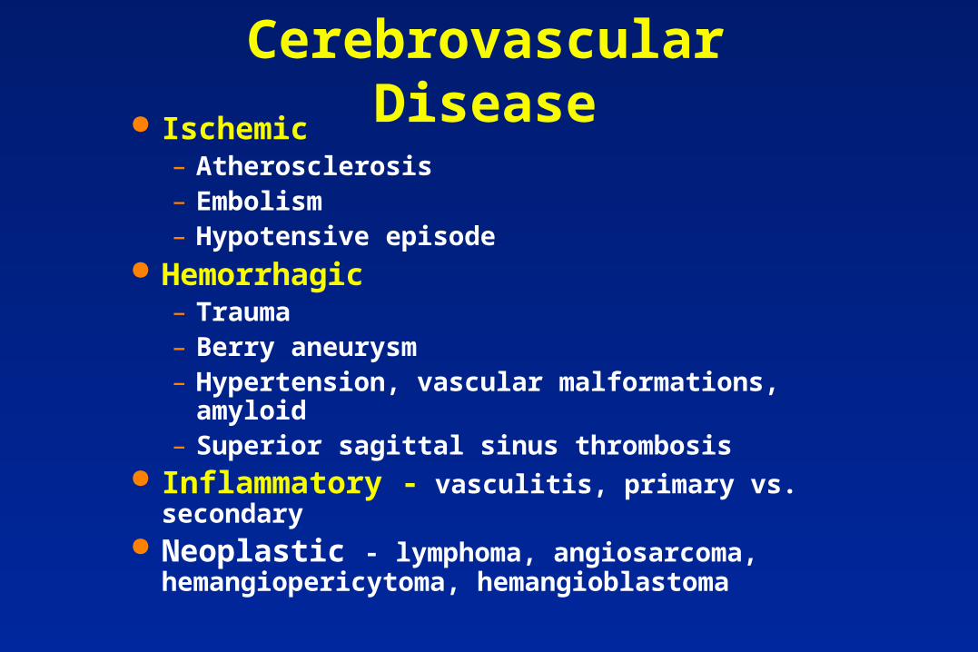
Cerebrovascular Disease Ischemic
– Atherosclerosis– Embolism– Hypotensive episode
Hemorrhagic– Trauma– Berry aneurysm– Hypertension, vascular malformations, amyloid– Superior sagittal sinus thrombosis
Inflammatory - vasculitis, primary vs. secondary Neoplastic - lymphoma, angiosarcoma,
hemangiopericytoma, hemangioblastoma
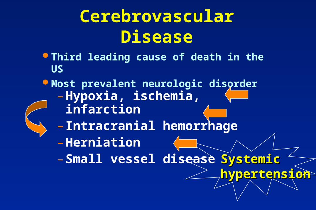
Cerebrovascular Disease
Third leading cause of death in the US Most prevalent neurologic disorder
Systemic Systemic hypertensionhypertension
– Hypoxia, ischemia, infarction– Intracranial hemorrhage– Herniation– Small vessel disease
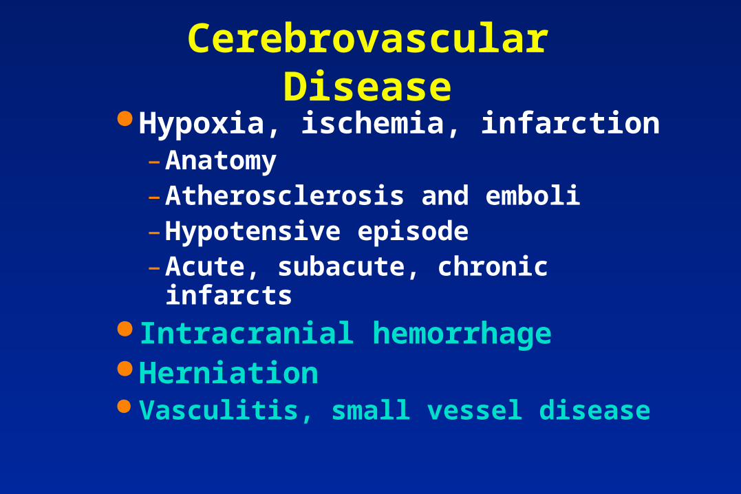
Cerebrovascular Disease Hypoxia, ischemia, infarction
– Anatomy– Atherosclerosis and emboli– Hypotensive episode– Acute, subacute, chronic infarcts
Intracranial hemorrhage Herniation Vasculitis, small vessel disease
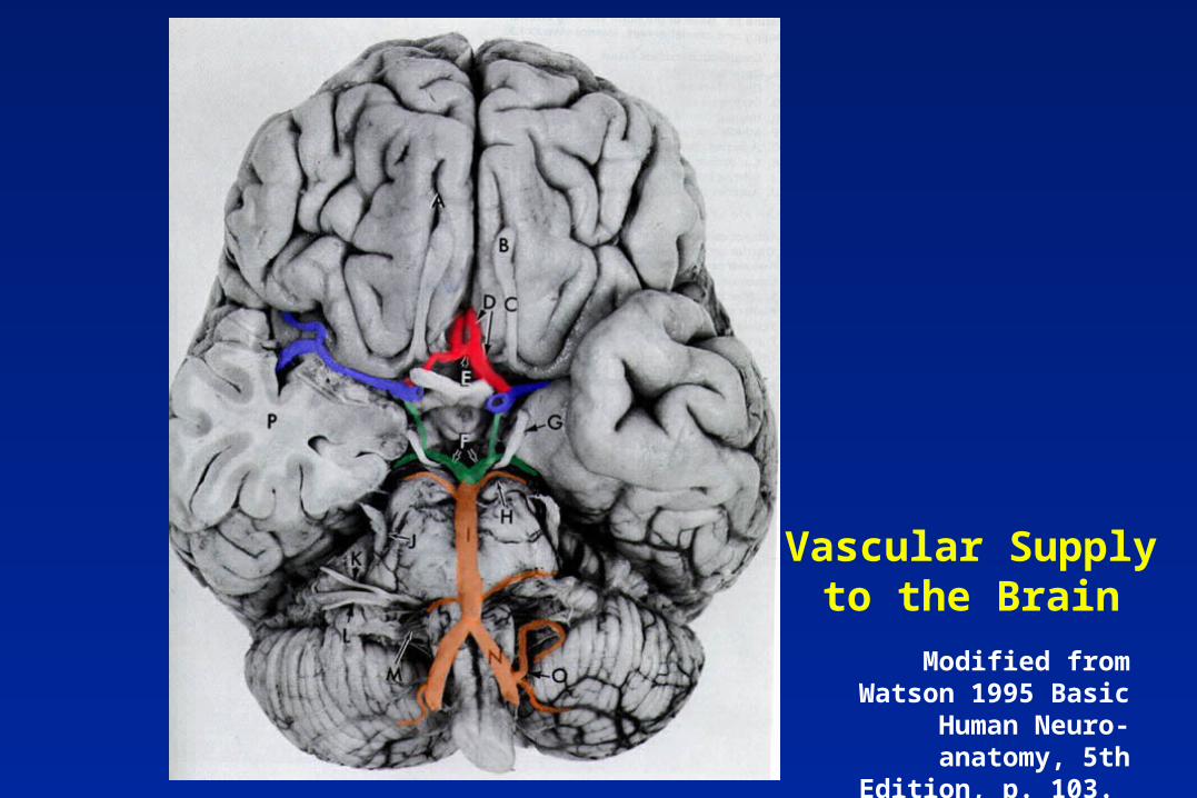
Vascular Supply to the Brain
Modified from Watson 1995 Basic Human Neuro-
anatomy, 5th Edition, p. 103. Little, Brown & Co.
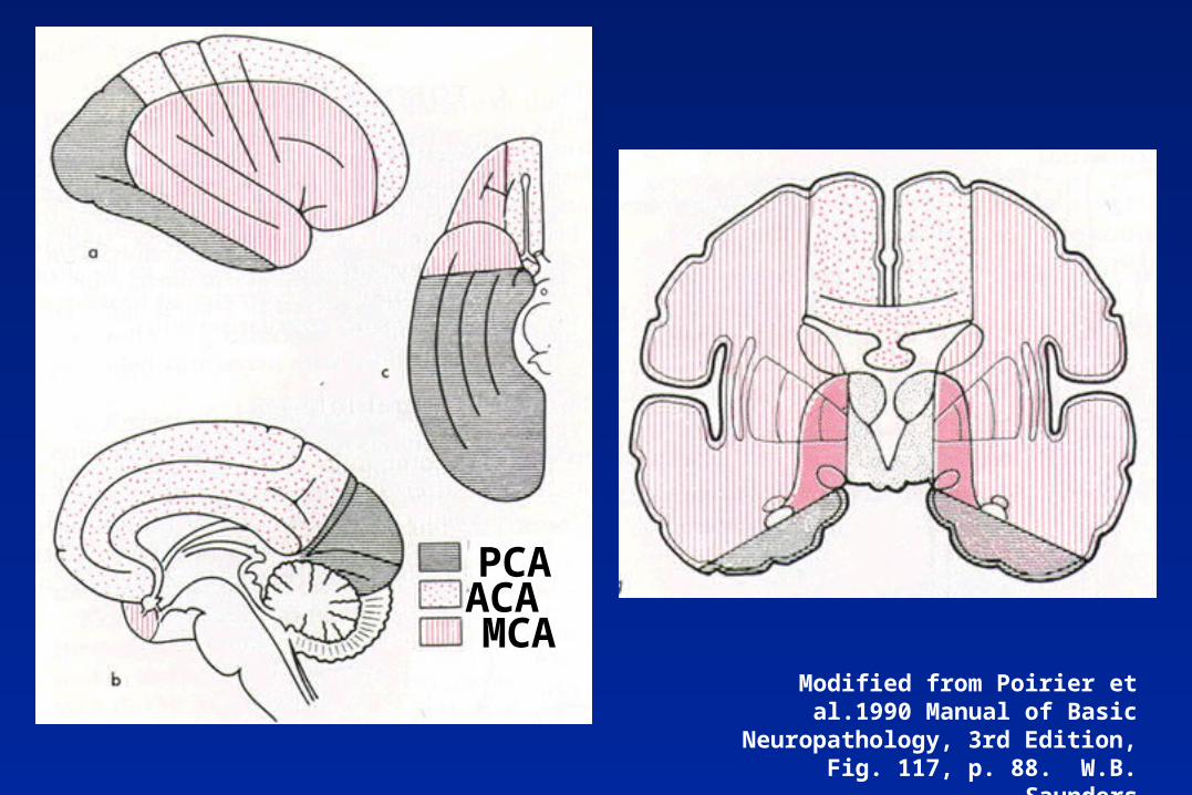
Modified from Poirier et al.1990 Manual of Basic Neuropathology, 3rd Edition,
Fig. 117, p. 88. W.B. Saunders
ACA PCA
MCA
Anatomic Considerations Vascular anatomy
– Circle of Willis and anastomoses (Figs. 109-110 - Poirier)
– Internal carotid-middle cerebral artery– Watershed zone
Rigid brain case and herniation (Robbins p. 1298)
– Falx– Tentorium– Foramen Magnum
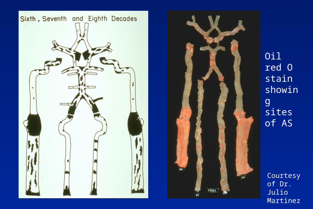
Courtesy of Dr. Julio Martinez
Oil red O stain showing sites of AS
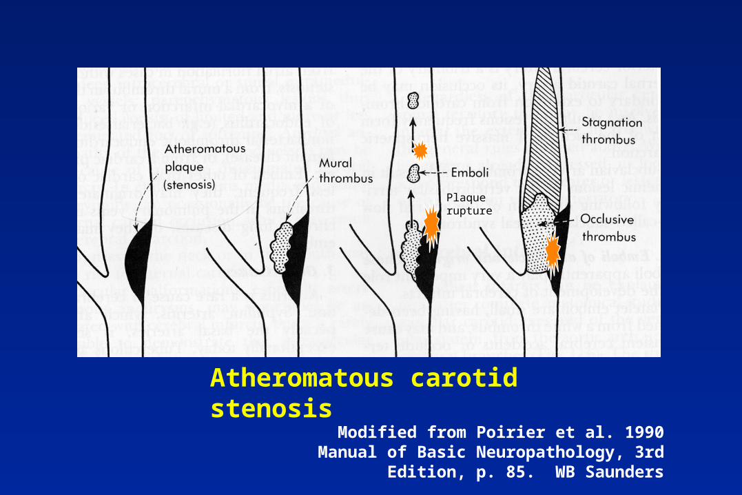
Plaque rupture
Atheromatous carotid stenosis
Modified from Poirier et al. 1990 Manual of Basic Neuropathology, 3rd Edition, p. 85. WB Saunders
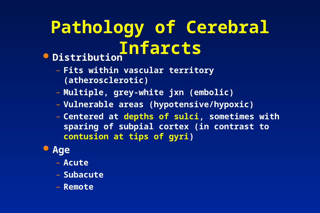
Distribution– Fits within vascular territory (atherosclerotic)
– Multiple, grey-white jxn (embolic)
– Vulnerable areas (hypotensive/hypoxic)
– Centered at depths of sulci, sometimes with sparing of subpial cortex (in contrast to contusion at tips of gyri)
Age– Acute
– Subacute
– Remote
Pathology of Cerebral Infarcts
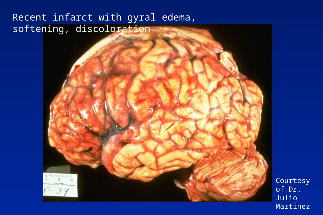
Courtesy of Dr. Julio Martinez
Recent infarct with gyral edema, softening, discoloration
Courtesy of Dr. Julio Martinez
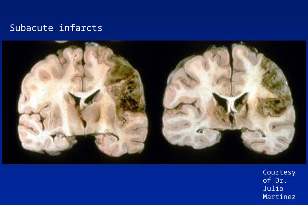
Subacute infarcts
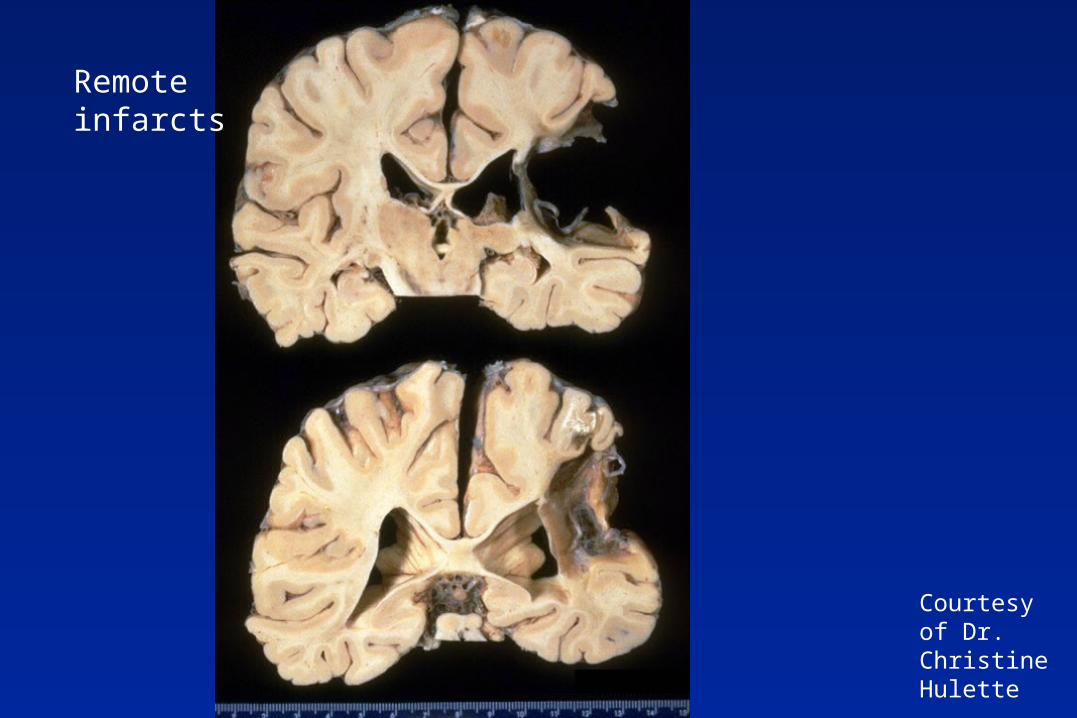
Courtesy of Dr. Christine Hulette
Remote infarcts
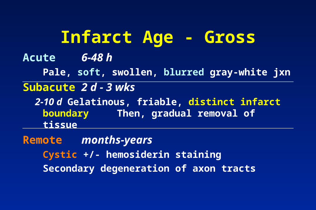
Infarct Age - GrossAcute 6-48 h
Pale, soft, swollen, blurred gray-white jxn
Subacute 2 d - 3 wks2-10 d Gelatinous, friable, distinct infarct boundary
Then, gradual removal of tissue
Remote months-yearsCystic +/- hemosiderin staining
Secondary degeneration of axon tracts
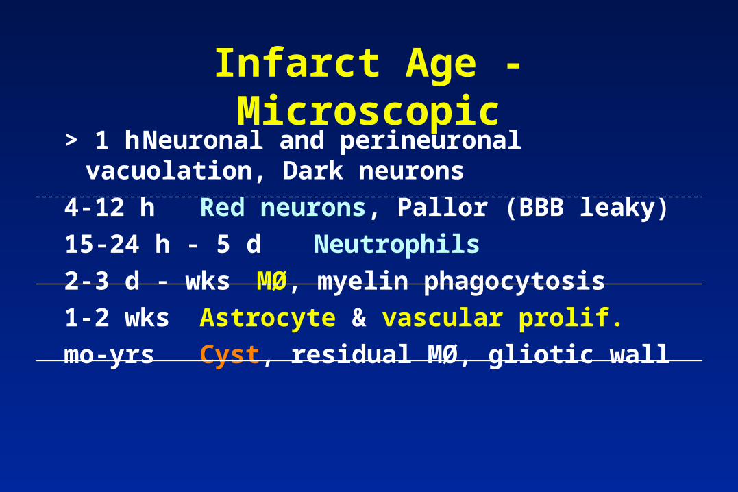
Infarct Age - Microscopic> 1 h Neuronal and perineuronal
vacuolation, Dark neurons
4-12 h Red neurons, Pallor (BBB leaky)
15-24 h - 5 dNeutrophils
2-3 d - wks MØ, myelin phagocytosis
1-2 wks Astrocyte & vascular prolif.
mo-yrs Cyst, residual MØ, gliotic wall
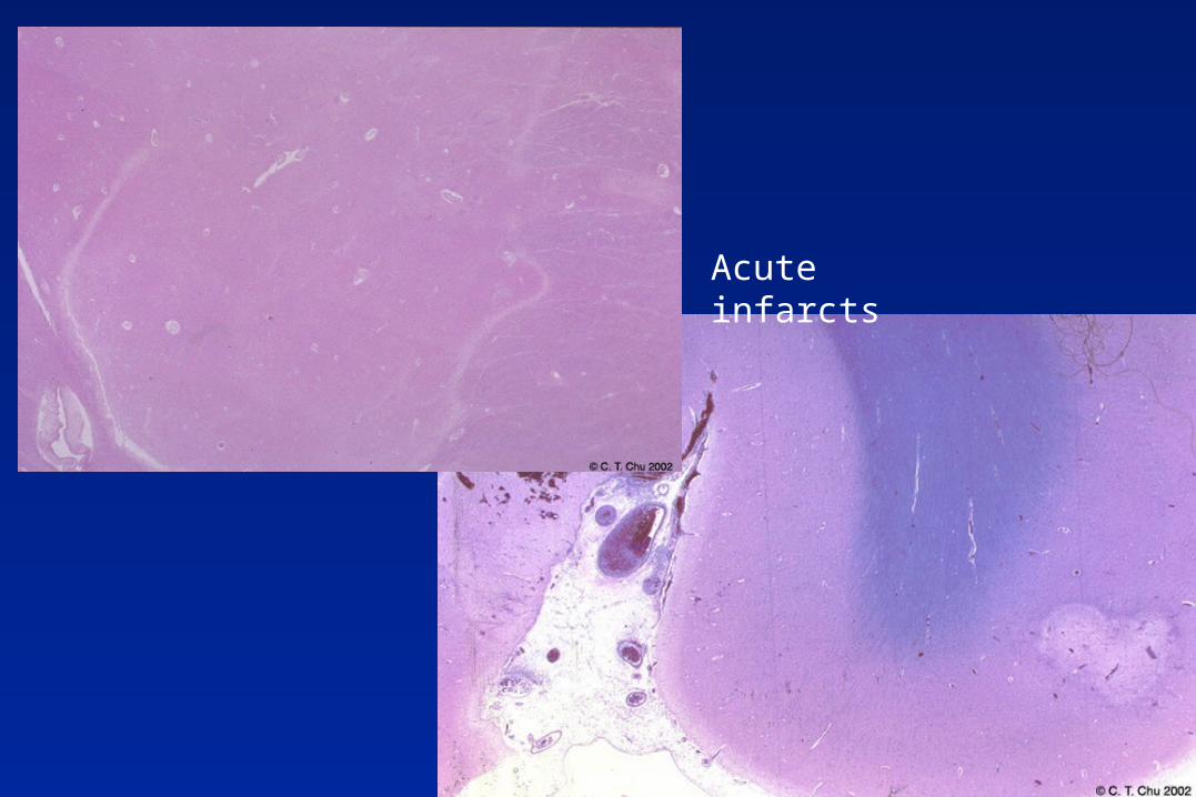
Acute infarcts
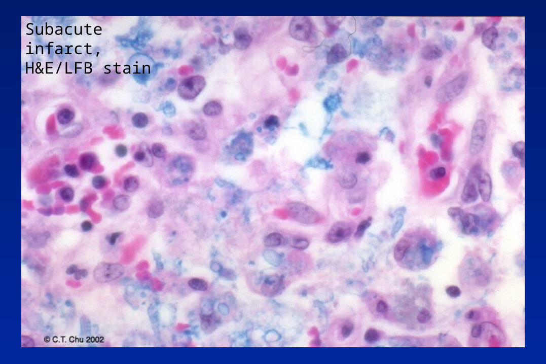
Subacute infarct, H&E/LFB stain
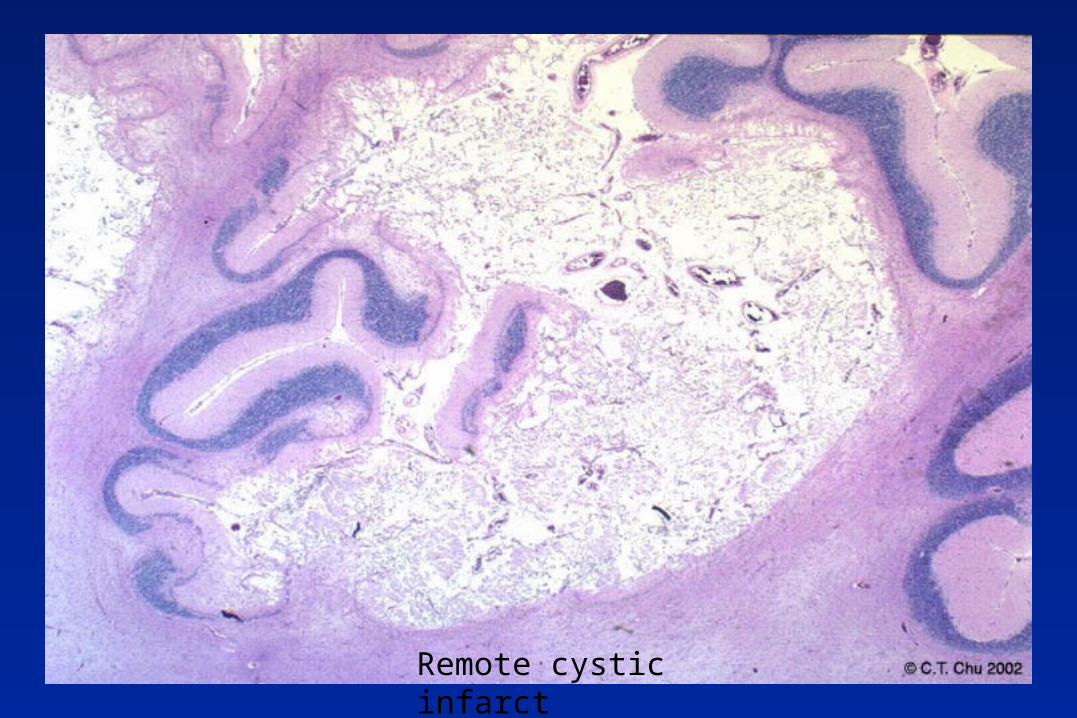
Remote cystic infarct
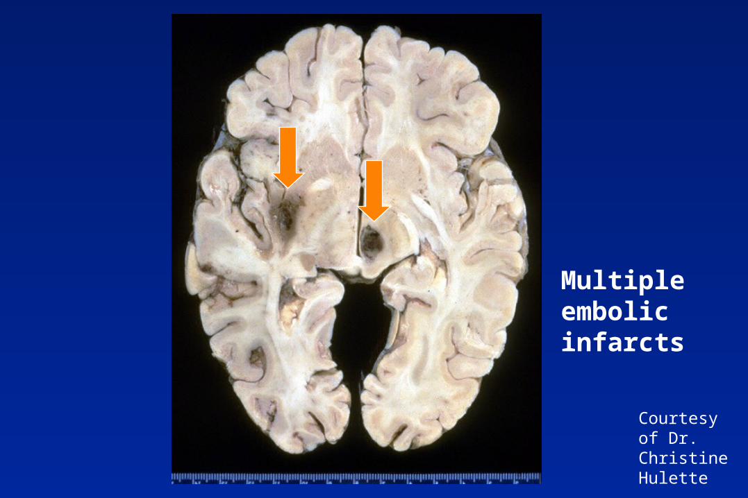
Multiple embolic infarcts
Courtesy of Dr. Christine Hulette
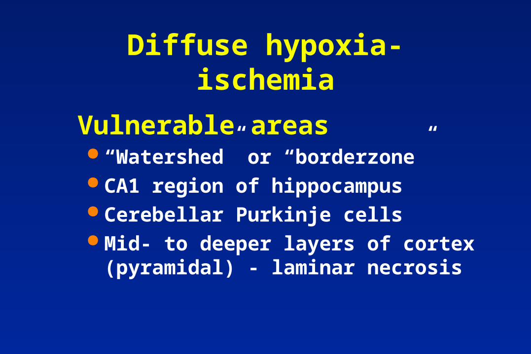
Diffuse hypoxia-ischemia
“Watershed” or “borderzone” CA1 region of hippocampus Cerebellar Purkinje cells Mid- to deeper layers of cortex
(pyramidal) - laminar necrosis
Vulnerable areas
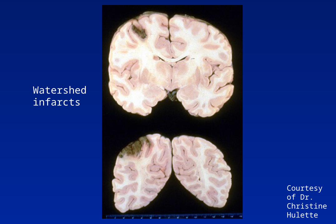
Courtesy of Dr. Christine Hulette
Watershed infarcts
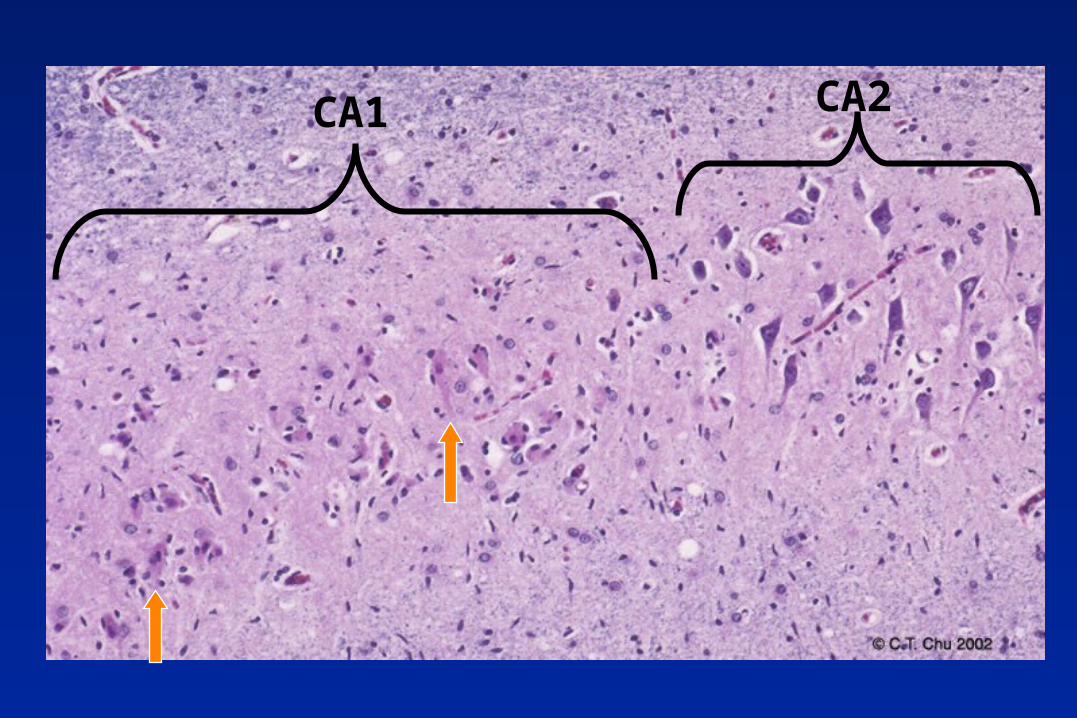
CA1 CA2
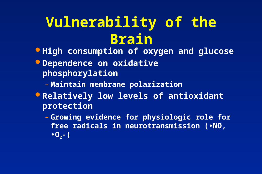
Vulnerability of the Brain High consumption of oxygen and glucose Dependence on oxidative phosphorylation
– Maintain membrane polarization Relatively low levels of antioxidant
protection– Growing evidence for physiologic role for free
radicals in neurotransmission (•NO, •O2-)
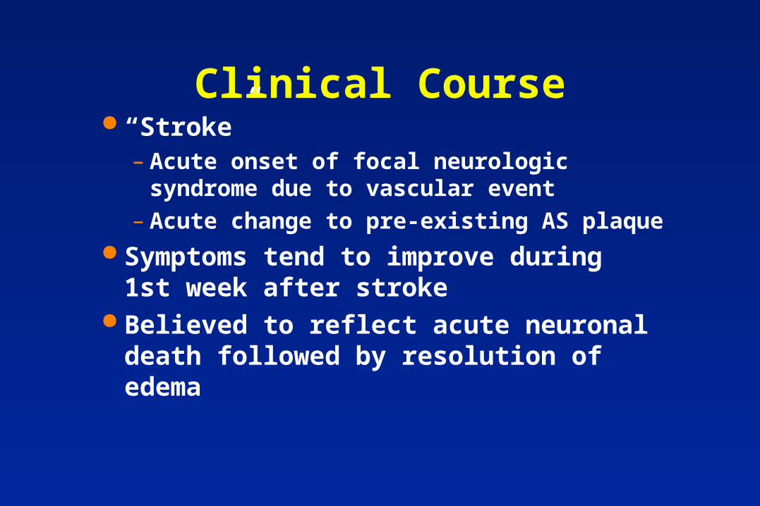
Clinical Course “Stroke”
– Acute onset of focal neurologic syndrome due to vascular event
– Acute change to pre-existing AS plaque Symptoms tend to improve during 1st
week after stroke Believed to reflect acute neuronal death
followed by resolution of edema
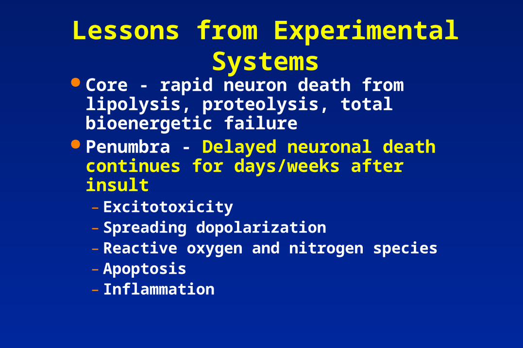
Lessons from Experimental Systems Core - rapid neuron death from lipolysis,
proteolysis, total bioenergetic failure Penumbra - Delayed neuronal death
continues for days/weeks after insult– Excitotoxicity– Spreading dopolarization– Reactive oxygen and nitrogen species– Apoptosis– Inflammation
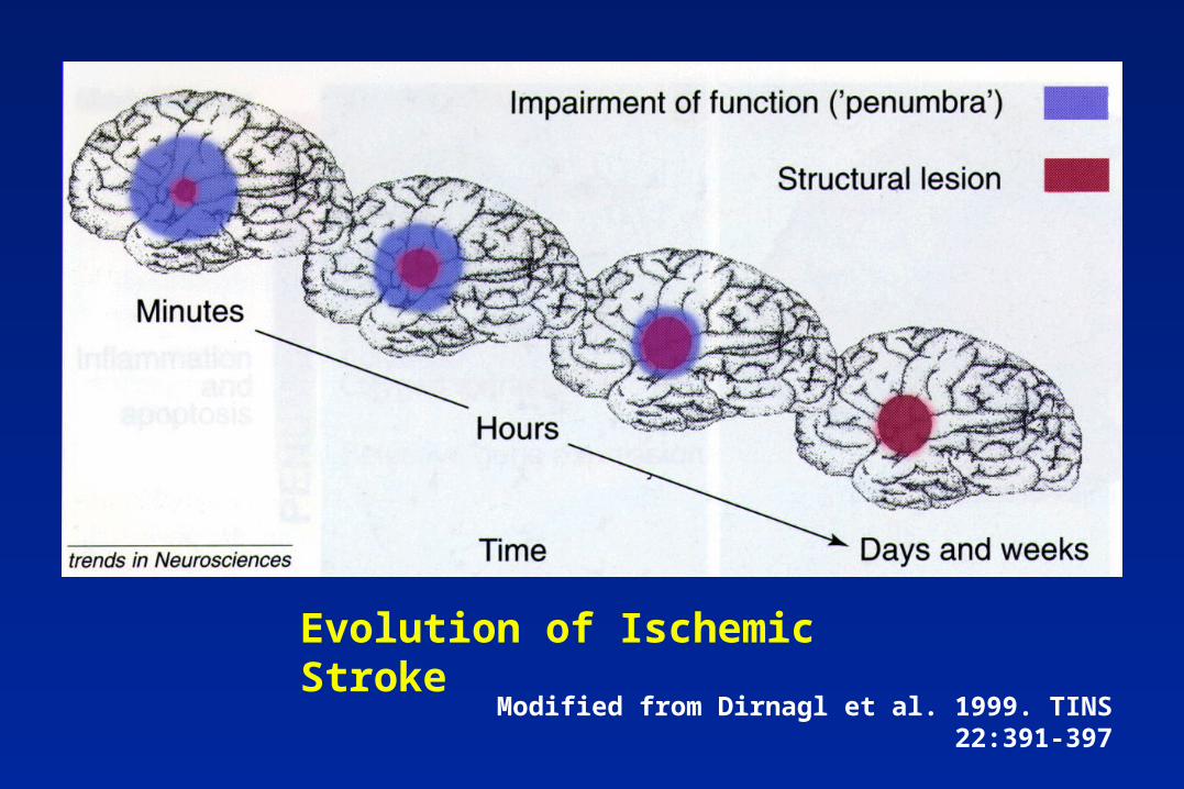
Evolution of Ischemic Stroke
Modified from Dirnagl et al. 1999. TINS 22:391-397
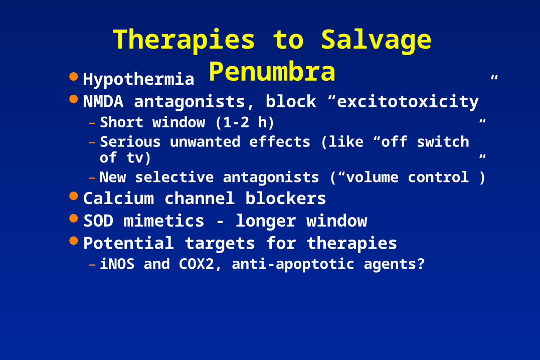
Therapies to Salvage Penumbra Hypothermia NMDA antagonists, block “excitotoxicity”
– Short window (1-2 h)– Serious unwanted effects (like “off switch” of tv)– New selective antagonists (“volume control”)
Calcium channel blockers SOD mimetics - longer window Potential targets for therapies
– iNOS and COX2, anti-apoptotic agents?
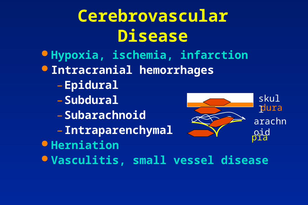
Cerebrovascular Disease
Hypoxia, ischemia, infarction Intracranial hemorrhages
– Epidural– Subdural– Subarachnoid– Intraparenchymal
Herniation Vasculitis, small vessel disease
pia
arachnoid
skulldura
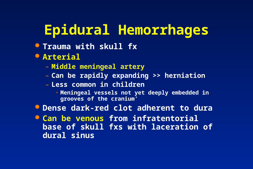
Epidural Hemorrhages Trauma with skull fx Arterial
– Middle meningeal artery– Can be rapidly expanding >> herniation– Less common in children
• Meningeal vessels not yet deeply embedded in grooves of the cranium’
Dense dark-red clot adherent to dura Can be venous from infratentorial base of skull
fxs with laceration of dural sinus
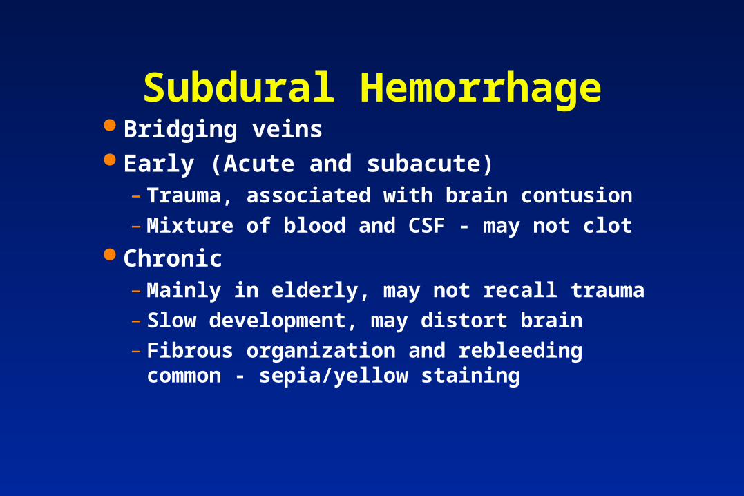
Subdural Hemorrhage Bridging veins Early (Acute and subacute)
– Trauma, associated with brain contusion– Mixture of blood and CSF - may not clot
Chronic– Mainly in elderly, may not recall trauma– Slow development, may distort brain– Fibrous organization and rebleeding common -
sepia/yellow staining
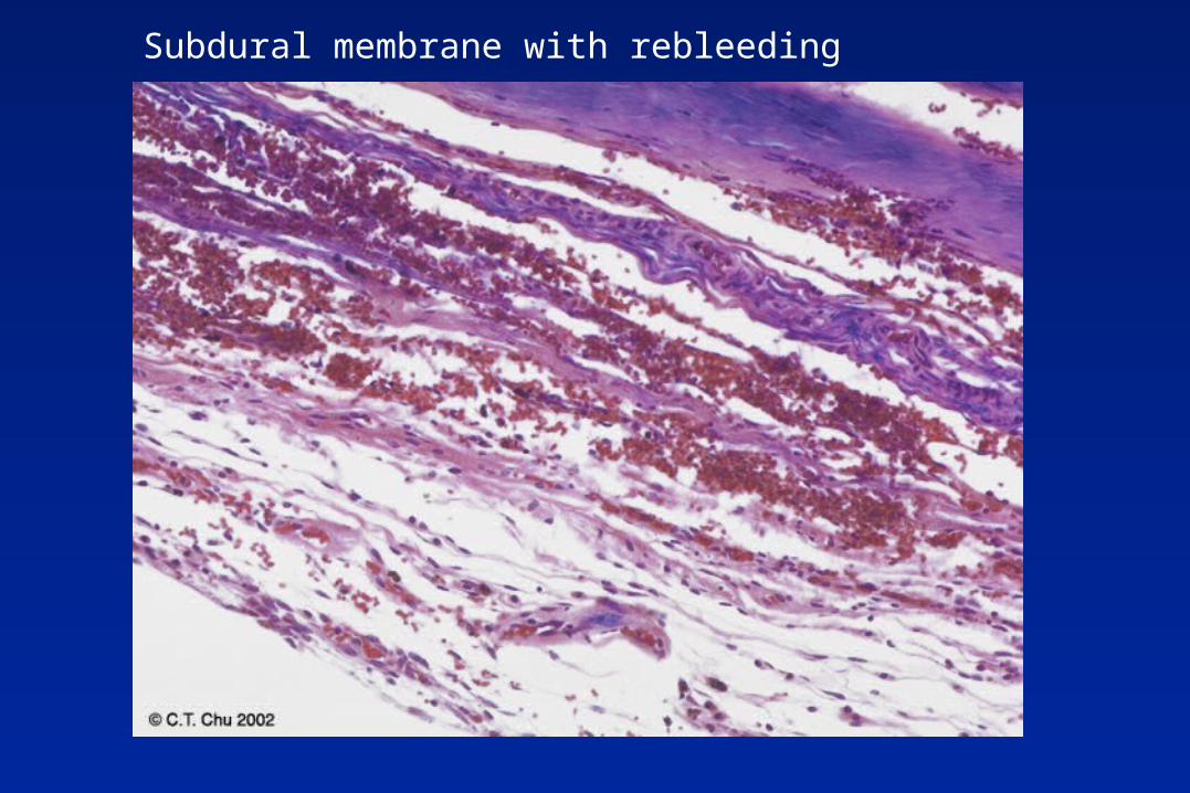
Subdural membrane with rebleeding
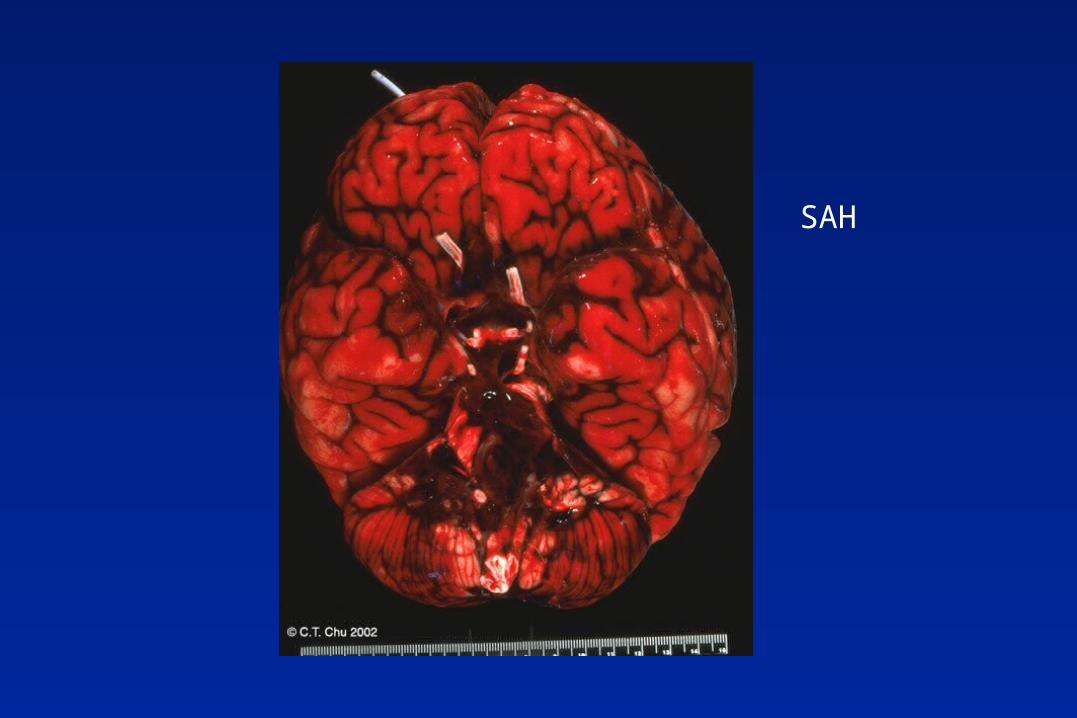
SAH
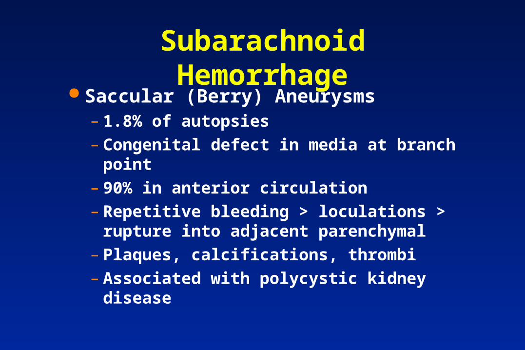
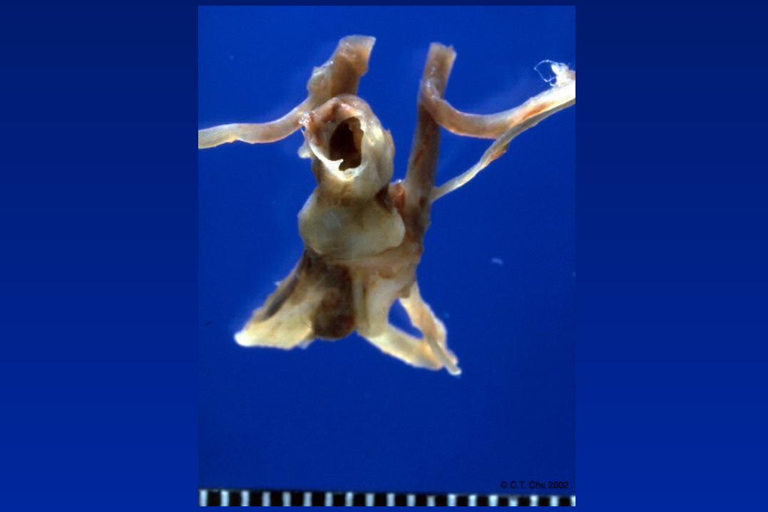
Subarachnoid Hemorrhage Saccular (Berry) Aneurysms
– 1.8% of autopsies– Congenital defect in media at branch point– 90% in anterior circulation– Repetitive bleeding > loculations > rupture
into adjacent parenchymal– Plaques, calcifications, thrombi– Associated with polycystic kidney disease
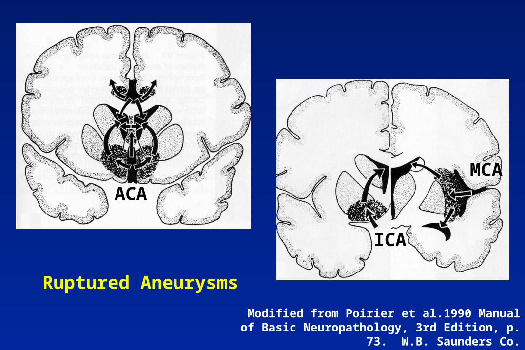
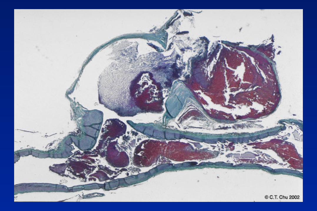
Ruptured Aneurysms
Modified from Poirier et al.1990 Manual of Basic Neuropathology, 3rd Edition, p. 73. W.B. Saunders Co.
ACA
ICA
MCA
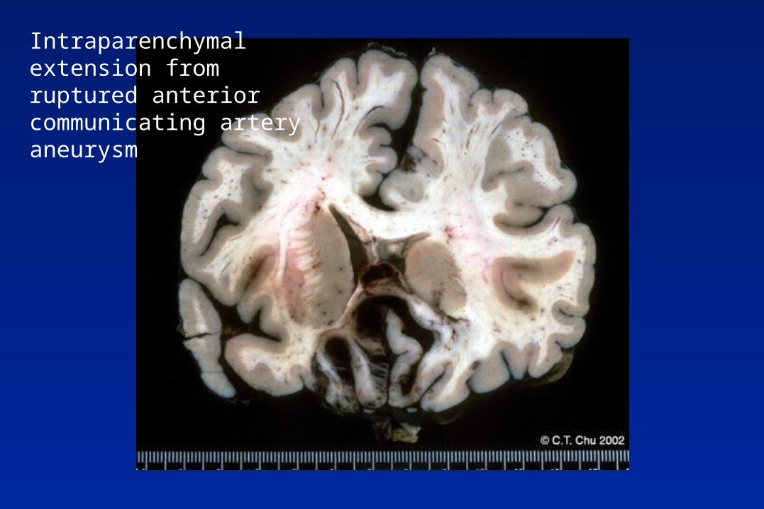
Intraparenchymal extension from ruptured anterior communicating artery aneurysm
Intraparenchymal Hemorrhage
15% mortality Arterial hypertension - 80% of cases Vascular malformations Amyloid angiopathy Neoplasms
Other intracranial aneurysms
Seldom present as SAH Fusifirm atherosclerotic aneurysms
– Basilar artery– Compression of adjacent structures– Infectious and post-traumatic
Mycotic, traumatic, dissecting– Usually involve anterior circulation
Arterial dissection
Young adults - IC, MCA, vertebral, basilar Hyperextension injury - may be “trivial” Spontaneous dissection
– Arteritis, AS, HTN, birth control pill, Marfan’s, cystic medial necrosis, fibromuscular dysplasia, Ehlers-Danlos
– Focal absence, splitting, fraying of internal elastic membrane
– 33% no identifiable pathology
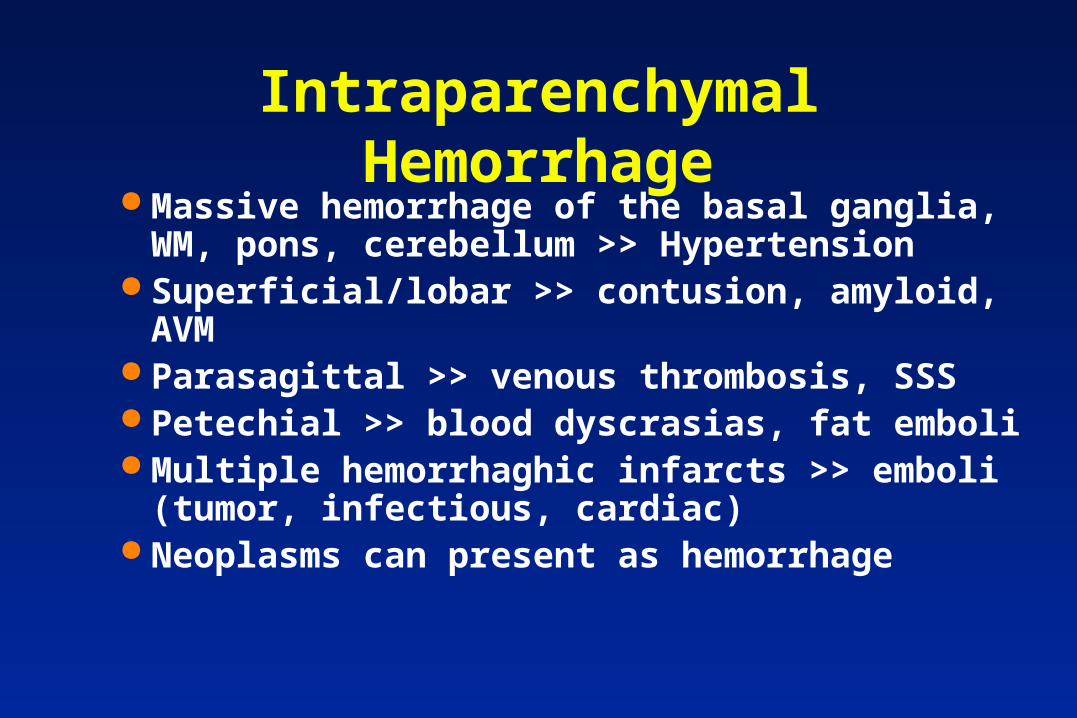
Intraparenchymal Hemorrhage Massive hemorrhage of the basal ganglia, WM,
pons, cerebellum >> Hypertension Superficial/lobar >> contusion, amyloid, AVM Parasagittal >> venous thrombosis, SSS Petechial >> blood dyscrasias, fat emboli Multiple hemorrhaghic infarcts >> emboli
(tumor, infectious, cardiac) Neoplasms can present as hemorrhage
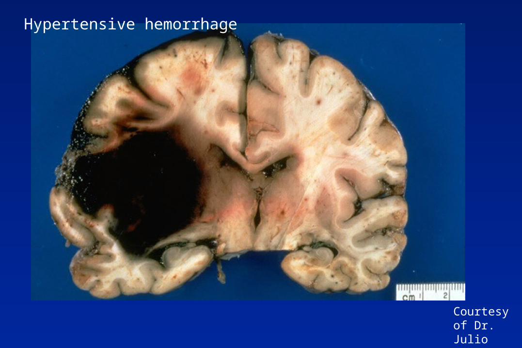
Courtesy of Dr. Julio Martinez
Hypertensive hemorrhage
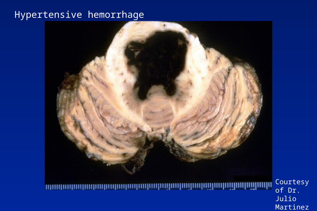
Courtesy of Dr. Julio Martinez
Hypertensive hemorrhage
Surgical Pathology Hemorrhages
Usual dx - clotted blood– May see erythrophagocytosis, fibrovascular
organization, subdural membrane > then can call organizing hemorrhage/hematoma
Look for brain tissue and note in report If present, look for underlying cause
– Congophilic angiopathy (-APP, cystatin C)
– Tumor
– AVM
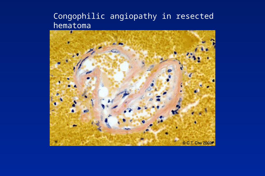
Congophilic angiopathy in resected hematoma
CNS Vascular Malformations
Arteriovenous malformation (AVM) Cavernous hemangioma Capillary telangiectasia - pons Venous angioma (varices)
Arteriovenous malformation Medusa-like lesions with potential for
rupture Most over hemispheric surface of MCA Multiple lesions occasionally seen with
Rendo-Osler-Weber disease or Wyburn-Mason syndrome
Sx: seizures, focal deficits, increased ICP, catastrophic hemorrhage
AVM - Pathology Vessels vary in caliber Core may exclude brain parenchyma, but
feeding and draining vessels interdigitate with intervening brain
Presence of abnormal arteries possessing internal elastic lamina is diagnostic
“Arterialized” veins from the high pressure
Evidence of prior hemorrhage
Arteriovenous malformation
In children, deep AVMs draining into the great vein of Galen can cause cardiac decompensation from shunting
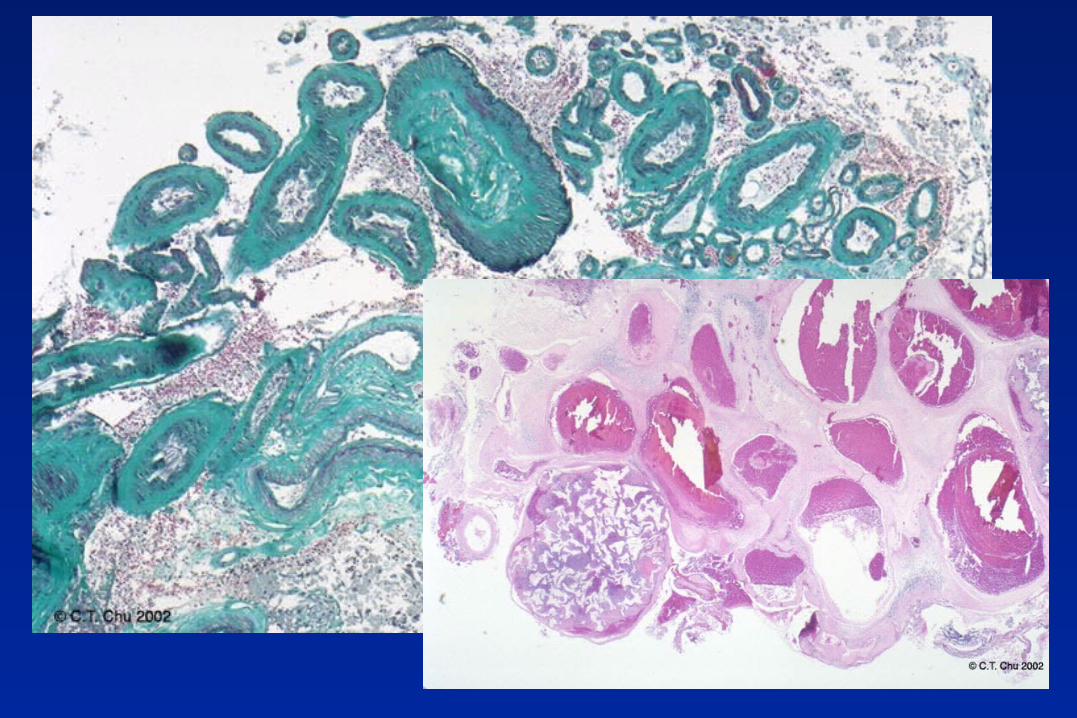
Cavernous Malformations
Compact spherical calcified mass Most often affect subcortical areas, but also
hindbrain Multiple lesions frequent Recently recognized that it can be
transmitted as an autosomal dominant trait Typically present with seizures.
Hemorrhages common, but usually small
Cavernous Malformations
Honeycomb of compact vessels, often collagenized
No muscle or elastic lamina Closely packed, no intervening brain Surrounding brain shows extensive
hemosiderin and iron laden macrophages/astrocytes - dark MR signal
Venous Infarction
Hemorrhagic lesions involving parasagittal meninges, cortex, WM
Superior sagittal sinus thrombosis– Centrum ovale and overlying cortex,
meninges, usually symmetric Great vein of Galen
– Periventricular and thalamic regions
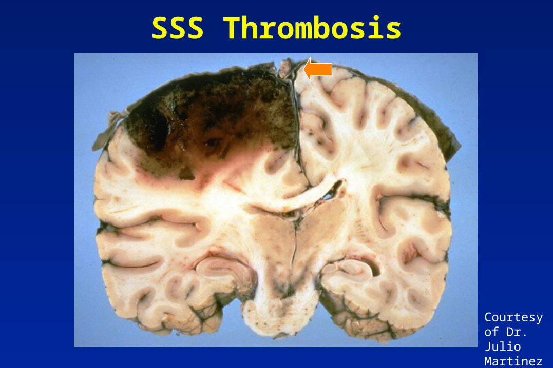
SSS Thrombosis
Courtesy of Dr. Julio Martinez
Brain tumors presenting with hemorrhage
Classically associated with oligodendroglioma, choriocarcinoma, metastatic melanoma
However, any glioma can present with hemorrhage– Recent examples include GBM, anaplastic
ependymoma
Post-operative hematoma from incompletely excised tumors - clinical history often not given
Cerebrovascular Disease
Hypoxia, ischemia, infarction Intracranial hemorrhages Herniation
– Symptoms– Anatomic basis
Vasculitis, small vessel disease

Herniation Rigid skull, tough inelastic dura
– Brain, CSF, blood Symptoms of increased pressure
– Headache– Papilledema - precedes herniation
Symptoms of transtentorial herniation– Remember anatomic basis
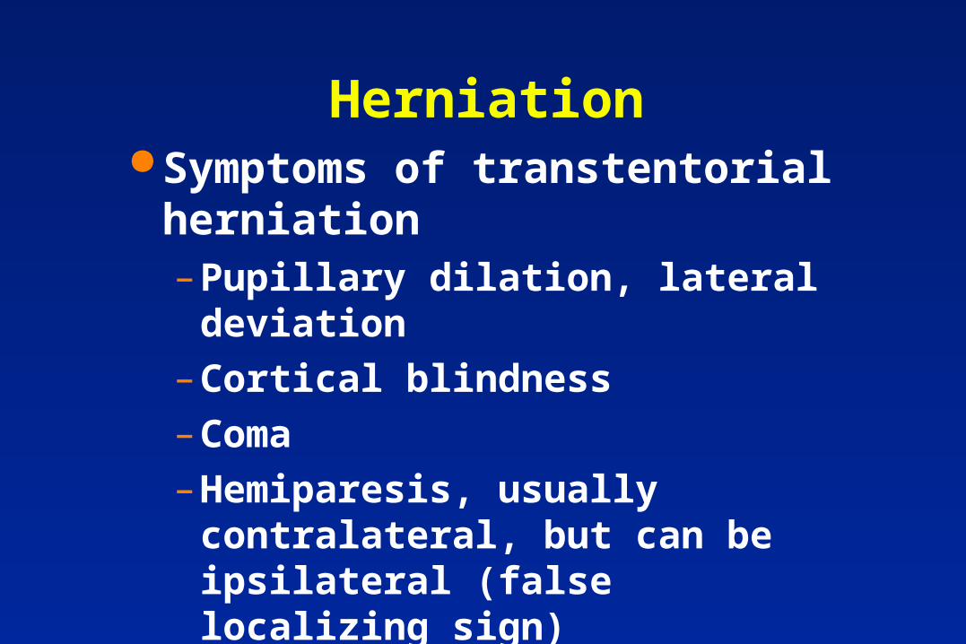
Herniation Symptoms of transtentorial
herniation– Pupillary dilation, lateral deviation
– Cortical blindness
– Coma
– Hemiparesis, usually contralateral, but can be ipsilateral (false localizing sign)
Hydrocephalus, Duret hemorrhages of pons
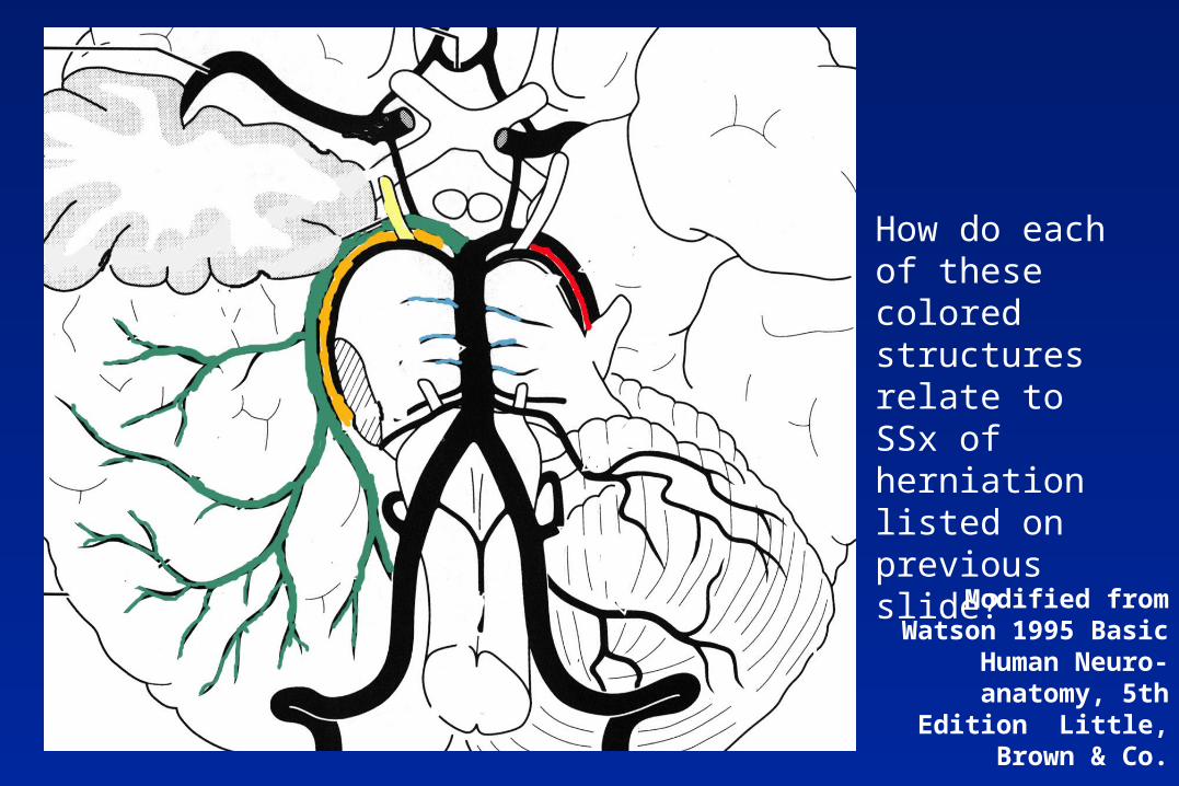
Modified from Watson 1995 Basic Human Neuro-
anatomy, 5th Edition Little, Brown & Co.
How do each of these colored structures relate to SSx of herniation listed on previous slide?
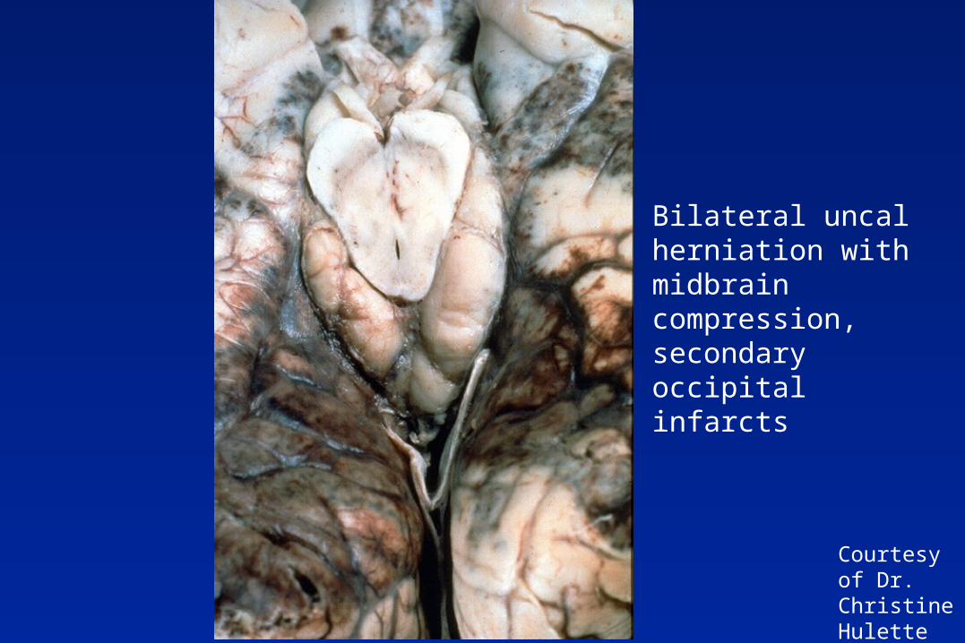
Courtesy of Dr. Christine Hulette
Bilateral uncal herniation with midbrain compression, secondary occipital infarcts
Cerebrovascular Disease
Hypoxia, ischemia, infarction Intracranial hemorrhages Herniation Vasculitis, small vessel disease
– Temporal arteritis– Microvascular diseases
• HTN, amyloid angiopathy, primary angiitis of the CNS
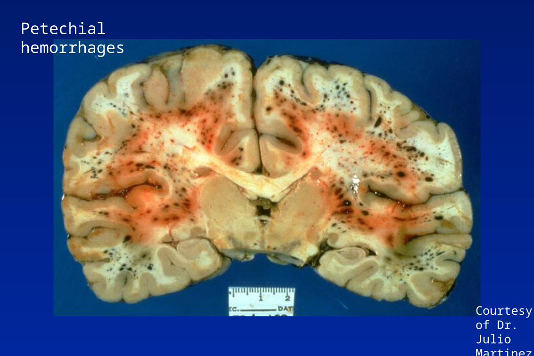
– Petechial hemorrhages
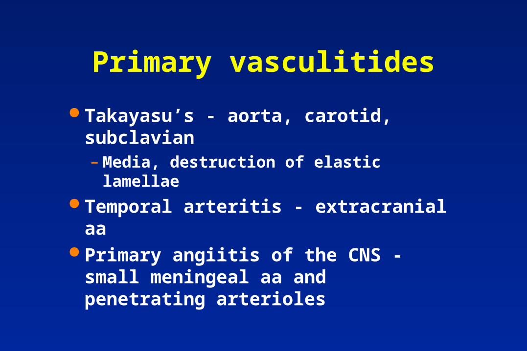
Primary vasculitides
Takayasu’s - aorta, carotid, subclavian– Media, destruction of elastic lamellae
Temporal arteritis - extracranial aa Primary angiitis of the CNS - small
meningeal aa and penetrating arterioles
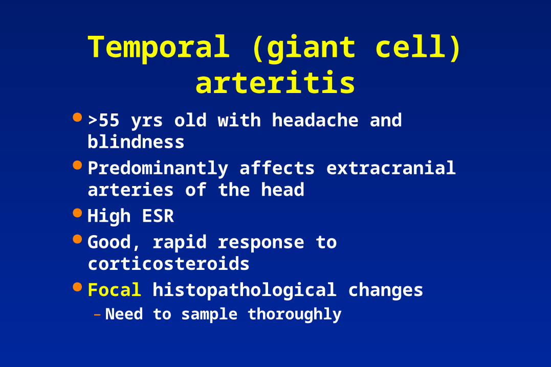
Temporal (giant cell) arteritis
>55 yrs old with headache and blindness Predominantly affects extracranial
arteries of the head High ESR Good, rapid response to corticosteroids Focal histopathological changes
– Need to sample thoroughly
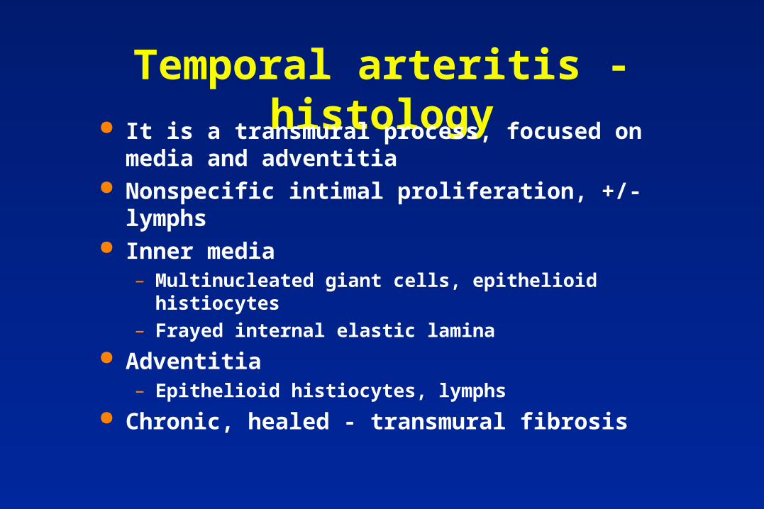
Temporal arteritis - histology It is a transmural process, focused on media and
adventitia Nonspecific intimal proliferation, +/- lymphs Inner media
– Multinucleated giant cells, epithelioid histiocytes
– Frayed internal elastic lamina
Adventitia– Epithelioid histiocytes, lymphs
Chronic, healed - transmural fibrosis
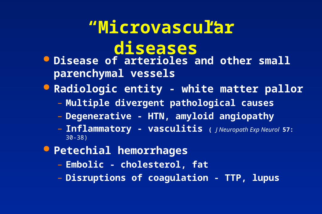
“Microvascular diseases” Disease of arterioles and other small parenchymal
vessels Radiologic entity - white matter pallor
– Multiple divergent pathological causes
– Degenerative - HTN, amyloid angiopathy – Inflammatory - vasculitis ( J Neuropath Exp Neurol 57: 30-38)
Petechial hemorrhages– Embolic - cholesterol, fat
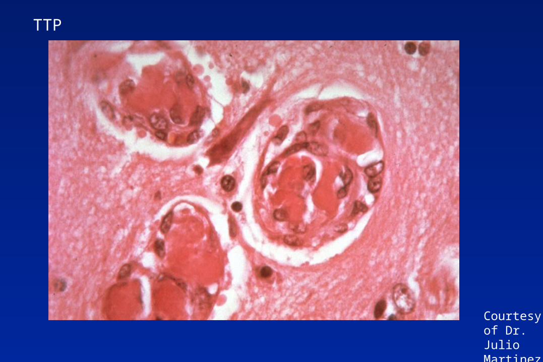
– Disruptions of coagulation - TTP, lupus
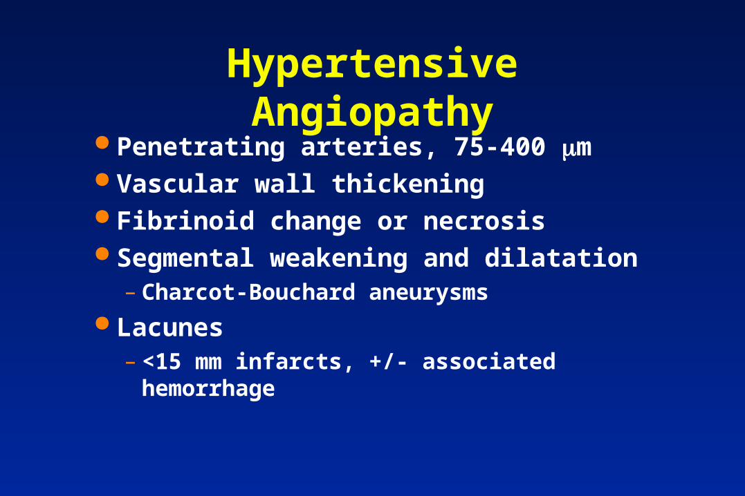
Hypertensive Angiopathy Penetrating arteries, 75-400 m Vascular wall thickening Fibrinoid change or necrosis Segmental weakening and dilatation
– Charcot-Bouchard aneurysms Lacunes
– <15 mm infarcts, +/- associated hemorrhage
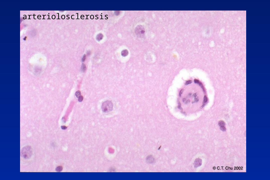
arteriolosclerosis
Primary angiitis of the CNS Noninfectious granulomatous angiitis or isolated
angiitis of the CNS Untreated - almost universally fatal Combination steroid and cytoxan ESR variable and not diagnostically useful, CSF
resembles chronic meningitis Transmural granulomatous or lymphocytic
inflammation, esp. intima, media Rule out infectious vasculitides
PACNS - DDx
Clinical mimics - hypertension, AD, amyloid angiopathy, glioma, antiphospholipid syndromes, moyamoya, fibromuscular dysplasia, cardiac myxoma embolism)
J Neuropath Exp Neurol 57: 30-38, 1998.
Pathologic DDx - viral infection, Hodkin’s, lymphomatoid granulomatosis, systemic rheumatic disorders ( SLE, sarcoid), drug hypersensitivity
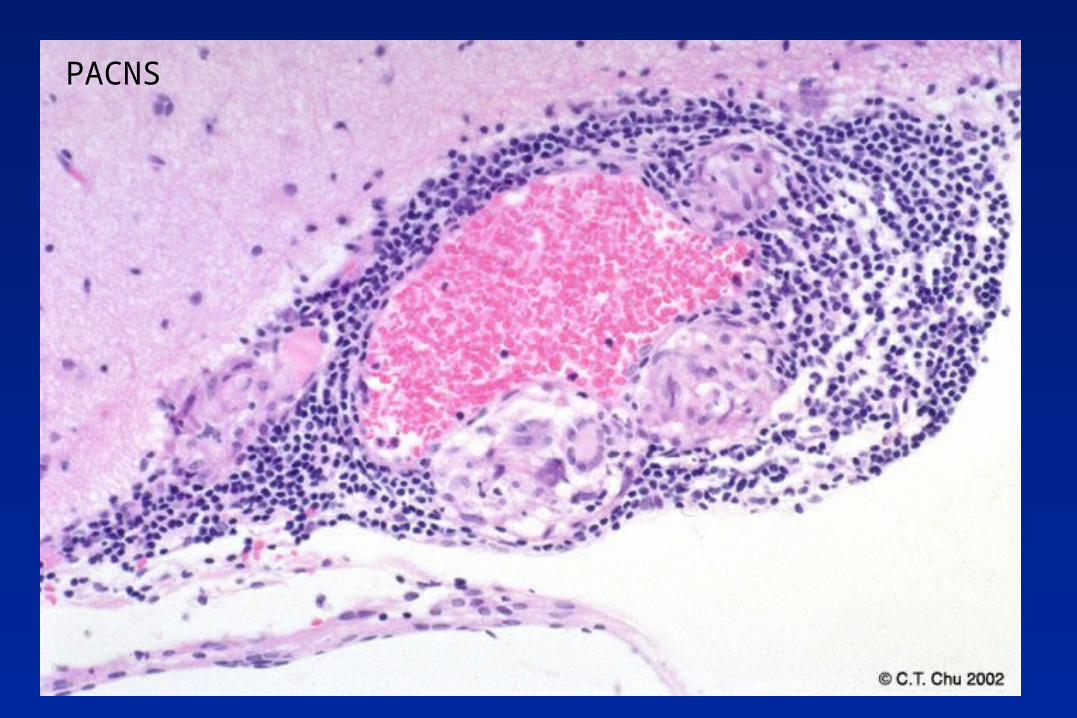
PACNS
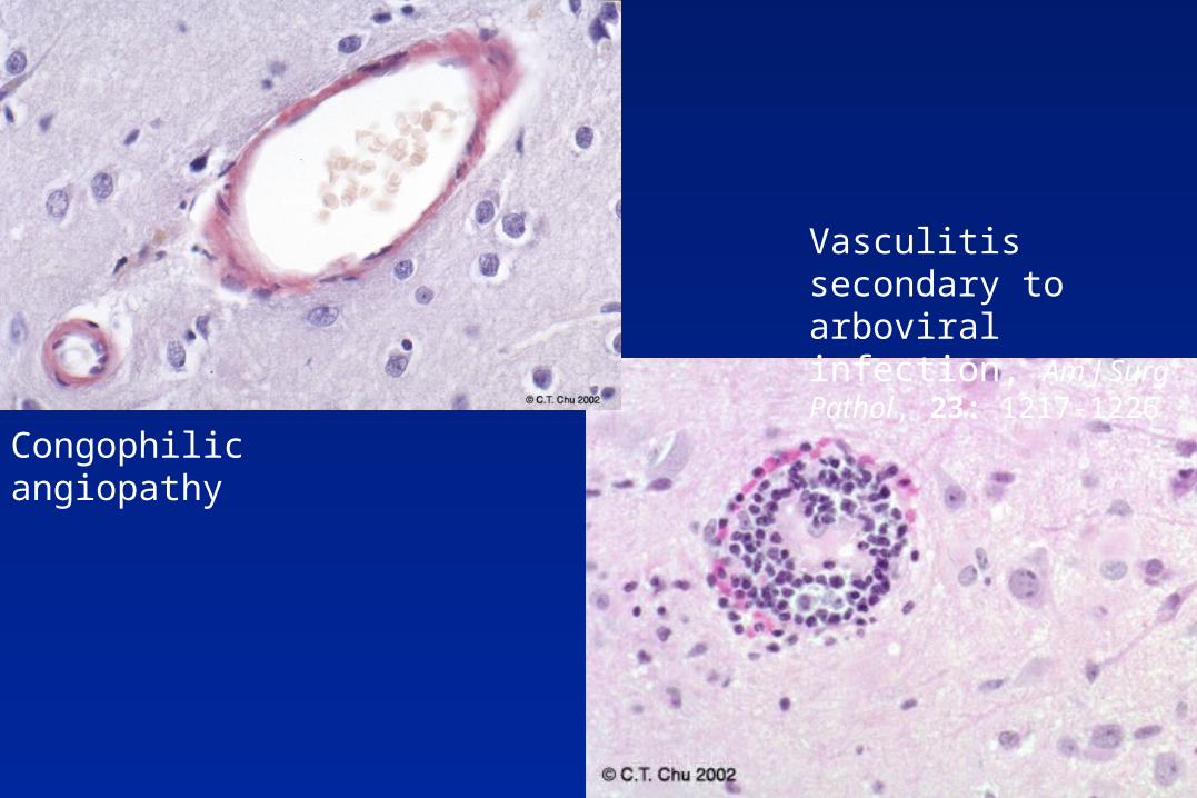
Congophilic angiopathy
Vasculitis secondary to arboviral infection, Am J Surg Pathol, 23: 1217-1226
Courtesy of Dr. Julio Martinez
Petechial hemorrhages
Courtesy of Dr. Julio Martinez
TTP
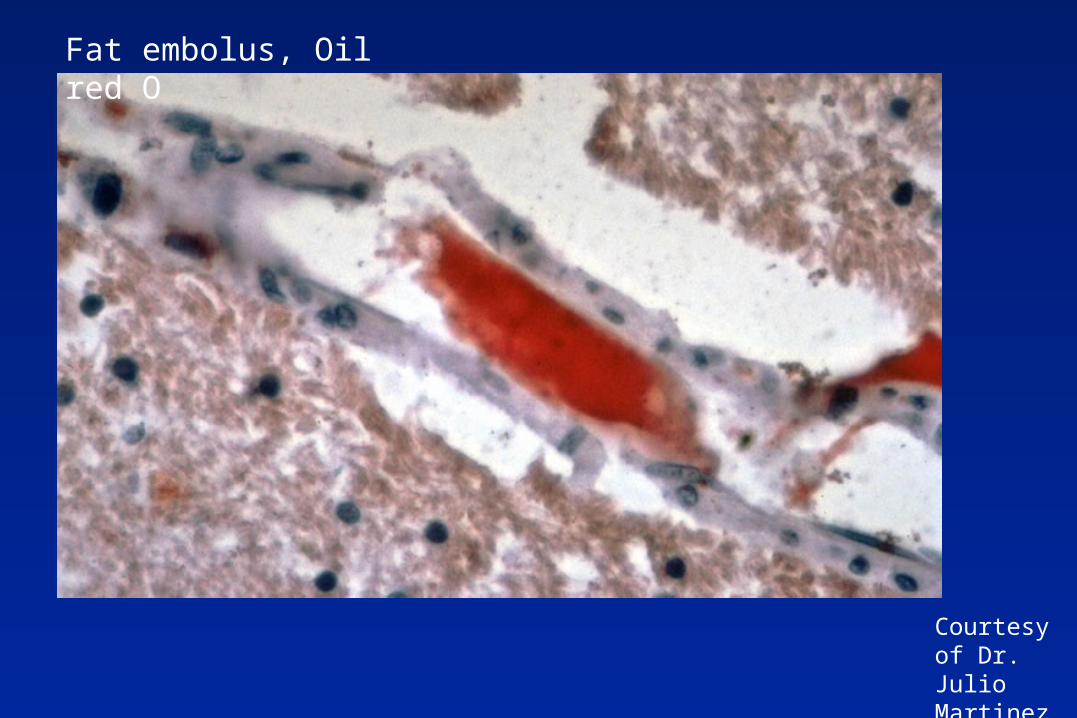
Fat embolus, Oil red O
Courtesy of Dr. Julio Martinez

Self quiz (see next slide) Which two panels show pathology related
to a common etiology (cause)? What panel results from trauma, what is
anatomic space occupied by the lesion, and what vessel is commonly involved?
Which panel reflects differential neuronal susceptibility to injury?
Which panel reflects a chronic process?
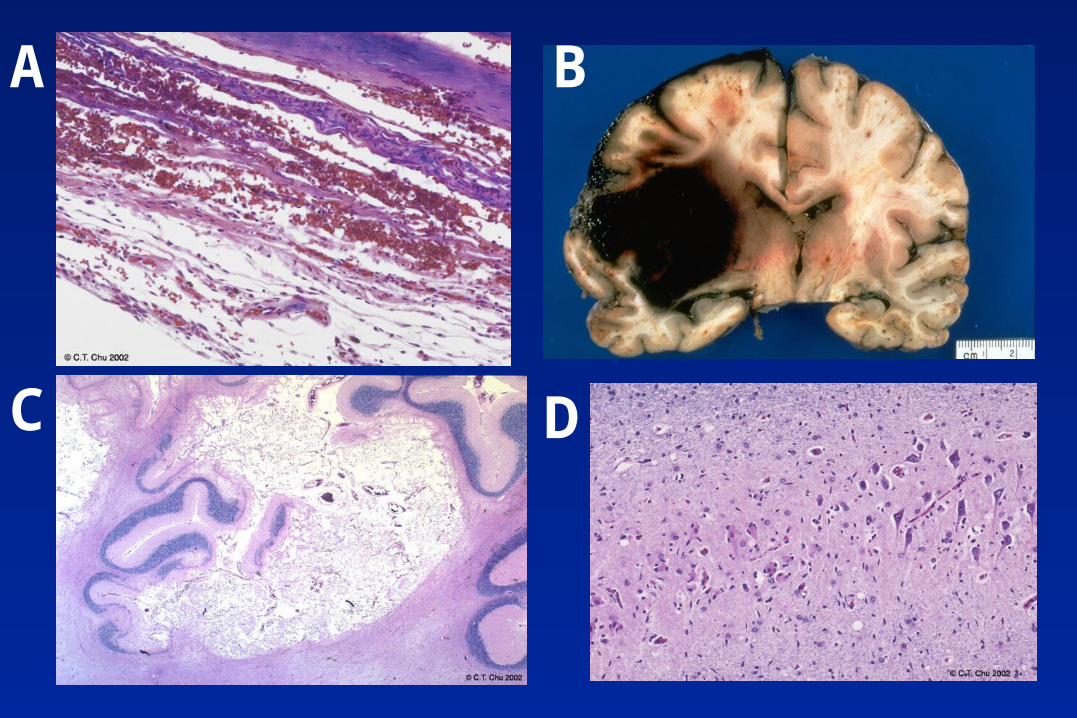
D
A B
C
Self quiz (answers) B shows hypertensive hemorrhage originating
in BG and C shows lacunar infarcts in the BG, also related to hypertension.
The subdural hemorrhage in A results from trauma, sometimes so mild it is not remembered, and involves bridging veins
D shows acute neuronal injury (red neurons) in the region of the hippocampus susceptible to hypotensive-hypoperfusion injury?
C shows remote cerebellar infarct
Recommended Reading
Manual of Basic Neuropathology by Poirier et al.
– Pp. 52-56, 58-61, Chapter 4. Robbins Pathologic Basis of Disease by Cotran, Kumar,
and Collins 6th Ed.
– Pertinent sections of Chapter 30 (CNS). Greenfield’s Neuropathology text - a must for all NP
fellows