varicella-zoster virus orf66 kinase ... - d …d-scholarship.pitt.edu/6493/1/erazo_a_etd2010.pdf ·...
TRANSCRIPT

0
VARICELLA-ZOSTER VIRUS ORF66 KINASE: REVEALING CRITICAL CELL-SPECIFIC ROLES OF THE KINASE AND ITS
TARGETING OF THE NUCLEAR MATRIX PROTEIN, MATRIN 3
by
Angela Erazo
B.S., Biology, Brooklyn College of the City University of New York, 2002
Submitted to the Graduate Faculty of
School of Medicine in partial fulfillment
of the requirements for the degree of
Doctor of Philosophy
University of Pittsburgh
2010

UNIVERSITY OF PITTSBURGH
SCHOOL OF MEDICINE
This dissertation was presented
by
Angela Erazo
It was defended on
May 26, 2010
and approved by
Donald B. DeFranco, Ph.D. Professor, Department of Pharmacology and Chemical Biology
Frederick L. Homa, Ph.D.
Associate Professor, Department of Microbiology and Molecular Genetics
Thomas E. Smithgall, Ph.D. Professor, Department of Microbiology and Molecular Genetics
Ora A. Weisz, Ph.D.
Professor, Departments of Medicine and Cell Biology and Physiology
Paul R. Kinchington, Ph.D. Professor, Departments of Ophthalmology and Microbiology and Molecular Genetics
ii
Copyright permission was granted for the use of parts of the following publications:
Erazo, A., M.B. Yee, N. Osterrieder, and P.R. Kinchington. 2008. Varicella-zoster virus open reading frame 66 protein kinase is required for efficient viral growth in primary human corneal stromal fibroblast cells. J. Virol 82: 7653-65 and Erazo, A. and P.R. Kinchington. 2010. Varicella-Zoster Virus open reading frame 66 protein kinase and its relationship to alphaherpesvirus US3 kinases. Curr Top Microbiol Immunol. In Press.
iii

VARICELLA-ZOSTER VIRUS ORF66 KINASE: REVEALING CRITICAL CELL-SPECIFIC ROLES OF THE KINASE AND ITS
TARGETING OF THE NUCLEAR MATRIX PROTEIN, MATRIN 3
Angela Erazo, Ph.D
University of Pittsburgh, 2010
Varicella-Zoster Virus (VZV) is the causative agent of chickenpox during primary infection and
herpes-zoster or shingles following reactivation from neuronal latency. The VZV ORF66 protein
kinase is a serine/threonine kinase and one of two VZV protein kinases. Its homologue in the
alphaherpesviruses are termed the US3 kinases, and through phosphorylation of targets affect
many events in infection, influencing processes such as survival of the infected cell to apoptosis,
the state of permissivity to gene expression, avoidance of immunity, modulating cellular
pathways affecting host actin dynamics, and influencing the nuclear structure and nuclear
membrane to enable assembly of virus components. ORF66 is not essential in most cell culture
but important for viral replication in T cells. In this work, we have found the ORF66 is critical
for viral growth in primary corneal fibroblasts and thus established a model for further
investigatation of cell-type dependent functions for ORF66. This finding may have important
applications for viral pathogenesis as VZV reactivates and causes infection of the eye in herpes
zoster ophthalmicus disease. Here we also describe a novel ORF66 cellular target, the nuclear
matrix protein, matrin 3. Specific matrin 3 phosphorylation is conserved for herpes simplex
virus - type 1 and pseudorabies virus US3 kinases. Thus, this finding may have important
implications for the role of ORF66/US3 function in common alphaherpesvirus strategies to
utilize host cell machinery in establishing a host cell environment conducive to viral replication.
ORF66/US3-induced phosphorylation of matrin 3 was needed for matrin nuclear retainment late
iv

in viral infection, suggesting that ORF66/US3 may have a role in modulating matrin 3 nuclear
functions needed for viral replication. The ORF66 kinase is clearly important for VZV growth in
certain cell types relevant to human disease and our studies underscore the diverse roles of this
protein in VZV infection.
v

TABLE OF CONTENTS
PREFACE.................................................................................................................................XIII
1.0 INTRODUCTION........................................................................................................ 1
1.1 VARICELLA-ZOSTER VIRUS DISEASE ...................................................... 1
1.1.1 Clinical Presentation & Complications....................................................... 1
1.1.2 Prevention & Treatment Strategies: Impact on VZV Epidemiology...... 3
1.2 VZV VIROLOGY................................................................................................ 5
1.2.1 Classification ................................................................................................. 5
1.2.2 Physical & Molecular Properties................................................................. 8
1.2.3 VZV Life cycle............................................................................................. 11
1.3 VZV TROPISM & PATHOGENESIS ............................................................ 17
1.4 VZV ORF66 KINASE ....................................................................................... 19
1.4.1 Introduction................................................................................................. 19
1.4.2 Genetics........................................................................................................ 20
1.4.3 ORF66 Structure & Characteristics ......................................................... 21
1.4.4 ORF66 targets ............................................................................................. 26
1.4.4.1 Autophosphorylation.......................................................................... 26
1.4.4.2 IE62 ...................................................................................................... 27
1.4.4.3 HDACS ................................................................................................ 29
vi

1.4.5 Additional cellular activities modulated by the ORF66 protein kinase. 31
1.4.5.1 MHC-1 surface presentation ............................................................. 31
1.4.5.2 IFN Signaling ...................................................................................... 33
1.4.5.3 Apoptosis ............................................................................................. 33
1.4.6 Alphaherpesvirus US3 kinase studies that guide the search for roles of
ORF66 34
1.4.6.1 US3 Kinases and Inhibition of Apoptosis ......................................... 34
1.4.6.2 US3 Modulation of HDAC ................................................................. 35
1.4.6.3 Nucleocapsid Egress ........................................................................... 36
1.4.6.4 Alteration of the host cytoskeleton.................................................... 38
1.4.6.5 Concluding Remarks .......................................................................... 39
1.5 STATEMENT OF GOALS............................................................................... 41
2.0 VARICELLA-ZOSTER VIRUS ORF66 PROTEIN KINASE IS REQUIRED
FOR EFFICIENT VIRAL GROWTH IN PRIMARY HUMAN CORNEAL
FIBROBLASTS........................................................................................................................... 43
2.1 ABSTRACT........................................................................................................ 44
2.2 INTRODUCTION ............................................................................................. 45
2.3 MATERIALS & METHODS ........................................................................... 48
2.3.1 Cells. ............................................................................................................. 48
2.3.2 VZV and derivation of rVZV..................................................................... 49
2.3.3 Antibodies & Immunological Procedures................................................. 51
2.3.4 Analaysis of viral growth............................................................................ 52
2.3.5 Virion purification and characterization.................................................. 53
vii

2.3.6 Apoptosis Assay........................................................................................... 54
2.4 RESULTS ........................................................................................................... 54
2.4.1 ORF66 is required for efficient growth in PCF cells ............................... 54
2.4.2 Derivation of VZV expressing IE62 protein mutated at the critical
ORF66 phosphorylation site. .................................................................................... 59
2.4.3 VZV.IE62(S686A)2 IE62 remains nuclear in the presence of functional
ORF66. 62
2.4.4 Tegument proteins of VZV.IE62(S686A)2 ................................................ 64
2.4.5 Growth comparisons of VZV.IE62(S686A)2 and rVZV lacking
functional ORF66 in PCF cells ................................................................................. 67
2.4.6 Detection of apoptosis levels in recombinant VZV-infected corneal
fibroblasts ................................................................................................................... 68
2.5 DISCUSSION..................................................................................................... 71
3.0 THE ALPHAHERPESVIRUS ORF66/US3 KINASES DIRECT SPECIFIC
PHOSPHORYLATION OF THE NUCLEAR MATRIX PROTEIN, MATRIN 3 .............. 77
3.1 ABSTRACT........................................................................................................ 78
3.2 INTRODUCTION ............................................................................................. 79
3.3 MATERIALS & METHODS ........................................................................... 83
3.3.1 Cell culture .................................................................................................. 83
3.3.2 Viruses.......................................................................................................... 83
3.3.3 Protein identification by MALDI TOF..................................................... 86
3.3.4 Plasmid construction .................................................................................. 87
3.3.5 Drug treatments .......................................................................................... 88
viii

3.3.6 Transfections & Infections ......................................................................... 89
3.3.7 Antibodies & Immunological Procedures................................................. 89
3.3.8 Immunoblot analysis & Immunoprecipitation......................................... 91
3.3.9 GST fusion protein purification & In vitro kinase assay......................... 92
3.4 RESULTS ........................................................................................................... 93
3.4.1 PKA-substrate profiles show drastic differences between VZV, HSV,
&PRV 93
3.4.2 The 125 kDa protein represents a host cell target ................................... 96
3.4.3 Matrin 3 is a conserved phosphorylation target for the alphaherpesvirus
ORF66/US3 kinases.................................................................................................... 97
3.4.4 Identification of matrin 3 target residues for the ORF66/US3 kinases 101
3.4.5 ORF66-induced matrin 3 phosphorylation is through a non-PKA
dependent pathway .................................................................................................. 104
3.4.6 Matrin 3 localization in VZV-infected cells............................................ 109
3.4.7 Matrin 3 efficient nuclear retention is dependent on US3 kinase
expression.................................................................................................................. 111
3.5 DISCUSSION................................................................................................... 113
4.0 FUTURE DIRECTIONS & GENERAL SUMMARY ......................................... 122
APPENDIX................................................................................................................................ 135
REFERENCES.......................................................................................................................... 136
ix

LIST OF TABLES
Table 1. Human Herpesviruses ...................................................................................................... 7
Table 2. Mammalian alphaherpesviruses....................................................................................... 8
Table 3. In vivo and in vitro protein substrates of the ORF66/US3 kinases................................ 40
x

LIST OF FIGURES
Figure 1-1. VZV genome............................................................................................................... 9
Figure 1-2. VZV ORF66 kinase putative catalytic subdomains .................................................. 23
Figure 1-3. Alphaherpesvirus ORF66/US3 putative catalytic subdomains ................................. 24
Figure 2-1. IE62 cellular distribution and GFP expression in VZV-infected cells ..................... 57
Figure 2-2. Progeny virus growth curves reveal a requirement for ORF66 for VZV growth in
PCFs.............................................................................................................................................. 58
Figure 2-3. DNA characterization of BAC DNA and of VZV-infected cell of the recombinants
VZV.71.S686A and VZV.IE62(S686A)2 ..................................................................................... 61
Figure 2-4. IE62 localizes to the nucleus in VZV.IE62(S686A)2 - infected cells ....................... 63
Figure 2-5. VZV.IE62(S686A)2 does not incorporate IE62 into the virion tegument................. 66
Figure 2-6. Progeny growth curves of VZV.IE62(S686A)2, VZV-66, and ORF66 mutants in
PCFs.............................................................................................................................................. 68
Figure 2-7. Apoptosis in PCF and MRC-5 cells infected with VZV and kinase negative mutants
....................................................................................................................................................... 70
Figure 3-1. Conserved ORF66/US3-dependent phosphorylation of a 125kDa protein by
alphaherpesviruses displaying different PKA-substrate profiles.................................................. 95
Figure 3-2. The 125 kDA 66-specific PKA-substrate is a host cell protein ................................ 97
xi
Figure 3-3. VZV ORF66 kinase activity induces specific matrin 3 phosphorylation ............... 100
Figure 3-4. Matrin 3 is a conserved phosphorylation target for the US3 kinases during infection
..................................................................................................................................................... 101
Figure 3-5. Matrin 3 a.a. T150 is the major phosphorylation site for VZV ORF66 and HSV-1
US3 kinase .................................................................................................................................. 103
Figure 3-6. The 125kDa PKA-substrate protein is phosphorylated through a non-PKA pathway
..................................................................................................................................................... 107
Figure 3-7. ORF66 does not target matrin 3 under the conditions required for IE62................ 108
Figure 3-8. VZV infection leads to subtle differences in matrin 3 cellular localization ............ 110
Figure 3-9. HSV-1 and PRV US3 kinase expression leads to matrin 3 nuclear retention or
shuttling to the nucleus during infection..................................................................................... 112
Figure 4-1. List of candidate VZV ORF66 targets ..................................................................... 126
Figure 4-2. VZV ORF66 targets ORF 4 & ORF9 in vitro......................................................... 127
Figure 4-3. VZV ORF66 kinase activity induces actin rearrangement in infected cells ........... 129
Figure 4-4. VZV ORF66 kinase activity induces greater phosphorylation of nuclear PKA-
substrate proteins. ....................................................................................................................... 131
Figure 4-5. GST66 is detected by PKA-substrate antibody....................................................... 132
xii

PREFACE
I have many people to thank for having reached this point in my education. First, I would
like to thank my thesis advisor, Dr. Paul Kip Kinchington. I am very fortunate to have had him
as my mentor; his guidance, flexibility, good nature, and enthusiasm for science has played a
large part in my growth as a scientist that I did not know I was capable of. I also thank my past
mentors, whom have provided key lessons that I will continue to use as I develop in my career. I
sincerely thank my committee members Dr. Homa, Dr. Smithgall, Dr. Weisz, & Dr. Defranco for
their support, & recommendations throughout the years. I also thank members of the
Kinchington lab – Amie Eisfeld, Srividya Ramachandran, Michael Yee, and JP Vergnes for their
friendship and assistance. You have been wonderful people to work with & I will miss you very
much. Last, but very importantly, I would like to thank my family - especially my mother &
father. I am very lucky to have been raised by them and their model of incredible work ethic.
My father has always been proud of me & taught me the importance of reading very early on.
My mother, whom I can only aspire to become a person of her character, thank you for loving
me so much and never hesitating to listen to me & support me. Thanks to my sisters, Kathy &
Vanessa for their support and for keeping my life interesting. I am grateful for my niece Sarah
who doesn’t know yet how important she is, and for being my ray of sunshine on the best and
worst of days, & my grandmother Clotilde for her love and for starting everything.
xiii

xiv

1.0 INTRODUCTION
1.1 VARICELLA-ZOSTER VIRUS DISEASE
1.1.1 Clinical Presentation & Complications
Varicella-Zoster Virus (VZV) is the causative agent of chickenpox during primary infection and
herpes-zoster or shingles following reactivation. VZV is a highly communicable agent, spread
through respiratory aerosols or direct contact with vesicular lesions (140). In healthy children,
the virus typically is a benign disease, affecting those between 1-9 years of age. VZV in adults is
often a more severe disease including complications such as insterstitial pneumonia (140).
Following an incubation period of 14-15 days, symptoms first appear as fever, headache, and
general malaise concurrent with an itchy rash that progresses from a macular rash to a vesicular
lesion that eventually crusts over 1-2 weeks later (67, 140). In immunocompromised patients
(e.g. bone marrow, liver, or renal transplant patients) varicella infection is more likely to cause
severe disseminated disease (67). The virus then undergoes latency within neurons of the cranial
nerve ganglia & dorsal root ganglia - potentially along any level of the neuraxis (140).
Complications associated with chickenpox are serious bacterial infections (40) as well as central
nervous system complications such as cerebellar ataxia, meningitis, and meningoencephalitis.
Importantly, VZV can also lead to vasculopathies which can present as a stroke. Furthermore,
1

untreated vasculopathies which result from productive VZV replication in small and/or large
cerebral arteries can lead to mortality in 25% of untreated patients (140).
VZV then enters sensory nerve fibers innervating the skin and travels retrogradely up the
nerve to sensory ganglia to undergo neuronal latency usually for decades in its host, and
reactivates in the form of herpes zoster (“shingles”). During reactivation, the virus travels in an
anteretrograde direction from the sensory ganglia to the nerves of the skin leading to a localized
dermatomal rash. Zoster symptoms are characterized by unilateral radicular pain, itching,
numbness, or hypersensitivity to touch (allodynia). Days later a vesicular rash develops in the
area (usually 1-3 dermatomes) and persists for 7-10 days, followed crusting of lesions which
usually heal within 4 weeks of rash onset (140, 153). Zoster is also infectious till the point where
the lesion is crusted over. Systematic dissemination can occur in immunocompromised patients
(e.g. hematologic malignancy or iatrogenic immunosuppression), leading to multidermatomal
involvement (140).
Zoster has the potential to be a serious, morbid, debilitating disease. Zoster incidence &
severity increases with advancing age and/or immunosuppression. A serious neurological
complication of herpes zoster (HZ) is postherpetic neuralgia (PHN) which affects 10-30 % of
zoster patients (214) and can range from mild to excruciating pain. PHN is characterized by
constant severe stabbing or burning pain, as well as allodynia in a majority of affected patients
(153) that persists for 3 months and potentially years after resolution of rash. Thoracic zoster is
the most common form followed by ophthalmicus zoster which follows VZV reactivation from
the trigeminal ganglia. Other rare complications have been reported including VZV myelitis
which can be fatal in immunocompromised patients such as AIDS patients, VZV vasculopathy,
and zoster sin herpete (reactivation without rash) (63, 140).
2

Herpes zoster ophthalmicus (HZO) occurs when reactivation occurs from the ophthalmic (V-1)
division of the trigeminal nerve, which is affected approximately 20 times more often than other
divisions of the TG (156). Keratitis most often occurs in HZO patients, potentially leading to
blindness if untreated. An indicator of involvement of the eye during reactivation is
Hutchinson’s sign, where rash develops on the tip of the nose. Other ophthalmic complications
include VZV acute retinal necrosis and progressive outer retinal necrosis, which is the second
most common opportunistic retinal infection in AIDS patients in North America (140).
1.1.2 Prevention & Treatment Strategies: Impact on VZV Epidemiology
Treatment for varicella infection depends on stage of infection, patients’ age and immune status.
Chickenpox is usually a mild disease in children and there is usually no specific therapy
required. Antiviral agents used for the treatment of VZV infections are nucleoside analogues that
require phosphorylation by a viral thymidine kinase (TK) that act as viral DNA synthesis
inhibitors, such as acyclovir (ACV) and related drugs exhibiting higher bioavailability. In cases
where varicella symptoms are more serious, ACV is suggested 24h following appearance of skin
lesions, while intravenous ACV is effective in immunosuppressed patients even 72 hrs after
appearance of lesions. Foscarnet, also a viral DNA synthesis inhibitor, acts independently of TK
and can be used in cases of ACV-resistant VZV (145).
The aim of treatment for shingles is to reduce the acute symptoms of pain and limit
spread and duration of lesions, as well as prevent complications like PHN and HZO. In shingles
patients over the age of 50 or immunosuppressed patients, antiviral therapy (ACV, valacyclovir,
famciclovir) must be quickly started within 72 hrs after onset of skin lesions. Treatments for
PHN are varied and rarely lead to complete pain relief. Recommendations depend on harshness
3

of pain, patient mental status and preference. Treatments include topical analgesics,
anticonvulsants (e.g. gabapentin), tricyclic antidepressants (e.g. amitriptyline), opioids, and other
treatments (e.g.N-methyl-D aspartate receptor antagonists, intrathecal coricosteriod injection,
spinal stimulation and surgical removal of the painful skin area) whose efficacy are questionable
(145).
Fortunately, a varicella vaccine and zoster vaccine are available & aimed to prevent
disease and/or severity of disease. Before introduction of the VZV vaccine, virtually every child
in the US was afflicted with chickenpox usually between the 1 and 9 years of age. Varicella
infection was responsible for on average, 10,632 hospitalizations and 105 deaths per year (125).
In 1995, a live attenuated Oka/Merck VZV vaccine (Varivax, Merck) was approved in the US as
part of universal childhood varicella vaccination program (117). The vaccine is safe, effective,
and has high acceptance rates, reported to prevent varicella of any severity by 80-85%,
hospitalizations by 75-88%, and deaths by 74% (125). Approximately 2-3% of children and 30-
40% of adults experienced breakthrough varicella - infection after exposure to wild type VZV in
a vaccinee. Consequently, in 2006 a two-dose vaccine regimen was adopted (117).
The lifetime risk for having HZ is 30%, or approximately one million cases per year in
the US (117). In 2006, a new live attenuated VZV zoster vaccine ( Zostavax, Merck & Co
became available as a result of the US Shingles Prevention Study (154). The aim of the vaccine
was to boost VZV cell-mediated immunity (CMI) which according to the Hope-Simpson model,
maintains protection from HZ. The vaccine was found to reduced the burden of illness from HZ
by 61.1% and reduce incidence of HZ and PHN by 51.3% and 66.5% respectively (117, 154).
Currently it is recommended for people over the age of 60, including those who already have had
an episode of zoster or have chronic medical conditions. It is not yet recommended for
4

immunosuppressed patients, children, & pregnant women (117) as safety & effectiveness are
unknown. Furthermore, risk-benefit ratio is unknown for moderately immunosuppressed
patients. Hence, there are still concerns with the effects of VZV vaccinations. Duration of
protection by the varicella vaccine is also unknown. Additionally, because the VZV vaccine is
so effective, it is hypothesized that the general population is not receiving VZV CMI “boosters”
stemming from exposure to WT VZV. This may result in an increase of HZ in those under 50
yrs of age (117).
1.2 VZV VIROLOGY
1.2.1 Classification
VZV is a member of the family Herpesviridae which divides into three subfamilies, the alpha-,
beta-, and gamma- herpesviruses. VZV is an alphaherpesvirus and one of eight human
herpesviruses which span all three subfamilies (Table 1). While some herpesviruses have a broad
host range, most display a high degree of host specificity which has aided in their ability to
coevolve with their host. It is thought that herpesviruses infected an ancient host progenitor and
subsequent viruses underwent cospeciation within their host. Estimates indicate that the three
subfamilies diverged 180 to 220 million years ago, yet still there are 43 genes common to the
subfamilies. A fundamental biological feature of herpesviruses is their ability to establish
inapparent lifelong latent infection which can reactivate once or multiple times to cause and
transmit disease (43, 57). Characteristics of most herpesviruses also include viral replication and
5

capsid assembly occuring in the nucleus, and expression of a large number of proteins involved
in nucleic acid metabolism (e.g. thymidine kinase), DNA replication (e.g.DNA polymerase,
helicase/primase), and protein modification (e.g. kinases).
The αherperviruses are characterized as neurotrophic during their latency stage, & exhibit
a short replication cycle (~18hrs), efficient cell destruction & variable host range in vitro and in
vivo (135). In contrast to HSV-1, VZV is highly host-restricted in cell culture yet displays broad
tissue tropism in humans for reasons unknown (135). Mammalian αherpesviruses are further
subdivided into the genera Simplexvirus (α1-herpesviruses) & Varicellovirus (α2-herpesviruses)
(Table 2) (135). The simplexviruses have primates as their natural hosts, yet the varicelloviruses
infect a variety of mammals. Members of two other lineages in the αherpesviruses are Marek’s
disease virus (MDV) and infectious laryngotracheitis virus, both avian viruses (43).
6

Table 1. Human Herpesviruses
Human
Herpesvirus Synonym Subfamily Latency Disease
HHV-1 Herpes simplex virus type 1 α Neuron facial & labial lesions, HSK
HHV-2 Herpes simplex virus type 2 α Neuron genital lesions
HHV-3 Varicella-Zoster Virus α Neuron chickenpox & shingles
HHV-5 Human cytomegalovirus β
Monocytes,
lymphocytes
& others
birth defects, retinitis, pneumonia
HHV-6 HHV-6 β
T lymphocytes
& others
childhood roseola
HHV-7 HHV-7 β
T lymphocytes
& others
childhood roseola
HHV-4 Esptein-Barr virus γ B
lymphocytes
Burkitt’s lymphoma,
nasal pharyngeal carcinoma,
infectious mononucleosis
HHV-8 Kaposi’s Sarcoma-
Associated herpesvirus γ Unknown
Kaposi’s Sarcoma,
primary effusion lymphoma,
multicentric Castleman disease
7

Table 2. Examples of mammalian alphaherpesviruses
Lineage Virus Natural Host Disease in natural host
α1 HSV-1
HSV-2
B virus
SA8
Humans
Humans
Macaques
Baboons
Generally asymptomatic
Generally asymptomatic
Generally asymptomatic
Generally asymptomatic
α2 VZV
SVV
EHV-1
EHV-4
BHV-1
BHV-5
PRV
Humans
Old world monkeys
Horses
Horses
Cattle
Cattle
Pigs
Varicella/Herpes zoster
Varicella-like disease
Abortion
Rhinopneumonitis
Rhinotracheitis/vulvovaginitis
Meningoencephalitis
Aujeszky’s disease
1.2.2 Physical & Molecular Properties
Morphologically, the VZV virion is indistinguishable from HSV (prototypic αherpesvirus). The
virion is 150-200-nm in diameter and is pleiomorphic. VZV is a linear double-stranded DNA
virus that is encapsidated by an icosohedral nucleocapsid which consists of 162 capsomeres.
Capsids, in turn, are surrounded by a proteinaceous tegument layer and cell-derived lipid
envelope. The VZV genome is approximately 125 kbp nucleotides in length (44), and is closely
homologous to HSV-1, with an overall genome structure that is similar to other alphaherpesvirus
(αherpesvirus) members. Specifically, the VZV genome consists of a covalently linked unique
long (UL) and unique short (US) regions bounded by inverted repeat sequences IRL/TRL and
8

IRS/TRS. The UL is 104,836bp long and flanked by 88bp inverted repeats, while the US is
5232bp long flanked by 7320 bp inverted repeats (Figure 1-1).
Figure 1-1. VZV genome
The VZV genome consists of 125 kbp double-stranded DNA, made up of a UL and US region (shown as black bars), bounded by internal and terminal repeats (TRL and IRL – red, IRS and TRS – yellow). ORF62 location is depicted within the IRS and TRS regions. ORF66 location within the unique short region (US) is indicated by the black arrow.
The VZV genome contains two origins of replication, Oris sites, in the IRS/TRS region.
Concatameric DNA contains one cleavage site as typical of some α2 viruses - as opposed to two
sites in the α1 (HSV), thus leading to one cleavage site per VZV genome length. The US region
is directed in one of two directions 50% of the time, and the UL region exists in one direction
95% of the time and 5% in the opposite direction, therefore two VZV genomic isomers
predominate (177). The genome encodes 71 predicted open reading frames (ORF), and at least
68 unique VZV genes. Five of these genes (ORFs 1, 2, 13, 32, 57) have no HSV homolog, yet
some are found in EHV-1. ORFs 62, 63, and 64 are found within the IRS region, and are
duplicated as ORFs 71, 70, and 69, respectively, in the opposite direction within the TRS region.
9

ORF42 and ORF45 are the only two known splicing products from the same 5.7kbp primary
transcript.
The tegument layer connects the nucleocaspsid and lipid envelope and holds a cargo of several
VZV proteins, including ORFs 4, 9, 10, 47, 62, and 63 (54, 97, 100, 200). There is also evidence
for cellular proteins CDK1 & cyclin B1 in the VZV tegument (112), which is not unprecedented
as cellular proteins in the tegument have been reported for other herpesviruses (6, 47). Mostly
through studies in HSV & PRV, functions attributed to the tegument include targeting the virion
to and from the nucleus during entry and egress, recruiting of molecular motors utilized in virion
transport, regulation of cell & viral gene and protein expression, and directing virion assembly
during egress (91).
Lastly like other fusogenic viruses, herpesviruses encode for an array of glycoproteins
involved in the fusion process. VZV encodes for eight envelope proteins that are glycosylated:
gB, gC, gE, gH, gI, gK, gL, and gM (96, 224). It is unknown if gN is glycosylated. All of these
are conserved in HSV-1 in the UL region, yet VZV lacks many glycoproteins found in the HSV-1
US region. Notably, VZV does not encode a gD homolog which is essential for HSV-1
replication (135). Five glycoproteins are conserved amongst all herpesviruses gB, gH, gL, gM,
and gN suggesting that they provide critical functions for their respective viruses. Consequently,
in transient expression systems, a minimum combination of VZV gH and gL or gB and gE are
required for cell-cell fusion (39). Additionally, gE is conserved in all αherpesviruses, yet is only
essential for VZV replication and important for cell-cell spread and fusion (39, 122, 124). VZV
is highly cell-associated in vitro, and unlike HSV and PRV, VZV does not spontaneously release
infectious virions from cultured cells (215). Differences in use of glycoproteins during viral
fusion, the fact that VZV causes synctia within vesicles of infected skin, and its cell-associated
10

nature in vitro suggests that the VZV envelope machinery is more geared to accommodate cell-
cell fusion than HSV.
1.2.3 VZV Life cycle
Entry. The mechanism for VZV entry is not well understood but advances in our understanding
of VZV receptors have been made in recent years. In HSV, four cellular proteins - Herpesvirus
entry mediator A, nectin-1, nectin-2, and 3-O-sulfated heparan sulfate, are established receptors
involved in entry. This does not directly translate to VZV as these receptors all have been found
to interact with HSV gD, of which there is no VZV homologue (29, 134). However, similarly to
other herpesviruses, heparan sulfate proteoglycan is involved in the initial attachment of VZV
virions (232). Thus far, two host cell receptors have been implicated in mediating VZV entry.
Cation-independent mannose 6-phoshate receptors (MPRci) receptors have been implicated in
cell-free but not cell-associated VZV entry spread, since MPRci – deficient cells do not restrict
entry of cell-associated VZV. Four viral glycoproteins (gB, gE, gH, and gI) contain N-linked
oligosaccharides with mannose 6-phosphate groups (25). MPRci receptors are acquired during
virion trafficking on the convex face of the trans-Golgi network (TGN) that become transport
vesicles directed to late endosomes. In late endosomes, acidification leads to degradation of
VZV particles. This process is held responsible for the lack of efficient release of infectious
particles in cell culture and cell-to-cell VZV dissemination in the body. Interestingly, this
process is thought to occur in all VZV-infected cells excepted for cells of the suprabasal
epidermis (corneocytes) which downregulate expression of MPRci (61). It is at the outer
epidermis where infectious cell free VZV is secreted in lesions. The second receptor, Insulin
11
Degrading Enzyme (IDE), mediates cell-to-cell spread in VZV. IDE is a zinc metalloproteinase
that bind various ligands such as glucagon and insulin-like growth factor II, without activation of
its hydrolyzing activity. Moreover, IDE has a wide tissue distribution, and is present in the
cytoplasm as well as plasma membranes, including the surface of differentiated neurons (115).
Studies do indicate that other receptors may be involved in VZV entry since blocking of IDE did
not completely block VZV infection (115).
VZV DNA Replication & Regulatory Proteins. Investigating the kinetics of VZV
transcription has been difficult because low amounts of cell-free VZV prevent high multiplicity
synchronized infections. Nevertheless, VZV transcription likely follows the temporally-
controlled transcription cascade found in other herpesviruses (78), where genes are designated as
immediate early (IE), early (E), or late (L). It is thought that upon entry, tegument proteins - of
which three are IE proteins - are released and enter the nucleus with the nucleocapsid to activate
viral genes. IE gene transcription occurs first without the need for prior de novo protein
synthesis. These are transcribed in the nucleus, then translated in the cytoplasm and transported
back to the nucleus. This cycle repeats with E and L genes. VZV E gene transcription requires
IE proteins, and function in facilitating DNA replication and metabolism. Once DNA replication
ensues, VZV late proteins are made and locate to the nucleus for capsid assembly. Nucleocapsids
will then commence nuclear egress. VZV encodes homologues of the seven viral proteins
required for origin-dependent DNA synthesis in HSV. These include a viral DNA polymerase
catalytic subunit (ORF28) and accessory factor (ORF16), major DNA binding protein (ORF29),
a putative heterotrimeric helicase/priamse complex encoded by ORFs 6, 52, and 55, as well as an
origin binding protein ORF 51. Most αherpesvirus genes are expressed from their own
promoters. The model for VZV DNA replication posits that linear DNA from an infecting virion
12

circularizes in the nucleus prior to the intiation of replication. In the case of closed circular DNA
this would have happened just prior or post-packaging of DNA (177). DNA replication then
begins at one of the two OriS, where unwinding of the origin is likely facilitated by ORF29
enabling the recruitment of proteins involved in VZV DNA synthesis and a nick is introduced in
the replicating DNA and shifts to a rolling circle mechanism resulting in head to tail concatemers
that are cleaved at the novel L-S joint created during circularization (177). VZV encodes
homologues to four of five HSV IE genes, ORFs 4, 61, 62, and 63, yet only ORFs 4 (46), 62
(58), and 63 (150) have found to be expressed under IE conditions. ORF 61 is potentially an IE
protein as it is expressed very early in infection (1 hr PI) (174). Only ORF62 contains an
upstream TAATGARAT-like sequence, which in HSV is important for responsiveness to the
transcriptional activator VP16. Analysis of single human fibroblasts has demonstrated that a
productive cycle of VZV infection occurs between 9 and 12 hours, with viral DNA and
formation of replication compartments visible by 4 hours (174).
VZV transcription depends on host cell RNA polymerase and transcription factors, in
addition to several VZV proteins that have transactivating functions, many of which are
tegument proteins. Regulatory proteins include ORFs 4, 10, 61, 62/71, 63/70 (100). VZV also
encodes for two kinases ORFs 47 and 66, and as well as functional thymidine kinase. ORF 4 is a
a transcriptional activator of all gene classes as well as a co-activator of IE62-mediated
transactivation in transient expression (4, 96). VZV ORF 10 is the homologue of the essential
HSV VP16 protein, and shares some of its properties such as it tegument incorporation,
transactivating property, and binding to TAATGARAT motifs and cell proteins HCF-1 and Oct-
1. Unlike VP16, VZV ORF10 is a non-essential protein in cultured cells (32) and it
13

transactivates IE62 and not other IE genes (136). However, ORF10 is important for VZV
replication in skin xenografts in vivo (24).
ORF61 is the homologue of the multifunctional protein HSV-1 ICP0. In transient
expression assays the protein transactivated several viral and cell promoters, and repressed or
enhanced the promoters of ORF4 and IE62 (136, 146, 158). The transcription factor, specificity
protein 1 (Sp1), binds to the ORF61promoter, and these sites have been found to be important for
ORF61 contributions to VZV virulence in severe combined immunodeficiency ( SCID-hu skin)
xenografts. Additionally, ORF61 may be involved in inhibiting the chromatin silencing effects
of histone deacetylases, an important function of ICP0 (28, 211).
IE62 is a major tegument protein and is the key transcriptional activator of all VZV genes
and some cellular promoters. It is divided into 5 regions (I to V) based on alignments with other
αherpesvirus homologs. IE62 interacts with several cell transcription factors including Sp1,
upstream stimulatory factor (USF), TATA binding protein (TBP), TFIIB, human mediator
complex and upstream cis-activating elements in VZV promoters (157, 178, 225, 227). IE62 can
also directly bind DNA in region II through the DNA binding domain. This region is highly
conserved and responsible for IE62 homodimerization and repressor activities (218). Region III
contains the nuclear localization phosphorylation, and also sites for phosphorylation by VZV
kinases (99, 102). Region IV functions are unknown and Region V is the site of many mutations
in VZV vaccine strains (65).
ORF63 is a tegument protein that is the most abundantly found VZV transcript expressed
in latently infected human ganglia (34, 38). The protein has been reported to bind IE62 (119).
Additionally, ORF63 has been reported to act as a transcriptional repressor for VZV and cell
promoters (18, 50) and involved in inhibiting innate immune responses (2).
14
ORF 47 is a serine/threonine kinase that is conserved amongst all three herpesvirus
subfamilies & displays homology to HSV-1 UL 13 kinase and casein kinase II (152, 198). This
tegument protein is dispensable for VZV replication in fibroblasts and melanoma cells but is
important for replication in human T cells and skin xenografts in the SCID-hu mouse model cells
(73, 133, 199). ORF 47 autophosphorylates and phosphorylates gE, gI, ORF 62, 63, and 32 (73,
95, 148, 173, 184). Although the function of ORF47 kinase activity on IE62 is not well
delineated, ORF47 N-terminal binding to IE62 contribute to VZV replication in skin xenografts
(15).
VZV Egress. For all α-herpesviruses, nucleocapsids are assembled in the nucleus and
during egress an envelope is acquired as capsids pass through the inner nuclear envelope and into
the perinuclear space which is continuous with the lumen of the rough endoplasmic reticulum
(RER). VZV is thought to then undergo the de-envelopment – re-envelopement process where
nucleocapsids acquire a primary envelope at the the inner nuclear membrane, enter the
perinuclear space/RER lumen, and lose the envelope as it crosses the RER membrane - as
opposed to acquiring its final envelope at the inner nuclear membrane. Most VZV glycoproteins
tested sort to the TGN, supporting this model (212). In the TGN, tegument proteins interact with
the cytoplasmic domains of glycoproteins. During invagination of the nucleocapsid with the
TGN, the nucleocapsid then undergoes final envelopement, acquiring tegument and
glycoproteins. In cell culture, newly synthesized VZV are diverted to the MPR-mediated
endosomal pathway and eventually the transport vesicles become late endosomes, which then
through exocytosis release degraded noninfectious virions (70). Infectious virions are thought to
be the result of vesicles that have escaped this pathway, with most infection depending on cell to
cell fusion.
15

VZV Latency. VZV establishes a latent infection in neurons of sensory ganglia (93),
where it assumes a circular or concatameric state (27). Here VZV is present as 2-9 copies in 1 to
7% of individual neurons, therefore viral burden is predicted to be 30 to 3500 VZV DNA copies
per 100 ng of total ganglionic DNA (37-38, 93, 159), likely depending on the severity of primary
infection. Several transcripts corresponding to ORF 4, 21, 29, 62, 63, 66 have been found in
latently infected human ganglia, with the most predominant being ORF63 (36, 38, 92) as well
proteins encoded by these genes (36, 92, 118, 120). These proteins exhibit a cytoplasmic
localization in neurons, as compared to nuclear localization for most of these proteins during
lytic infection – this is thought be a mechanism to regulate nuclear transcriptional activator
activites and thus maintain latency. ORF66 functions may be critical in maintenance of latency,
as during VZV infection it functions in regulating IE62 localization and causes a dramatic IE62
nuclear exclusion late in infection (35, 52, 54). In contrast, HSV gene expression is tightly
regulated with silencing of most lytic expression concomitant with expression of latency
associated transcript (LAT) gene which encodes a 8.3 kb primary transcript and two stable
introns (2.0 kb and 1.5 kb) (94), whereas apparently VZV does not encode a LAT homologue.
While the molecular mechanisms of VZV reactivation are largely unknown, immune responses
also seem to play a more critical role in control of VZV reactivation compared to HSV (135).
According to the Hope-Simpson model reported in 1965, Hope-Simpson postulated that
following chickenpox infection, VZV elicits an immune response that impedes future VZV
reactivations from progressing to zoster. This level of immunity over time declines but is
boosted through subclinical infections stemming from exogenous exposures to VZV in the
environment (chickenpox or zoster patients) (79, 153). These boost help slow the age-related
16

decline in immune responses to VZV, yet eventually the patients’ immune response declines to a
critical level where previously “contained reversions” now advanced to cause herpes-zoster.
1.3 VZV TROPISM & PATHOGENESIS
VZV has a highly restricted host range and host-cell range in experimental settings, yet exhibits
broad tissue tropism in humans, its only natural reservoir. Primary VZV infection begins with
inoculation of the respiratory mucosal epithelium, and subsequent events have been based on a
mousepox model. This Fenner-Grose model dictates that during the first replication or primary
phase, VZV infects mononuclear cells in regional lymph nodes bringing the virus to
reticuloendothelial organs, e.g. liver. Here a second replication occurs leading to a second
viremia that brings the virus to the skin (68, 106). However, work by the Arvin group using the
SCID-hu mouse support a model where infected T cells mediate the transfer to the skin during
primary viremia, and the VZV incubation period reflects the time it takes to overcome innate
immune system barriers, e.g. IFNα, at the skin before a chickenpox lesion presents. VZV infects
the upper respiratory tract and regional lymph node where the virus targets peripheral blood
mononuclear cells (PBMC) (121, 219), particularly lymphocytes (105) although all
subpopulations have been found to be susceptible to infection (82). Dendritic cells are also
suspectible to infection and can mediate the transfer to T lymphocytes (1). Using the SCID-hu
mouse model VZV possessed a tropism for CD4 and CD8 T cells within thymus/liver
xenografts. Specifically tonsil CD4 T cells with activated memory subpopulations are highly
permissive for VZV infection in vitro (107), as well as those with skin homing markers
17

cutaneous leukocyte antigen (CLA) and chemokine receptor 4 (CCR4). Interestingly, more than
20% of mononuclear cells in the tonsils are CD3 T cells, and although B cells are also present,
VZV infection of B cells induces their apoptosis (105-106). Hence, this model holds that VZV
preferentially targets this cell type that is programmed for immune surveillance as a means of
VZV transmission to the skin. Interestingly, VZV infection of T cells did not trigger their fusion
in contrast to the cell-to-cell spread and polykaryocyte formation that is seen in skin cells,
suggesting T cells depend on infectious cell-free VZV during transfer to the skin (132). Through
studies in skin-xenografts, it was found that once VZV reaches the skin there is a progressive
lesion that eventually extends up to surface keratinocytes.
Further studies in the SCID-hu mouse model using dorsal root ganglia xenografts (230)
have contributed to the understanding of herpes zoster neuropathogenesis (230). VZV
reactivation is characterized by a lesion involving the dermatome innervated by the successfully
reactivating sensory ganglia. This is in contrast to HSV, where reactivation usually presents as a
more focal region of the affected skin innervated by the reactivating neuron. In the SCID DRG
xenografts, VZV genomic DNA, proteins, and virions were found both in neurons and satellite
cells of the DRG. Furthermore, VZV infection induced cell-cell fusion and polykaryon
formation between these two cell types . It is postulated that VZV reactivation may begin with
replication in one neuron which spreads through cell-to-cell fusion to neighboring satellite cells.
Subsequently, satellite cells would spread VZV to adjacent satellite cells surrounding a separate
neuron within the ganglion, and thus increase the likelihood for VZV to gain access to more
neurons within the ganglion and thus affect a large region within the dermatome. Past studies
have supported VZV infection within the ganglia prior to involvement in the skin, as patients
often present with neuralgia several days before the zoster rash appears.
18

1.4 VZV ORF66 KINASE
1.4.1 Introduction
The ORF66 protein kinase is one of two VZV protein kinases initially identified based on
genomic position and homology to herpes simplex virus (HSV) kinases and the presence of
classical structural motifs found common to all ser/thr kinases. Homologs of ORF66 are often
termed the US3 kinases, since they are found in the unique short region of the genome of all
sequenced neurotrophic αherpesviruses (and probably all αherpesviruses), but are absent in
members of beta and gamma herpesviruses. By influencing phosphorylation states, the key
means of reversible protein functional modulation, the US3 kinase family affect many events in
infection, such as resistance of the infected cell to apoptosis, the state of permissivity to gene
expression, avoidance of immunity, modulating cellular pathways affecting host actin dynamics,
and influencing the nuclear structure and nuclear membrane to enable assembly of virus
components. The ORF66 kinase is clearly important for VZV growth in certain cell types
relevant to human disease. Thus, interest in the ORF66 kinase and the search for its targets
continues.
19

1.4.2 Genetics
VZV ORF66 lies in the unique short region of the VZV genome (nucleotides 113,037 to 114218
in VZV Dumas, 113142 – 114323 in POka). Its genetic disruption in VZV, first reported by
Heineman et al 1995, established it as not required for growth in cell cultures used for VZV
propagation. In this regard, ORF66 mirrors similar US3 mutants of HSV, pseudorabiesvirus
(PRV) and marek’s disease virus (MDV). In the vaccine Oka background, ORF66 disruption
had no effect on viral growth rates, but in the parent Oka VZV background, disruption causes 3-
20 fold drop in peak growth levels compared to parental virus, depending on host cell type. As
the gI gene lies immediately downstream of ORF66, the complete ORF66 gene cannot be deleted
entirely without affecting gI expression, as ORF66 contains control elements in the gI promoter.
VZV mutants lacking ORF66 kinase activity do show more impaired growth in
certain cell types or in organ culture models, suggesting that the host cell dictates the importance
of the kinase to infection. VZV lacking ORF66 grow poorly in cultured T cells (199) and in
human thymus/liver xenografts in SCID-hu mice (133, 185-186). This has relevance to human
disease, since current models of VZV infection propose that tonsillar T cells transport VZV from
the tonsillar respiratory epithelium to the skin (106). In T cells, VZV lacking ORF66 kinase
show greater sensitivity to IFN-γ treatment, and increased levels of apoptosis. Electron
microscopic examination of such cells reveals an apparent defect in the formation of
nucleocapsids, but do not show the accumulation of abundant nucleocapsids at invaginations of
the inner nuclear membrane, as seen for US3 kinase-deficient HSV-1 and PRV (see section
1.4.6.3). Although we have noted accumulation of major capsid protein at the nuclear membrane
20

in VZV-infected cells expressing kinase dead ORF66 kinase (Eisfeld, A. unpublished results).
The molecular basis for the VZV phenotypes is not yet understood.
VZV without functional ORF66 also replicate poorly in primary corneal
fibroblasts obtained from human corneal stroma donor rims (54). This is significant to human
disease, as the cornea is often infected during zoster reactivating from the fifth cranial nerve.
Corneal fibroblasts were initially evaluated to investigate possible roles of the ORF66 kinase on
actin dynamics, as they develop prominent stress fibers when cultured on plastic support. Using
recombinant VZV in which GFP was tagged N-terminally to ORF66 kinase, a truncated form or
a kinase-inactive form, it was found that VZV without kinase were blocked for replication at a
stage following the initial round of replication following infection with infected human MRC-5
cells. VZV lacking ORF66 formed microfoci of GFP positive cells that subsequently fail to
expand over time. The basis for growth impairment is not yet known, but data suggested it was
not a result of differential regulation of apoptosis or regulation of cellular localization of IE62
(54).
1.4.3 ORF66 Structure & Characteristics
The ORF66 protein kinase, at 393 residues, has a predicted molecular weight of 44 kDa. It is a
phosphoprotein (202) that migrates at 55kDa as two forms which appear to be differentially
phosphorylated. There is no evidence to suggest alternative forms initiating at alternative ATG
residues, as seen for US3 kinases of HSV and PRV (164, 209). ORF66 has a conserved 286
residue kinase (catalytic) domain spanning amino acids 93 to 378 that has homology to all
Ser/Thr kinases, which consists of 12 subdomains that fold into a characteristic 3-dimensional
active core structure to transfer a γ-phosphate from ATP to the hydroxyl group of a specific S/T
21

residue within its protein substrate. These subdomains are remarkably invariant within the
eukaryotic protein kinase superfamily (71). A constraint-based multiple alignment tool illustrates
the putative 12 kinase subdomains in ORF66 using known subdomains (71) in cellular
eukaryotic kinases in (Figure 1-2). As shown in figure 1-3, these subdomains account for most
of the homology between αherpesviruses and as expected, kinase invariant residues are preserved
for those viruses listed. Conservative mutations (D206E and/or K208R) in the central catalytic
domain spans residues 203-211 disrupt kinase activity (99). Comparing ORF66 to the cellular
Ser/Thr kinases predicts the ATP binding residue is likely K122: a K122A mutation also
abrogates kinase activity (99). Using the entire gene in blast searches, the closest cellular
homologs are human serine/threonine kinase 9 (also known as cyclin dependent kinase –like 5)
and the yeast the cell cycle regulator cdc28 (127, 186). However, the amino terminal region of
the protein has a high ratio of acidic residues, as found in all US3 kinases and also in the p21-
activated kinases upstream of Cdc42/Rac pathways. The precise role of the acidic domain is not
clear.
22

EASVALCA--------EARVGINKA---GFVILKTFTPGAE KA GFAFACMDSKTCEHVVI KAKEDFLKKWESPAQNTAHLD-------QFERIKTLGTGSFGRVMLVKHKETGNHYAMKI -------------------ME-------NFQKVEKIGEGTYGVVYKARNKLTGEVVALKK KAKRSLAPRFDLPDMKETKYTVDKRFGMDFKEIELIGSGGFGQVFKAKHRIDGKTYVIKR * * * *
GQRQGTAT--------- TVLRALTHPSVVQLKGTFT---------------------- EA LDKQKVVKLKQIEHTLNEKRILQAVNFPFLVKLEFSFK---------------------- IRLDTETE-GVPSTAIREISLLKELNHPNIVKLLDVIH---------------------- VKYNNEKA-------EREVKALAKLDHVNIVHYNGCWDGFDYDPETSDDSLESSDYDPEN * * *
-------YNKMTCLILPRY-RTDLYCYL--AAKRNLPICDILAIQRSVLRALQYLHNNSI -------DNSNLYMVMEYVPGGEMFSHL--RRIGRFSEPHARFYAAQIVLTFEYLHSLDL -------TENKLYLVFEFL-HQDLKKFMDASALTGIPLPLIKSYLFQLLQGLAFCHSHRV SKNSSRSKTKCLFIQMEFCDKGTLEQWIEKRRGEKLDKVLALELFEQITKGVDYIHSKKL *
IHR KSE FINHPGDVCVG AACFPVDINANRYYGWAGTIATNSP A-RDPYGP DI NI DFG ELL IYRDLKPENLLIDQQGYIQVTDFGFAK-RVKG--RTWT-LCGTPEYLAPEIIL-SKGYNK LHRDLKPQNLLINTEGAIKLADFGLAR-AFGVPVRTYTHEVVTLWYRAPEILLGSKYYST IHRDLKPSNIFLVDTKQVKIGDFGLVT--SLKNDGKRTRSKGTLRYMSPEQIS-SQDYGK ** * * *** * ** *
AV SAGIVLFEMATGQNSLFERDGLDGNCDSERQIKLIIRRSGTHPNEFPINPTSNLR DIW AVDWWALGVLIYEMAAG-------------------YPPFFADQPIQIYEKIVS------ AVDIWSLGCIFAEMVTR-------------------RALFPGDSEIDQLFRIFRTLGTPD EVDLYALGLILAELLHV-------------CDTAFETSKFFTDLRDGIISDIFD------ ** * *
RQ-YIGLAKRSSRKPGSRPLWTNLYE-----LPIDLEYLICKMLSFDARH -----SAE RP -----GKVRFPSH------------------FSSDLKDLLRNLLQVDLTKRFGNLKNGVN EVVWPGVTSMPDYKPSFPKWARQDFSKVVPPLDEDGRSLLSQMLHYDPNKRI-----SAK ---------------------------------KKEKTLLQKLLSKKPEDRP-----NTS * * *
VLLNHSVFQTLPDPYPNPMEVGD------------------------------------- DIKNHKWFATTDWIAIYQRKVEAPFIPKFKGPGDTSNFDDYEEEEIRVSINEKCGKEFSE AALAHPFF----------QDVTKP-VPHLRL----------------------------- EILRTLTVWK-----------KSP-----------------EKNE-----RHTC------
VZV 66PKA‐c αCDK2PKR
VZV 66PKA‐c αCDK2PKR
VZV 66PKA‐c αCDK2PKR
VZV 66PKA‐c αCDK2PKR
VZV 66PKA‐c αCDK2PKR
VZV 66PKA‐c αCDK2PKR
VZV 66PKA‐c αCDK2PKR
I II
III IV
V VIA
VIB VII VIII
IX X
XI
Figure 1-2. VZV ORF66 kinase putative catalytic subdomains
A constraint based multiple alignment tool (COBALT) was used to perform an alignment of VZV ORF66 kinase with Protein Kinase A catalytic subunit alpha (PKA-c α), cyclic-dependent kinase 2 (CDK2), and Protein Kinase R (PKR – Eukaryotic translation initiation factor 2-α kinase). Known catalytic subdomains for these cellular kinases (filled yellow/gray boxes), domain boundaries were extended to a best fit for ORF66 kinase (grey/yellow-outlined boxes). Stars denote shared residues. Invariant catalytic subdomain residues are shown in red. Subdomains regions are denoted by roman numerals above alignments.
23

I
II III IV
V VIA
VIB VII VIII
IX
X
XI
Figure 1-3. Alphaherpesvirus ORF66/US3 putative catalytic subdomains
Illustration of kinase catalytic subdomains in the HSV and PRV US3 kinases (colored boxes – denoted above by roman numerals) based on the putative ORF66 kinase catalytic subdomains outlined in figure 1-2. The red vertical boxes highlight invariant residues conserved in all kinases. The horizontal black box in region VIB is the catalytic loop region of the kinase. The vertical box in region XI is the likely consensus motif (his-x-Aromatic-hydrophobic) that is found 9-13 residues downstream from the invariant arginine of this subdomain.
24

A significant fraction of ORF66 kinase is insoluble in most buffers designed to solubilize
the protein without disturbing its kinase activity. ORF66 solubility is increased in higher pH
buffers, as found for HSV-1 US3 kinase, and our optimal buffer used to solubilize GST-tagged
ORF66 from baculovirus-infected cells contains 20mM Tris-HCl pH 8.5, 50mM KCL, 1mM
EDTA, 1mM DTT, 1%NP40, and 0.5% DOC. Kinase activity in vitro is optimal in 20mM
Hepes pH 7.5, 50mM Mn2+, and 50mM KCl. ORF66 is not inhibited by 10μg/ml heparin (which
effectively block casein kinase II activity), so this is included in assays. 10mM Mg2+ can also be
used as the cation in the ORF66 in vitro kinase assay (52).
The cellular localization of ORF66 protein has an unusual distribution. While initial
studies using ORF66 specific antibodies first indicated ORF66 as a cytoplasmic protein (202),
studies from our lab using epitope tagged or functional GFP-ORF66 fusions indicate both
nuclear and cytoplasmic distribution in VZV-infected cells and in cells expressing the kinase
autonomously, with nuclear forms predominating (53, 99, 185). Nuclear ORF66 shows a
discrete and distinct punctate nuclear accumulation, forming rings of puncta surrounding the
infected cell nucleolus (53, 99). Characterization of these ORF66 speckles is in progress, and
these appear dynamic (Eisfeld, A., and P.R. Kinchington manuscript in preparation). Functional
ORF66 also associates with replication compartments early in infection in MRC-5 cells, whereas
kinase dead (kd) forms (D206E, K208R) accumulate in both nuclear replication compartments
and nuclear rim of late stage VZV-infected cells, colocalizing with major capsid protein (MCP) (
Eisfeld, A. and P.R. Kinchington manuscript in preparation). This suggests that ORF66 kinase
activity influences its own cellular distribution, and may be associated with capsid assembly
and/or egress.
25

1.4.4 ORF66 targets
Only one target of ORF66 has been extensively characterized – IE62. The two sites targeted
strongly suggest ORF66 is a basophilic kinase that phosphorylates ser/thr residues preceded by
multiple arginine or lysine residues, particularly at -2 and -3 positions. This is consistent with
target motifs of PRV and HSV US3 kinases determined by in vitro peptide substrates, and are
optimal at (R)n-X-(S/T)-Y-Y, where n is >2, S/T is the target site where either serine or threonine
is phosphorylated, X can be absent or any amino acid but preferably Arg, Ala, Val, Pro, or Ser,
and Y is similar to X except that it cannot be an absent amino acid, proline, or an acidic residue.
The optimal consensus sequence is similar except that X is not absent and n is ≥3 (9). However,
studies on the US3 kinase suggest the optimal motif is overly restrictive, and sites of
phosphorylation with much lower matches to the consensus have been reported on lamin C
(138). Of particular note is that both VZV ORF66 and HSV-1 US3 kinase target motifs overlap
that targeted by Protein kinase A (PKA). Using antibodies to the phosphorylated
serine/threonine in the PKA target motif, we will show that novel substrates are detected in
extracts of VZV-infected cells that are not found in VZV kinase-deficient infected cells,
suggesting the kinase targets multiple cellular proteins or induces activation of cellular kinases
that target phospho-PKA motifs. Interestingly, by western blot the antibody identifies radically
different protein profiles in the same cell type infected with VZV, HSV and PRV (Section 3.0).
1.4.4.1 Autophosphorylation
Protein kinase autophosphorylation is frequently employed to uphold the specificity of kinase
functions, thus it is not surprising that ORF66 autophosphorylates. Disruption of the kinase
26

catalytic domain or the ATP binding residue result in a poor 32P- incorporation into the protein
within VZV-infected cells and in in vitro reactions with purified kinase. The sites of
phosphorylation remains to be determined. Assuming kinase targeting of serines preceded by
basophilic residues, likely candidate sites are located at KRS331SRK and RHRPS368. However,
mutagenesis studies indicate that the S331 residue is not required for kinase activity (186). VZV
ORF66 has no obvious equivalent to the S147 autophosphorylation residue found to be the site
of HSV-1 US3 autophosphorylation (87), and it is not yet known if ORF66 is phosphorylated by
ORF47. The US3 kinase is phosphorylated by the UL13 kinase (89) in HSV-1 infected cells.
1.4.4.2 IE62
ORF66 kinase targets IE62, the major regulatory protein of VZV. IE62 is a nuclear
transcriptional regulatory protein that drives VZV transcription by interacting with
transcriptional activators, components of the mediator complex and members of the general
factors involved in recruitment of RNA pol II complex (178). While the underlying mechanisms
by which IE62 acts are not resolved, its ability to partly substitute for HSV ICP4 infers that both
IE62 and ICP4 act in a similar manner. The targeting of IE62 by the VZV ORF66 kinase was
serendipitously discovered in studies to examine the influence of ORF47 kinase on IE62
functions, which had previously been shown (148). Cells transfected to express IE62 with or
without the ORF47 kinase showed IE62 as a predominantly nuclear protein. The IE62 nuclear
localization signal (NLS) is a classical SV40-like signal high in arg/lys rich residues mapping to
residues 677-85 (102). However, IE62 coexpressed with the ORF66 kinase showed accumulation
of abundant cytoplasmic forms of IE62, mirroring that seen in late stage VZV-infected cells.
While IE62 is nuclear early in VZV infection before ORF66 is expressed, IE62 levels build in
the cytoplasmic compartment as ORF66 accumulates, until some infected cell nuclei appear
27

devoid of IE62. Cytoplasmic IE62 does not form in cells infected with VZV lacking functional
ORF66 kinase, establishing that kinase activity was required. The sites of phosphorylation on
IE62, mapped using plasmids expressing IE62 peptides in ORF66 transfected and VZV-infected
cells, are predominantly restricted to IE62 residues S686 and S722 (52). ORF66 directly
phosphorylated IE62 in vitro, and bacterially expressed IE62 peptides with both or one serine
intact remained a target for purified VZV ORF66 kinase in vitro, whereas loss of both serines
abrogated the ability of IE62 to be an ORF66 target.
The ORF66 kinase–mediated regulation of IE62 has not been reported for the
corresponding proteins of other alphaherpesviruses, but it reflects the regulated nuclear import of
many cellular proteins through phosphorylation (72, 85). As phosphorylation is reversible, it can
enable multifunctional proteins to be controlled by their relocation to different cellular
compartments. The nuclear exclusion of IE62 in VZV infection enables the packaging of
abundant levels of IE62 into VZV virions, at about 50% of the level of the major capsid protein.
Virions obtained from VZV-infected cells lacking kinase show virtually no structural forms of
IE62 (98). It was concluded that nuclear exclusion of IE62 allows it to relocate to the trans-
Golgi network, where VZV tegument is added to the egressing nucleocapsid. Virion packaging
of IE62 may allow the introduction of preformed IE62 into the newly infected cell to promote the
first events of infection, although this remains to be formally shown. We will show the
importance of the targeting of S686 in the ORF66 driven relocation of IE62 using a VZV
recombinant containing S686A changes in both copies of IE62 in the VZV genome (Section 2.0).
Such virus expressed IE62 protein which did not relocate to cytoplasm or become packaged
during infection, despite the presence of a functional ORF66 kinase (54).
We postulate that this interaction may come to play during VZV latency. VZV infects
28

sensory nerve endings during varicella and establishes latency in neural nuclei in dorsal root
ganglia (DRG). In contrast to HSV-1, where there is predominant silencing of protein
expression and expression of non-coding latency associated RNA transcripts (LATs), VZV
latency is characterized by expression of several lytic mRNAs and some regulatory proteins
which show nuclear exclusion. Transcripts and proteins of ORF62 and the ORF66 kinase have
been reported in human latently infected tissue (34-35), and IE62 shows predominantly
cytoplasmic distribution (35, 118). It has been proposed that VZV latency is maintained by
preventing nuclear functions of the regulatory proteins through nuclear exclusion. Our discovery
may mechanistically explain IE62 nuclear exclusion during latency.
Interestingly S686 in IE62, which immediately follows the nuclear import signal, is
highly conserved in virtually all the alphaherpesvirus ICP4/IE62 homologs. This suggests that
their cellular localization may also be regulated by phosphorylation. Indeed, cotransfection
studies suggest US3 kinases reduce nuclear import of the corresponding IE62/ ICP4 homolog
(Yee and Kinchington, in preparation). However, HSV-1 ICP4 shows cytoplasmic forms as
infection progresses, but these are more reliant upon the functionality of the HSV ICP27 protein
(191).
1.4.4.3 HDACS
Transcription is strongly influenced by the chromatin state of the template DNA that, in turn, is
under an elaborate control system that modulates histone binding and condensation. A key
component is the reversible post translational modification of histones through the addition and
subtraction of acetyl groups to their lysine tails. In general, permissive gene expression is
promoted by histone acetyl transferases which acetylate histones to lessen DNA binding and
29

condensation. Silencing of expression is partly driven by their deacetylation, mediated by histone
deacetylases (HDACS). HDACs are an ancient family of enzymes that have a major role in
numerous biological processes. Eleven different HDAC isoforms have been identified in
mammalian genomes and these are classified into four different families: class I (HDAC1, 2, 3
and 8), class II, (HDAC4, 5, 6, 7, 9 and 10), sirtuin class III and class IV (HDAC11) (69, 190).
Because HDACs lack intrinsic DNA-binding activity, they are recruited to target genes through
direct association with transcription regulatory proteins. HDAC activity is controlled by
phosphorylation by numerous cellular kinases (160). This blocks their deacetylase functions and
promotes a cellular permissive state of transcription. The herpesviral infected cell is favored by
a pro-active transcriptional state in which deacetylation is inhibited. In HSV-1 infected cells,
multiple mechanisms are involved in the inactivation of HDAC activity, for example HSV-1
ICP0 dislodges the LSD1/CoREST/REST complex from HDAC1 and HDAC2, disrupting the
silencing effects of this repressor complex on viral promoters. More recently, it was reported that
HDAC-1 and 2 showed novel forms which were induced by the US3 kinase, and that cells
expressing the US3 kinase showed a more activated transcriptonal state (162-163).
We have been part of the recent work suggesting that HDAC-1 and 2 are also modulated
in VZV-infected cells in an ORF66-dependent manner (213). HDAC-1 and 2 show novel slower
mobility forms in SDS-PAGE gels of VZV-infected cell extracts that are not apparent if the
ORF66 kinase is deleted. The slower form is differentially phosphorylated and is also seen in
cells expressing the ORF66 protein kinase autonomously by transfection or by transduction with
ORF66 expressing adenoviruses. Mapping of the sites of HDAC phosphorylation identified a
specific phosphorylated residue in the C terminal domain of both proteins which are preceded by
basic residues at -2 and -3 positions, consistent with PKA target motifs.
30

Functional consequences of ORF66 activity affecting HDACs has been suggested from
studies using the HDAC inhibitors sodium butyrate. At 1mM, this inhibitor relieves some of the
attenuation of the ORF66 negative VZV as compared to parental virus. Thus it seems that a
prime function of the kinase is to regulate cell permissivity at the transcriptional level through
interactions with HDAC-1 and 2 and possibly other HDACs.
1.4.5 Additional cellular activities modulated by the ORF66 protein kinase
1.4.5.1 MHC-1 surface presentation
The ORF66 protein kinase appears to mediate VZV-encoded immune evasion strategies. In the
host, viral and cellular antigenic peptides are presented on the cell surface for CD8+ T cell
recognition in conjunction with the major histocompatibility complex type I or MHC-I. Most
herpesviruses have mechanisms to reduce surface presentation of MHC-I coupled viral antigens,
presumably to allow prolonged survival of the cell in the presence of a developed immune
system. In MHC biogenesis, antigenic peptides generated by the host 26S proteasome are
actively transported to the ER lumen by the Transporter of Antigen Presentation (TAP),
composed of a heterodimer of TAP-1 and TAP-2. TAP is inhibited by many viruses, because its
inhibition affects MHC-I A and B, the main antigen presenters, but not MHC-I types that are
needed to signal to natural killer (NK) cells. TAP is blocked in HSV-1 infected cells by the
immediate early protein ICP47 (76), of which there is no homolog in VZV. Varicelloviruses are
reported to have a second gene that blocks TAP, of which the bovine herpesvirus UL49.5 is the
most well characterized (104). However, we and others have not seen evidence that VZV ORF
9.5 has similar activities (53). In the ER lumen, MHC-I heavy chain (Hc) bound to beta 2-
microglobulin is stabilized by several chaperones (tapasin, ERp57 and calreticulin) until it
31

couples with TAP and the antigenic peptide. The peptide loading complex can be disrupted or
actively inhibited by some viral MHC-I modulators (e.g. human Cytomagalovirus US2) (62).
Once loaded, the antigenic peptide is processed to high affinity forms, which mature through the
secretory pathway via the Golgi to the cell surface. Cis- to medial- Golgi transport is concurrent
with conversion of high mannose glycan side chains to complex endoglycosidase-H (endo H)
resistant forms.
It is not surprising that VZV downmodulates surface antigen presentation, as first
reported by Abendroth et al (1). VZV has lymphotropic parameter in its human pathogenesis,
and can sustain infection in multiple cell types over a prolonged period, including professional
antigen presenting cells and chronic antigen-expressing neurons during latency. The lack of an
ICP47 homolog suggests VZV uses novel mechanisms to mediate this block. Following an
initial report by Abendroth et al in which reduced MHC-I expression in ORF66 expressing cells,
we reported that surface MHC-I was reduced in ORF66 expressing cells mediated by
transfection, adenovirus mediated transduction or in recombinant viruses expressing GFP tagged
forms of ORF66, but were not downregulated to the same extent in the corresponding conditions
when the expressed kinase was disrupted or abrogated (53). In both adenovirus transduced and
in VZV-infected cells, the ORF66 kinase delays Golgi-processing of MHC-I and induces the
accumulation of endoglycosidase H sensitive MHC-1 forms, suggesting that it blocks either at
the assembly stage or the Golgi maturation step prior to cis to medial Golgi processing. In VZV
infections without ORF66 kinase, MHC-1 processing is still partly blocked as compared to
control cells, suggesting that additional mechanisms exist for VZV to block surface MHC-I . In
this respect, VZV is like many herpesviruses, and employs overlapping mechanisms. This is
currently under further study.
32

1.4.5.2 IFN Signaling
Schaap et al demonstrated that expression of ORF66 correlated with a differential level of
signaling following IFNγ treatment of VZV-infected T cells. Specifically, the formation of
phospho-Stat in T cells following IFNγ binding to its receptor was significantly diminished with
ORF66 expression as compared to VZV infections lacking functional ORF66 (186). It is not yet
resolved as to how ORF66 blocks this activity, but it is notable that Roizman and colleagues
have recently indicated that the HSV-1 US3 kinase may phosphorylate IFN-γRα to prevent its
signaling (116).
1.4.5.3 Apoptosis
Arvin and colleagues also demonstrated that ORF66 protein kinase modulates the apoptosis of T
cells that mediate dissemination of VZV from respiratory sites of infection to skin (106). VZV
lacking ORF66 grew to levels 2 logs lower than parental virus in cultured human T cells but not
in MeWo cells, and the expression of the kinase conferred a marginal growth advantage in skin
xenografts . T cells infected with a kinase-inactive G102A mutant showed increased levels of
active caspase 3, the executioner protease in apoptosis, suggesting that loss of the ORF66 kinase
correlated with VZV inability to check the development of apoptosis from VZV infection in this
cell type (186). These findings imply that ORF66 has an important function in extending the
survival of infected T cells until they are able to home in on the skin (185). Therefore, inhibition
of apoptosis may be the contributing function or the defining function of ORF66 needed for
VZV propagation in T cells. It has also been reported that VZV modulates the PI3K/Akt
pathway, involved in regulation of apoptosis. Expression of ORF66 transiently or by VZV-
infected MeWo cells is involved in a pro-survival signaling by activation of Akt, indicated by
33

the increase of Akt phosphorylation at serine 473, that decreased when 66 was not expressed
(171). This may also partly explain the growth deficit of VZV in this cell type (133). However
results from studies of ORF66 kinase deficient infections in human corneal fibroblasts, which are
very restrictive for such mutants, indicated no significantly increased levels of apoptosis. Recent
work has revealed that the role of apoptosis in HSV-1 infection is more important in highly
replicating or transformed cells than in primary cell lines (149).
1.4.6 Alphaherpesvirus US3 kinase studies that guide the search for roles of ORF66
Several roles of ORF66 may be speculated from identified roles of the HSV and PRV US3
kinases, since there are clear structural similarities. In addition to the kinase catalytic domain, all
have a high proportion of acid residues in the amino terminal region, although this region is
functionally ill-defined. Here, we summarize the known features of the US3 kinases of other
alphaherpesviruses.
1.4.6.1 US3 Kinases and Inhibition of Apoptosis
Viral perturbation of the host cell often triggers apoptosis, and herpesviruses have mechanisms to
block programmed cell death and thus extend cell survival time to allow for viral replication (5).
HSV-1 US3 kinase has been the most extensively examined in its role in blocking apoptosis,
although the US3 kinases of others such as VZV, HSV-2, and PRV may have similar activities.
US3 kinases block apoptosis by the virus as well as by a variety of ectopic treatments. HSV-1
US3 is one of four blockers of apoptosis (in addition to UL39, glycoproteins gD, gJ). Kinase
activity is required in HSV and PRV suggesting that cellular components are phosphorylated.
The possible targets include the pro-apoptotic protein Bad, whose phosphorylation (and
34

inactivation) was found to be US3-dependent (22). Recent studies indicate HSV-1 US3 kinase
acts at a post-mitochondrial level, since US3, but not its inactive form US3 K220N, inhibited the
cleavage and activation of procaspase 3, the zymogen form of caspase 3 - a major effector of the
pro-apoptotic pathway. This was also phosphorylated in vitro by US3(10).
Two forms of HSV-1 US3 have been seen initating at different ATGs and these appear to
differ in their ability to block apotosis. A US3 blocked apoptosis but US3.5 did not, even though
both were able to localize in mitochondria (161) . PRV also encodes two forms, a long (US3a)
and short isoform (US3b) differing by an additional N terminal 54 residues from US3a that
encodes a mitochondrial localization signal. However both block apoptosis, suggests that the
mitochondrial signal and localization of the protein may only be partly responsible for the
increase in its anti-apoptotic function (59). There is evidence for the block in apoptosis to be
downstream of cell signaling pathways activated by US3. HSV-1 US3 activates PKA, and PKA
activation by forskolin inhibits apoptosis (9). In sum, evidence points to the US3 kinases as one
means to prevent programmed cell death, but several cellular targets may be involved.
1.4.6.2 US3 Modulation of HDAC
Inhibition of histone deacetylases is needed for efficient viral gene expression. Although ICPO
is thought to be the key player in blocking genomic silencing, HSV-1 US3 may contribute by
post translationally modifying HDAC1 and HDAC2 (163, 165). HSV-1 US3.5 shares this ability
(161). US3 and 3.5 enable viral or host gene expression from restrictive cells and enhance
expression from permissive cells transduced with baculovirus carrying CMV immediate early
promoter-driven genes (162). While phosphorylation regulates HDAC 1 and 2 enzymatic
35

activity, it is not yet clear how US3 or ORF66 facilitates viral gene expression, but the suspicion
is that HDAC1 and 2 are direct phosphorylation targets of US3 in infection .
1.4.6.3 Nucleocapsid Egress
Herpesvirus nucleocapsids assemble in the nucleus, and DNA packaged nucleocapsids pass
through the inner (INM) and outer nuclear (ONM) membranes to the cytoplasm in an
envelopment/de-envelopment fusion mechanism. Capsids then acquire most of the tegument and
their final envelope as they bud through membranes of the trans-Golgi network (60). In both
HSV and PRV infection, deletion of the US3 kinase only moderately affects growth rates and
infectious virus production in culture. However, EM analyses reveal that such mutants
accumulate capsids in extended folds in between the INM and ONM, or perinuclear space (103,
176). The US3 kinase thus facilitates egress of the capsid from the intranuclear space. US3
kinase mediated phosphorylation of several viral and cellular proteins have been implicated in
this process, including UL34, a type 2 integral membrane protein that localizes to the INM, and
UL31, both which are critical regulators of primary envelopment of nucleocapsids (88, 139, 169,
179). In HSV-1 infection, the UL34 forms a complex with UL31 and displays a smooth
localization along the nuclear envelope, while in the absence of US3 kinase activity, the complex
forms aberrant punctate accumulations at the nuclear membrane (175-176). UL34 was the first
established target of the US3 kinase, but studies now indicate that UL34 phosphorylation at the C
terminal end does not appear to be directly involved (89) in directing the normal localization of
the UL34-UL31 complex. The US3 kinase does phosphorylate UL31 in vitro (88) and
prevention of phosphorylation at the serine rich N terminus of UL31 leads to virions
accumulating in the perinuclear space. Thus the US3 specific phosphorylation of UL31 may
36

facilitate virion nuclear egress (139). The VZV homolog of UL34 and UL31 do not have
obvious corresponding regions.
Additional roles of the US3 kinase in nucleocapsid egress stem from recent studies
indicating the kinase phosphorylates nuclear membrane forms of the major glycoprotein gB at
the cytoplasmic tail. HSV with gB altered at the site of US3 mediated phosphoryation shows the
same phenotype to the US3 kinase deletions, in that nucleocapsids accumulate in perinuclear
invaginations that protrude into the nucleoplasma. The US3 kinase may modulate gB-mediated
fusion events at the ONM to allow de-envelopment during nuclear egress (221). With regards to
VZV ORF66, VZV gB does have predictable IE62-like potential motifs for phosphorylation in
the cytoplasmic tail which bear similarities to that targeted by US3, but it is not yet known if
ORF66 phosphorylates gB. Schaap-nutt indicated no obvious accumulation of VZV
nucleocapsids at the nuclear rim in T cells, but rather showed vastly decreased nucleocapsid
formation (186).
The US3 kinase also modulates host components of the nuclear envelope, including
lamin A/C and emerin, an integral nuclear membrane protein which associates with lamin
proteins (109, 138). The US3 kinase leads to the redistribution of these proteins in HSV-1
infection, which normally lie just inside the nuclear membrane and act as a barrier for virions
budding into the INM. In the presence of US3 kinase, emerin becomes hyperphosphorylated,
increasing its mobility during infection. It was postulated that this causes dissociation of emerin
and lamin A/C to facilitate the nuclear egress of nucleocapids . In the case of lamin A/C, it is
theorized that US3 is involved in a careful balance in breaching the lamina network barrier to
ease virion access to budding sites at the INM yet maintaining laminar structure (138).
37

1.4.6.4 Alteration of the host cytoskeleton
Many viruses restructure the host cell cytoskeleton to promote viral inter and intracellular spread.
The HSV-1, PRV and MDV US3 kinases have joined the group of increasing viral effector
proteins reported to manipulate the host cell cytoskeleton (197). PRV US3 has been shown to
disassemble the actin cytoskeleton and induce novel formation of actin and microtubule-
containing cell projections in both the context of viral infection and in US3-transfected cells (56,
208). Viral particles found within these dynamic projections where shown to move directionally
towards the tip of projections as infection progressed (56) enabling more efficient spread to
adjacent cells. This may allow more efficient infection in the presence of virus neutralizing
antibodies. The US3-mediated actin disassembly also induced loss of cell-cell contacts and
disassembly of focal adhesion which may be important to PRV spread (206). Interestingly, US3
was found to induce activation of group A p21-activated kinases through a threonine residue in
the activation loop, and induce phosphorylation of PAK1 and PAK2 in vitro. These host proteins
are players in Rho GTPase signaling pathways involved in actin disassembly and lamepodia or
filopodia formation. PAK1 and PAK2 were found to be required for infection-induced actin cell
projections and disassembly, respectively (207).
In HSV-2, US3 kinase induced cell rounding and dissolution of actin stress fibers in
transfected cells and US3 expressing cell lines. Dominant active forms of the RhoGTPases - Rac
and Cdc42, co-transfected with US3 kinase reduced cell rounding, suggesting that US3 affects
the Cdc42/Rac signaling pathway. Interestingly, the Rho family proteins regulate various aspects
of actin dynamics and can activate PAKs (143). Transient dissassembly of the actin
cytoskeleton was also seen for MDV in infections and was dependent on expression of US3.
Addition of cytochalasin D, which inhibits G actin repolymerization, blocked MDV plaque
38
formation. This taken together with the growth defects observed for US3-deficient MDV and the
US3-dependent actin disassembly, support the idea that actin but not microtubule restructuring is
important for virus intercellular spread (189). In contrast to PRV, MDV US3 kinase activity was
not needed for actin remodeling in transfected cells (188), indicating US3-dependent actin
disassembly may be dependent on its structure. With regard to VZV, my work found that
cellular stress fibers are reduced in wild type VZV- infected cells and are more prominent and
abundant in VZV-infected cells if the kinase is disrupted. Thus it seems likely that a common
target for ORF66 and the US3 kinases is the modulation of the actin-based cytoskeleton. This is
of interest for future studies.
1.4.6.5 Concluding Remarks
VZV ORF66 clearly has important multifunctional roles in the infectious process which are
highly cell type dependent. Thus it is likely that the critical functions of the kinase are inducing
the phosphorylation of cellular targets or the induction of cellular signaling pathways that drive
the altered phosphorylation states of cellular proteins. It is clear that VZV has both novel
functions as well as targets that are common to those targeted by other US3 kinases in other
alphaherpesviruses. What those common pathways and cellular targets are remain to be
resolved.
39

Table 3. In vivo and in vitro protein substrates of the ORF66/US3 kinases
Specific US3 kinase and its protein target are noted. Evidence of US3 induced phosphorylation in vivo or in vitro is denoted by a plus (+) sign or not done (ND). Functions associated with phosphorylation of each protein are also listed.
αherpesvirus ORF66/US3 phosphorylated
protein substrate
In vivo / In vitro
target?
Function
VZV (52, 54, 99, 102) IE62 (ICP4) +/+ IE62 cytoplasmic accumulation / IE62 tegument inclusion
VZV, HSV, PRV Matrin 3 +/ND ? VZV(210), HSV-1(165) HDAC 1 &2 +/ND Block HDAC
transcriptional repression HSV-1 (86, 221) gB +/+ Downregulate gB surface
expression, promote virion nuclear egress
HSV-1 (88, 139) UL31 +/+ promote virion nuclear
egress HSV-1 (88, 169, 179) UL34 +/+ ?
HSV-1 (138) Lamin A/C +/+ Disrupt nuclear lamina, promote virion nuclear
egress HSV-1 (109) Emerin +/ND Disrupt nuclear lamina,
promote virion nuclear egress
HSV1,2 (41, 88) US9 +?/+ ? HSV-1 (88, 168, 196) ICP22 +?/+ ?
HSV-2 (42) UL12 ND/+ ? HSV-2 (144) Cytokeratin 17 +?/+ Cell morphological changes
HSV-1 (22, 88) Bad +/+ Block apoptosis HSV-1 (21, 88) Bid ND/ +/- Block apoptosis,
Mediates protection from granzyme B cleavage of Bid
HSV-1 (116) IFNRα +/ND Inhibit activation of IFN-γ genes
HSV-1 (9) PKA +/ND PKA activation PRV (207) PAK1 and PAK2 +/+ Actin projection formation
& stress fiber dissasembly HSV-1 (10) Procaspase 3 ND/+ Block activation of
procaspase 3 and apoptosis HSV-1 (196) cdc25C phosphatase ND/+ Enhance interaction with
ICP22, optimize viral gene expression
40

1.5 STATEMENT OF GOALS
VZV ORF66 kinase is responsible for the direct phosphorylation of IE62 and induced
phosphorylation of cellular proteins HDAC 1 & 2. Additionally it is involved in the modulation
of diverse cell signaling pathways such as causing surface MHC-I downregulation, inhibiting
apoptosis and IFN-γ signaling pathways. Morever, it is known that ORF66 is essential to viral
growth in T cells. The US3 kinases have been implicated in the phosphorylation of multiple viral
and cellular proteins in other αherpesviruses. In sum, the ORF66/US3 kinases are a group of
multifunctional proteins important for establishing an optimal cellular environment for viral
growth. The mechanisms of how ORF66 modulates cell signaling pathways are unknown since
specific targets of this kinase and the US3 kinases as a group are mostly unidentified. The goals
of this thesis work are to discover novel protein substrates for VZV ORF66 kinase to further
define the specific roles of this kinase during the VZV life cycle. These finding may also further
highlight the potential of ORF66 kinase as a target for VZV antiviral therapy.
Specific Aim 1: In chapter 2, we explore whether ORF66 expression is needed for VZV growth
in primary stromal corneal fibroblasts. Thus far, ORF66 has been reported to be non-essential
for VZV growth except in T cells. This study was based on our observations that VZV
expressing ORF66kd did not replicate efficiently in corneal fibroblasts. We hypothesize that
either loss of VZV growth in this PCF is due to ORF66 kinase inability to function in
phosphorylation of IE62 at the S686 site, or the inability to impede VZV-induced apoptosis. For
this purpose, we constructed a recombinant VZV with IE62 serine to alanine mutation at the 66
targeted phosphorylation residue and also characterized this virus for IE62 localization and
virion incorporation.
41

Specific Aim 2: The basis for the studies in chapter 3, was the observation that the residue
directly phosphorylated by ORF66 on IE62 is similar to that of PKA-substrate motif. We
hypothesize that we can utilize an antibody that recognizes proteins phosphorylated at serines or
threonines within PKA-substrate-like motifs to find novel ORF66 substrate proteins. Once a
candidate protein was identified – matrin 3, we confirmed its phosphorylation induced by ORF66
and tested if it is a conserved target for the alphaherpesvirus US3 family, as well as characterized
the 66-targeted phosphorylation site and determined changes in subcellular localization of matrin
3. In these studies, we utilize VZV or adenovirus recombinant proteins expressing functional
ORF66 kinase or mutated ORF66 that is kinase-inactive, in addition to HSV and PRV US3-
expressing or US3 null viruses.
42

2.0 VARICELLA-ZOSTER VIRUS ORF66 PROTEIN KINASE IS REQUIRED FOR
EFFICIENT VIRAL GROWTH IN PRIMARY HUMAN CORNEAL FIBROBLASTS
Angela Erazo1, 2†, Michael B. Yee2†, Nikolaus Osterrieder4 and Paul R. Kinchington2, 3*
Graduate Program in Molecular Virology and Microbiology1, and Departments of Ophthalmology2,
Microbiology and Molecular Genetics3, School of Medicine, University of Pittsburgh, Pittsburgh, PA;
and Department of Microbiology and Immunology4, Cornell University, Ithaca, NY.
Note: For this manuscript, I performed all immunofluorescence and image correction, as
well as MRC-5 growth curves, NHS growth curve in Figure 2-6, growth curve enumeration by
flow cytometry, and apoptosis experiments. I also did all statistical analyses, and wrote the
manuscript. Recombinant VZV, virion purification and characterization, figure 2-3, 2-5, and
PCF growth curve in figure 2-2 were done by M.B.Y..
43

2.1 ABSTRACT
Varicella-zoster virus (VZV) open reading frame (ORF) 66 encodes a serine/threonine protein
kinase that is not required for VZV growth in most cell types, but is needed for efficient growth
in T cells. The ORF66 kinase affects nuclear import and virion packaging of IE62, the major
regulatory protein, and is known to regulate apoptosis in T cells. Here, we further examined the
importance of ORF66 using VZV recombinants expressing green fluorescent protein (GFP)-
tagged functional and kinase-negative ORF66 proteins. VZV with truncated or kinase-
inactivated ORF66 protein were marginally reduced for growth and progeny yields in MRC-5
fibroblasts, but were severely growth and replication impaired in low passage primary human
corneal stromal fibroblasts (PCF). To determine if the growth impairment was due to ORF66
kinase regulation of IE62 nuclear import, recombinant VZV were made that expressed IE62 with
alanine residues at S686, the suspected target by which ORF66 kinase blocks IE62 nuclear
import. IE62 S686A expressed by the VZV recombinant remained nuclear throughout infection
and was not packaged into virions. However, the mutant virus still replicated efficiently in PCF
cells. We also show that inactivation of the ORF66 kinase resulted in only marginally increased
levels of apoptosis in PCF cells, which could not fully account for the cell-specific growth
requirement of ORF66 kinase. Thus, the unique short region VZV kinase has important cell
type-specific functions which are separate from those affecting IE62 and apoptosis.
44

2.2 INTRODUCTION
Varicella-zoster virus (VZV) is a highly communicable human alphaherpesvirus that causes
chickenpox following a primary infection and herpes zoster (“shingles”) following reactivation
from neuronal latency. Current models (106-108) predict that VZV infects many cell types in
the course of primary pathogenesis, including epithelial cells, fibroblasts, keratinocytes, T
lymphocytes, dendritic cells, monocytes and sensory neurons (33, 137). Furthermore, VZV
reactivation from latency leads to viral replication in neuronal and non-neuronal cells in the
sensory ganglion, and in skin tissues at multiple locations belonging to the affected dermatome.
Reactivation may also lead to VZV replication in ocular tissues. Reactivation involving the
ophthalmic division (division V1) of the trigeminal nerve occurs in approximately 10-25% of
zoster cases, a condition second only in frequency to thoracic zoster (155). It causes herpes
zoster ophthalmicus (HZO), a condition that leads to severe ocular diseases in both anterior and
posterior compartments, which may ultimately threaten vision . Two-thirds of HZO patients
report serious ocular complications, most commonly involving the cornea(155). VZV DNA,
antigen and replication have been found in the corneal epithelium at the ocular cell surface, as
well as in stromal keratocytes that sparsely populate the stroma to form an interconnected
network in the clear extracellular matrix (141, 216). VZV has also been found in the vitreous
fluids and the retinal layers (155, 216). As such, VZV genes affecting viral replication in ocular
cell types have important implications for ocular disease.
VZV encodes two serine/threonine (S/T) specific protein kinases, from ORF66
and ORF47, which have been shown to be dispensable for replication in many cultured cells used
for VZV growth (73-74). Both kinases, however, have important roles that are needed for viral
growth in specific cell types (14, 80). Disruption or inactivation of the ORF66 protein kinase
45

leads to minimal impairment of VZV growth in MeWo cells, but significant growth defects in T
cells, both in culture (199) and in human thymus/liver xenografts in severe combined
immunodeficiency (SCID-hu) mice (133, 185-186). The functional roles directing the growth
requirement of ORF66 in T cells are not yet clear. ORF66 affects several host cell processes,
including pathways that lead to the downregulation of surface class I major histocompatibility
complex (MHC-I)-associated antigen presentation (1, 53). In T cells, ORF66 modulates the
interferon (IFN) γ-induced activation of the IFN signaling pathway (186), and inhibits virus-
induced apoptosis (57, 58). Apoptosis is also inhibited by ORF66 orthologues (US3 kinases) in
several alphaherpesviruses, including herpes simplex virus type 1 (HSV-1), HSV-2, and
pseudorabiesvirus (PRV) (9-10, 48). Loss of ORF66 reduced VZV nucleocapsid production in T
cells (186), but did not lead to the accumulation of virions in the nuclear membrane seen in HSV
and PRV lacking US3. HSV US3 is thought to be involved in the breakdown of the nuclear
lamina to facilitate primary envelopment and egress of the nucleocapsid across the nuclear
membrane (109, 138). Several additional functions have been attributed to specific US3 kinases,
but it is not known if ORF66 shares these functions. These include modulation of the
cytoskeletal actin structure (56), and in HSV-1, modification of histone deacetylase function
(162).
The only well-characterized target of the ORF66 protein kinase is the IE62
regulatory protein, orthologous to the well-studied HSV ICP4. IE62 is encoded by a diploid
gene (ORF62 and ORF71) and is a 1310 residue phosphoprotein with strong transcriptional
transactivator functions. It activates all VZV genes studied to date and can positively or
negatively autoregulate its own promoter in transfection assays, depending on the cell type (157-
158) . Functions of IE62 are predicted to be similar to HSV-1 ICP4, because IE62 can
46
complement HSV-1 ICP4 deletion mutants and partly replace ICP4 in the context of the HSV-1
genome (51). ICP4 facilitates the recruitment of cellular activating and basal transcriptional
factors to viral promoters to enhance RNA polymerase II-mediated transcription (228). In VZV
infections, IE62 is at first nuclear, but transitions to the cytoplasm as infection progresses to a
predominantly cytoplasmic distribution at late stage infection. Unlike HSV-1 ICP4, IE62 is an
abundant tegument protein incorporated at levels approximately 50% of the major capsid protein
(100). Virion packaging requires the ORF66 kinase, as kinase-negative mutant VZV fail to
accumulate IE62 in virions (99-100). In co-expression studies, ORF66 kinase phosphorylates
IE62 directly in vitro at two sites, mapped to serine residues 686 (S686) and S722. IE62 protein
containing a S686A mutation does not accumulate in the cytoplasm in the presence of the
ORF66 kinase, strongly suggesting that the ORF66 kinase phosphorylation of S686, immediately
adjacent to the IE62 nuclear localization signal, drives IE62 exclusion from the nucleus (52, 99).
Recent studies have suggested that the ORF66 kinase is not the only means by which IE62 can
accumulate in the cytoplasm, since T cells infected with VZV lacking ORF66 kinase still show
cytoplasmic forms of IE62 (185). This suggests that a host cell component may be involved in
the cytoplasmic accumulation of IE62 in infection.
Recently, we detailed the development of recombinant VZV which express
enhanced green fluorescent protein (GFP) tagged to the amino terminus of either functional or
truncated ORF66 protein (53). Using these and a VZV expressing a kinase-inactivated ORF66
protein, we show that VZV lacking ORF66 kinase activity fail to grow in PCF cells. The ORF66
growth requirement is, however, separate from its role in regulating IE62 nuclear import to
enable virion packaging. Furthermore, while cells infected with VZV expressing inactivated
ORF66 show higher levels of apoptosis in PCF cells, this does not account for the failure of
47

ORF66 kinase- negative VZV to replicate in this cell type. PCF cells represent the first non-
lymphocytic cell type in which the kinase has a critical role for VZV replication.
2.3 MATERIALS & METHODS
2.3.1 Cells.
MRC-5 (human lung fibroblasts) (ATCC, Manassas, VA) and MeWo cells (human melanoma
cell line; kindly provided by C. Grose, University of Iowa, Iowa City) were maintained and used
to grow VZV as described previously (52). Primary corneal stromal fibroblasts (PCF) were
derived through the Tissue Culture and Morphology Core Module of the Department of
Ophthalmology at the University of Pittsburgh. All primary cells were isolated under protocols
approved by the University of Pittsburgh Committee for Oversight on Research Involving the
Dead (CORID). Briefly, discarded donor rims of human corneas for transplantation were
delivered under anonymous condition. The scleral, endothelial and epithelial tissues were
removed by mechanical debridement using forceps. Washed corneal stroma was cut horizontally
to expose internal stromal tissue to media. Sections were then treated with 1 mg/ml collagenase
for 30 s and then washed with 10% fetal bovine serum (FBS) in Dulbecco’s Modified Eagle
Medium (DMEM) media before placement into plastic culture dishes overlaid with a glass
coverslip. Following incubation at 37oC under media for 2-3 days, cells on the coverslip and
plastic support were trypsinized and passaged twice before being mixed with other donor
fibroblasts and frozen in aliquots under liquid nitrogen. PCF cells were used in experimental
studies within 5-9 passages and were maintained in Dulbecco’s Modified Eagle Medium
48

Nutrient Mixture F-12 (Ham) (1:1) containing sodium bicarbonate and sodium pyruvate
(Invitrogen) supplemented with 10% FBS and antibiotic/antimycotic mixture (100 U/ml
Penicillin G, 100 μg/ml Streptomycin , and 0.25 μg/ml Fungizone; Gemini Bio-Products).
2.3.2 VZV and derivation of rVZV
VZV used here is based on the parent of Oka vaccine (pOka). The construction and derivation of
VZV.GFP-66 and VZV.GFP-66s from pOka-based cosmids have been detailed previously (53).
These express amino-terminal GFP-tagged functional ORF66 protein or one that has been
disrupted by insertion of stop codons at residue 84, respectively. The same cloning procedure
detailed in that study was used to derive VZV.GFP-66kd, which contains GFP-tagged ORF66
protein with two highly conservative changes in the catalytic loop domain of the kinase (D206E,
K208R) (98). The derivation of a rescuant VZV was carried out by subcloning a unique SgrAI -
AvrII fragment from the pSpe23 cosmid that was used to derive VZV.GFP-66kd into a modified
pUC19 derivative containing sites for SgrAI and AvrII (53). The AvrII-BamHI fragment within
this construct that contains the ORF66 promoter and amino terminal portion of ORF66 with the
inactivating mutations was replaced with the same corresponding DNA fragment containing
functional GFP-tagged ORF66 (53). The SgrAI-AvrII fragment was then reengineered back into
the pSpe23 cosmid to generate pSpe23GFP66rsc, which was then used in conjunction with
pFsp73, pSpe14 and pPme2 to derive rVZV designated VZV.GFP-66Rsc.
Additional rVZV were derived from viral DNA cloned as a bacterial artificial
chromosome (BAC) containing the entire pOka genome (204). To generate S686A changes in
ORF62 and ORF71, we conducted mutagenesis using the markerless two-step recombination
49

system, detailed previously (205). All oligonucleotides were obtained from IDT, Inc.
(Coralville, IA) and were SDS-PAGE purified by the manufacturer. PCR was performed with
Expand proofreading polymerase (Roche Diagnostics, Indianapolis, IN). Two oligonucleotides
used for mutagenesis were S686AF 5’-
GTGTGTCCACCGGATGATCGTTTACGAACTCCGCGCAAGCGCAAGGCTCAACCGGTC
GAGAGCAGAAGCCTCCTCGACAAAGGATGACGACGATAAGTAGGG-3’ and S686AR
5’-
CGACGGGTGTCTCCCTAATCTTGTCGAGGAGGCTTCTGCTCTCGACCGGTTGAGCCT
TGCGCTTGCGCGGAGTTCGTAAACAACCAATTAACCAATTCTGATTAG-3’ (underlined
residues mark the altered sequence discussed in the text). The two oligonucleotides were used to
PCR amplify the kanamycin resistance cassette (kanr) from the plasmid pEP-kanS2 (205) to add
the IE62 flanking sequences required for homologous recombination and mutagenesis. The gel
purified PCR product was then electroporated into E. coli SW105 (a kind gift of N. Copeland,
NCI Frederick MD) containing the pOka VZV BAC. Recombination was induced by heating to
42oC for 15 min, and growth of recombinants were selected by plating at 32oC on LB agar plates
containing 15 μg/ml kanamycin (kan) and 25 μg/ml chloramphenicol (chm). DNA of individual
colonies was mapped for the correct insertion of the cassette (the first recombination occurred in
ORF71) by restriction analyses using HindIII digestion. Resolution of the construct to remove
the kanr cassette was achieved by transformation of the plasmid pBAD-I-SceI into the SW105
cells containing the modified VZV BAC, which was then growth-selected on LB-agar plates
additionally containing 50µg/ml ampicillin. I-SceI expression was induced by growth in liquid
culture containing 1% arabinose, and a second round of recombination was induced by heating
cultures to 42oC for 15 min. Colonies growing at 32oC on LB agar containing chm, amp and
50

arabinose were checked for loss of the kanr cassette by replica plating individual colonies onto
plates containing kanamycin. E.coli containing the altered BAC were then subjected to a second
round of mutagenesis with the same PCR fragment, selecting for homologous recombination into
ORF62. The BAC DNAs were transfected into MeWo cells using Lipofectamine 2000
(Invitrogen Corp, Carlsbad, CA) and derived VZV plaques were visible after 3-5 days of
incubation at 35oC. VZV containing a single change in ORF71 was termed VZV.71.S686A,
whereas rVZV with mutations in both IE62 genes was termed VZV.IE62(S686A)2. The
insertion of AgeI sites into both genes was confirmed by restriction analyses. The presence of
the changes in VZV was carried out through Southern blotting of AgeI digested infected cell
genomic DNA (gDNA), purified from VZV-infected MeWo cells using Qiagen’s Blood and Cell
Culture DNA Midi Kit (Valencia, CA) onto a positively charged nylon membrane (Nytran
SuPerCharge, Schleicher & Schuell, Keene, NH). The 1018 bp fragment isolated from an AgeI
digest of pKCMV62 DNA was used as a probe following radiolabeling with dCTP-[α-32P]
(Perkin Elmer, Waltham, MA) and random prime labeling kit (Roche Diagnostics Corp.,
Indianapolis, IN).
2.3.3 Antibodies & Immunological Procedures
Rabbit polyclonal antibodies to IE62 (99) and to VZV proteins ORF 4, 61, and 63 were detailed
previously (97). Previously detailed anti-peptide antibodies to ORFs 10 (97) and 29 (101) were
also used in this work. Antibodies to ORF 9 (26) were a kind gift of W.T. Ruyechan, SUNY at
Buffalo NY and antibodies to ORF 47 were a kind gift of C. Grose (University of Iowa, Iowa
City, IA). Mouse antibodies to β-actin (Sigma, St. Louis, MO) and to ORF40 major capsid
protein (MCP) (Virusys Corp., Sykesville, MD) were purchased commercially. All
51
immunofluorescent staining procedures were carried out as described previously (53), and
antibodies were detected using goat anti-rabbit immunoglobulin G-antibody conjugated to Alexa
Fluor 546 or goat anti-mouse immunoglobulin G-antibody conjugated to Alexa Fluor 488.
Immunofluorescence was visualized using a Nikon Eclipse TE2000-E epifluorescence
microscope equipped with a xenon lamp.
2.3.4 Analaysis of viral growth
VZV for growth rate studies was prepared from aliquoted and titrated stocks of virus in MeWo
cells stored in liquid nitrogen. Stocks were 80-95% infected at the time of preparation, to
minimize addition of uninfected cells. Viral growth rates were determined using similar
procedures detailed elsewhere (53). Briefly, confluent monolayers of cells were established, and
infected with cell-associated VZV at an estimated 300 infectious centers (IC) per well. Serial
dilutions of the infecting virus were immediately titrated on MeWo cell monolayers to determine
the exact titer of the inoculum. At specific times, cells were trypsinized, diluted in fresh media
and added to fresh MeWo monolayers. At 4-5 days post-infection (p.i.) plaques were quantified
using either autofluorescence of GFP, or were immunofluorescently stained for IE62. Statistical
analysis to compare overall growth curves was done using Dunnett’s multiple comparison test
using GraphPad Prism software v. 4.0. For growth curve enumeration by flow cytometry, a
similar setup was established, except that trypsinized cells were immediately fixed in 1%
paraformaldehyde, washed in 1X PBS and counted using a FACSaria cytometer/cell sorter
(Becton-Dickinson). GFP gates were set using mock infected cell controls.
52

2.3.5 Virion purification and characterization
Virion purification was performed as described previously (21). Briefly, monolayers of MeWo
cells infected with VZV were grown at 35°C until showing ~80% cytopathic effect. Harvested
cells were washed with ice-cold PBS containing a protease inhibitor cocktail (Complete EDTA-
free; Roche Diagnostics, Indianapolis, IN), and subjected to dounce homogenization to obtain
cytoplasmic extract. Virion particles were also collected from the cell-free media by high speed
centrifugation and combined with cytoplasmic fractions. Virions were purified by velocity
gradient ultracentrifugation for 2 hours (hrs) at 17,000xg on 5-15% Ficoll gradients made in
PBS. The diffuse virion band migrating approximately 2/3rd down the tube was harvested,
concentrated by centrifugation, and resuspended overnight at 4oC in PBS containing protease
inhibitors. Virions were then subjected to 10-50% sucrose gradients made in PBS, which were
then fractionated using a piston gradient fractionator. For protein analyses, proteins were
precipitated from the fractions with 10% trichloroacetic acid, washed with ice-cold acetone and
resuspended in 100 μl protein gel loading buffer for SDS-PAGE. For immunoblotting, proteins
were separated by SDS-PAGE, transferred to Immobilon-P membrane (Millipore, Billerica, MA)
and probed with rabbit polyclonal antibodies to ORF10 to identify the peak virus fractions.
These were pooled for further studies for VZV proteins. Virion samples for protein stain were
run on 4-15% Tris-HCl linear gradient polyacrylamide SDS gels (Bio-Rad Laboratories,
Hercules, CA), and stained with SYPRO Ruby Protein Gel Stain (Invitrogen, Carlsbad, CA).
Proteins were visualized using a Bio-Rad Fluor-S MultiImager.
53

2.3.6 Apoptosis Assay
Confluent cells were seeded onto 6 well plates, and infected with VZV at a multiplicity of
infection (MOI) of approximately 0.02 or mock infected. At times indicated, cells were
harvested and stained according to manufacturers’ instructions for APC Annexin V (BD
Pharmingen). Briefly, cells were washed twice in cold PBS, resuspended in the supplied binding
buffer, and counted. Approximately 1 X 10⁵ cells were then stained with APC Annexin V and 7-
Amino-Actinomycin D (7-AAD). Stained cells were analyzed by flow cytometry and GFP gates
were set using mock infected cell controls. Cells were characterized and analyzed using BD
FACSDiva v4.1 software. Experimental values are the fraction of GFP positive cells showing
APC Annexin V positive stain as a percentage of the total GFP positive cells. Values represent
the mean of three identical experiments. Statistical analysis was done by Student’s t-test using
GraphPad Prism software v4.0.
2.4 RESULTS
2.4.1 ORF66 is required for efficient growth in PCF cells
The VZV ORF66 protein kinase is categorized as a non-essential gene in culture, yet recent
studies indicate it is needed for VZV growth in T cells . To explore the growth properties of
VZV with or without ORF66 kinase in other cell types, we employed two fluorescent VZV
recombinants, which express a GFP-tagged and functional ORF66 protein or one that is
disrupted at residue 84. GFP has no apparent effect on VZV growth when compared to parent
54

pOka virus (53). We also generated VZV which expressed the full length ORF66 protein in
which the kinase was inactivated by two point mutations in the catalytic loop (VZV.GFP-66kd),
as well as a rescuant of this change (VZV.GFP-66Rsc) in which the two point mutations were
reversed. These mutations abrogated the ability of ORF66 to induce accumulation of
cytoplasmic IE62 or phosphorylate IE62 peptides in co-expression assays (99). The rescuant
was phenotypically indistinguishable from VZV.GFP-66 and parental Oka in all assays
examined.
IE62 produced by viruses expressing functional ORF66 protein in both MRC-5 cells (Fig.
2-1A) and PCF cells (Fig. 2-1B) showed predominantly cytoplasmic forms in most GFP-positive
cells (Fig. 2-1A. a,c,j,l & Fig. 2-1B. a,c,j,l). In contrast, IE62 expressed by VZV.GFP-66kd and
VZV.GFP-66s displayed a predominantly nuclear localization in both cell lines in GFP positive
cells (Fig. 2-1A. d,f,g,i & Fig. 2-1B. d,f,g,i). GFP-66s showed a diffuse cellular localization
(Fig. 2-1A. h,i & Fig. 2-1B. h,i), but GFP-66kd protein exhibited a distinct predominantly
nuclear localization in both cell lines (Fig. 2-1A. e,f & Fig. 2-1B. e,f) that was clearly different
from functional kinase produced by VZV.GFP-66, suggesting that the kinase activity of ORF66
affected its cellular localization. Some cells displayed marked nuclear rim accumulations of
GFP-66kd.
In addition to differences in cellular distribution patterns, it was obvious that ORF66-
negative VZV formed tiny plaques on PCFs that involved far fewer cells. In MRC-5 cells,
plaque size of all four viruses increased in size during the duration of the study (up to 7 days
post-infection), although plaques of VZV not expressing kinase were slightly smaller (data not
shown). In PCF cells, plaques of the VZV lacking the kinase formed very small centers of
infection at day 1-2 that involved 10-20 cells, but these failed to increase further in size,
55
suggesting that infections were initially established but failed to progress. We also found severe
growth impairment of a previously described ORF66 kinase-deficient VZV made in the Vaccine
Oka background . To quantify this more accurately, we determined yields of progeny virus on
both cell types for all four viruses. In MRC-5 cells, VZV without the kinase were marginally
impaired for virus growth as compared to VZV.GFP-66, yielding 0.4 to 0.7 log reduction at 96
hours (hrs) post-infection (p.i.) (Fig 2-2A). Maximal titers of VZV.GFP-66s and VZV.GFP-
66kd were lower (7.5 X 10³ and 4.8 X10³ IC, respectively) than that of VZV.GFP-66 (2.0 X10⁴
IC) and VZV.GFP-66Rsc (2.2 X10⁴ IC), at 96 hrs p.i. However, the overall growth curve
differences compared to VZV.GFP-66 were not statistically significant (p>.05). In contrast, a
much greater impairment for growth of ORF66-negative viruses was observed in PCF cells.
While VZV.GFP-66 and VZV.GFP-66Rsc reached maximum titers of 3.2 X 10⁴ and 4.0 X 10⁴
IC respectively at 96 hrs p.i., VZV.GFP-66s and VZV.GFP-66kd maximal titers peaked at 48 hrs
(1.1 X 10³ and 4.3 X 10² IC, respectively) and declined thereafter (Fig. 2-2A). Extension of the
growth curves to seven days did not lead to any increases in viral titer (data not shown). These
results indicate that PCF cells do not permit efficient growth of VZV if the ORF66 kinase is not
functional.
The growth impairment in PCF cells was also detected when flow cytometry was used to
quantify GFP-positive cells as an indicator of infection. In MRC-5 cells infected with
VZV.GFP-66, greater than 40% of cells showed GFP positive signals by day 9 p.i. (Fig.2-2B).
ORF66 kinase-negative viruses showed a reduced rate of increase in the number of GFP-positive
cells, with VZV.GFP-66kd showing marginally greater impairment. However, in PCF cells,
VZV with non-functional kinase showed only a very small increase in the number of GFP
positive cells at day 1, which then subsequently diminished. VZV.GFP-66 infection resulted in a
56

reduced rate of increase of GFP positive cells as compared to that seen in MRC-5 cells, but this
reflects a slightly reduced growth rate in PCF cells (see Figure 2-2a) rather than a loss of GFP
positive cells in flow cytometry. We did not find there was untoward significant loss of cell
structure due to mechanical damage in these studies. Thus, these results reflect growth curve
studies and further establish that PCF cells are unable to support VZV replication in the absence
of functional ORF66 protein kinase.
Figure 2-1. IE62 cellular distribution and GFP expression in VZV-infected cells
IE62 cellular distribution and GFP expression in VZV-infected cells. MRC-5 cells (A) or PCF cells (B) were infected at an MOI of 0.0005 with MeWo cell-grown VZV.GFP-66 (panels a-c); VZV.GFP-66kd, expressing complete GFP-tagged kinase-inactivated ORF66 protein (panels d-f); VZV.GFP-66s, expressing GFP tagged to residues 1- 84 of ORF66 (panels g-i); or VZV.GFP-66Rsc (panels j-l). Cells were fixed with 4% paraformaldehyde at 3 days p.i. and immunostained with rabbit anti-IE62, which was then detected with anti-rabbit Alexa Fluor 546 (panels a,d,g,j). ORF66 expression was determined using GFP autofluorescence (panels b,e,h,k). The merge panels (panels c,f,i,l) are overlays of GFP (green), IE62 (red) and nuclei stained with Hoescht dye (blue). Fluorescence images were taken using a 40X objective.
57

Figure 2-2. Progeny virus growth curves reveal a requirement for ORF66 for VZV growth in PCFs
Progeny virus growth curves reveal a requirement for ORF66 for VZV growth in PCF cells. (A) Growth curves were performed as detailed in Methods, and the titers of the progeny VZV in MRC-5 cells (top) and PCF cells (bottom) are shown. Confluent monolayers were infected at an MOI of 0.001 with VZV.GFP-66 (▲), VZV.GFP-66kd (▼), VZV.GFP-66s (●), or VZV.GFP-66Rsc (□). The inoculum (Inoc) of each virus was immediately determined upon the setup of the studies at time 0 h. At 24, 48, 72 and 96 h post-infection, infected cells were trypsinized and titrated onto subconfluent monolayers of MeWo cells. Plaque formation and enumeration were assessed by fluorescent microscopy at 4-5 days post-infection. X- axis values represent hours post-infection when cells were harvested and Y-axis values represent average total number of infectious centers at each time point. Each data point was determined in quadruplicate, and results represent the mean +/- SE of these values. (B) GFP-positive infected cell numbers fail to increase in PCF cells if the ORF66 protein is disrupted. MRC-5 cells (top) or PCF cells (bottom) in confluent monolayers in 35 mm dishes were infected with VZV.GFP-66 (▲), VZV.GFP-66kd (▼), VZV.GFP-66s (●) at an MOI of 0.001, or mock infected (■). At the day after infection indicated (X-axis), cells were harvested and counted by flow cytometry, gating for GFP-positive cells. The ratio of GFP-positive cells to the total number of cells in the harvested monolayers is expressed as a percentage (Y-Axis). Values represent the mean +/- SE of three parallel but independent values.
58

2.4.2 Derivation of VZV expressing IE62 protein mutated at the critical ORF66
phosphorylation site.
The most well characterized target of the ORF66 kinase is IE62, the VZV major regulatory
protein. In transfection studies, IE62 expressed alone is largely nuclear, but when it is
coexpressed with the ORF66 kinase, IE62 accumulates predominantly in the cytoplasm. We
have previously shown that the accumulation of cytoplasmic IE62 requires the ORF66 kinase
activity, and that the kinase phosphorylates IE62 both in vivo and in vitro at two residues, S686
and S722. However, IE62 containing S686A changes (but not IE62 containing S722A
mutation), were resistant to ORF66 induced cytoplasmic accumulation, and remained
predominantly nuclear in cells expressing the ORF66 protein kinase . While it was concluded
that ORF66 kinase phosphorylated IE62 at residue S686 to induce its cytoplasmic accumulation,
the interaction has not been addressed in the context of VZV infection. It is clear that VZV-
infected cells not expressing the ORF66 kinase show only nuclear IE62 (Fig. 2-1A, B) and loss
of the kinase abrogates virion packaging of the IE62 protein (52). Thus it was possible that the
requirement of the kinase in PCF cells reflected a loss of the cytoplasmic accumulation and
virion packaging of IE62.
To test this possibility, we developed recombinant VZV in which the IE62 residue
targeted by the kinase, S686, was altered to alanine (VZV.IE62(S686A)2). Our previous studies
predict that IE62 with this change would not be regulated by the ORF66 kinase during VZV
infection. The mutations were generated in a VZV pOka BAC, and were designed to
simultaneously introduce a marker AgeI site with the S686A mutation (Fig. 2-3A). In the
parental BAC, AgeI digestion produced a 2-molar 1018 bp fragment, representing an internal
fragment of ORF62 and ORF71 (Fig. 2-3B). DNA of BACs engineered to contain the S686A
59

mutation in only the ORF71 gene showed a loss of one copy of the 1018 bp fragment, while
DNA from BACs containing S686A mutations in both the ORF62 and ORF71 genes showed loss
of both 1018 bp fragments (Fig. 2-3B). The introduction of the mutation resulted in the
generation of two smaller AgeI DNA fragments of 605 and 413 bp. While these could not be
easily identified in ethidium bromide stained gels (Fig. 2-3B) due to small size and comigration
with other DNA fragments, we confirmed their presence by Southern blot analyses. Viruses
derived from each BAC were used to generate infected cell DNA, and AgeI digests were
examined by Southern blotting and probing with a radiolabeled 1018 bp AgeI DNA fragment
(Fig. 2-3C). As expected, a prominent 1018 bp fragment was detected in pOka, one copy was
reduced to 605 and 413 bp fragments in VZV with the ORF71 mutation, and both copies were
converted to the smaller fragments in DNA of VZV.IE62(S686A)2. VZV with single and
double copies of the S686A mutations grew and formed plaques that were similar in size and
appearance to that formed by BAC-derived pOka VZV in MeWo cells. We then confirmed the
presence of the S686A mutations by DNA sequencing.
60

Figure 2-3. DNA characterization of BAC DNA and of VZV-infected cell of the recombinants
VZV.71.S686A and VZV.IE62(S686A)2
DNA characterization of BAC DNA and of VZV-infected cell DNA of the recombinants rVZV.71.S686A and VZV.IE62(S686A)2. (A) Schematic representation of the mutagenesis of IE62 residue S686. The top portion represents IE62 and its nuclear localization signal (NLS), with the presence of the existing AgeI sites. Part of the DNA sequence and encoded amino acids of the target region in the parental DNA and in mutant DNA are shown underneath. The DNA sequence of the alterations induced by mutagenesis that introduce the novel AgeI site is underlined. (B) 1.2% agarose gel showing DNA fragments generated by AgeI digestion of the VZV BAC DNAs derived from pOka and BACs containing the mutation in ORF71(S) or in both ORF62 and ORF71 (Dbl). M is the DNA size marker (1Kb plus, Invitrogen). The abundant 5 Kbp DNA seen in the mutant BACs represents the pBAD-I-SceI plasmid used to select for loss of the kanr cassette. The positions of the 1018 bp fragment in the pOka BAC and
61
the resultant smaller digestion fragments in the mutants are arrowed. (C) Southern blot of rVZV-infected MeWo cell genomic DNA, digested with AgeI and probed with a dCTP-[α-32P]-labeled 1018 bp fragment obtained from an AgeI digest of pKCMV62. The autoradiograph shows DNA of VZV pOka, the VZV.71.S686A and VZV.IE62(S686A)2. The approximate sizes of DNA markers are indicated to the left.
2.4.3 VZV.IE62(S686A)2 IE62 remains nuclear in the presence of functional ORF66.
The prediction was that IE62 expressed from VZV.IE62(S686A)2 would remain nuclear
throughout viral infection, because IE62 could not be phosphorylated by the ORF66 protein
kinase at the site affecting its cellular distribution (52). While VZV.GFP-66 formed abundant
cytoplasmic forms of IE62 in MeWo cells, VZV.IE62(S686A)2 expressed IE62 that remained
exclusively nuclear at all stages of infection, even in cells expressing the late ORF40 major
capsid protein (Fig. 2-4A). VZV.71.S686A produced IE62 that showed an intermediate
phenotype (data not shown). Extension of these studies to MRC-5 and PCF cells revealed that
IE62 remained exclusively nuclear in both cell types, fully overlapping the Hoescht nuclear stain
(Fig. 2-4B). Thus, it is clear that residue S686 of IE62 is critical for ORF66 to regulate IE62
nuclear import in the context of viral infection of several cell types.
The predominantly nuclear localization of IE62 in VZV.IE62(S686A)2-infected cells is
similar to that seen for ORF66 kinase-negative VZV. To confirm the integrity of the ORF66
protein kinase, we sequenced the ORF and its promoter and also evaluated the ORF66 gene in
VZV.IE62(S686A)2 by PCR amplifying it, placing it into an expression vector and then
examining it for the ability to induce cytoplasmic forms of IE62 in co-expression studies. As the
kinase retained the ability to regulate the cellular localization of IE62 (data not shown), we
conclude that the exclusively nuclear phenotype of IE62 was due to a loss of the IE62 S686 site
rather than a spurious kinase-inactivating mutation in the ORF66 protein.
62

Figure 2-4. IE62 localizes to the nucleus in VZV.IE62(S686A)2 - infected cells
(A) Scanned images of VZV.GFP-66 and VZV.IE62(S686A)2-infected MeWo cell plaques, immunostained with rabbit anti-IE62 and mouse anti-major capsid protein (MCP), which was then detected with anti-rabbit Alexa Fluor 546 or anti-mouse Alexa Fluor 488, respectively . The merge panels are overlays of IE62 (red) and MCP(green). (B) Immunofluorescence images of VZV.IE62(S686A)2-infected MRC-5 (top row) or PCF cells (bottom row) immunostained for IE62 (red). Nuclei detected using Hoescht stain (blue) are displayed in the merge images. All cells were fixed at 3 days p.i. with 4% paraformaldehyde and images were taken using a 40X objective.
63

2.4.4 Tegument proteins of VZV.IE62(S686A)2
Previous studies demonstrated that genetic disruption of the ORF66 kinase in VZV resulted in
the IE62 protein remaining completely nuclear throughout infection, and simultaneously
abrogated the packaging of IE62 into virions, leading us to conclude that the cytoplasmic
distribution of IE62 during infection was required for its virion packaging (98). The
predominantly nuclear IE62 seen in VZV.IE62(S686A)2-infected cells would also be predicted to
abrogate IE62 packaging into VZV.IE62(S686A)2 virions. To examine this possibility, we
infected MeWo cells with VZV.IE62(S686A)2 or VZV.GFP-66 and used methods established
previously to obtain purified virions from the cytoplasmic extracts (98). We consistently
obtained lower levels (approximately 1/3rd to 1/5th) of VZV.IE62(S686A)2 virions compared to
virions obtained from VZV.GFP-66 infected cells, based on total protein content of the Ficoll
gradient-purified virion fraction, although virion bands migrated to the same relative position in
the gradients. Comparison of Ficoll gradient-purified virions to cell extracts of VZV.GFP-66
(Fig. 2-5A), showed that virions contained a prominent polypeptide of 155 kDa, the major capsid
protein, and had a virion protein profile that was similar to that previously reported. We noted
that two polypeptides of 175 kDa and 180 kDa seen only in infected cell extracts (arrowed in
Fig. 2-5A) were likely the predominant forms of IE62 seen in infected cells in a past study.
Comparison of 15% of the total Ficoll gradient virion preparations from VZV.GFP-66 with 30%
of the total virion preparation from VZV.IE62(S686A)2 -infected cells revealed that both virions
had similar protein profiles, with the exception that the 175 kDa polypeptide was not seen in
VZV.IE62(S686A)2 virions. This is the size of virion-packaged IE62 reported previously (98).
To further assess virion protein content, virions were then purified on sucrose gradients,
which were subsequently fractionated. Fractions containing virions (identified by probing SDS-
64

PAGE separated proteins in each fraction for ORF10 protein) were restricted to three fractions
from the central region of each gradient. The total protein stain for VZV.GFP-66 virions was
highly similar to that seen from the Ficoll gradient, although the low levels of virions obtained
for VZV.IE62(S686A)2 virions did not permit a total protein comparison.. It is possible that
virions produced by VZV.IE62(S686A)2 may have reduced stability, although this has not been
investigated further. Based on the levels of the ORF10 major tegument protein, proteins of the
sucrose gradient-purified virions were normalized, and probed for additional VZV proteins. The
absence of non-structural proteins encoded by ORF29 (the homologue of HSV ICP8, which is
also non-structural) and ORF61 established that the preparations were not contaminated with
infected cell material (Fig. 2-5B). Virions of VZV.IE62(S686A)2 showed no reactivity to IE62
specific antibodies, although IE62 was present in VZV.GFP-66-infected whole cell extracts and
virions. This supports the conclusion that the major immediate early regulatory protein is not
packaged into VZV.IE62(S686A)2 virions, and that virion packaging of IE62 required IE62
localization to the cytoplasm.
The VZV tegument contains additional VZV regulatory proteins encoded by ORFs 4, 10,
47, 62, and 63 . The ORF9 protein, which has recently been shown to interact with IE62, is also
a suspected structural protein, based on its orthologue, the major tegument protein VP22 in HSV-
1 . We conjectured that the abrogation of cytoplasmic IE62 accumulation may also impair the
tegument incorporation of some of these proteins, particularly the proteins encoded by ORFs 4,
9, 47 and 63, which are known to physically interact with IE62. Probing of blots of ORF10-
normalized virion proteins of VZV.GFP-66 and VZV.IE62(S686A)2 revealed that ORF4 and
ORF47 proteins were present at similar levels for each virus (Fig. 2-5B). The ORF9 protein was
incorporated into virions as predicted, and multiple forms of the protein were seen in both
65

infected cell extracts and in virions. Interestingly, we consistently observed from two studies
that VZV.IE62(S686A)2 virions exhibited increased levels of ORF9 and ORF63 proteins.
Interestingly, we did not detect actin as a component of VZV virions from either virus. From this
work, we conclude that the mutation of S686A in both copies of IE62 abrogated the
incorporation of IE62 into virions. While virion packaging of other tegument-associated
regulatory proteins did not require virion IE62, some appeared to compensate for the absence of
tegument IE62.
Figure 2-5. VZV.IE62(S686A)2 does not incorporate IE62 into the virion tegument
(A) Sypro Ruby staining of purified VZV virion particles harvested from infected MeWo cells following separation on a 4-15% Tris-HCl linear gradient SDS gel. A MeWo-infected cell extract is also shown (cell ext), with arrows indicating two suspected forms of IE62. Virion particles were obtained from cells infected with VZV.GFP-66 (P) or VZV.IE62(S686A)2 (686). Virions are shown following purification after the 5-15% Ficol gradient step (Ficol 1), and after the second fractionation on 10-50% sucrose gradients (Sucrose 2). Arrows depict the 175 kDa protein in the VZV.GFP-66 virion fractions. B) Immunoblot analysis of cell extracts and sucrose gradient purified virions that were equalized based on the abundance of ORF10 protein, and then probed with rabbit antibodies to VZV proteins derived from ORFs 4, 9, 10, 29, 47, 61, 62, 63 and β-actin. The HSV homologs of some VZV proteins are indicated in brackets. M indicates the marker lane.
66

2.4.5 Growth comparisons of VZV.IE62(S686A)2 and rVZV lacking functional ORF66 in
PCF cells
The previous data established that VZV.IE62(S686A)2 showed a similar phenotype to VZV
deficient in functional ORF66. However, comparative reassessment of growth curves of
VZV.IE62(S686A)2 revealed differences to VZV.GFP-66kd, and VZV.GFP-66s in PCF cells
(Fig. 2-6). Specifically, by 96 hrs p.i., VZV.IE62(S686A)2 grew to titers in PCF cells (2.2 X10³
IC) that were only marginally lower than those of VZV.GFP-66 (5.2 X10³ IC). In contrast,
VZV.GFP-66s and VZV.GFP-66kd showed little increase in progeny virus titers subsequent to
24 hrs p.i., and at 96 hrs p.i, titers were 3.4 X 101 and 7.3 X 101 IC, respectively. The difference
between titers of ORF66-negative viruses and VZV.GFP-66 were statistically significant
(p<.01), mirroring the results shown in Fig.2-2A. As VZV.IE62(S686A)2 clearly replicated in
PCF cells, we conclude that the critical ORF66 function required for productive VZV growth in
PCF cells is independent of its functions in regulating IE62 nuclear import through
phosphorylation of the IE62 S686 site. We presume that virions produced in PCF cells by
VZV.IE62(S686A)2 and by ORF66 kinase-negative VZV do not incorporate IE62 in VZV
virions, as IE62 would not localize to the site of tegumentation in the cytoplasm of these cells.
67

Figure 2-6. Progeny growth curves of VZV.IE62(S686A)2, VZV-66, and ORF66 mutants in PCFs
Growth curves were performed as detailed in Fig. 2, using confluent PCF cell monolayers infected with VZV.GFP-66 (▲), VZV.GFP-66kd (▼), VZV.GFP-66s (●), or VZV.IE62(S686A)2 (◊) after infection at an MOI of 0.001. To quantify plaques of VZV.IE62(S686A)2, cells were fixed in 4% paraformaldehyde at 4-5 days p.i., permeabilized and immunostained with primary rabbit anti-62 antibodies so that plaques could be enumerated using fluorescent microscopy. Inoculant data values represent direct seeding onto MeWo cells on the same day PCF cells were infected. X-axis values represent hrs p.i. when cells were harvested and Y-axis values represent infectious centers. Each data point was done in quadruplicate and represent the mean +/- SE of these values. Growth curves are representative of two independent experiments.
2.4.6 Detection of apoptosis levels in recombinant VZV-infected corneal fibroblasts
Several alphaherpesvirus US3 kinases orthologous to ORF66 have been shown to block
apoptosis in response to both the stress of viral infection and to exogenous agents or treatments.
Recent studies have indicated that ORF66 inhibits apoptosis in VZV-infected T cells (186), as a
fraction of cells infected with VZV lacking ORF66 show higher levels of caspase-3 activation
(185). In the context of the corneal stroma, keratocytes are highly sensitive to apoptosis
following injury (130, 217, 220). To test the role of apoptosis in the PCF cell-specific growth
requirement for ORF66, we assessed MRC-5 or PCF infected cells for the early apoptosis marker
Annexin V and the viability stain 7-AAD. Gating for GFP fluorescence enabled assessment of
68

VZV-infected cells in which the ORF66 promoter was active (Fig.2-7). In MRC-5 cells infected
with VZV.GFP-66, indicators of apoptosis were seen in mean levels of 6.5, 10.5, and 18.9 % of
GFP-expressing cells at 24, 48, and 72 hrs p.i., respectively. Cells infected with VZV.GFP66-kd
displayed almost equivalent levels (9.2, 9.5 and 18.7%) and VZV.GFP-66s levels were slightly
lower (6.4, 4.8, and 13.5 %, respectively). Statistical evaluation revealed no significant
difference in numbers of cells showing APC Annexin V signal between VZV.GFP-66 and the 66
kinase mutants at any time. In contrast, PCF cells infected with VZV.GFP-66kd showed
significantly higher levels of apoptosis (17.5, 13.6, and 19.7 %) as compared to VZV.GFP-66
(8.2, 8.5 and 11.1%) at the 24 and 48 hr time points, although the levels of apoptosis of cells
infected with VZV.GFP-66s were not statistically significant at any time point (11.3, 10.5, and
14.0% at 24, 48 and 72 hr p.i.). Longer times of incubation (96 hrs) did not result in any further
increase in levels of apoptosis. Of importance, the total levels of cells undergoing apoptosis
under all conditions were in the minority, as seen previously in VZV-infected T cells (185). GFP
negative cells present in VZV-infected cultures did not show variations in levels of Annexin V
staining (data not shown). These results indicate that corneal fibroblasts infected with VZV
lacking ORF66 kinase activity do not have dramatically increased levels of apoptosis, although
the slightly increased levels of apoptosis in VZV.GFP-66kd may be a contributing factor to its
growth phenotype. We conclude that apoptosis cannot fully account for the almost complete loss
of viral growth in this cell type.
69

Figure 2-7. Apoptosis in PCF and MRC-5 cells infected with VZV and kinase negative mutants
MRC-5 (top) and PCF cells (bottom) were infected at a MOI of 0.02 with VZV.GFP-66, VZV.GFP-66kd, VZV.GFP-66s, or mock infected. At 24, 48, and 72 hrs p.i., cells were carefully harvested and stained with APC Annexin V, and the viability stain, 7-Amino-Actinomycin D (7-AAD). Cells for analysis were gated on GFP-positive cells for infection. The X-axis shows the fraction of GFP-positive cells that stained APC Annexin V-positive as a percentage of total GFP-positive cells. Results represent a mean +/- SE of values from three separate but identical experiments. Statistical analysis was done by Student’s t-test. Asterisks represent the level of statistical significance: p<.05(*), or p<.001(**) compared to VZV.GFP-66 values.
70

2.5 DISCUSSION
In this work, we report the first non-lymphocytic cell line in which the ORF66 protein
kinase is required for efficient VZV replication. We also show that ORF66 kinase-mediated
phosphoryation of IE62 residue S686 is necessary and sufficient for IE62 cytoplasmic
accumulation and virion packaging in the context of VZV infection. However, this activity is
not the PCF cell-specific requirement for the kinase. Our data also indicate that the loss of the
protein kinase does not result in a marked increase in VZV-induced apoptosis in the cell types
studied, although it might contribute to growth impairment of VZV.GFP-66kd in PCF cells.
Therefore, the ORF66 protein kinase has at least one additional critical role in VZV infection of
this cell type.
This work contributes to the changing concept that ORF66 is required for VZV
growth in certain cell types. It can thus be grouped with additional VZV genes that, when
deleted, result in VZV with cell type-specific or conditional growth patterns, including
glycoprotein I (gI) (31), ORF17 (183), ORF49 (180) and ORF63 (30). In addition, it has
become clear that many VZV genes deemed not essential for VZV growth in cultured cells are,
nevertheless, required for VZV growth in human tissue in SCID–hu mice models of
pathogenesis. An example is gI, which is not required for VZV growth in MeWo cells, but is
needed for growth in Vero cells and for efficient VZV replication in skin and T cells in the
SCID-hu mouse model (131). A second example is the non-essential ORF10 gene, orthologous
to the VP16 of HSV-1 which is required for growth in human skin implants (24). Third, deletion
of the ORF47 kinase does not affect growth in culture, even though abnormal virions are formed.
Such viruses show severe impairment of VZV growth in both T cells (132-133)) and skin (15)
implants in SCID-hu mice. Regarding ORF66, while its disruption was initially reported to have
71
no influence on growth of the VZV vaccine strain in cultured MeWo cells (74), its disruption in
the pOka background marginally reduced replication in this cell type (185). As such, it was not
unexpected to find that our mutants disrupted for ORF66 kinase expression exhibited reduced
cell-to-cell spread and infectious virus yields in MRC-5 cells (53). The additional VZV detailed
here expressing complete but kinase-inactive ORF66 protein also has some growth impairment
in MRC-5 cells. However, the lack of growth in human corneal fibroblasts of both ORF66
kinase-negative VZV was not expected and highlights important cell type-specific functions for
the kinase. Our data suggest that ORF66 kinase-negative mutants are able to enter PCF cells, as
we detected small foci of infection in which both IE62 and the ORF66 kinase were expressed.
This suggests the block is at a later stage of the infectious cycle after early gene expression.
Previously, it was shown that viruses disrupted for ORF66 expression or kinase activity by
mutation of the ATP binding region (G102A) abrogated virus replication in T cells in the SCID-
hu thy/liv mouse model (185), and in cultured T cells. It was further demonstrated that
functional ORF66 was needed to modulate host cell signaling pathways in vitro in human
tonsillar CD4+ T cells, which have been proposed to be involved in the transfer of infectious
virus from circulation to sites on the skin in this mouse model of VZV pathogenesis (108). We
conclude that the important ORF66-encoded function involves the unique short kinase
phosphorylating host or viral proteins in PCF cells. Identification of an easily cultured non-
lymphocytic cell type in which the kinase is required opens up avenues to investigate these
targets.
Since the PCF specific function of ORF66 likely involves phosphorylation of a target, it
was logical to investigate the only well-defined target, IE62. Our work confirmed that S686 was
the critical residue by which the kinase induces cytoplasmic IE62 in the context of VZV
72

infections and established its importance in the assembly and packaging of IE62 into virions.
We also established that virion incorporation of proteins encoded by ORFs 4, 9, 10, 47, and 63
do not require IE62 in the tegument for packaging. The proteins from ORFs 4, 9, 47, and 63
have been suggested to physically interact with IE62, but it seems unlikely that these interactions
with IE62 are required for their virion incorporation. The increased levels of ORF9 and ORF63
in the virion in the absence of structural IE62 is reminiscent of several reports in which absence
of one tegument protein is compensated by an increase in the incorporation of other tegument
components (128) or even host proteins such as actin (129). McKnight et al., in their
investigation of the essential α-TIF tegument protein speculated that protein-protein interactions
in the tegument allows a certain level of flexibility of packaging, and noted that larger virion-
fusion protein mutants were still packaged into virions (128).
Our work has separated ORF66 phosphorylation of IE62 S686 from the PCF cell-specific
function of the kinase. VZV.IE62(S686A)2 expresses a functional kinase but showed the IE62
phenotypes of ORF66-negative VZV, yet displayed only slightly diminished growth by 96 hrs.
This also suggests that IE62 tegument inclusion is not required for viral growth in PCF cells. A
similar conclusion was proposed from studies in tonsillar T cells, such that the ORF66 function
critical for VZV growth is independent of its effects on IE62. It was reported that in T cells,
IE62 locates to the cytoplasm in a VZV ORF66 stop mutant or ORF66 kinase-inactive mutants
(185). This suggests that IE62 sub-cellular localization is controlled by an ORF66-independent
mechanism in tonsillar T cells, possibly a cellular kinase that regulates IE62 nuclear import. It is
not known if IE62 incorporates into virions from T cells infected with ORF66-deficient VZV.
We also show that the disruption of the kinase does not result in greatly increased levels
of apoptosis in PCF cells that would account for the loss of VZV replication. Modulation of
73

apoptotic pathways seems to be conserved for many alphaherpesvirus US3 kinases, although the
mechanisms have not been fully elucidated. It has been suggested that the kinase enables
maintenance of survival of the infected cell long enough to allow virion production and spread
(5). Keratocytes are known to be highly susceptible to apoptosis in the corneal stroma,
particularly following a corneal wound or infection (220). Our data show that stromal fibroblasts
derived from corneas show only significantly more apoptosis at 24 and 48 hr following infection
with VZV expressing the kinase-dead protein. The fraction of cells showing apoptosis are similar
to that seen in T cells (185). However, unlike that study, we found that the levels of apoptosis in
VZV expressing ORF66 stop mutants were not significantly different from those in cells infected
by VZV with functional kinase. The significantly higher apoptosis in VZV.GFP-66kd-infected
PCF cells may reflect binding of the kinase-dead protein to a pro-survival factor that is normally
phosphorylated, leading to dominant negative effects. Interestingly, in HSV-1, anti-apoptotic
activity stems from the amino terminus of US3 in transfected cells induced for programmed cell
death (161, 165). Thus, while ORF66 kinase activity may have a role in inhibition of apoptosis
in PCF, it is unlikely to be the ORF66 function responsible for the severe growth deficiency of
VZV lacking functional ORF66 in PCF cells.
The critical target(s) of the kinase in this cell type remain to be defined, and our current
studies are aimed at determining if the VZV kinase shares the same functions attributed to other
alphaherpesvirus US3 kinases. The US3 kinases of HSV-1, Marek’s disease virus (MDV), and
PRV have been implied to modulate the nuclear membrane to facilitate de-envelopment of
nucleocapsids into the cytoplasm, since absence of US3 kinases causes irregular nucleocapsid
accumulations in the perinuclear space. It has been suggested that phosphorylation of lamin A/C
and emerin hyperphosphorylation aid in nuclear lamina breakdown that is required for proper
74

virion nuclear egress. Our recent studies (Eisfeld and Kinchington, unpublished studies) indicate
that nuclear rim concentrates of GFP-66kd co-localize with nucleocapsid proteins. Work by
Schaap-Nutt et al. suggests that complete virion formation is not affected in T cells infected with
VZV encoding ORF66 stop mutants, and there is no similar nucleocapsid accumulation in the
perinuclear regions (186). Rather, they reported lower levels of nucleocapsids in infected cells.
It is possible that in PCF cells the kinase may have a more important role in virion egress than in
T cells, and studies are ongoing using the GFP tag to cell sort and concentrate the initially
infected PCF cells for electron microscopy studies of nucleocapsid maturation at the nuclear
membrane.
It is possible that the ORF66 kinase augments activity of a cellular protein kinase which
has different levels of activation or expression in different cell types. The US3 kinase of HSV-1
overlaps the targets of protein kinase A, which may be reduced in PCF cells (9). ORF66 also
modulates the induction of the IFN pathway, as the level of Stat1 phosphorylation in a small
fraction of tonsillar T cells is higher in response to IFN-γ when the kinase is not expressed (186).
In SCID-hu mouse skin xenografts, VZV lesions are surrounded by a defined region of
uninfected epidermal cells positive for Stat1 phosphorylation (triggered in the IFN signaling
pathway), yet actual VZV-containing lesions downregulate IFN-α. Although ORF66 only has a
modest effect on VZV titers in skin xenografts infected with pOka VZV66 stop mutants (186), it
is thought that inhibition of IFN signal transduction may promote the survival of infected
tonsillar T cells. Thus, it is possible that PCF cells express higher levels of IFN and that this
may exert an antiviral effect. Human corneal epithelial cells have been found to increase
transcriptional expression of various cytokines including IFN-β in response to HSV-1 infection
in vitro (113). Interestingly, addition of recombinant human IFN-α2a can inhibit VZV
75

replication in human corneal stromal fibroblasts cultures (166). Investigation of the role of
interferon pathway modulation by ORF66 in PCF is an area under study.
In conclusion, VZV deficient in functional ORF66 protein kinase expression is severely
growth-impaired in human corneal stromal fibroblasts, but this is not due to the kinase mediated
phosphorylation of IE62 residue S686, that drives cytoplasmic accumulation and virion
packaging of IE62. While our data suggests that there is a role for ORF66 in inhibition of viral
induced apoptosis in PCF cells, this effect seems unlikely to be solely responsible for the
complete growth deficiency. This work establishes that ORF66 may be critical for productive
VZV growth in some non-lymphocytic cell types and additional primary cell cultures are now
being explored.
76

3.0 THE ALPHAHERPESVIRUS ORF66/US3 KINASES DIRECT SPECIFIC
PHOSPHORYLATION OF THE NUCLEAR MATRIX PROTEIN, MATRIN 3
Angela Erazo1, 2, Michael B. Yee2, and Paul R. Kinchington2, 3*
Graduate Program in Molecular Virology and Microbiology1, and Departments of Ophthalmology2,
Microbiology and Molecular Genetics3, School of Medicine, University of Pittsburgh, Pittsburgh, PA.
Note: For this manuscript I performed all experiments, plasmid construction, and immunfluorescence
image correction, and created adenovirus & baculovirus recombinants. I also made all figures and wrote
the manuscript. VZV recombinants and HSV US3 mutants were created by MBY. PRV recombinants
were a gift from Dr. B. Banfield.
77

3.1 ABSTRACT
Alphaherpesviruses express a protein kinase termed US3 in herpes simplex virus type 1 (HSV-1)
and ORF66 in varicella zoster virus (VZV), that affects multiple viral and host cell processes,
although targets of the kinases remain poorly studied. The HSV-1 US3 substrate motif overlap to
that of cellular protein kinase A (PKA) prompted use of an antibody specific for phosphorylated
PKA substrates to address possible US3/ORF66 targets. While HSV-1, VZV and
pseudorabiesvirus induced very different profiles of substrates, most species were US3/ORF66
kinase dependent. The predominant VZV 125KDa species was identified was Matrin 3, one of
the major nuclear matrix proteins. Matrin 3 was phosphorylated by coexpression with the VZV
ORF66 kinase alone but not by point mutant innactivated kinase. It was also phosphorylated by
HSV-1 and PRV in a US3 dependent manner. The phosphorylation occurred primarily at residue
T150, which was not a major residue targeted by PKA, and as T150 phosphorylation was not
blocked by inhibitors of PKA nor induced by forskolin activation of PKA, we conclude that
herpesvirus-induced matrin 3 phosphorytlation at this site is PKA-independent. However,
purified VZV ORF66 kinase did not phosphorylate matrin 3 in vitro, suggesting additional
intermediate cellular kinase(s) were involved. In VZV-infected cells matrin 3 showed subtle
nuclear differences in localization in the absence of the ORF66 kinase, while a pronounced
78
cytoplasmic location in late stage cells infected with US3 negative HSV-1 and PRV suggests
matrin 3 phosphorylation is required for its nuclear localization late in infection. This work thus
defines a new conserved substrate protein of this kinase group.
3.2 INTRODUCTION
All mammalian and avian herpesviruses studied to date include one to several proteins that are
predicted to be protein kinases, based on the presence of conserved signature domains found in
all cellular kinases. Two such protein kinases are found in the genomes of the
alphaherpesviruses- that termed UL13 in herpes simplex virus types 1 and 2 (HSV-1, HSV-2)
has recognizable homologs in all known mamalian herpesviruses, while the second, termed US3
in HSV and ORF66 in VZV and its close relative Simian Varicella Virus, are only in members of
the alphaherpesvirus subfamily. Studies of the VZV ORF66 and US3 kinases of HSV-1 and
pseudorabies virus (PRV) have revealed them as all basophilic serine/threonine (S/T) kinases, in
which the preceeding residues of the target serine or threonine are composed of multiple arginine
and/or lysine residues.
79

Multiple functions have been associated with various members of the alphaherepsvirus
US3 kinases. In VZV, PRV, HSV-1 and HSV-2, the kinase appears to be not essential for viral
growth in most cell types. However kinase deficient viruses are unable to replicate in certain cell
types and invariably show significant attenuation in animal models of disease. Functions
attributed include establishing an environment conducive to viral replication by modulation of
the nuclear membrane facilitating virion nuclear egress (86, 109, 138-139, 221), inhibition of
apoptosis (10, 21-22, 88), restructuring cytoskeletal networks to aid in intercellular spread (56,
143, 165, 189, 207), chromatin regulation through modification of histone deacetylases 1 and 2
(HDAC 1 and 2) (165), and downregulation of interferon gamma (IFN-γ) signaling (116). The
HSV-1 and PRV US3 kinase show multiple forms: US3 in HSV-1 includes a predominant 481
a.a. full length US3 kinase and a second form that can initiate at an alternate ATG to encode for
the US3.5 kinase lacking the amino-terminal 76 a.a. of US3. PRV US3 also encodes for two
forms of its US3 kinase, differing by an additional amino-terminal 54a.a. in its long form. For
HSV and PRV, these forms have demonstrated to have different kinase function capabilities (20,
161). The 393 residue VZV ORF66 kinase is only seen as one form in VZV infected cells.
VZV lacking ORF66 or its kinase activity shows modest growth impairment in several
tissue culture cell types, and severe impairment of VZV growth in T cells (133, 185-186, 199)
and in primary corneal stromal fibroblasts (54), leading it to become increasingly recognized for
its importance for viral replication. ORF66 is also expressed during the VZV neuronal latent
state in human sensory ganglia that is established after the primary infection, varicella, or
chickenpox (35). ORF66 kinase has one known direct phosphorylated viral target, which is the
VZV major regulatory transcriptional activator and immediate early 62 (IE62) protein (52). IE62
phosphorylation by ORF66 leads to nuclear exclusion of IE62 and its redirection to the trans-
80

Golgi network for incorporation into the assembling VZV tegument (52, 54, 98-99). Its
detection in neurons may explain the apparent cytoplasmic location of neuronal IE62 seen during
latency (35). More recently, VZV has been shown to have a role in transcriptional regulation
through an ability to induce the hyperphosphorylation of HDAC1 & 2 (210). This feature,
initially recognized for HSV-1 US3, is theorized to be viral attempts to resist transcriptional
silencing of viral DNA by the infected host, since chemical inhibition of HDAC activity relieves
the growth defects seen for ORF66 deficient VZV. VZV ORF66 contributes to immune evasion
tactics by modulating apoptosis and IFN-γ pathways in infected human tonsil T cells (186), and
displays a mechanism to inhibit viral antigen presentation by reducing class I major
histocompatibility complex surface expression (1, 53) in VZV-infected cells.
Collectively, the US3 kinases and ORF66 kinase are basophilic S/T kinases. The
HSV and PRV US3 kinase motif, determined by optimal peptide substrates, is characterized by
(R)nX-(S/T)-Y-Y where n >2, Ser/Thr is the phosphorylated site, and X can be absent or
preferably Arg, Ala, Val, Pro, or Ser and Y cannot be an absent amino acid, Pro, or an acidic
residue (167) (110-111). The known ORF66 kinase sites on IE62 suggest it also target serines or
threonines carboxy-terminal to multiple basic residues ( RKRKS686QPV and KRRVS722EPV).
Furthermore, such motifs are similar to the target motifs of cAMP-dependent protein kinase
(PKA), defined by RRX(S/T). Bennetti et al. used this overlap to show that multiple proteins
were similarly targeted by US3 and PKA, and that US3 may induce PKA activity in HSV-1
infected cells (9). PKA is found ubiquitously in eukaryotic organisms and has pleitropic effects
and many substrates, directing phosphorylation of metabolic enzymes, ion channels, regulatory
& structural proteins, and transcription factors (193). In its inactive form, PKA is a tetrameric
holoenzyme composed of catalytic subunits (C) bound to a regulatory subunit dimer.
81

Cooperative binding of cAMP to the regulatory subunits releases the active C subunits that in
turn can phosphorylate specific S/T residues on substrate proteins (195). Benneti et al. suggests
that US3 activates PKA and can parallel its anti-apoptotic function through phosphorylation of
common protein substrates. One study in VZV reports evidence for increased expression of the
catalytic subunit of PKA early in VZV infection, as well as phosphorylation of some PKA
substrates (49).
Despite accumulating evidence that ORF66 kinase is critical for growth in a
certain cell types and has a role in mitigating host cell defense mechanisms, much is unknown
about further viral and host cell targets. Here, we described a novel host cell phosphorylation
target for this kinase family, the nuclear matrix protein matrin 3 (MATR3). The nuclear matrix
is a fibrogranular network within the nuclear interior characterized as a proteinaceous subnuclear
non-histone fraction resistant to high salt, detergent and nuclease treatment of the interphase cell
(147). Proteins forming the nuclear matrix are participant in many nuclear functions, including
DNA replication & transcription, since they serve as a structural framework for these activities &
their binding to key protein factors (3, 11, 83). Approximately 12 major proteins make up this
internal matrix, composed of Lamins A, B, and C, the nucleolar protein B-23, residual
components of nuclear ribonuclear proteins, and 8 nuclear matrins (Matrin 3, 4, D, E, F, G, 12, &
13) (147). Here we report the nuclear matrix protein, matrin 3 as an unprecedented protein target
of the alphaherpesvirus US3 kinase family.
82
3.3 MATERIALS & METHODS
3.3.1 Cell culture
MRC-5 (human diploid lung fibroblasts), human embryonic kidney (Hek) 293 , Hek 293T
(ATCC, Manassas, VA) and MeWo cells (human melanoma cell line; a gift from C. Grose,
University of Iowa, Iowa City) were maintained as described previously (52). SF9 cells
(Invitrogen Corp.) were grown at 28ºC in Grace’s Insect Medium (Gibco) supplemented with
10% fetal bovine serum (FBS) & gentamicin (50μg/ml) (Gibco) & antibiotics as detailed by the
supplier.
3.3.2 Viruses
(i) VZV. VZV used here are based on the parent of the Oka vaccine strain and were amplified in
MeWo cells as detailed previously (98). Recombinant VZV.GFP-66, VZV.GFP-66kd (kinase
dead), VZV.GFP-66s (truncated ORF66 protein), and rescuant VZV were constructed from
pOka-based cosmids as described previously (53-54). VZV.GFP-66kd contains two
conservative mutations (D206E, K208R) within the catalytic loop domain to inactivate kinase
activity (99). VZV.GFP-66s was constructed with a stop codon at 84 a.a. of the 393 a.a. ORF66
kinase, to form a truncated protein that is also kinase inactive.
(ii) HSV-1. The HSV-1 RE strain has been detailed by us previously. Recombinant wild-type
(containing US3 or 3.5 with an N-terminal GFP tag) and US3 deleted HSV-1 were generated as
follows. Plasmids were developed following PCR amplifying the HSV-1 RE genomic template
using the GC-RICH PCR Proofreading System (Roche, Indianapolis, IN) under hot start
83

conditions and primers US3F 5' -
GGGAATTCATGGCCTGTCGTAAGTTTTGTCGCGTTTAC - 3' or US3.5F 5' -
GGGAATTCATGTACGGAAACCAGGACTAC - 3', each in conjunction with US3R 5' -
GGAAGATCTTCATTTCTGTTGAAACAGCGGCAA - 3'. The PCR products for US3 and
US3.5 were digested with BglII and EcoRI and ligated to BamHI/EcoRI digested pEGFP-C so as
to be downstream and in frame of EGFP. These plasmids express the EGFP tagged versions of
each kinase from the human cytomegalovirus immediate early (hCMV IE) promoter (pEGFP-
US3 and pEGFP-US3.5, respectively). To derive HSV-1 US3null, the US3.5 construct was
collapsed with Xho I, which removed the N terminal part of the US3 coding sequence encoding
residues 1 to 169 and placed the remaining sequence out of frame with respect to EGFP and
without an initating ATG. Any aberrant protein that might be unpredictably expressed would not
retain sufficient domain to be kinase functional. For derivation of recombinant viruses, all three
plasmids were treated to replace the CMV IE promoter with DNA containing the US3 promoter
immediately upstream of the US3 ATG, which was PCR amplified using primers US3PF 5' -
GCGCCCTAGGGCTAGCTCGCCGCACCGTGAGTGCCA - 3' and US3PR 5' -
GCCATTAATATTAATGCCGCGAACGGCGATCAGAGGGTCAGT - 3'. The PCR product
was digested with Ase I and Nhe I and ligated to constructs which had been digested with Ase I
and Nhe I to remove the CMV IE promoter. Viruses were derived by cotransfecting sspI-
linearized constructs with RE DNA and were identified and plaque purified based on gain of
EGFP fluorescence as detailed previously (172). Insertion of GFP and the integrity or deletion
of US3 coding sequences were confirmed by Southern blot analysis of viral DNA.
(iii) PRV. Wild-type PRV (Becker strain) was described previously. PRV-US3 null virus
(PRV645) was constructed by co-transfecting purified PRV DNA (Kaplan strain, kindly
84
provided by Dr. T. C. Mettenleiter, Federal Centre for Virus Diseases of Animals, Insel Riems,
Germany) and SpeI linearized plasmid, pML7 into PK15 cells. Virus produced after co-
transfection was plated on PK15 cells, and plaques expressing EGFP were identified. Virus was
isolated from EGFP-expressing plaques and subjected to three rounds of purification. Southern
blot analysis was performed on PRV645 genomic DNAs to verify that the appropriate
recombination events had occurred. US3 protein expression was examined in PRV645 and wild-
type PRV Kaplan strain (PRV-WT) infected cell lysates by western blot to confirm the absence
or presence of US3 expression, respectively. Virus titer was determined on vero cells as detailed
previously.
(iv) Baculoviruses. Baculoviruses expressing glutathione S-transferase (GST) tagged ORF66
(GST.66) or GST.66kd were derived using the BaculoGold system (BD pharmingen, San Diego,
Calif.) Full length HA tagged ORF66 or ORF66kd were excised using BstZ 17I and PstI sites
from pGK2-HA66 or pGK2-HA66kd which contain an HCMV IE promoter and IE1
polyadenylation site, as well as encode a HA epitope (YPYDVPDYA) (52). HA66kd is a kinase
inactive mutant containing two highly conservative changes in the catalytic loop domain of the
kinase (D206E, K208R) (99). These were inserted into pAcGHLT-A using SmaI and PstI sites.
This vector contains a 6xHis tag and a glutathione S-transferase (GST) tag upstream of the
multiple cloning site. Resulting positive clone plasmid DNA were cotransfected with baculogold
DNA in Cellfectin reagent (Invitrogen) and Grace’s insect media (serum/antibiotic free)
(Invitrogen) into SF9 cells. After 5 days, primary supernatant was collected (1º virus). GST66 or
GST66kd expressing viruses were then amplified in SF9 cells. A control baculovirus expressing
GST was also utilized. Cells infected with later stocks of these viruses were checked for proper
protein expression by immunoblot anaylsis using HA or GST-specific antibodies.
85

(v) Adenoviruses. Replication-incompetent adenoviruses expressing GFP-HA-66 (Ad.GFP-66)
or GFP-HA-66kd (Ad.GFP-66kd) containing tetracycline or doxycycline – responsive cassettes
were constructed using the Adeno X Tet-Off system (Clonetech) described previously (53).
These were used in conjunction with an Adeno.Tet-off (Ad.Toff) regulatory virus expressing a
tetracycline-controlled transactivator as previously detailed (53) .
3.3.3 Protein identification by MALDI TOF
Confluent Hek293 cells were infected with Ad.GFP-66 at an MOI of 0.05, and then amplified by
transferring infected cells to two 50% confluent 175-cm2 flasks. Cells were harvested when
showing signs of cytopathic effect at 50%. Cells were lysed for at least 30 min’ with three 10
second sonication pulses on ice using a solubilization buffer (20mM Tris-HCl pH 7.0, 0.5M KCl,
1mM EDTA, 1% NP40 with protease inhibitor cocktail (Complete, Roche Diagnostics) and
phosphatase inhibitors (20mM NaF, 1mM Na4P2O7 ·10 H2O, 1mM Na3VO4). Soluble fractions
were collected and added at 4oC to Protein G agarose beads (Sigma, Saint Louis, MO) bound to
rabbit α-phospho-PKA substrate antibody (1:10 for IP) for immunoprecipitation. The beads
were then washed with a modified RIPA buffer (20mM TrisHCl pH 7.4, 1% NP40, 150mM
NaCl, 1mM EDTA) with a final wash in low salt buffer (20mM TrisHCL pH7.4 100mM KCl)
and lastly resuspended in SDS-sample buffer and boiled. Samples were then seperated by 6%
sodium-dodecyl sulfate-polyacrylamide gel electrophoresis (SDS-PAGE). The gel was then
stained with Bio-Safe Coomassie G-250 stain (BioRad, CA). The region approximately between
the 100 and 150kDA marker was cut out of the gel and sent for LC tandem MS/MS at the
86

University of Pittsburgh Proteomics Core Facility. Returned candidate proteins were
subsequently identified through the use of specific antibodies.
3.3.4 Plasmid construction
Plasmids expressing maltose binding protein (MBP) and the MBP-IE62 peptide fusion of IE62
residues 571 to 733 were described previously (52). To generate an MBP fused to the full length
matrin 3, a commercial plasmid of the matrin 3 gene was purchased from InVitrogen. The gene
was placed in frame with MBP protein using the vector pMalC2 and a PCR amplifications of 2
PCR products forming full length Matrin 3 using the proofreading polymerase combination sold
as Expand (Roche). MBP-Matrin 3 protein was purified using amylose affinity
chromatography, as recommended by the manufacturer.
An HA tagged version of Matrin 3 with or without single residue point mutations was
prepared using Splicing by overlap (SOE) PCR with the proofreading polymerase Expand. All
Matrin 3 constructs were developed using the terminal PCR oligonucleotides Matrin3Start (5’-
gcgcaattgtcgaccacc atgtccaagtcattccagc-3’) and Matrin 3 end (5’- gcgggatccttaactagt
ttccttcttctgtctgcgttcttctgccaat-3’ ) so that the resulting constructs were cut with MfeI and BamHI
(sites shown in bold) and then cloned into the EcoRI and BamHI sites in the vector PGK2-HA
detailed previously (53) so as to be in frame with an N terminal HA tag. Candidate residues
were mutated using SOE of left and right sides of Matrin 3 gene with the terminal primers and
the following pairs, followed by reamplification of the entire matrin 3 gene with the terminal
primers and a 1:1 mix of left and right ends as substrates. Primers to mutate T150A and insert a
novel silent AscI site for identification were T150Fasc (5’-
87

cttaaaaggaggcgcgcggaagaaggccctacc-3’) and T150Rasc (5’-ggtagggccttcttccgcgcgcctccttttaag-
3’): those to mutate S188A also inserted a novel silent NsiI site S188Fnsi (5’-
cactttagaagagatgcatttgatgatcgtggt-3’) and S188Rnsi (5’-accacgatcatcaaatgcatctcttctaaagtg-3’):
mutation of S592A, S596A and S598A simultaneously and insert a silent BsiWI site were
nucS123fbsiw (5’-ctgaaaaaagataaagcccgaaaaagagcgtac gctccagatggcaaagaa and nucS1Rbsiw (5’-
ttctttgccatctggagcgtacgctctttttcgggctttatcttttttcag-3’): and primers to mutate S596A and S598A
and insert a novel BsiW I site were nucS23Fbsiw (5’-gataaatcccga
aaaagagcgtacgctccagatggcaaagaa -3’) and nucS23Rbsiw 5’-
ttctttgccatctggagcgtacgctctttttcgggatttatc-3’). Pimers to mutate S596A and insert a BsiWI site
were nucS2FbsiW (5’-gataaatcccgaaaaagacgctactctccagatggcaaagaa-3’) and nucS2RbsiW (5’-
ttctttgccatctggagagtacgctctttttcgggatttatc-3’). All constructs were identified for expression in
transfected cells followed by immunoblotting with HA specific antibodies.
3.3.5 Drug treatments
Forskolin (Fisher BioReagents, Fair Lawn, New Jersey) and the PKA inhibitor (PKI) 14-22
amide (EMD Biosciences, San Diego, CA)were purchased commercially . Forskolin and PKI
1422 amide were reconstituted in DMSO and minimum essential media (MEM), respectively.
PKI 1422 amide treatment was started 2 hours prior to infection & continued until time of cell
harvest. Forskolin treatment was started at the time of infection & continued until the time of
cell harvest as indicated in the figure and text.
88

3.3.6 Transfections & Infections
For transfections for optimal expression, Hek 293T were seeded onto poly-lysine-coated 6-well
culture dishes. Cells were transfected with DNA using Fugene HD (Roche, Indianapolis, IN)
and as recommended by the manufacturer. For transfection – infection experiments, MeWo cells
were transfected with DNA using Fugene HD, and then infected the following day with either
HSV-1 using MOI of 5, or overlaid with VZV infected cells at a 10:1 ratio uninfected to
infected. Adenovirus infections were performed in MRC-5 cells that were incubated under low
serum conditions (Minimum Essential Media, 1% FBS, plus antibiotics) for 1 day prior to
infection. Infections where done at an MOI of 5 for Ad.GFP66, Ad.GFP66kd, or Ad.Vector and
Ad.Toff at an MOI of 2.5 under low serum conditions. Infections for immunoblot analysis were
performed by binding of virus at room temperature, change of media, and subsequent transfer to
37°C, for the times indicated in the figures. For HSV and PRV immunofluorescence (IF)
analyses, the binding step was done at 4°C using chilled cells and 4°C media, with subsequent
wash, addition of warm media and transfer to 37°C, then fixed with 4% paraformaldehyde at 18
hrs p.i. (HSV) or 11 hrs p.i. (PRV). For VZV IF analyses, cells were infected with VZV at an
approximate ratio of 1 infected to 25 uninfected cells, and then immediately transferred to 37°C.
Cells were fixed with 4% paraformaldehye at 36hrs p.i.. All VZV infections were performed
using titrated stocks of virus in MeWo cells stored in liquid nitrogen.
3.3.7 Antibodies & Immunological Procedures
Rabbit polyclonal antibodies to VZV proteins IE62 and ORF4 were detailed previously (97, 99).
Antibodies to VZV ORF 9 (26) were a kind gift of W.T. Ruyechan, SUNY at buffalo, NY. The
89

mouse monoclonal antibody that recognizes the 9 amino acid epitope (YPYDVPDYA) from the
influenza protein hemaglutinin (HA) has been described previously (52) and used at 1:600 for
immunoblot analyses or approximately 4μl per immunoprecipitation. Mouse monoclonal
antibodies to α-tubulin (Sigma-Aldrich, St. Louis, MO) and α-Ku-86 (Santa Cruz
Biotechnology, Inc, Santa Cruz, CA), were purchased commercially & used at 1:8000 and 1:500,
respectively for immunoblot analysis. Monoclonal antibody pools that recognize HSV-1gB were
a kind gift from William F. Goins, University of Pitsburgh) and was used at 1:6000 for
immunoblot. Mouse α-ICP4 (Virusys, Taneytown, MD) was used at 1:1000 for IF. PRV goat α-
UL34 (a kind gift from Dr. Bruce Banfield) was used at 1:1000 for immunoblot and 1:500 for
IF. Rabbit monoclonal α-phospho-PKA-substrate antibody was purchased commercially (Cell
Signaling Technology, Danvers, MA). This p-PKA-sub antibody was used at a dilution of 1:800
for immunoblot analysis and 1:50 for IP. Rabbit α-matrin 3 antibody (Bethyl Laboratories, Inc,
Montgomery, TX) was purchased commercially and used at 1:3000 for IF and 1:8000 for
immunoblot. Goat α-glutathione S-transferase (GST) (GE Healthcare,Piscataway, NJ) was
purchased commercially, and used at 1:20,000 for immunoblot analysis. Rabbit α-MBP (New
England Biolabs, Inc, Beverly, MA) was purchased commercially, & used at 1:15000 dilution
for immunoblots. Secondary antibodies used in immunoblot analysis used were goat α-mouse,
goat α-rabbit, and donkey α-goat conjugated to horseradish peroxidase (ICN-Cappel, Aurora,
OH) were purchased commercially and used between 1: 20,000 -1:40,000.
All immunofluorescent staining procedures were carried out as described previously (53).
Briefly, MRC-5 cells were grown on glass coverslips or on 2-well slides. At the indicated time
point, cells were fixed with 4% paraformaldehyde, permeabilized with 0.2% Triton-X, and
antibody incubations done in heat-inactivated goat serum or donkey serum. For VZV and HSV
90

IF staining, antibodies were detected using goat α--rabbit immunoglobulin G antibody
conjugated to Alexa Fluor 546 or goat α-mouse immunoglobulin G antibody conjugated to Alexa
Fluor 647. For PRV IF staining, donkey α-rabbit immunoglobulin G antibody conjugated to
Alexa Fluor 555 or donkey α-goat conjugated to Alexa Fluor 647 (Invitrogen Corp).
Immunofluorescence was visualized using an Olympus Fluoview FV1000 confocal microscope
equipped with a 60X oil objective. Images are of one slice of Z-stacks acquired in 0.48μm steps.
3.3.8 Immunoblot analysis & Immunoprecipitation
For analysis of protein expression, infected or transfected cells, whole cell lysates, soluble
fractions or protein bound to IgG beads were treated with SDS- sample buffer and separated by
sodium dodecyl sulfate – polyacrylamide gel electrophoresis (SDS-PAGE) and transferred to
PVDF membranes (Immobilon- P membrane , Millipore, Billerica, MA) as previously
described (52). When soluble fractions were needed, lysates were treated with protease
inhibitors (Complete protease inhibitor cocktail (Complete EDTA-free; Roche Diagnostics,
Indianapolis, IN) and phosphatase inhibitors (20mM NaF, 1mM Na4P2O7 ·10 H2O, 1mM
Na3VO4). Where indicated Immobilon-P membranes were stripped by using 0.2 M NaOH for 5
minutes and rinsed with distilled H2O, then reblotted. Following exposure to film, digital images
were acquired using an Epson Perfection 4990 photo scanner with Silver Fast Ai and Adobe
Photoshop CS imaging software.
For immunoprecipitations, equivalent numbers of cells per condition were washed
with ice-cold 1X phosphate-buffered saline (PBS) and lysed using a solubilization buffer (20mM
Tris-HCl pH 7.0, 0.5M KCl, 1mM EDTA, 1% NP40) plus protease and phosphatase inhbitors for
30 minutes on ice. During the lysing period, cells were sonicated on ice 3 times in 30 second
91

pulses. Soluble fraction was collected by centrifugation at 16.1 RCF for 35 min, then incubated
with antibody-bound protein G beads for 3-4 hours. Protein G agarose beads bound to antibodies
were prepared by washing with 1XPBS and incubated with mouse or rabbit antibodies for 3 hrs.
Following incubation of Protein G beads to soluble fractions, beads bound to antigen where now
extensively washed in solubilization buffer with a final wash in 1X PBS, and lastly resuspended
in SDS-sample buffer and boiled. When specified in figure legend, blocking peptide (Bethyl
Laboratories, Inc, Montgomery, TX) against matrin 3 antibody was used. Here, antibody bound
beads were prepared, washed, then were incubated with excess blocking peptide (5-fold more
peptide than antibody) for 45 min’. This preparation was then incubated with soluble fractions,
with subsequent steps as described above.
3.3.9 GST fusion protein purification & In vitro kinase assay
GST proteins were purified from SF9 infected cells by first solubilizing infected SF9 cells in
radioimmunoprecipitation (RIPA) buffer (20mM Tris-HCL pH8.5, 50mM KCl, 1mM EDTA,
1mM DTT, 1% NP-40, and 0.5% deoxycholate). These were sonicated on ice in the presence of
protease and phosphatase inhibitors. Soluble fractions were then collected and combined with
glutathione beads (Glutathione Uniflow Resin) (BD Biosciences) for 3 hrs or overnight. GST
beads were then collected and washed extensively in 2M KCl (pH7.5), followed by RIPA buffer,
and finally kinase buffer (pH7.5) for a total wash time of 1hr. GST fusion proteins were then
eluted overnight at 4ºC with glutathione elution buffer containing 0.1% Triton X-100 and protein
levels were measured by Bradford assays. MBP proteins were constructed and purified as
detailed previously (52) except inductions were carried out in E. coli BL21 cells at 30C for 3 hrs.
92

In vitro kinase assay. Approximately 2 μg of GST or GST fusion protein were
incubated with 2μg of MBP or MBP fusion proteins in 70ul kinase assay buffer (20mM Hepes-
KOH (ph7.5), 50mM KCl, 10mM MgCl2, and 5ug/ml Heparin) and 5μCi of [γ-32P] ATP (6000
Ci/mmol) for 25 min’ at 35ºC. Kinase assays were normalized so that they were performed in
equal reaction volumes. Reactions were stopped by addition of SDS-PAGE sample buffer.
Proteins were then separated on 7% SDS-PAGE gel and then transferred to an Immobilon-P
membrane. Phosphorylated bands were then detected by autoradiography directly from
Immobilon-P membranes. Following exposure, membranes were then subject to immunoblot
using Rabbit α-MBP or Goat α-GST antibodies to ensure equivalent protein levels. For PKA
kinase assasys, PKA catalytic subunit (New England Biolabs, Inc, Beverly, MA) was purchased
commercially and in vitro kinase assays were performed as recommended by manufacturer.
Briefly, approximately 2 μg of PKA and MBP fusion proteins were incubated using PKA
reaction buffer or ORF66 kinase assay buffer for 30 min’ at 30°C. Reactions were then stopped
with SDS-sample buffer. Where indicated, protein G beads bound to HA-tagged matrin 3
proteins were subject to a PKA in vitro kinase assays.
3.4 RESULTS
3.4.1 PKA-substrate profiles show drastic differences between VZV, HSV, &PRV
The ORF66 phosphorylation motifs found in IE62 are similar to the consensus motifs for HSV
US3 and, by extrapolation from the work of Benetti and Roizman, were suspected to also overlap
the substrates of PKA. We thus speculated that a phosphospecific (p) PKA-substrate antibody
93
would identify candidate protein substrates of ORF66 kinase. The p-PKA-substrate profile of
whole cell extracts (WCE) of MeWo cells infected with VZV expressing functional ORF66
(VZV.GFP-66) or a point mutated kinase-dead (kd) ORF66 (VZV.GFP-66kd) revealed a modest
number of protein species (approximately 9) in ORF 66-expressing VZV infected cells that were
absent in VZV ORF66kd-expressing cells. Intriguingly, the predominant species was a 125 kDa
species (Fig. 3-1a). The predominant 125 kDa species was also observed in several cell types
infected with VZV, including MRC-5s, human foreskin fibroblasts and primary human corneal
fibroblasts, and was also seen in cells infected with VZV lacking the expression of the ORF47
protein kinase (data not shown). Comparison to wild type HSV-1 and HSV-1 expressing only
the the US3.5 form in the same cell type revealed many more species that were absent from HSV
not expressing either form of the US3 kinase (ΔUS3), and several species that were present in
HSV infected cells that were kinase independent. It has been reported that US3 and US3.5 drive
PKA activation (9). Furthermore, the same cell type infected with PRV and PRV not expressing
its US3 kinase (ΔUS3) revealed a very different profile of species that was US3 dependent.
Species in the 125kDa region were less obvious yet distinguishable over other areas (Fig 3-1a.).
To demonstrate equal infections, WCE were also probed for VZV ORF9 (a tegument protein),
HSV-1 gB (an envelope glycoprotein) and PRV UL34 (membrane associated phosphoprotein)
(Fig 3-1b.). We conclude that the p-PKA-substrate profiles differed extensively for these three
alphaherpesviruses and may indicate that each kinase has very different influences on the host
cell targets.
94

A. B.
Figure 3-1. Conserved ORF66/US3-dependent phosphorylation of a 125kDa protein by
alphaherpesviruses displaying different PKA-substrate profiles
MeWo cells were infected at an MOI of 0.1 with VZV.GFP-66 (66), or VZV.GFP-66kd expressing kinase-inactivated ORF66 protein (66kd) or mock infected. Cells were infected at an MOI of 1 with HSV-1 (US3), HSV-EGFP-US3.5 expressing truncated US3 which lacks the N teriminal 76 a.a. of the kinase (US3.5), HSV-EGFP-XhoΔUS3 that does not express US3 (ΔUS3), PRV (US3), or PRV.ΔUS3 that does not express US3 (ΔUS3). WCE were harvested 20hrs p.i. and separated by 7% SDS-PAGE gel and immunoblot analysis of proteins is shown. Membranes were probed with (A) rabbit α-p-PKA-substrate antibody, then stripped and reprobed for loading controls (LC) mouse α-tubulin (VZV & PRV) and α-Ku-86 (HSV-1) antibody. (B) Immunoblot analysis of viral proteins to show equal infections (rabbit α-VZV ORF9, mouse α-HSV gB, or goat α-PRV UL34 antibodies). Protein size markers in kDa are specified to the left.
95

3.4.2 The 125 kDa protein represents a host cell target
We have previously detailed the use of replication defective adenoviruses expressing EGFP and
HA tagged forms of the ORF66 protein kinase (53). To determine if the 125 kDa species seen in
VZV-infected cells was a cellular protein, Hek293 cells were infected with replication-deficient
adenoviruses expressing ORF66 (Ad.66) or ORF66kd (Ad.66kd), together with a regulatory
virus, Ad.Toff, and in the absence or presence of doxycycline to turn off expression of ORF66
and 66kd. WCE extracts analyzed by western blot & probed with a p-PKA substrate antibody
show the appearance of a 125 kDa species when ORF66 was expressed. Its absence in all
controls, including when ORF66 gene expression was turned off with doxycycline, or in
conditions of kinase inactive forms (Fig 3-2.) establishes the 125 kDa species as a host cell
protein. In this study, Hek293 cells show a consitutively expressed 135 kDa species that was not
ORF66 dependent. Antibody to HA showed expression of the HA tagged 66 and 66kd proteins.
96

Figure 3-2. The 125 kDA 66-specific PKA-substrate is a host cell protein
MRC-5 were coinfected with replication-deficient adenoviruses Ad.Toff which expresses a tetracycline-controlled transactivator at an MOI of 2.5 and one of the following : Ad.GFP-66 expressing functional ORF66 (Ad.66), Ad.GFP-66kd expressing kinase-inactive ORF66 (Ad.66kd), or Ad.Vector at an MOI of 5 or mock infected. Doxcycline was added (+) to turn off ORF66/66kd gene expression. Cells were harvested 1 day p.i. and immunoblot analysis was done of WCE. Membranes were probed with rabbit α-p-PKA-substrates, then stripped and reprobed with mouse α-HA to analyze for ORF66/66kd expression. Mouse α-tubulin was used to evaluate equal amount of total protein levels. Protein size markers in kDa are specified to the left.
3.4.3 Matrin 3 is a conserved phosphorylation target for the alphaherpesvirus
ORF66/US3 kinases.
To identify the 66-induced p-PKA-substrate 125 kDa protein(s), the 125 kDa species in
adenovirus infected ORF66 expressing HeK293 cells was scaled up and immunoprecipitated
97

with p-PKA substrate antibody. SDS-PAGE gel fragments stained with colloidal coomassie blue
without fixation for the 100kDa to 140kDa species were sent for LC MS/MS analysis. A
returned target candidate was the nuclear matrix protein, matrin 3.
The identification of matrin 3 as the 125 kDa species differentially phosphorylated by
VZV ORF66 was then confirmed using commercial matrin 3 specific antibodies. Anti-Matrin 3
immunoprecipitated extracts of Hek 293 cells transduced with Ad.66 or Ad.66kd at an MOI of
0.3, followed by western blot using the p-PKA-sub specific antibody (Fig 3-3a) revealed that
immunoprecipitated matrin 3 was differentially recognized by p-PKA-substrate proteins in an
ORF66 kinase-dependent manner. In a similar experiment in which anti PKA-sub reactive
proteins were immunoprecipitated & probed for matrin 3, results were similar in that the ORF
66-induced 125 kDa p-PKA-sub protein was recognized by the matrin 3 specific antibodies. We
then confirmed that matrin 3 was differentially phosphorylated in a 66-dependent manner in
VZV infection. MeWo cells infected with VZV.GFP-66 or kinase-inactive mutants (VZV.GFP-
66kd and VZV.GFP-66s) were analyzed in a similar manner, in which extracts were
immunoprecipitated for either matrin 3 (Fig. 3-3B, top panel) or PKA-sub proteins (Fig. 3-3B
middle panel) and probed for PKA-sub reactive proteins or matrin 3, respectively. These
approaches revealed that functional ORF66 expression in the context of VZV infection induced
phosphorylation of multiple forms of matrin 3 during VZV infection, although matrin 3 was
present in the IP fraction for all VZV infections (Fig. 3-3B lower panel). These studies also
indicated that VZV infection did not radically alter global levels of matrin 3 as a result of
infection, since similar amounts of matrin 3 were seen by western blot and from
immunprecipitates.
98

A final approach to establish matrin 3 was the detected 125kDa protein and not a
similarly sized protein contaminating the IP fraction used a matrin 3 blocking peptide. The
blocking peptide contains the epitope that is recognized by the matrin 3 antibody, thus blocks the
antibody-antigen interaction. An IP was done on VZV-infected cells using matrin 3 or p-PKA
substrate antibody in the presence or absence of a blocking peptide added to the soluble fractions
containing functional ORF66 kinase. The peptide efficiently blocked α-matrin 3 from binding
matrin 3 (Fig. 3-3c left) and this prevented immunoblot detection of a 125kDa PKA-sub
antibody in the presence of ORF66 kinase (Fig. 3-3c right); hence, matrin 3 is the ORF66-
dependent 125kDa p-PKA substrate protein and not a contaminating phosphorylated protein.
These approaches were then extended to examine US3 kinase-dependent matrin 3
phosphorylation in cells infected with HSV-1 and PRV. Immunoprecipitates of matrin 3 that
were subsequently probed for p-PKA- sub antibody revealed that matrin 3 was phosphorylated in
cells infected with HSV expressing US3 or US3.5, and in PRV infected cells expressing US3,
but were not phosphorylated or only marginally phosphorylated in cells infected with each virus
lacking its respective US3 kinase (ΔUS3). Fig 3-4a (left top panel) cells probed with PKA-
substrate showed that the 125 kDa protein was strongly detected when functional HSV US3 or
US3.5 was present, but diminished in ΔUS3 virus infected cells. Global matrin 3 levels did not
change during HSV or PRV infection (Fig. 3-4a left middle panel). Similar findings resulted in
PRV- infected cells, where phosphorylation in the 125 kDa region occurred in cells infected with
PRV expressing functional US3 (Fig. 3-4b left panels). PRV ΔUS3 reproducibly showed a small
level of matrin 3 phosphorylation (Fig. 3-4b right top panel). We conclude from these data that
matrin 3 is a conserved phosphorylation target for the VZV, HSV and PRV US3 kinases.
99

Figure 3-3. VZV ORF66 kinase activity induces specific matrin 3 phosphorylation
(A) Hek 293 cells were infected with Ad.GFP-66 or Ad.GFP-66kd at an MOI of 0.3 or mock infected. Soluble fractions (sol) & immunoprecipitation (IP) products were separated on a 7% gel and analyzed by immunoblot (WB). IPs were performed using rabbit α-matrin, followed by probing of membranes with rabbit α-PKA-substrate antibody (top panel), or IPs were performed using rabbit α-PKA-substrate followed by probing of membranes with rabbit α-matrin 3 antibody (bottom panel). (B) MRC-5 cells were infected with VZV.GFP-66 (66), VZV.GFP-66kd (66kd), or VZV.GFP-66s expressing a.a. 1 to 84a of ORF66 (66s) at an MOI of 0.01 or mock infected, then harvested 2 days p.i. & separated on a 7% SDS-PAGE gel, then transferred to a PVDF membrane. Immunoprecipitations followed by immunoblot analysis (top & middle panel) were performed as described above. The top membrane was then stripped and reprobed for Matrin 3 to analyze levels of matrin 3 in IP fractions (bottom panel). Protein size markers in kDa are specified to the left. (C) MRC-5 were infected with VZV expressing 66 or 66kd at an MOI of 0.01 or mock infected and harvested at 2 days p.i.. Cleared lysates where then immunoprecipitated using rabbit α-matrin 3 antibody in addition to matrin 3 blocking peptide (BP) where indicated. Proteins were separated on a 6% SDS-PAGE gel and membranes were then probed with rabbit α-matrin 3 antibody (left panel) to test for efficiency of IPs and the blocking peptide, or to analyze for phosphorylation of matrin 3 with rabbit α-PKA-substrate antibody (right panel).
100

Figure 3-4. Matrin 3 is a conserved phosphorylation target for the US3 kinases during infection
MRC-5 cells were infected with (A) HSV-1 (US3), HSV-EGFP-US3.5 (US3.5) or HSV-EGFP-XhoΔUS3 (ΔUS3) at an MOI of 0.1 or (B) PRV (US3) or PRV.ΔUS3 (ΔUS3) at an MOI of 1 or mock infected, then harvested 1 day p.i.. Cleared lysates were immunoprecipitated using rabbit α-matrin 3 antibody. Whole cell extracts (Cell Ext.) or immunoprecipitation (IP) products were separated by 7% (HSV) or 6% (PRV) SDS-PAGE gel and analyzed by immunoblot. Membranes were probed with rabbit α-PKA-substrate antibody, and then stripped and reprobed using rabbit α-matrin 3 antibody as indicated on the autoradiograph above. Cell extracts were also analyzed for viral protein expression using mouse α-HSV gB (A) or goat α-PRV UL34 antibodies (B). Protein size markers in kDa are specified to the right.
3.4.4 Identification of matrin 3 target residues for the ORF66/US3 kinases
Screening of matrin 3 for the phosphorylation target residue(s) of matrin 3 was based on the
manual identification of serine and threonine residues immediately preceded by multiple basic
residues. To identify the key residues targeted by ORF66 for phosphorylation, a series of HA
101

tagged matrin 3 mutants was generated in which the full length matrin 3 was expressed from the
CMV IE promoter (HA-Matr3) with key S/T residues changed to alanine. S/T residues chosen
reflected sites that fit the PKA-substrate motif or those that showed similarity to the US3
phosphorylation motif or IE62-target-like motifs. 293T cells were transfected with each HA
tagged construct either alone or cotransfected with CMV IE driven EGFP-66, harvested the next
day, followed by immunoprecipitation of soluble fractions using HA monoclonal antibodies.
Immunoprecipitates were then analyzed by western blot and probed with p-PKA-sub or HA
antibody. All constructs expressed in cells without the ORF66 kinase showed no detectable
phosphorylation, as detected using the p-PKA sub antibody (Fig 3-5a), but when coexpressed
with functional ORF66 kinase, all showed strong phosphorylation of matrin 3 with the exception
of the HA-Matr3 - T150A mutant (T150A). This construct was expressed at the same levels, but
showed only trace levels of phosphorylation (Fig. 3-5a). These results strongly indicate that the
T150 residue is the major ORF66 dependent phosphorylation site on matrin 3. Matrin 3 with the
T150A mutation showed identical cellular localization to wild type and all other mutants, with a
strong nuclear localization, suggesting the differences in phosphorylation were not due to
aberrant cellular localization due to misfolding or aggregation (data not shown). As such we
conclude that T150A changes remove the predominant residue targeted for phosphorylation.
The T150 mutation was subsequently examined for phosphorylation in the context of
wild type VZV and HSV infections. MeWo cells were tranfected with HA vector, HA-Matr3-
T150A, or HA-Matr3, and the next day infected with VZV or HSV-1. 24 h post infection, the
HA tagged Matrin 3 protein was assessed for specific ORF66 directed phosphorylation as just
detailed. The T150A mutant was found to display greatly diminished levels of phosphorylation
compared to HA-Matr3 in both VZV and HSV-1 infections (Fig. 3-5b). Thus these results are
102

consistent with an ORF66/US3 directed phosphorylation of matrin 3 at residue T150 by both
VZV and HSV-1.
Figure 3-5. Matrin 3 a.a. T150 is the major phosphorylation site for VZV ORF66 and HSV-1 US3
kinase
(A) Immunoblot analysis of Hek 293T cells transfected with plasmids expressing HA alone, HA tagged to full length matrin 3 (HA-Matr3), or one of the following five HA tagged matrin 3serine/threonine to alanine mutants: S188A, T150A, S592/596/598A, S596/598A or S596A (top panels). Alternatively, cells were cotransfected with one of the HA tagged proteins and EGFP.66 (bottom panels). The next day cells were harvested and cleared lysates where immunoprecipitated using mouse α-HA antibody, followed by separation of IP products on a 6% SDS-PAGE gel and transfer to PVDF membranes. Membranes were probed with rabbit α-PKA-substrate antibody to analyze matrin 3 phosphorylation or with mouse HA antibody to analyze equivalent HA immunoprecipitation. Protein size markers in kDa are specified to the left. (B) Immunoblot analysis of a MeWo cell transfection-infection. Cells were transfected with plasmids expressing HA alone, or HA matrin 3 containing a T150A mutation (T150A), or HA tagged to
103

full length matrin 3 (HA-Matr3), and the following day infected with VZV (top panels) at an MOI of 0.3 or HSV-1 (bottom panels) at an MOI of 5. Cells were harvested 1 day p.i. and clear lysates were then immunoprecipitated using mouse α-HA antibody. IP products were separated by 6% SDS-PAGE gel and analyzed by immunoblot using rabbit α-PKA-substrate or mouse α-HA antibodies.
3.4.5 ORF66-induced matrin 3 phosphorylation is through a non-PKA dependent
pathway
As outlined in the introduction, the HSV-1 US3 kinase has been shown to induce the activation
of PKA (9). The possibility existed that matrin 3 phosphorylation was the result of a similar
ORF66 mediated activation of PKA. To address this question, cells were infected with Ad.GFP-
66 or Ad.GFP-66kd in the absence or presence of increasing doses of a PKA-inhibitor (PKI), 14-
22 amide. Cells were pretreated with PKI 2 hours prior to infection, and treatment continued till
the time of harvest. An immunoblot of cell extracts probed for p-PKA-substrates showed that
most detectable PKA substrate bands were essentially unchanged with the presence of 30-50uM
of PKI, but at 60uM PKI, the p-PKA-sub antibody still detected the 125kDa species and a
47KDa species without recognition of most other PKA-substrate bands species (Fig. 3-6a).
Probing for HA shows that ORF66 or ORF66kd were similarly expressed in infected cells. These
results indicate the inhibition of PKA signaling did not prevent phosphorylation of the 125 kDa
PKA-substrate band/matrin 3.
Further studies utilized the well established PKA activator forskolin, which acts
by activating adenylate cyclase, leading to increases in intracellular cAMP and activation of
PKA. Assessment of extracts of cells infected with VZV.GFP-66 or VZV.GFP-66kd in the
presence of forskolin or DMSO control added at the time of infection show that the p-PKA-sub
antibody recognized several novel protein species following forskolin treatment, with 37 and 60
104

kDa proteins being the most apparent. However the 66 kinase – dependent 125 kDa species
identified as matrin 3 was virtually unchanged in the absence or presence of forskolin (Fig.3-6b).
To ensure that matrin 3 phosphorylation was unaffected by forskolin treatment as detected by the
p-PKA-substrate antibody, matrin 3 immunoprecipitates were analyzed and found to show no
increase in detection of phosphorylated matrin 3 with forksolin treatment (Fig. 3-6b lower
panels). Taken together with the inhibitor studies, these results suggest PKA did not play a
significant role in the 66-directed matrin 3 phosphorylation.
It is known that matrin 3 can be targeted by PKA in neurons (64). We subsequently
addressed whether the matrin 3 T150 residue affected PKA directed phosphorylation of matrin 3.
The premise was that if PKA was not involved in the 66-dependent PKA phosphorylation, then a
mutation at T150 should not affect PKA direct phosphorylation of matrin 3. To investigate this,
an in vitro kinase assay was performed using purified PKA protein kinase and
immunoprecipitated HA tagged matrin 3 proteins obtained from transfected cells. Mutants
chosen for this assay were those that fit the PKA-substrate target motif RRXS/T. Tagged matrin
3 proteins were precipitated using HA-antibodies and these showed extensive phosphorylation in
vitro when incubated with approximately 2 μg of PKA catalytic subunit alpha (Fig. 3-6c). HA-
Matr3, S596A, and T150A mutants all demonstrate similar levels of phosphorylation by PKA.
Interestingly, matrin 3 with S188A mutations showed diminished phosphorylation compared to
HA-Matr3. This data supports the conclusion that ORF66 dependent phosphorylation of matrin
3 is not mediated through PKA, and suggests that the S188 site on matrin 3 accounts for a
significant amount of the PKA-specific phosphorylation of matrin 3 in vitro.
To test if ORF66 kinase could directly phosphorylate matrin 3, we employed an in vitro
kinase assay. Here, ORF66 or ORF66kd are fused to GST and matrin 3 is fused to MBP. GST
105

proteins were produced in SF9 using recombinant baculoviruses and MBP proteins expressed in
E. coli BL21, and proteins were subsequently affinity purified. Equal amounts of GST, GST.66
or GST.66kd were incubated alone or with either MBP, MBPmatr3, or a positive control-
MBP62p under optimal kinase assay conditions. After 25 minutes, reaction mixtures stopped
using SDS sample buffer and were separated by SDS-PAGE gel, then transferred to an
Immobilon-P membrane. These were then exposed to film and subsequently probed for GST or
MBP proteins. The autoradiograph shows a band at 75kDa for GST.66 (Fig. 3-7, lane 2, 6, 10,
14), that was much diminished in GST.66kd (Fig 3-7lane 3, 7, 11, 15). This was expected as
ORF66 has previously been reported to autophosphorylate (52). As shown previously (52),
MBP62p is extensively phosphorylated by GST66 (Fig. 3-7, lane 10) and this phosphorylation is
much diminished when the protein was combined with GST66kd (Fig. 3-7, lane 11). No
phosphorylation is detected for GST or MBP proteins alone (Fig. 3-7, lane 1, 4, 5, 8, 9, 12, 13).
MBPmatr3 migrated as two bands, one at approximately 170 kDa & 125kDa, neither of which
were appreciably phosphorylated by any of the GST fusion proteins (Fig. 3-7, lanes 12-15 ).
These results indicate that matrin 3 is not an in vitro ORF66 target under the conditions optimal
for MBP62p phosphorylation. Because MBPmatr3 is bacterially expressed, we wanted to ensure
that the protein was in a folded form that allowed phosphorylation. Thus, MBPmatr 3 was
incubated with PKA which is known to phosphorylate matrin 3 (64). Results showed that both
MBPmatr3 bands were phosphorylated by PKA, hence indicating that MBPmatr3 was in a
properly folded form to serve as target of PKA in both PKA reaction buffer (Fig. 3-7, lane 17) as
well as optimal 66 kinase buffer (Fig. 3-7, lane 18). Together, this data suggests that 66-
dependent phosphorylation of matrin 3 is not a direct ORF66 target and phosphorylation occurs
through a non-PKA signaling pathway.
106

Figure 3-6. The 125kDa PKA-substrate protein is phosphorylated through a non-PKA pathway
(A) MRC-5 cells were pre-treated with a PKA-substrate inhibitor, 14-22 amide (PKI), 2 hours prior to infection. Cells were then infected with Ad.Toff at an MOI of 2.5 and Ad.GFP-66 (66) or Ad.GFP-66kd (66kd) at an MOI of 5 with or without PKI, or mock infected. Dose of PKI at increasing μM levels are indicated at the top of the figure. An immunoblot was done of whole cell extracts. The membrane was first probed with mouse α-HA antibody, stripped, then reprobed with rabbit α-PKA-substrate antibody.
107
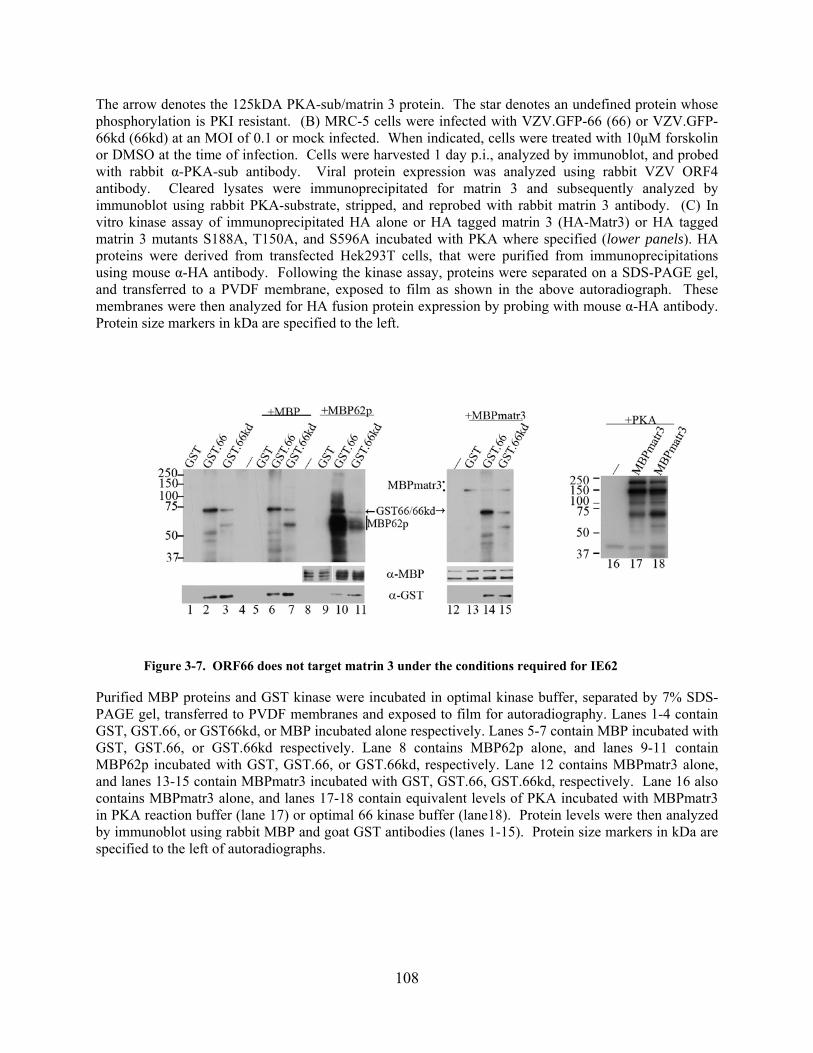
The arrow denotes the 125kDA PKA-sub/matrin 3 protein. The star denotes an undefined protein whose phosphorylation is PKI resistant. (B) MRC-5 cells were infected with VZV.GFP-66 (66) or VZV.GFP-66kd (66kd) at an MOI of 0.1 or mock infected. When indicated, cells were treated with 10μM forskolin or DMSO at the time of infection. Cells were harvested 1 day p.i., analyzed by immunoblot, and probed with rabbit α-PKA-sub antibody. Viral protein expression was analyzed using rabbit VZV ORF4 antibody. Cleared lysates were immunoprecipitated for matrin 3 and subsequently analyzed by immunoblot using rabbit PKA-substrate, stripped, and reprobed with rabbit matrin 3 antibody. (C) In vitro kinase assay of immunoprecipitated HA alone or HA tagged matrin 3 (HA-Matr3) or HA tagged matrin 3 mutants S188A, T150A, and S596A incubated with PKA where specified (lower panels). HA proteins were derived from transfected Hek293T cells, that were purified from immunoprecipitations using mouse α-HA antibody. Following the kinase assay, proteins were separated on a SDS-PAGE gel, and transferred to a PVDF membrane, exposed to film as shown in the above autoradiograph. These membranes were then analyzed for HA fusion protein expression by probing with mouse α-HA antibody. Protein size markers in kDa are specified to the left.
Figure 3-7. ORF66 does not target matrin 3 under the conditions required for IE62
Purified MBP proteins and GST kinase were incubated in optimal kinase buffer, separated by 7% SDS-PAGE gel, transferred to PVDF membranes and exposed to film for autoradiography. Lanes 1-4 contain GST, GST.66, or GST66kd, or MBP incubated alone respectively. Lanes 5-7 contain MBP incubated with GST, GST.66, or GST.66kd respectively. Lane 8 contains MBP62p alone, and lanes 9-11 contain MBP62p incubated with GST, GST.66, or GST.66kd, respectively. Lane 12 contains MBPmatr3 alone, and lanes 13-15 contain MBPmatr3 incubated with GST, GST.66, GST.66kd, respectively. Lane 16 also contains MBPmatr3 alone, and lanes 17-18 contain equivalent levels of PKA incubated with MBPmatr3 in PKA reaction buffer (lane 17) or optimal 66 kinase buffer (lane18). Protein levels were then analyzed by immunoblot using rabbit MBP and goat GST antibodies (lanes 1-15). Protein size markers in kDa are specified to the left of autoradiographs.
108

3.4.6 Matrin 3 localization in VZV-infected cells
Protein kinases often induce subcellular localization changes following phosphorylation of their
target substrates in order to modulate function. This is certainly exemplified by ORF66 kinase
phosphorylation of IE62, where phosphorylation induces radical changes in subcellular
localization. In this case, IE62 is excluded from the nucleus and localized in the cytoplasm
which is a requirement for IE62 tegument incorporation (52, 54, 98). We then asked whether
ORF66-specific phosphorylation of matrin 3 lead to changes in the protein’s intracelluar
localization. MRC-5 cells were infected with VZV.GFP-66Rsc, VZV.GFP-66kd, or mock
infected. In mock-infected cells matrin 3 exhibited dominantly nuclear localization, with
exclusion from what appeared to be nucleoli (Fig. 3-8a (panels i and iv) and 3-8d (panel i)). This
is in accordance with previously described subcellular localizations for matrin 3 in other cell
types. In infected cells expressing ORF66, matrin 3 was largely retained in the nucleus with a
slightly more diffuse intranuclear localization (Fig. 3-8d (panel ii)), even late in infection as
depicted in the shown synctia (Fig. 3-8 b (panels i and iv)). In cells expressing the kinase-
inactive ORF66kd, matrin 3 was still retained in the nucleus but many cells displayed multiple
areas within the nucleus that were devoid of matrin 3 (Fig. 3-8c (panels i and iv) and Fig. 3-8d
panel iii), yet were filled with IE62 and followed early replication compartment patterns.
Additionally, few cells also exhibited matrin localizing in 2-10 nuclear punctae. As reported
previously, IE62 was cytoplasmic - nuclear excluded in ORF66-expressing cells (Fig. 3-8b panel
iii & iv) and exclusively nuclear in ORF66kd-expressing cells (Fig. 3-8c panel iii & iv).
Furthermore, GFP.66 showed both cytoplamic and nuclear localization (Fig. 3-8b panel ii and
iv), compared to the more predominant nuclear localization of GFP.66kd (Fig. 3-8c panel ii and
iv). Overall, our immunofluorescence analysis indicates that matrin 3 localizes to the nucleus
109

late into VZV infection, and presents subtle differences in its intranuclear localization when
kinase activity is abrogated.
Figure 3-8. VZV infection leads to subtle differences in matrin 3 cellular localization
Immunofluorescence analysis of MRC-5 cells (A) mock infected or infected with (B) VZV.GFP-66Rsc or (C) VZV.GFP-66kd at an MOI of 0.003. Cells were fixed with 4% paraformaldehyde 3 days p.i. and immunostained with rabbit anti-matrin 3 antibody (A-C (panel i) detected with α-rabbit Alexa Fluor 546, D) or mouse anti-IE62 (A-C (panel iii) detected with α-mouse Alexa Fluor 647. ORF66 expression was determined by GFP autofluorescence (A-C (panel ii). The merge image (A-C (panel iv)) are overlays of matrin 3 (gray), GFP (green), IE62 (red), and nuclei stained with Hoechst dye (blue). (D) Single cell representative images depicting matrin 3 localization. Infection conditions are indicated in the image. Fluorescence images were taken using a 60X objective.
110

3.4.7 Matrin 3 efficient nuclear retention is dependent on US3 kinase expression
Our previous data had indicated that ORF66/US3 – dependent matrin 3 phosphorylation was
conserved for the alphaherpesvirus, and shared a common phosphorylation site. We extended our
immunofluorescence analysis to HSV-1 and PRV, to investigate whether US3 kinase activity had
any influence on matrin 3 cellular localization. MRC-5 cells were synchronously infected with
HSV-1, HSV-1 expressing GFP.US3.5 (HSV.US3.5), HSV that did not express US3
(HSV.ΔUS3), PRV, PRV that did not express US3 (PRV.ΔUS3) or mock infected. At 18hrs
p.i., cells expressing HSV US3 (Fig. 3-9 B) or US3.5 (Fig. 3-9C) displayed matrin 3
predominantly in the nucleus (Fig. 3-9B,C panels i & iv), although many instances of nuclear
condensation were visible for US3.5-expressing cells. Localization did not differ substantially
from mock-infected cells (Fig. 3-9A) except matrin 3 exhibited a more diffuse nuclear
localization, in addition to what appears to be loss of most detectable nucleoli. Redistribution of
nucleolar proteins have been reported previously in HSV infected cells (19). However, when
HSV US3 was not expressed, cytoplasmic accumulation of matrin 3 were consistently observed
in addition to nuclear localization (Fig. 3-9D panel i & iv). To visualize HSV infection, an ICP4
IF stain was included in our analysis. HSV-1 ICP4 was observed in all cells, and exhibited a
mainly nuclear localization in all infected cells (Fig. 3-9B-Dpanel iii), with some cytoplasmic
accumulations in HSV.ΔUS3 infected cells (Yee, MB unpublished results). GFP.US3.5 located
in the cytoplasm and nucleus of infected cells (Fig. 3-9B panel ii), and GFP alone was expressed
from the US3 native promoters in HSV.ΔUS3 (Fig. 3-9C panel ii). In PRV infected cells 11 hrs
p.i., matrin 3 again displayed a mostly nuclear localization late in infection (Fig. 3-9E panel i &
iv) although some cytoplasmic forms where noted. Interestingly, in the absence of PRV US3
expression, matrin 3 exhibited nuclear exclusion in multiple cells, with accumulation now in the
111

cytoplasm of infected cells (Fig. 3-9F panel i & iv). All cells were infected as displayed by
UL34 stain (Fig.3-9 E &F panel iii), and GFP expression was evident from the PRV US3 native
promoter in PRV.ΔUS3- infected cells (Fig. 3-9F panel ii). Our results reveal that both HSV and
PRV US3 modulate matrin 3 cellular localization, and US3 expression is necessary for efficient
matrin 3 nuclear localization.
Figure 3-9. HSV-1 and PRV US3 kinase expression leads to matrin 3 nuclear retention or shuttling
to the nucleus during infection
112

Immunofluorescence analysis of MRC-5 cells (A) mock infected or infected with (B) HSV-1, (C) HSV.US3.5, (D) HSV.ΔUS3 (E) PRV, or (F) PRV.ΔUS3 at an MOI of 5. Cells were fixed with 4% paraformaldehyde 18hrs p.i.(HSV) or 11hrs p.i (PRV), then immunostained with rabbit α-matrin 3 antibody (A-F (panel i)) detected with α-rabbit Alexa Fluor 546 (HSV) or α-rabbit Alexa Fluor 555 (PRV), mouse α-HSV ICP4 (A-D (panel iii)) detected with α-mouse Alexa Fluor 647, or goat anti-PRV UL34 (E & F (panel iii)) detected with α-goat Alexa Fluor 647. GFP-US3.5 expression or US3 promoter activity in HSV.ΔUS3 and PRV.ΔUS3-infected cells was determined using GFP autofluorescence (A-F (panel ii)). The merge image (A-F (panel iv)) are overlays of matrin 3 (gray), GFP (green), ICP4 (magneta) or PRV UL34 (magneta), and nuclei stained with Hoecht dye (blue). Fluorescence images were taken using a 60X objective.
3.5 DISCUSSION
Increasingly, the multifunctional roles for ORF66 kinase are realized, as well as the kinases’ cell-
type dependent importance for proper VZV growth and replication in the hosts. This is in
alignment with the multifunctional nature of the alphaherpesvirus US3 kinases. ORF66 kinase
activity is key in regulating IE62 cellular localization & tegument inclusion, and modulates
multiple cellular activities involved in immune evasion strategies, as well as induces
phosphorylation of proteins involved in silencing viral gene expression. Additionally, ORF66 is
critical for VZV growth in specific cell types. Nevertheless, many specific protein targets are yet
to be determined. In this work, we reveal that matrin 3 is an indirect phosphorylation target of
ORF66, and is a conserved target of the ORF66 orthologues, HSV-1 and PRV US3 kinases. Our
data also suggests that a function of this phosphorylation is to maintain matrin 3 nuclear
localization. This is the first report, to our knowledge, of matrin 3 protein modification by any
virus.
Matrin 3 is a 847 a.a. 125 kDa slightly acidic protein with a predicted weight of 95kDa
(7). The N-terminus contains a large number of free hydroxyl groups (26 of the first 100
113

residues) as characteristic of lamin proteins, in addition to an acidic C-terminus found in many
nuclear proteins, and two tandem RNA recognition motifs (7). Specific functions for matrin
proteins are mostly unknown, yet there are reports proposing intriguing roles for matrin 3 in host
cell processes. Recent studies have implicated it as a component of functional nuclear
microenvironments of chromatin replication & transcription (123). Moreover, matrin 3 has been
found to interact with hnRNP-L, an RNA splicing regulator, & SAFB, involved in RNA
processing & chromatin organization (229). Matrin 3 can bind DNA at scaffold/matrix
attachment regions (S/MAR) sites which influence chromatin structure by associating with
matrix-binding proteins that anchor chromatin into loop domains. Interestingly, matrin 3 is also
part of a three protein complex including p54nrb and PSF that prevent the nuclear export of
nuclear dsRNAs that are hyperedited by members of the ADAR (adenosine deaminase that act
on RNA) enzyme family (45, 231). In rat neurons, activation of the glutamate receptor, NMDA
by PKA induces matrin 3 phosphorylation. This phosphorylation in turn leads to matrin 3
degradation and subsequent neuronal death (64). Lastly, matrin 3 has been linked to the genetic
disorders such as one type of autosomal-dominat distal myopathy, VCPDM, and found in
decreased levels in fetal Down’s syndrome brains (13, 192).
The goal of our analysis was to uncover novel phosphorylation targets of ORF66
kinase and get a clearer understanding of the roles of ORF66 in VZV infection. The
phosphorylation motif for ORF66 is unknown, yet the target sites in IE62 are well-described.
Based on the similarity between these sites and that of the cellular kinase PKA phosphorylation
motif, we took advantage of a phosphospecific PKA-substrate antibody to find novel ORF66
protein targets. Interestingly, we found that these alphaherpesviruses varied widely in number
and molecular weights sizes of bands that were induced with infection (Figure 3-1). These
114

proteins may be direct ORF66 or US3 targets, direct PKA targets, or downstream
phosphorylation targets of activated PKA signaling pathways. HSV-1 displayed the strongest
induction of phosphorylation of PKA-substrate proteins, agreeing with previous findings that
US3 induced the phosphorylation of proteins fitting PKA substrate specificity, in addition to
their model which predicts that HSV-1 infection activates PKA and may share protein substrates
(9). VZV is reported to increase expression of the PKA catalytic subunit as well as induce
detection of PKA-substrate bands. Although, some of the phosphorylated sizes were of similar
molecular weight, it is unknown whether the same proteins are detected as these proteins have
not been identified (49). Notably, in our study a novel ORF66-dependent 125 kDa PKA-
substrate was the dominant protein present. Importantly, although profiles between the
alphaherpesviruses differed, there was conservation of ORF66/US3-dependent phosphorylation
of the 125 kDa protein/matrin 3 (Figure 3-1, 3-3, & 3-4). This leads to the prediction that
because phosphorylation of this specific protein is conserved, it may be important for
alphaherpesvirus replication. Also in figure 3-1, HSV.ΔUS3-infected cells, a faster migrating
band for gB protein was observed and likely represents an unphosphorylated gB fraction (86,
221). gB migrates at a similar molecular weight as matrin 3, and this may account for some
phosphorylation seen in HSV-1.ΔUS3 in the 125kDa region of cell extracts on longer exposures
of autoradiographs (Figure 3-4a). Some gB phosphorylation in cells infected with HSV-1
expressing inactive US3 has been reported previously using PKA-substrate antibody (86).
We also observed that expression of ORF66 using adenovirus vectors, strongly
induced phosphorylation of the 125 kDa protein, indicating that this band represented a cellular
protein (Figure 3-2). Other host proteins bands were detected with expression of ORF66, albeit
less strongly than the 125kDa protein but are of interest for future identification. Our mass
115

spectrometry analysis identified the protein as matrin 3. Interestingly, the results for this region
of the gel did not identify additional proteins in this region except for keratin contaminants
which are common for LC/MS/MS methods. Identification of matrin 3 and the observation that
for all 3 viruses this nuclear matrix protein is phosphorylated in an ORF66/US3 – dependent
fashion provides much evidence that matrin 3 is the 125kDa-PKA-substrate protein. While
matrin 3 has not been linked as a modified protein of any virus, interaction of virus with matrix
proteins have been described for HIV, SV40, PRV, HCMV and HPV (8, 66, 142, 182, 187, 201),
and notably for HSV-1 (16-17, 23, 138, 223, 226). For most of our analysis at least two forms
were detected in whole cell extracts and 2 or more in IP fractions. Importantly, both matrin 3
and p-PKA substrate antibodies detect a stronger faster migrating band, and a slower migrating
125kDa band in cell extracts. Because IP forms are detected by the p-PKA substrate antibody,
we believe that these likely represent different phosphorylated forms. The fact that IP fractions
are enriched for matrin 3, combined with the use of specific PKA-substrate antibody, we predict
leads to additional forms of this protein revelead in this fraction. Differentially phosphorylated
matrin 3 isoforms have been documented in rat cells, with a 130kDa and 123kDa matrin 3
localizing to the nucleus and extracellular compartments, respectively (75). Differing matrin 3
forms with slightly different isoelectric points have been isolated from neurons (64). Whether a
specific function is attributed to these forms or whether they represtent phosphoryation status in
the process of turnover or degradation fragments is unclear. Two transcricpts have been recorded
for matrin 3, differing at the 5’UTR but encoded the same protein (NCBI). Nevertheless,
alternate protein modifications should still be considered.
One of the objectives for analyzing 66-dependent phosphorylation of matrin 3
serine/threonine to alanine mutants was to explore potential functions of this specific matrin 3
116

targeting. Matrin 3 contains two recognition RNA recognition motifs, matrin-type zinc fingers,
and potential nuclear localization signal (a.a. 710-718). We chose candidate ORF66 sites with 2
or more basic residues adjacent and N-terminal to a serine or threonine. Interestingly, S592, 596
and 598 were within the human homologue region of the identified chicken matrin 3 bipartite
nuclear localization signal (77). Our data shows that the T150 mutation to alanine drastically
dimished phosphorylation detected for the HA-tagged matrin 3 mutant (Figure 3-5.). However,
there was still some phosphorylation detected for this mutant indicating the possibility for
additional matrin 3 residues that are phosphorylated in a 66-dependent manner. It is intriguing
that different alphaherpesviruses would share a common target and phosphorylation site on that
target protein; this may be due to the nature of similar phosphorylation target motifs for the US3
kinase. This would predict that this phosphorylation would have similar outcomes on matrin 3
function.
Activation of PKA-substrates during viral infection has been reported for HCV (55), and
activated PKA pathways have been exploited by many viruses including adenoviruses, HIV
(114, 203), HCMV (90), and HSV (222) to enhance infection. However our data suggest that
PKA does not directly phosphorylate matrin 3 at the T150 major phosphorylation site (Figure 3-
6), although matrin has been shown here and in previous studies to be a PKA target. Inhibition
of PKA with a highly specific inhibitor, 14-22 amide, did not prevent detection of the
phosphorylated 125kDa protein, although other PKA-substrates proteins were no longer
detected. Activation of PKA using forskolin also did not increase matrin 3 phosphorylation and
minimally increased its phosphorylation in ORF66kd-expressing infected cells (Figure 3-6.).
Moreover, in vitro phosphorylation of HA-Matr-T150A mutant by PKA did not differ from the
non-mutated form of matrin 3, yet there was reduced phosphorylation for HA-Matr-S188A.
117

These results indicate that PKA does phosphorylate matrin 3 at the S188 site in vitro and
potentially other sites, but does not target the T150 site which is the 66-dependent matrin 3
phosphorylation site. Thus these data suggests that PKA or PKA signaling pathways have a
minor role in the 66-dependent phosphorylation of matrin 3. We did note a prospective 60kDa
likely viral protein that was phosphorylated in response to forskolin treatment in ORF66kd
expressing cells. This indicates that there is a protein which is phosphorylated in response to
activation of PKA pathways that is dependent on VZV infection. We then explored whether
matrin 3 was directly phosphorylated by ORF66 in vitro. Surprisingly, ORF66 did not
phosphorylate MBPmatr3 under the same in vitro kinase assay conditions for which ORF66
phosphorylates MPP62p (Figure 3-7), while PKA did phosphorylate matrin 3 under these same
conditions. PKA phosphorylation of matrin 3 may reflect targeting of other S/T sites other than
those sites phosphorylated (T150) in response to ORF66 expression. This data does suggest that
ORF66 does not directly phosphorylate matrin 3. However, this does not exclude the possibility
that matrin 3 may be phosphorylated by ORF66 under different kinase assay conditions, or
require an additional nuclear protein; this will be the subject of further investigation.
ORF66 kinase has demonstrated the ability to regultate VZV IE62 localization and this
determines IE62 inclusion in the virion tegument. HSV US3 kinase activity is known to cause
the redistribution of a nuclear matrix protein, lamin A/C, involved in breaching the laminar
network needed for proper nucleocapsid envelopment yet still maintaining laminar structure
(138). In our immunofluorescence study, matrin 3 remained nuclear in VZV infection with a
more diffuse localization of the protein compared to the mock-infected cells. In contrast, many
cells in VZV.GFP-66kd expressing cells displayed regions devoid of matrin 3 of which were
filled with IE62 protein (Figure 3-8). This is reminiscent of the findings for lamin A/C that
118

normally forms a reticular pattern in infected cells, yet in the absence of US3 kinase activity
leads to larger intranuclear regions devoid of lamin A/C (138). Thus, similarly to what happens
to lamin A/C distribution, ORF66 may also be responsible for maintaining a balance between
disruption of a major component of the nuclear matrix yet retaining matrin 3 at replication
compartments, where viral replication and transcription occurs. This implies that ORF66 kinase
activity may be necessary for matrin 3 interaction with components of early replication
compartments, such as IE62. HSV infection also causes reorganization of another nuclear matrix
protein, nuclear mitotic apparatus (NuMA) protein and is excluded from replication
compartments, with redistribution dependent on viral DNA synthesis (226). In HSV-1 and PRV
immunofluorescence analysis, matrin 3 showed a diffusely nuclear localization in infected cells,
as well as in HSV.EGFP-US3.5 expressing cells. In contrast, subtle yet reproducible
cytoplasmic accumulation of matrin 3 was visible in cells that did not express US3 kinase in
HSV-1. These differences were likely not due to apoptotic cells, as HSV-1 US3.5 like US3-null
viruses do not possess the US3 function in inhibiting apoptosis (162), yet matrin 3 was still
predominantly nuclear. Cytoplasmic accumulation were even greater in PRV-infected cells that
lacked US3 with some cells displaying nuclear exclusion of matrin 3 (Figure 3-9), and for both
viruses the difference between US3 and US3-non-expressing infected cells was most
distinguishable late in infection. Together these findings point to a role for the US3s in the
active retention of matrin 3 in the nucleus or increased shuttling of matrin 3 to the nucleus.
Regulation of matrin 3 cellular localization by phosphorylation has been reported previously
where the use of a general kinase inhibitor staurosporine leads to matrin 3 accumulation in the
cytoplasm (77). It is possible that phosphorylation by US3 may be key for matrin 3 interaction
with cellular proteins that are retained in the cytoplasm. In the absence of US3, matrin 3 may be
119

lost to the cytoplasm due to the nuclear component reorganization that occurs in HSV infection .
Secondly, matrin 3 may be interacting with a viral protein that is also a US3 target which
localizes to the cytoplasm late in infection in the absence of US3 expression. The nuclear
exclusion seen in PRV.ΔUS3 may be explained by this latter scenario or an active export of
matrin 3 to the cytoplasm. We also observed that in infection with VZV, HSV-1 & PRV, global
matrin 3 levels remained unchanged as visualized by western blot. This suggests that matrin 3 is
a very stable protein and supports the idea that matrin 3 is a necessary protein for
alphaherpesvirus infection. This will be an interesting question to be addressed for future siRNA
studies.
Our studies thus far have indicated that matrin 3 is specifically phosphorylated in an
ORF66/US3 kinase-dependent manner, and the kinases’ function in either modulating
intranuclear localization or nuclear-cytoplasmic shuttling. This leads us to question the potential
functions of matrin 3 in alphaherpesviral infection, and how the ORF66/US3 specific matrin 3
phosphorylation contributes to this function. However, matrin 3 cellular functions are not clearly
defined but recent evidence points to a multifunctional nature for this protein. Nuclear matrix
has long been reported to be the site of organization for numerous nuclear processes such as
DNA replication, DNA loop attachment, transcription and RNA processing (3, 11-12, 83).
Matrin 3 specifically locates in close proximity to nascent DNA replication and transcription
zones within the nucleus. Thus, as matrin 3 is a major nuclear matrix protein, is may serve as a
platform for these essential genomic functions. Furthermore, we can conceive that matrin 3 is
also needed to be present at sites of viral replication and transcription. Recently, a comparable
role has been proposed for lamin a/c as serving as a scaffold for HSV genomes and nuclear
factors that reduce heterochromatin assembly on viral promoters (194). Matrin may also serve
120
as part the nuclear matrix that is involved as part of the site of capsid assembly due to findings
that capsid proteins localized to nuclear matrix fractions (16, 23). If ORF66 is involved in capsid
assembly, then this may explain the reduced intranuclear assembly of nucleocapsids in T cells
from SCID-hu T cell xenografts infected with VZV lacking ORF66. Matrin 3 is also involved as
part of a complex that retains hyperedited nuclear dsRNAs in the nucleus and to prevent their
export to the cytoplasm for translation. dsRNAs have been identified for HSV (84). The
ORF66/US3 kinases may potentially be involved in regulating interaction of matrin 3 with the
p54nrb-PSF complex and modulate the balance between nuclear accumulation and release of
viral dsRNA to the cytoplasm. Lastly, matrin 3 degradation in neurons leading to neuron death
may be in response to the hyperphosphorylatin of matrin 3 detected using PKA-substrate
antibody. This leads to the speculation of whether ORF66 phosphorylates matrin 3 leading to its
degradation during VZV reactivation in neurons, and in turn causing the neuronal death or
damage that is sometimes associated with herpes zoster (153).
In summary, this study describes a novel cellular target for ORF66 kinase, the
essential nuclear matrix protein –matrin 3, that is specifically phosphorylated in a 66-dependent
manner through a non-PKA pathway. This is a conserved target for two other alphaherpesvirus
US3 kinases from HSV-1 and PRV. This phosphorylation functions in modulation of matrin 3
intranuclear localization, or in the case of HSV and PRV, in efficiently maintaining matrin 3
nuclear localization. This is the first report of a viral manipulation of this nuclear matrix
protein.
121

4.0 FUTURE DIRECTIONS & GENERAL SUMMARY
Overall, the results presented here offer new insights for roles in ORF66 in VZV biology as well
as revealing a novel host protein target with potentially important implications for ORF66
manipulation of cellular biology. Previously, ORF66 was regarded as a non-essential protein for
VZV replication, yet it was obvious that this kinase must be important for VZV pathogenesis as
it specifically targeted the VZV major transcriptional activator, IE62, and its direct
phosphorylation determined IE62 virion incorporation. In recent years, VZV ORF66 has
demonstrated that it is indeed a multifunctional protein in that its kinase activities are aimed at
both viral and cellular proteins. These diverse roles include downregulation of surface MHC-1
in fibroblasts, modulation of interferon and apoptosis pathways, and targeting of HDAC 1 and 2.
Furthermore ORF66 is important for growth in human T cells, which are believed to be key for
VZV pathogenesis as they can transfer virions to sites in the skin in SCID-hu mouse models.
Corneal Fibroblasts
The work presented here describes the first non-lymphocytic cell line, primary corneal
fibroblasts (54), for which ORF66 kinase activity is important for VZV growth. Although our
studies found that the basis for impaired growth was not an inability to phosphorylate IE62 at the
major S686 site nor increased apoptosis, this study establish a new cell culture model for
exploration of cell-type dependent functions of ORF66. For future studies, the IFN-α/β
pathways have yet to be explored. It is thought that inhibition of IFN signal transduction may
122

promote the survival of infected tonsillar T cells. Thus, it is possible that PCF cells express
higher levels of IFN, exerting an antiviral effect when ORF66 is inactive or not expressed.
Human corneal epithelial cells have been found to increase transcriptional expression of various
cytokines including IFN-β in response to HSV-1 infection in vitro. Interestingly, addition of
recombinant human IFN-α2a can inhibit VZV replication in human corneal stromal fibroblasts
cultures. Thus, investigating whether there are differences in cytokine transcription induction in
PCF cells compared to another VZV permissive cell line could point out possible ORF66
substrates.
In infected PCF cells, small VZV foci were produced when ORF66 was not expressed or
inactivated. This suggests that there may be a block in late gene expression, therefore analyzing
at what point the delay is occuring may highlight potential viral proteins that are important VZV
targets for efficient PCF infection. Morevover, PCF expressing 66kd may have deficiencies in
proper virion maturation or trafficking during nuclear egress. gB is a leaky late protein that is
involved in nuclear egress of virion in HSV-infected cells and is phosphorylated by US3 kinase
in addition to likely cellular kinases at the same site . Interestingly VZV gB contains a potential
candidate ORF66 phosphorylation motif at RSRRS495VP and potentially another at the C
terminus.
Ultimately, the lack of growth for 66-inactive VZV in PCF is cell-type dependent and the
likelihood is that there is a cellular protein that aids in ORF66 kinase functions that is absent or
altered in PCF cells, or alternatively an efficient VZV antagonist that is present or altered in this
cell type. Hence, a focus should be placed in outlining what signaling pathways and specific
cellular proteins are targeted in VZV- infected versus VZV expressing 66kd in PCFs as
compared to more permissive cell lines.
123

Viral cell Targets
While ORF66 has been implicated in diverse cell processes, only one direct target
is known for this kinase, IE62. Thus a major endeavour of my studies was to find novel ORF66
cellular or viral protein targets. As a result, a set of candidate viral proteins were chosen that fit
a phosphorylation motif based on IE62 direct targeting for further investigation using in vitro
kinase assays (Figure 4-1) . Using aforementioned MBP fused to viral proteins and GST-66 and
GST-66kd, results showed that both VZV ORF4 and ORF9 were direct ORF66 targets in vitro
(Figure 4-2). Interestingly both of these proteins are viral tegument proteins and both contain a
proline at the +2 position following the putative phosphorylated serine/threonine - both
characteristics shared with the IE62 protein. ORF 4 is an IE phosphoprotein and major
transactivator, post-transcriptional regulator of gene expression, and partner in mRNA export.
IE4 is divided into four regions, one which is an arginine rich region subdivided into 3 units –Ra,
Rb, & Rc. This argine rich region is important for transactivation properties & protein-protein
interactions. Intriguingly, the putative phosphorylation site is located within the Rb region
which binds strongly to RNA in vitro (151), and is adjacent to IE4 nuclear localization signal.
Therefore, it is important that further studies pinpoint the site of IE4 phosphorylation by ORF66
to begin to understand possible functions of this phosphorylation. ORF9 is a tegument protein
and displays limited homology to its homologue HSV VP22. This protein is not well
characterized but has been reported to bind IE62 in complex with tubulin, possibly as a means of
recruitment for virion incorporation (26). Interestingly, my studies with HA tagged ORF9
indicate that ORF9 displays multiple different localizations. This same observation has been
reported for VP22 (126). Namely for ORF9 - cytoplasmic accumulations, perinuclear speckling,
124

rod-shaped filaments were noted. When cells were co-transfected with EGFP66, ORF9
predominantly displayed a cytoplasmic localization (data not shown). This suggests that ORF66
can modulate ORF9 subcellular localization. In VZV infection, ORF9 displayed cytoplasmic,
nuclear, and at times rod shaped filaments as well as what appeared to be mitotic chromatin
binding, yet it was unclear if there were localization differences in kinase-inactive cells. In
VP22, these rod-shaped bundles reflect VP22 binding to microtubules causing their stabilization
and in turn bundling. Thus, this implicates ORF66 in altering ORF9 intracellular localization,
interactions with microtubules and any downstream effects resulting from that disruption.
Challenges were encountered in in vivo phospholabeling of transfected cells, since ORF4 and
ORF9 are phosphoproteins in addition to the observation that ORF66 seems to increase amounts
of these proteins, therefore alternate methods should be considered. However, investigation of
these interactions may reveal undescribed viral protein targets for the alphaherpesvirus US3
kinases as well as further understandings of the role of the ORF66 kinase in VZV infection.
125
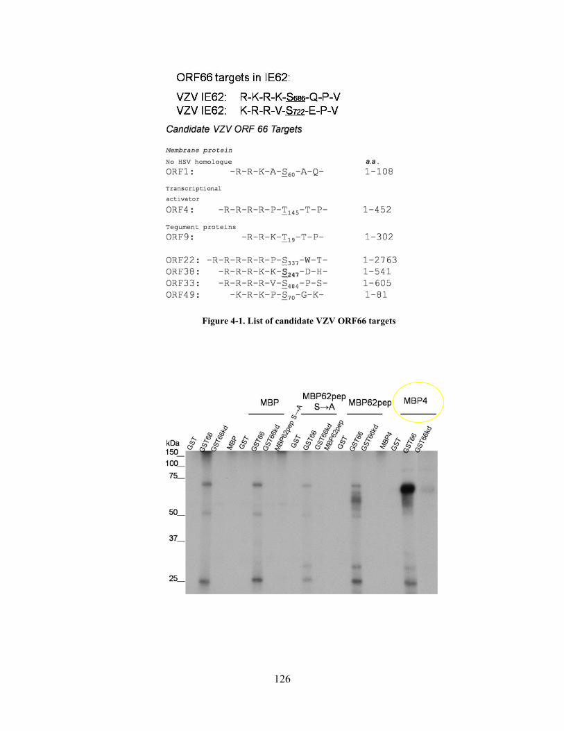
Figure 4-1. List of candidate VZV ORF66 targets
126

Figure 4-2. VZV ORF66 targets ORF 4 & ORF9 in vitro
Autoradiograph showing results of an in vitro kinase assay. Maltose binding protein (MBP) ORF9 and ORF4 fusion proteins were incubated alone or in equal amounts with GST, GST66, or GST66kd under optimal ORF66 kinase assay condtions described in section 3.3.9.
Actin.
Viruses interact with cytoskeletal elements at almost every step of the infectious cycle.
The cytoskeleton itself is a diffusion barrier that must be overcome for viruses to transport itself
within the cell (197). Actin is a globular structural protein that polymerizes into helical actin
filaments forming a network within the cell. This dynamic structure provides mechanical
support, determines cell shape, and is involved in cell mobility and formation of cell-cell
junctions. Viruses often depolymerize actin to alter the diffusion properties of the cytoplasm and
facitilitate viral egress or induce actin polymerization to propel viruses to adjacent cells (197).
Actin has also been implicated in nuclear activities such as transcription, chromatin remodeling
127

and modeling of the nucleoskeleton (170). Interstingly, HSV, MDV and PRV US3 kinases all
manipulate cellular signaling pathways in restructuring actin networks to form projections or
network disassembly (See Section 1.4.6.4). In PRV, the US3 kinases actually target PAK1 and
PAK2 cellular kinases which in turn have roles in the Rho GTPase signaling pathways involved
in actin dissaembly and lamellopodia and filopodia formation. This concurs with a common
theme as exemplified by US3 relationship with PKA, ORF66 phosphorylation of HDACs, and
matrin 3; ORF66/US3 functions in direct phosphorylation of certain targets, but also seems to
engage key host proteins for a common downstream effect especially when modulating cellular
processes. Results showed that VZV-infected cells disorganized normally stratified filamentous
actin (F-actin) filaments 24 hrs p.i., yet when ORF66 was kinase-inactivated actin cytoskeletal
networks remained mainly intact (Figure 4-3). ORF66 expressed in the absence of other VZV
proteins using Ad.GFP-66 or Ad.GFP-66kd, induced cell rounding and cellular extensions when
ORF66 was functional and loss of stratified F-actin, yet ORF66kd expressing cells did not
significantly alter overall cell morphology and F-actin filamentous where readily detected (data
not shown). These findings strongly suggest that ORF66 does alter cell signaling pathways
responsible for the dissolution of actin filaments. Initial studies should be directed at first
delineating whether these effects are not solely because of differences in degree of infection
between VZV and 66-inactive VZV, or in the case of adenoviruses a phenomenon due to
apoptosis or protein overexpression, in addition to a concentration on what components of Rho
GTPase pathways are modulated by 66 kinase activity. Single-cell live cell imaging experiments
using VZV with fluorescently tagged capsid proteins in a 66 or 66kd background, together with
delivered commercially available fluorescent phallodin or transiently expressed actin-GFP may
shed light on whether actin projections are produced in VZV with potential roles for intercellular
128

spread. Because, VZV seems to place heavy restrictions on production of cell free virions, it is is
highly probable that the virus would utilize the cytoskeletal network for intercellular spread and
hijacking this cellular component is amongst the shared functions of the ORF66/US3 kinase
family.
Figure 4-3. VZV ORF66 kinase activity induces actin rearrangement in infected cells
MRC-5 cells were infected with VZV.GFP-66 or VZV.GFP-66kd. Cells were fixed 24hrs p.i and fixed with 4% paraformaldehyde and stained with Alexa Fluor-546 phalloidin to visualize for F-actin (red). ORF66/66kd is visualized by GFP fluorescence (green). PKA-substrates & Matrin 3
Our investigation for ORF66 phosphorylation targets using a phosphospecific PKA-
substrate as a tool to identify potential candidates fitting an IE62-like motif, lead to
identification of Matrin 3. VZV ORF66 expression did not lead to the phosphorylation of
129

multiple PKA-substrate proteins as noted for HSV and PRV, yet ORF66 kinase expression did
lead to detection of protein bands of approximately 40kDa, 55kDa, and diffuse set of bands of
65-75kDa. Identification of these proteins by LC/MS in the context of VZV infection and
adenovirus infection may highlight interesting new ORF66 viral or host targets. One possibility
is that these proteins are all part of a signaling pathway that is activated by ORF66, or
components of different pathways with PKA as a common upstream activator, although our
forskolin studies would indicate that the latter is not the case. Using this antibody in IF studies,
p-PKA-substrates were strongly noted in the nucleus in VZV.GFP-66-infected cells as opposed
to mostly cytoplasmic accumulation in VZV.GFP-66kd. Although this may represent the matrin
3 target, it may also indicate that ORF66 is targeting other nuclear PKA-substrate proteins
(Figure 4-4). Use of this antibody in IF maybe useful in future studies to further investigate
ORF66 targets.
A separate observation made during this investigation was that purified GST tagged
ORF66 was detected as a PKA-substrate in western blots, but not kinase-inactive ORF66 (Figure
4-5). This suggests that ORF66 is a PKA-substrate or that the antibody may be detecting ORF66
autophosphorylation . Similarly, this antibody was used to reveal the HSV -1 US3
autophosphorylation motif, and this self regulation was needed for some of US3’s physiological
functions (87, 181). Thus, this tool may also serve to find phosphorylation sites on ORF66
which may impart regulation of the multiple functions of this kinase. Interestingly, the antibody
also detected a PKA-substrate band within the gel region for which MBP62p is usually detected
when MBP62p or MBP62pDM and GST66 were incubated together. This suggests that the
antibody detected a phosphorylated residue on MBP62p other than the ORF66 phosphorylation
sties – S686 and S722.
130

Figure 4-4. VZV ORF66 kinase activity induces greater phosphorylation of nuclear PKA-substrate
proteins.
MRC-5 cells were infected with VZV.GFP-66 or VZV.GfP-66kd and fixed 2days p.i. with 4% paraformaldehyde. Cells were then immunostained with rabbit anti-p-PKA-substrate antibody and visualized using Alexa Fluor 546 anti-rabbit and fluorescence microscopy. ORF66 or ORF66kd expression is visualized by GFP fluorescence (green) and nuclei are stained with Hoechst dye (blue)
131

Figure 4-5. GST66 is detected by PKA-substrate antibody.
Purified GST, GST66 or GST66kd were subjected to an in vitro kinase assay alone or incubated with MBP62p or MBP62pDM (62p double mutant-S686A, S722A). SDS-PAGE separated proteins were transferred to PVDF membranes and probed with rabbit- α-PKA-substrate and visualized by autoradiography for antibody detected proteins. The star denotes MBP62 or 62-DM peptide.
Lastly, this work has uncovered the nuclear matrix protein, matrin 3, as a novel ORF66
cellular protein phosphorylation target. This is a first report of any virus targeting this
component of the nuclear matrix, and reveals a common target for the alphaherpesvirus US3
kinases. The latter point is highly indicative that targeting of this protein is important at a point
in the alphaherpesvirus life cycle. Moreover, this work strongly suggests that this induced matrin
3 phosphorylation is involved in regulation of matrin 3 subnuclear localization in VZV-infected
cells, or matrin 3 nuclear retainment late into infection in HSV-1 and PRV-infected cells.
Although ORF66 and US3 kinase functions are not essential for viral replication in most cell
culture, our studies with corneal fibroblasts, and studies in SCID-hu xenografts amongst others
(56, 81, 181, 186), demonstrate that ORF66/US3 functions indeed are important for optimal viral
growth when models closer to a natural host are used. Hence, future studies should be directed
at understanding the role of matrin 3 in VZV infection, potentially by using matrin 3 siRNA.
While data demonstrated that the matrin 3 T150 a.a. was the major ORF66/US3 specific
132

phosphorylation site, there was a small fraction of phosphorylation detected in HA-Matrin-
T150A by the PKA-substrate antibody. This indicates that there is still potential for ORF66 at
another site(s) on matrin 3 that are not detected by the PKA-substrate antibody due to sensitivity
or because the site does not fit the PKA-substrate consensus site. Finding matrin 3 in vivo
phosphorylation sites in the context of VZV infection versus infection lacking 66 kinase may
unveil differential phosphorylation that may have been missed by our current analysis.
Additionally, this phosphorylation may serve to increase solubility of the protein, as matrin 3 is a
major nuclear matrix protein and this would likely aid in capsid egress from the nucleus. Lamin
A/C is phosphorylated at multiple sites by US3 expression in vitro which aids in its partial
solubilization, though there is only one consensus site on the protein (138). Matrin 3 has also
been implicated in a nuclear complex containing p54nrb and PSF that prevent the nuclear export
of nuclear hyperedited dsRNAs (45). An interesting question to be pursued is to test whether
ORF66 has any functional consequences on the formation of this complex or on the retention of
these hyperedited RNAs. If hyperedited dsRNAs occur in VZV infection either made accidently
or by design, ORF66 regulation of this process may serve as a means to prevent defective viral
RNAs from being translated or to override a natural cellular response. These studies open up
many interesting avenues of investigation for the role of ORF66 in VZV infection. Matrin 3
targeting by ORF66 may represent a large gradient of matrin 3 involvement in VZV
pathogenesis, from involvement of a nuclear scaffold needed for VZV replication to potentially
clinical consequences such as neuron death in chronic zoster. Overall, our studies contribute to
the notion that ORF66 is critical for efficient VZV replication in a cell type dependent manner
and unveils a novel ORF66 cellular protein phosphorylation target that is conserved for the
alphaherpesvirus families, suggesting an added role of the kinase as a mediator of VZV DNA
133

replication and transcriptional activities. Discovery of novel viral and host cell targets, as well as
understanding the intricacies of each of these phosphorylation events and their functional effects
will shed light on the complex and remarkably diverse roles of ORF66 in VZV infection &
pathogenesis.
134

APPENDIX
PUBLICATIONS
1. Eisfeld, A.J., M.B. Yee, A. Erazo, A. Abendroth, and P.R. Kinchington. 2007. Downregulation of class 1 major histocompatibility complex surface expression by varicella-zoster virus involves open reading frame 66 protein kinase-dependent and –independent mechanisms. J. Virol 81: 9034-49
2. Erazo, A., M.B. Yee, N. Osterrieder, and P.R. Kinchington. 2008. Varicella-zoster virus open reading frame 66 protein kinase is required for efficient viral growth in primary human corneal stromal fibroblast cells. J. Virol 82: 7653-65 3. Cockrell S.K., M.E. Sanchez, A. Erazo, and F.L. Homa. 2009. Role of the UL25 protein in herpes simplex virus DNA encapsidation. J. Virol. 83: 47-57 4. Walters, M.S., A. Erazo, P.R. Kinchington, and S. Silverstein. 2009. Histone deacetylases 1 and 2 are phosphorylated at novel sites during varicella-zoster virus infection. J. Virol. 83: 11502-13 5. Erazo, A. and P.R. Kinchington. 2010. Varicella-Zoster Virus open reading frame 66 protein kinase and its relationship to alphaherpesvirus US3 kinases. Curr Top Microbiol Immunol. In Press. 6. Erazo, A., M.B. Yee, and P.R. Kinchington. 2010. The alphaherpesvirus ORF66/US3 kinases direct specific phosphorylation of the nuclear matrix protein, matrin 3. In Preparation.
135

REFERENCES
1. Abendroth, A., I. Lin, B. Slobedman, H. Ploegh, and A. M. Arvin. 2001. Varicella-zoster virus retains major histocompatibility complex class I proteins in the Golgi compartment of infected cells. J Virol 75:4878-88.
2. Ambagala, A. P., and J. I. Cohen. 2007. Varicella-Zoster virus IE63, a major viral latency protein, is required to inhibit the alpha interferon-induced antiviral response. J Virol 81:7844-51.
3. Anachkova, B., V. Djeliova, and G. Russev. 2005. Nuclear matrix support of DNA replication. J Cell Biochem 96:951-61.
4. Arvin, A. M. 2001. Varicella-zoster virus: molecular virology and virus-host interactions. Curr Opin Microbiol 4:442-9.
5. Aubert, M., and J. A. Blaho. 2001. Modulation of apoptosis during herpes simplex virus infection in human cells. Microbes Infect 3:859-66.
6. Bechtel, J. T., R. C. Winant, and D. Ganem. 2005. Host and viral proteins in the virion of Kaposi's sarcoma-associated herpesvirus. J Virol 79:4952-64.
7. Belgrader, P., R. Dey, and R. Berezney. 1991. Molecular cloning of matrin 3. A 125-kilodalton protein of the nuclear matrix contains an extensive acidic domain. J Biol Chem 266:9893-9.
8. Ben-Porat, T., R. A. Veach, M. L. Blankenship, and A. S. Kaplan. 1984. Differential association with cellular substructures of pseudorabies virus DNA during early and late phases of replication. Virology 139:205-22.
9. Benetti, L., and B. Roizman. 2004. Herpes simplex virus protein kinase US3 activates and functionally overlaps protein kinase A to block apoptosis. Proc Natl Acad Sci U S A 101:9411-6.
10. Benetti, L., and B. Roizman. 2007. In transduced cells, the US3 protein kinase of herpes simplex virus 1 precludes activation and induction of apoptosis by transfected procaspase 3. J Virol 81:10242-8.
11. Berezney, R., and D. S. Coffey. 1975. Nuclear protein matrix: association with newly synthesized DNA. Science 189:291-3.
12. Berezney, R., K. S. Malyavantham, A. Pliss, S. Bhattacharya, and R. Acharya. 2005. Spatio-temporal dynamics of genomic organization and function in the mammalian cell nucleus. Adv Enzyme Regul 45:17-26.
13. Bernert, G., M. Fountoulakis, and G. Lubec. 2002. Manifold decreased protein levels of matrin 3, reduced motor protein HMP and hlark in fetal Down's syndrome brain. Proteomics 2:1752-7.
136

14. Besser, J., M. Ikoma, K. Fabel, M. H. Sommer, L. Zerboni, C. Grose, and A. M. Arvin. 2004. Differential requirement for cell fusion and virion formation in the pathogenesis of varicella-zoster virus infection in skin and T cells. J Virol 78:13293-305.
15. Besser, J., M. H. Sommer, L. Zerboni, C. P. Bagowski, H. Ito, J. Moffat, C. C. Ku, and A. M. Arvin. 2003. Differentiation of varicella-zoster virus ORF47 protein kinase and IE62 protein binding domains and their contributions to replication in human skin xenografts in the SCID-hu mouse. J Virol 77:5964-74.
16. Bibor-Hardy, V., A. Dagenais, and R. Simard. 1985. In situ localization of the major capsid protein during lytic infection by herpes simplex virus. J Gen Virol 66 ( Pt 4):897-901.
17. Bibor-Hardy, V., M. Pouchelet, E. St-Pierre, M. Herzberg, and R. Simard. 1982. The nuclear matrix is involved in herpes simplex virogenesis. Virology 121:296-306.
18. Bontems, S., E. Di Valentin, L. Baudoux, B. Rentier, C. Sadzot-Delvaux, and J. Piette. 2002. Phosphorylation of varicella-zoster virus IE63 protein by casein kinases influences its cellular localization and gene regulation activity. J Biol Chem 277:21050-60.
19. Calle, A., I. Ugrinova, A. L. Epstein, P. Bouvet, J. J. Diaz, and A. Greco. 2008. Nucleolin is required for an efficient herpes simplex virus type 1 infection. J Virol 82:4762-73.
20. Calton, C. M., J. A. Randall, M. W. Adkins, and B. W. Banfield. 2004. The pseudorabies virus serine/threonine kinase Us3 contains mitochondrial, nuclear and membrane localization signals. Virus Genes 29:131-45.
21. Cartier, A., E. Broberg, T. Komai, M. Henriksson, and M. G. Masucci. 2003. The herpes simplex virus-1 Us3 protein kinase blocks CD8T cell lysis by preventing the cleavage of Bid by granzyme B. Cell Death Differ 10:1320-8.
22. Cartier, A., T. Komai, and M. G. Masucci. 2003. The Us3 protein kinase of herpes simplex virus 1 blocks apoptosis and induces phosporylation of the Bcl-2 family member Bad. Exp Cell Res 291:242-50.
23. Chang, Y. E., and B. Roizman. 1993. The product of the UL31 gene of herpes simplex virus 1 is a nuclear phosphoprotein which partitions with the nuclear matrix. J Virol 67:6348-56.
24. Che, X., L. Zerboni, M. H. Sommer, and A. M. Arvin. 2006. Varicella-zoster virus open reading frame 10 is a virulence determinant in skin cells but not in T cells in vivo. J Virol 80:3238-48.
25. Chen, J. J., Z. Zhu, A. A. Gershon, and M. D. Gershon. 2004. Mannose 6-phosphate receptor dependence of varicella zoster virus infection in vitro and in the epidermis during varicella and zoster. Cell 119:915-26.
26. Cilloniz, C., W. Jackson, C. Grose, D. Czechowski, J. Hay, and W. T. Ruyechan. 2007. The varicella-zoster virus (VZV) ORF9 protein interacts with the IE62 major VZV transactivator. J Virol 81:761-74.
27. Clarke, P., T. Beer, R. Cohrs, and D. H. Gilden. 1995. Configuration of latent varicella-zoster virus DNA. J Virol 69:8151-4.
28. Cliffe, A. R., and D. M. Knipe. 2008. Herpes simplex virus ICP0 promotes both histone removal and acetylation on viral DNA during lytic infection. J Virol 82:12030-8.
29. Cocchi, F., M. Lopez, L. Menotti, M. Aoubala, P. Dubreuil, and G. Campadelli-Fiume. 1998. The V domain of herpesvirus Ig-like receptor (HIgR) contains a major
137

functional region in herpes simplex virus-1 entry into cells and interacts physically with the viral glycoprotein D. Proc Natl Acad Sci U S A 95:15700-5.
30. Cohen, J. I., T. Krogmann, S. Bontems, C. Sadzot-Delvaux, and L. Pesnicak. 2005. Regions of the varicella-zoster virus open reading frame 63 latency-associated protein important for replication in vitro are also critical for efficient establishment of latency. J Virol 79:5069-77.
31. Cohen, J. I., and H. Nguyen. 1997. Varicella-zoster virus glycoprotein I is essential for growth of virus in Vero cells. J Virol 71:6913-20.
32. Cohen, J. I., and K. Seidel. 1994. Varicella-zoster virus (VZV) open reading frame 10 protein, the homolog of the essential herpes simplex virus protein VP16, is dispensable for VZV replication in vitro. J Virol 68:7850-8.
33. Cohen JI, S. S. 2001. Varicella-Zoster Virus and Its Replication, p. 2707-2729. In D. M. K. a. P. M. Howley (ed.), Fields Virology, 4th ed. Lippincott Williams & Wilkins, Philadelphia, Pa.
34. Cohrs, R. J., and D. H. Gilden. 2007. Prevalence and abundance of latently transcribed varicella-zoster virus genes in human ganglia. J Virol 81:2950-6.
35. Cohrs, R. J., D. H. Gilden, P. R. Kinchington, E. Grinfeld, and P. G. Kennedy. 2003. Varicella-zoster virus gene 66 transcription and translation in latently infected human Ganglia. J Virol 77:6660-5.
36. Cohrs, R. J., M. P. Hurley, and D. H. Gilden. 2003. Array analysis of viral gene transcription during lytic infection of cells in tissue culture with Varicella-Zoster virus. J Virol 77:11718-32.
37. Cohrs, R. J., J. J. Laguardia, and D. Gilden. 2005. Distribution of latent herpes simplex virus type-1 and varicella zoster virus DNA in human trigeminal Ganglia. Virus Genes 31:223-7.
38. Cohrs, R. J., J. Randall, J. Smith, D. H. Gilden, C. Dabrowski, H. van Der Keyl, and R. Tal-Singer. 2000. Analysis of individual human trigeminal ganglia for latent herpes simplex virus type 1 and varicella-zoster virus nucleic acids using real-time PCR. J Virol 74:11464-71.
39. Cole, N. L., and C. Grose. 2003. Membrane fusion mediated by herpesvirus glycoproteins: the paradigm of varicella-zoster virus. Rev Med Virol 13:207-22.
40. Cowan, M. R., P. A. Primm, S. M. Scott, T. J. Abramo, and R. A. Wiebe. 1994. Serious group A beta-hemolytic streptococcal infections complicating varicella. Ann Emerg Med 23:818-22.
41. Daikoku, T., R. Kurachi, T. Tsurumi, and Y. Nishiyama. 1994. Identification of a target protein of US3 protein kinase of herpes simplex virus type 2. J Gen Virol 75 ( Pt 8):2065-8.
42. Daikoku, T., Y. Yamashita, T. Tsurumi, and Y. Nishiyama. 1995. The US3 protein kinase of herpes simplex virus type 2 is associated with phosphorylation of the UL12 alkaline nuclease in vitro. Arch Virol 140:1637-44.
43. Davison, A. 2000. Molecular evolution of alphaherpesviruses, p. 25-50. In A. Arvin, Gershon,A (ed.), Varicella-Zoster Virus Virology and Clinical Management. University Press, Cambridge.
44. Davison, A. J., and J. E. Scott. 1986. The complete DNA sequence of varicella-zoster virus. J Gen Virol 67 ( Pt 9):1759-816.
138

45. DeCerbo, J., and G. G. Carmichael. 2005. Retention and repression: fates of hyperedited RNAs in the nucleus. Curr Opin Cell Biol 17:302-8.
46. Defechereux, P., S. Debrus, L. Baudoux, B. Rentier, and J. Piette. 1997. Varicella-zoster virus open reading frame 4 encodes an immediate-early protein with posttranscriptional regulatory properties. J Virol 71:7073-9.
47. del Rio, T., C. J. DeCoste, and L. W. Enquist. 2005. Actin is a component of the compensation mechanism in pseudorabies virus virions lacking the major tegument protein VP22. J Virol 79:8614-9.
48. Deruelle, M., K. Geenen, H. J. Nauwynck, and H. W. Favoreel. 2007. A point mutation in the putative ATP binding site of the pseudorabies virus US3 protein kinase prevents Bad phosphorylation and cell survival following apoptosis induction. Virus Res 128:65-70.
49. Desloges, N., M. Rahaus, and M. H. Wolff. 2008. The phosphorylation profile of protein kinase A substrates is modulated during Varicella-zoster virus infection. Med Microbiol Immunol 197:353-60.
50. Di Valentin, E., S. Bontems, L. Habran, O. Jolois, N. Markine-Goriaynoff, A. Vanderplasschen, C. Sadzot-Delvaux, and J. Piette. 2005. Varicella-zoster virus IE63 protein represses the basal transcription machinery by disorganizing the pre-initiation complex. Biol Chem 386:255-67.
51. Disney, G. H., and R. D. Everett. 1990. A herpes simplex virus type 1 recombinant with both copies of the Vmw175 coding sequences replaced by the homologous varicella-zoster virus open reading frame. J Gen Virol 71 ( Pt 11):2681-9.
52. Eisfeld, A. J., S. E. Turse, S. A. Jackson, E. C. Lerner, and P. R. Kinchington. 2006. Phosphorylation of the varicella-zoster virus (VZV) major transcriptional regulatory protein IE62 by the VZV open reading frame 66 protein kinase. J Virol 80:1710-23.
53. Eisfeld, A. J., M. B. Yee, A. Erazo, A. Abendroth, and P. R. Kinchington. 2007. Downregulation of class I major histocompatibility complex surface expression by varicella-zoster virus involves open reading frame 66 protein kinase-dependent and -independent mechanisms. J Virol 81:9034-49.
54. Erazo, A., M. B. Yee, N. Osterrieder, and P. R. Kinchington. 2008. Varicella-zoster virus open reading frame 66 protein kinase is required for efficient viral growth in primary human corneal stromal fibroblast cells. J Virol 82:7653-65.
55. Farquhar, M. J., H. J. Harris, M. Diskar, S. Jones, C. J. Mee, S. U. Nielsen, C. L. Brimacombe, S. Molina, G. L. Toms, P. Maurel, J. Howl, F. W. Herberg, S. C. van Ijzendoorn, P. Balfe, and J. A. McKeating. 2008. Protein kinase A-dependent step(s) in hepatitis C virus entry and infectivity. J Virol 82:8797-811.
56. Favoreel, H. W., G. Van Minnebruggen, D. Adriaensen, and H. J. Nauwynck. 2005. Cytoskeletal rearrangements and cell extensions induced by the US3 kinase of an alphaherpesvirus are associated with enhanced spread. Proc Natl Acad Sci U S A 102:8990-5.
57. Flint, S., Enquist LW, Rancaniello V, Skalka AM. Principles of Virology. ASM, Washington DC.
58. Forghani, B., R. Mahalingam, A. Vafai, J. W. Hurst, and K. W. Dupuis. 1990. Monoclonal antibody to immediate early protein encoded by varicella-zoster virus gene 62. Virus Res 16:195-210.
139

59. Geenen, K., H. W. Favoreel, L. Olsen, L. W. Enquist, and H. J. Nauwynck. 2005. The pseudorabies virus US3 protein kinase possesses anti-apoptotic activity that protects cells from apoptosis during infection and after treatment with sorbitol or staurosporine. Virology 331:144-50.
60. Gershon, A. A., D. L. Sherman, Z. Zhu, C. A. Gabel, R. T. Ambron, and M. D. Gershon. 1994. Intracellular transport of newly synthesized varicella-zoster virus: final envelopment in the trans-Golgi network. J Virol 68:6372-90.
61. Gershon, M. D., and A. A. Gershon. 2010. VZV Infection of Keratinocytes: Production of Cell-Free Infectious Virions In Vivo. Curr Top Microbiol Immunol.
62. Gewurz, B. E., E. W. Wang, D. Tortorella, D. J. Schust, and H. L. Ploegh. 2001. Human cytomegalovirus US2 endoplasmic reticulum-lumenal domain dictates association with major histocompatibility complex class I in a locus-specific manner. J Virol 75:5197-204.
63. Gilden, D. 2000. Postherpetic neuralgia and other neurologic complications, p. 299-316. In A. Arvin, Gershon,A (ed.), Varicella-Zoster Virus Virology and Clincal Management. University Press, Cambridge.
64. Giordano, G., A. M. Sanchez-Perez, C. Montoliu, R. Berezney, K. Malyavantham, L. G. Costa, J. J. Calvete, and V. Felipo. 2005. Activation of NMDA receptors induces protein kinase A-mediated phosphorylation and degradation of matrin 3. Blocking these effects prevents NMDA-induced neuronal death. J Neurochem 94:808-18.
65. Gomi, Y., H. Sunamachi, Y. Mori, K. Nagaike, M. Takahashi, and K. Yamanishi. 2002. Comparison of the complete DNA sequences of the Oka varicella vaccine and its parental virus. J Virol 76:11447-59.
66. Greenfield, I., J. Nickerson, S. Penman, and M. Stanley. 1991. Human papillomavirus 16 E7 protein is associated with the nuclear matrix. Proc Natl Acad Sci U S A 88:11217-21.
67. Grose, C. 2000. Pathogenesis of primary infection, p. 123-141. In A. Arvin (ed.), Varicella-Zoster Virus Virology & Clinical Management. University Press, Cambridge.
68. Grose, C. 1981. Variation on a theme by Fenner: the pathogenesis of chickenpox. Pediatrics 68:735-7.
69. Haberland, M., R. L. Montgomery, and E. N. Olson. 2009. The many roles of histone deacetylases in development and physiology: implications for disease and therapy. Nat Rev Genet 10:32-42.
70. Hambleton, S., M. D. Gershon, and A. A. Gershon. 2004. The role of the trans-Golgi network in varicella zoster virus biology. Cell Mol Life Sci 61:3047-56.
71. Hanks, S. K., and T. Hunter. 1995. Protein kinases 6. The eukaryotic protein kinase superfamily: kinase (catalytic) domain structure and classification. Faseb J 9:576-96.
72. Harreman, M. T., T. M. Kline, H. G. Milford, M. B. Harben, A. E. Hodel, and A. H. Corbett. 2004. Regulation of nuclear import by phosphorylation adjacent to nuclear localization signals. J Biol Chem 279:20613-21.
73. Heineman, T. C., and J. I. Cohen. 1995. The varicella-zoster virus (VZV) open reading frame 47 (ORF47) protein kinase is dispensable for viral replication and is not required for phosphorylation of ORF63 protein, the VZV homolog of herpes simplex virus ICP22. J Virol 69:7367-70.
74. Heineman, T. C., K. Seidel, and J. I. Cohen. 1996. The varicella-zoster virus ORF66 protein induces kinase activity and is dispensable for viral replication. J Virol 70:7312-7.
140

75. Hibino, Y., T. Usui, Y. Morita, N. Hirose, M. Okazaki, N. Sugano, and K. Hiraga. 2006. Molecular properties and intracellular localization of rat liver nuclear scaffold protein P130. Biochim Biophys Acta 1759:195-207.
76. Hill, A., P. Jugovic, I. York, G. Russ, J. Bennink, J. Yewdell, H. Ploegh, and D. Johnson. 1995. Herpes simplex virus turns off the TAP to evade host immunity. Nature 375:411-5.
77. Hisada-Ishii, S., M. Ebihara, N. Kobayashi, and Y. Kitagawa. 2007. Bipartite nuclear localization signal of matrin 3 is essential for vertebrate cells. Biochem Biophys Res Commun 354:72-6.
78. Honess, R. W., and B. Roizman. 1974. Regulation of herpesvirus macromolecular synthesis. I. Cascade regulation of the synthesis of three groups of viral proteins. J Virol 14:8-19.
79. Hope-Simpson, R. E. 1965. The Nature of Herpes Zoster: A Long-Term Study and a New Hypothesis. Proc R Soc Med 58:9-20.
80. Hu, H., and J. I. Cohen. 2005. Varicella-zoster virus open reading frame 47 (ORF47) protein is critical for virus replication in dendritic cells and for spread to other cells. Virology 337:304-11.
81. Imai, T., K. Sagou, J. Arii, and Y. Kawaguchi. Effects of phosphorylation of herpes simplex virus 1 envelope glycoprotein B by Us3 kinase in vivo and in vitro. J Virol 84:153-62.
82. Ito, Y., H. Kimura, S. Hara, S. Kido, T. Ozaki, Y. Nishiyama, and T. Morishima. 2001. Investigation of varicella-zoster virus DNA in lymphocyte subpopulations by quantitative PCR assay. Microbiol Immunol 45:267-9.
83. Jackson, D. A. 2003. The principles of nuclear structure. Chromosome Res 11:387-401. 84. Jacquemont, B., and B. Roizman. 1975. RNA synthesis in cells infected with herpes
simplex virus. X. Properties of viral symmetric transcripts and of double-stranded RNA prepared from them. J Virol 15:707-13.
85. Jans, D. A., and S. Hubner. 1996. Regulation of protein transport to the nucleus: central role of phosphorylation. Physiol Rev 76:651-85.
86. Kato, A., J. Arii, I. Shiratori, H. Akashi, H. Arase, and Y. Kawaguchi. 2009. Herpes simplex virus 1 protein kinase Us3 phosphorylates viral envelope glycoprotein B and regulates its expression on the cell surface. J Virol 83:250-61.
87. Kato, A., M. Tanaka, M. Yamamoto, R. Asai, T. Sata, Y. Nishiyama, and Y. Kawaguchi. 2008. Identification of a physiological phosphorylation site of the herpes simplex virus 1-encoded protein kinase Us3 which regulates its optimal catalytic activity in vitro and influences its function in infected cells. J Virol 82:6172-89.
88. Kato, A., M. Yamamoto, T. Ohno, H. Kodaira, Y. Nishiyama, and Y. Kawaguchi. 2005. Identification of proteins phosphorylated directly by the Us3 protein kinase encoded by herpes simplex virus 1. J Virol 79:9325-31.
89. Kato, A., M. Yamamoto, T. Ohno, M. Tanaka, T. Sata, Y. Nishiyama, and Y. Kawaguchi. 2006. Herpes simplex virus 1-encoded protein kinase UL13 phosphorylates viral Us3 protein kinase and regulates nuclear localization of viral envelopment factors UL34 and UL31. J Virol 80:1476-86.
90. Keller, M. J., A. W. Wu, J. I. Andrews, P. W. McGonagill, E. E. Tibesar, and J. L. Meier. 2007. Reversal of human cytomegalovirus major immediate-early
141

enhancer/promoter silencing in quiescently infected cells via the cyclic AMP signaling pathway. J Virol 81:6669-81.
91. Kelly, B. J., C. Fraefel, A. L. Cunningham, and R. J. Diefenbach. 2009. Functional roles of the tegument proteins of herpes simplex virus type 1. Virus Res 145:173-86.
92. Kennedy, P. G., E. Grinfeld, and J. E. Bell. 2000. Varicella-zoster virus gene expression in latently infected and explanted human ganglia. J Virol 74:11893-8.
93. Kennedy, P. G., E. Grinfeld, and J. W. Gow. 1998. Latent varicella-zoster virus is located predominantly in neurons in human trigeminal ganglia. Proc Natl Acad Sci U S A 95:4658-62.
94. Kent, J. R., W. Kang, C. G. Miller, and N. W. Fraser. 2003. Herpes simplex virus latency-associated transcript gene function. J Neurovirol 9:285-90.
95. Kenyon, T. K., J. I. Cohen, and C. Grose. 2002. Phosphorylation by the varicella-zoster virus ORF47 protein serine kinase determines whether endocytosed viral gE traffics to the trans-Golgi network or recycles to the cell membrane. J Virol 76:10980-93.
96. Kinchington P.R., C. J. I. 2000. Viral Proteins, p. 74-104. In G. A. Arvin A (ed.), Varicella-Zoster Virus. Cambridge University Press 2000, Cambridge, United Kingdom.
97. Kinchington, P. R., D. Bookey, and S. E. Turse. 1995. The transcriptional regulatory proteins encoded by varicella-zoster virus open reading frames (ORFs) 4 and 63, but not ORF 61, are associated with purified virus particles. J Virol 69:4274-82.
98. Kinchington, P. R., K. Fite, A. Seman, and S. E. Turse. 2001. Virion association of IE62, the varicella-zoster virus (VZV) major transcriptional regulatory protein, requires expression of the VZV open reading frame 66 protein kinase. J Virol 75:9106-13.
99. Kinchington, P. R., K. Fite, and S. E. Turse. 2000. Nuclear accumulation of IE62, the varicella-zoster virus (VZV) major transcriptional regulatory protein, is inhibited by phosphorylation mediated by the VZV open reading frame 66 protein kinase. J Virol 74:2265-77.
100. Kinchington, P. R., J. K. Hougland, A. M. Arvin, W. T. Ruyechan, and J. Hay. 1992. The varicella-zoster virus immediate-early protein IE62 is a major component of virus particles. J Virol 66:359-66.
101. Kinchington, P. R., G. Inchauspe, J. H. Subak-Sharpe, F. Robey, J. Hay, and W. T. Ruyechan. 1988. Identification and characterization of a varicella-zoster virus DNA-binding protein by using antisera directed against a predicted synthetic oligopeptide. J Virol 62:802-9.
102. Kinchington, P. R., and S. E. Turse. 1998. Regulated nuclear localization of the varicella-zoster virus major regulatory protein, IE62. J Infect Dis 178 Suppl 1:S16-21.
103. Klupp, B. G., H. Granzow, and T. C. Mettenleiter. 2001. Effect of the pseudorabies virus US3 protein on nuclear membrane localization of the UL34 protein and virus egress from the nucleus. J Gen Virol 82:2363-71.
104. Koppers-Lalic, D., M. C. Verweij, A. D. Lipinska, Y. Wang, E. Quinten, E. A. Reits, J. Koch, S. Loch, M. Marcondes Rezende, F. Daus, K. Bienkowska-Szewczyk, N. Osterrieder, T. C. Mettenleiter, M. H. Heemskerk, R. Tampe, J. J. Neefjes, S. I. Chowdhury, M. E. Ressing, F. A. Rijsewijk, and E. J. Wiertz. 2008. Varicellovirus UL 49.5 proteins differentially affect the function of the transporter associated with antigen processing, TAP. PLoS Pathog 4:e1000080.
142

105. Koropchak, C. M., S. M. Solem, P. S. Diaz, and A. M. Arvin. 1989. Investigation of varicella-zoster virus infection of lymphocytes by in situ hybridization. J Virol 63:2392-5.
106. Ku, C. C., J. Besser, A. Abendroth, C. Grose, and A. M. Arvin. 2005. Varicella-Zoster virus pathogenesis and immunobiology: new concepts emerging from investigations with the SCIDhu mouse model. J Virol 79:2651-8.
107. Ku, C. C., J. A. Padilla, C. Grose, E. C. Butcher, and A. M. Arvin. 2002. Tropism of varicella-zoster virus for human tonsillar CD4(+) T lymphocytes that express activation, memory, and skin homing markers. J Virol 76:11425-33.
108. Ku, C. C., L. Zerboni, H. Ito, B. S. Graham, M. Wallace, and A. M. Arvin. 2004. Varicella-zoster virus transfer to skin by T Cells and modulation of viral replication by epidermal cell interferon-alpha. J Exp Med 200:917-25.
109. Leach, N., S. L. Bjerke, D. K. Christensen, J. M. Bouchard, F. Mou, R. Park, J. Baines, T. Haraguchi, and R. J. Roller. 2007. Emerin is hyperphosphorylated and redistributed in herpes simplex virus type 1-infected cells in a manner dependent on both UL34 and US3. J Virol 81:10792-803.
110. Leader, D. P. 1993. Viral protein kinases and protein phosphatases. Pharmacol Ther 59:343-89.
111. Leader, D. P., A. D. Deana, F. Marchiori, F. C. Purves, and L. A. Pinna. 1991. Further definition of the substrate specificity of the alpha-herpesvirus protein kinase and comparison with protein kinases A and C. Biochim Biophys Acta 1091:426-31.
112. Leisenfelder, S. A., P. R. Kinchington, and J. F. Moffat. 2008. Cyclin-dependent kinase 1/cyclin B1 phosphorylates varicella-zoster virus IE62 and is incorporated into virions. J Virol 82:12116-25.
113. Li, H., J. Zhang, A. Kumar, M. Zheng, S. S. Atherton, and F. S. Yu. 2006. Herpes simplex virus 1 infection induces the expression of proinflammatory cytokines, interferons and TLR7 in human corneal epithelial cells. Immunology 117:167-76.
114. Li, P. L., T. Wang, K. A. Buckley, A. L. Chenine, S. Popov, and R. M. Ruprecht. 2005. Phosphorylation of HIV Nef by cAMP-dependent protein kinase. Virology 331:367-74.
115. Li, Q., M. A. Ali, and J. I. Cohen. 2006. Insulin degrading enzyme is a cellular receptor mediating varicella-zoster virus infection and cell-to-cell spread. Cell 127:305-16.
116. Liang, L., and B. Roizman. 2008. Expression of gamma interferon-dependent genes is blocked independently by virion host shutoff RNase and by US3 protein kinase. J Virol 82:4688-96.
117. Liesegang, T. J. 2009. Varicella zoster virus vaccines: effective, but concerns linger. Can J Ophthalmol 44:379-84.
118. Lungu, O., C. A. Panagiotidis, P. W. Annunziato, A. A. Gershon, and S. J. Silverstein. 1998. Aberrant intracellular localization of Varicella-Zoster virus regulatory proteins during latency. Proc Natl Acad Sci U S A 95:7080-5.
119. Lynch, J. M., T. K. Kenyon, C. Grose, J. Hay, and W. T. Ruyechan. 2002. Physical and functional interaction between the varicella zoster virus IE63 and IE62 proteins. Virology 302:71-82.
120. Mahalingam, R., M. Wellish, R. Cohrs, S. Debrus, J. Piette, B. Rentier, and D. H. Gilden. 1996. Expression of protein encoded by varicella-zoster virus open reading
143

frame 63 in latently infected human ganglionic neurons. Proc Natl Acad Sci U S A 93:2122-4.
121. Mainka, C., B. Fuss, H. Geiger, H. Hofelmayr, and M. H. Wolff. 1998. Characterization of viremia at different stages of varicella-zoster virus infection. J Med Virol 56:91-8.
122. Mallory, S., M. Sommer, and A. M. Arvin. 1997. Mutational analysis of the role of glycoprotein I in varicella-zoster virus replication and its effects on glycoprotein E conformation and trafficking. J Virol 71:8279-88.
123. Malyavantham, K. S., S. Bhattacharya, M. Barbeitos, L. Mukherjee, J. Xu, F. O. Fackelmayer, and R. Berezney. 2008. Identifying functional neighborhoods within the cell nucleus: proximity analysis of early S-phase replicating chromatin domains to sites of transcription, RNA polymerase II, HP1gamma, matrin 3 and SAF-A. J Cell Biochem 105:391-403.
124. Maresova, L., T. J. Pasieka, and C. Grose. 2001. Varicella-zoster Virus gB and gE coexpression, but not gB or gE alone, leads to abundant fusion and syncytium formation equivalent to those from gH and gL coexpression. J Virol 75:9483-92.
125. Marin, M., H. C. Meissner, and J. F. Seward. 2008. Varicella prevention in the United States: a review of successes and challenges. Pediatrics 122:e744-51.
126. Martin, A., P. O'Hare, J. McLauchlan, and G. Elliott. 2002. Herpes simplex virus tegument protein VP22 contains overlapping domains for cytoplasmic localization, microtubule interaction, and chromatin binding. J Virol 76:4961-70.
127. McGeoch, D. J., and A. J. Davison. 1986. Alphaherpesviruses possess a gene homologous to the protein kinase gene family of eukaryotes and retroviruses. Nucleic Acids Res 14:1765-77.
128. McKnight, J. L., M. Doerr, and Y. Zhang. 1994. An 85-kilodalton herpes simplex virus type 1 alpha trans-induction factor (VP16)-VP13/14 fusion protein retains the transactivation and structural properties of the wild-type molecule during virus infection. J Virol 68:1750-7.
129. Michael, K., B. G. Klupp, T. C. Mettenleiter, and A. Karger. 2006. Composition of pseudorabies virus particles lacking tegument protein US3, UL47, or UL49 or envelope glycoprotein E. J Virol 80:1332-9.
130. Miles, D. H., M. D. Willcox, and S. Athmanathan. 2004. Ocular and neuronal cell apoptosis during HSV-1 infection: a review. Curr Eye Res 29:79-90.
131. Moffat, J., H. Ito, M. Sommer, S. Taylor, and A. M. Arvin. 2002. Glycoprotein I of varicella-zoster virus is required for viral replication in skin and T cells. J Virol 76:8468-71.
132. Moffat, J. F., M. D. Stein, H. Kaneshima, and A. M. Arvin. 1995. Tropism of varicella-zoster virus for human CD4+ and CD8+ T lymphocytes and epidermal cells in SCID-hu mice. J Virol 69:5236-42.
133. Moffat, J. F., L. Zerboni, M. H. Sommer, T. C. Heineman, J. I. Cohen, H. Kaneshima, and A. M. Arvin. 1998. The ORF47 and ORF66 putative protein kinases of varicella-zoster virus determine tropism for human T cells and skin in the SCID-hu mouse. Proc Natl Acad Sci U S A 95:11969-74.
134. Montgomery, R. I., M. S. Warner, B. J. Lum, and P. G. Spear. 1996. Herpes simplex virus-1 entry into cells mediated by a novel member of the TNF/NGF receptor family. Cell 87:427-36.
144

135. Mori, I., and Y. Nishiyama. 2005. Herpes simplex virus and varicella-zoster virus: why do these human alphaherpesviruses behave so differently from one another? Rev Med Virol 15:393-406.
136. Moriuchi, H., M. Moriuchi, S. E. Straus, and J. I. Cohen. 1993. Varicella-zoster virus open reading frame 10 protein, the herpes simplex virus VP16 homolog, transactivates herpesvirus immediate-early gene promoters. J Virol 67:2739-46.
137. Morrow, G., B. Slobedman, A. L. Cunningham, and A. Abendroth. 2003. Varicella-zoster virus productively infects mature dendritic cells and alters their immune function. J Virol 77:4950-9.
138. Mou, F., T. Forest, and J. D. Baines. 2007. US3 of herpes simplex virus type 1 encodes a promiscuous protein kinase that phosphorylates and alters localization of lamin A/C in infected cells. J Virol 81:6459-70.
139. Mou, F., E. Wills, and J. D. Baines. 2009. Phosphorylation of the U(L)31 protein of herpes simplex virus 1 by the U(S)3-encoded kinase regulates localization of the nuclear envelopment complex and egress of nucleocapsids. J Virol 83:5181-91.
140. Mueller, N. H., D. H. Gilden, R. J. Cohrs, R. Mahalingam, and M. A. Nagel. 2008. Varicella zoster virus infection: clinical features, molecular pathogenesis of disease, and latency. Neurol Clin 26:675-97, viii.
141. Muller, L. J., L. Pels, and G. F. Vrensen. 1995. Novel aspects of the ultrastructural organization of human corneal keratocytes. Invest Ophthalmol Vis Sci 36:2557-67.
142. Muller, W. E., T. Okamoto, P. Reuter, D. Ugarkovic, and H. C. Schroder. 1990. Functional characterization of Tat protein from human immunodeficiency virus. Evidence that Tat links viral RNAs to nuclear matrix. J Biol Chem 265:3803-8.
143. Murata, T., F. Goshima, T. Daikoku, H. Takakuwa, and Y. Nishiyama. 2000. Expression of herpes simplex virus type 2 US3 affects the Cdc42/Rac pathway and attenuates c-Jun N-terminal kinase activation. Genes Cells 5:1017-27.
144. Murata, T., F. Goshima, Y. Nishizawa, T. Daikoku, H. Takakuwa, K. Ohtsuka, T. Yoshikawa, and Y. Nishiyama. 2002. Phosphorylation of cytokeratin 17 by herpes simplex virus type 2 US3 protein kinase. Microbiol Immunol 46:707-19.
145. Mustafa, M. B., P. G. Arduino, and S. R. Porter. 2009. Varicella zoster virus: review of its management. J Oral Pathol Med 38:673-88.
146. Nagpal, S., and J. M. Ostrove. 1991. Characterization of a potent varicella-zoster virus-encoded trans-repressor. J Virol 65:5289-96.
147. Nakayasu, H., and R. Berezney. 1991. Nuclear matrins: identification of the major nuclear matrix proteins. Proc Natl Acad Sci U S A 88:10312-6.
148. Ng, T. I., L. Keenan, P. R. Kinchington, and C. Grose. 1994. Phosphorylation of varicella-zoster virus open reading frame (ORF) 62 regulatory product by viral ORF 47-associated protein kinase. J Virol 68:1350-9.
149. Nguyen, M. L., R. M. Kraft, and J. A. Blaho. 2007. Susceptibility of cancer cells to herpes simplex virus-dependent apoptosis. J Gen Virol 88:1866-75.
150. Nikkels, A. F., S. Debrus, C. Sadzot-Delvaux, J. Piette, B. Rentier, and G. E. Pierard. 1995. Immunohistochemical identification of varicella-zoster virus gene 63-encoded protein (IE63) and late (gE) protein on smears and cutaneous biopsies: implications for diagnostic use. J Med Virol 47:342-7.
145

151. Ote, I., M. Lebrun, P. Vandevenne, S. Bontems, C. Medina-Palazon, E. Manet, J. Piette, and C. Sadzot-Delvaux. 2009. Varicella-zoster virus IE4 protein interacts with SR proteins and exports mRNAs through the TAP/NXF1 pathway. PLoS One 4:e7882.
152. Overton, H. A., D. J. McMillan, L. S. Klavinskis, L. Hope, A. J. Ritchie, and P. Wong-kai-in. 1992. Herpes simplex virus type 1 gene UL13 encodes a phosphoprotein that is a component of the virion. Virology 190:184-92.
153. Oxman, M. N. 2009. Herpes zoster pathogenesis and cell-mediated immunity and immunosenescence. J Am Osteopath Assoc 109:S13-7.
154. Oxman, M. N., M. J. Levin, G. R. Johnson, K. E. Schmader, S. E. Straus, L. D. Gelb, R. D. Arbeit, M. S. Simberkoff, A. A. Gershon, L. E. Davis, A. Weinberg, K. D. Boardman, H. M. Williams, J. H. Zhang, P. N. Peduzzi, C. E. Beisel, V. A. Morrison, J. C. Guatelli, P. A. Brooks, C. A. Kauffman, C. T. Pachucki, K. M. Neuzil, R. F. Betts, P. F. Wright, M. R. Griffin, P. Brunell, N. E. Soto, A. R. Marques, S. K. Keay, R. P. Goodman, D. J. Cotton, J. W. Gnann, Jr., J. Loutit, M. Holodniy, W. A. Keitel, G. E. Crawford, S. S. Yeh, Z. Lobo, J. F. Toney, R. N. Greenberg, P. M. Keller, R. Harbecke, A. R. Hayward, M. R. Irwin, T. C. Kyriakides, C. Y. Chan, I. S. Chan, W. W. Wang, P. W. Annunziato, and J. L. Silber. 2005. A vaccine to prevent herpes zoster and postherpetic neuralgia in older adults. N Engl J Med 352:2271-84.
155. Pavan-Langston, D. 2000. Ophthalmic zoster, p. 276-298. In A. Arvin and A. Gershon (ed.), Varicella-Zoster Virus: virology and clinical management. Cambridge University Press, Cambridge, UK.
156. Pavan-Langston, D. 2000. Opthalmic zoster, p. 276-298. In A. M. a. G. Arvin, A (ed.), Varicella-Zoster Virus Virology and Clinical Management. Cambridge University Press, Camridge, UK.
157. Perera, L. P., J. D. Mosca, W. T. Ruyechan, G. S. Hayward, S. E. Straus, and J. Hay. 1993. A major transactivator of varicella-zoster virus, the immediate-early protein IE62, contains a potent N-terminal activation domain. J Virol 67:4474-83.
158. Perera, L. P., J. D. Mosca, M. Sadeghi-Zadeh, W. T. Ruyechan, and J. Hay. 1992. The varicella-zoster virus immediate early protein, IE62, can positively regulate its cognate promoter. Virology 191:346-54.
159. Pevenstein, S. R., R. K. Williams, D. McChesney, E. K. Mont, J. E. Smialek, and S. E. Straus. 1999. Quantitation of latent varicella-zoster virus and herpes simplex virus genomes in human trigeminal ganglia. J Virol 73:10514-8.
160. Pflum, M. K., J. K. Tong, W. S. Lane, and S. L. Schreiber. 2001. Histone deacetylase 1 phosphorylation promotes enzymatic activity and complex formation. J Biol Chem 276:47733-41.
161. Poon, A. P., L. Benetti, and B. Roizman. 2006. U(S)3 and U(S)3.5 protein kinases of herpes simplex virus 1 differ with respect to their functions in blocking apoptosis and in virion maturation and egress. J Virol 80:3752-64.
162. Poon, A. P., H. Gu, and B. Roizman. 2006. ICP0 and the US3 protein kinase of herpes simplex virus 1 independently block histone deacetylation to enable gene expression. Proc Natl Acad Sci U S A 103:9993-8.
163. Poon, A. P., Y. Liang, and B. Roizman. 2003. Herpes simplex virus 1 gene expression is accelerated by inhibitors of histone deacetylases in rabbit skin cells infected with a mutant carrying a cDNA copy of the infected-cell protein no. 0. J Virol 77:12671-8.
146
164. Poon, A. P., and B. Roizman. 2005. Herpes simplex virus 1 ICP22 regulates the accumulation of a shorter mRNA and of a truncated US3 protein kinase that exhibits altered functions. J Virol 79:8470-9.
165. Poon, A. P., and B. Roizman. 2007. Mapping of key functions of the herpes simplex virus 1 U(S)3 protein kinase: the U(S)3 protein can form functional heteromultimeric structures derived from overlapping truncated polypeptides. J Virol 81:1980-9.
166. Punda-Polic, V., W. J. O'Brien, and J. L. Taylor. 1999. Synergistic anti-varicella-zoster virus activity of interferon-alpha 2a and acyclovir in corneal cells. Zentralbl Bakteriol 289:203-10.
167. Purves, F. C., A. D. Deana, F. Marchiori, D. P. Leader, and L. A. Pinna. 1986. The substrate specificity of the protein kinase induced in cells infected with herpesviruses: studies with synthetic substrates [corrected] indicate structural requirements distinct from other protein kinases. Biochim Biophys Acta 889:208-15.
168. Purves, F. C., W. O. Ogle, and B. Roizman. 1993. Processing of the herpes simplex virus regulatory protein alpha 22 mediated by the UL13 protein kinase determines the accumulation of a subset of alpha and gamma mRNAs and proteins in infected cells. Proc Natl Acad Sci U S A 90:6701-5.
169. Purves, F. C., D. Spector, and B. Roizman. 1991. The herpes simplex virus 1 protein kinase encoded by the US3 gene mediates posttranslational modification of the phosphoprotein encoded by the UL34 gene. J Virol 65:5757-64.
170. Radtke, K., K. Dohner, and B. Sodeik. 2006. Viral interactions with the cytoskeleton: a hitchhiker's guide to the cell. Cell Microbiol 8:387-400.
171. Rahaus, M., N. Desloges, and M. H. Wolff. 2007. Varicella-zoster virus requires a functional PI3K/Akt/GSK-3alpha/beta signaling cascade for efficient replication. Cell Signal 19:312-20.
172. Ramachandran, S., J. E. Knickelbein, C. Ferko, R. L. Hendricks, and P. R. Kinchington. 2008. Development and pathogenic evaluation of recombinant herpes simplex virus type 1 expressing two fluorescent reporter genes from different lytic promoters. Virology 378:254-64.
173. Reddy, S. M., E. Cox, I. Iofin, W. Soong, and J. I. Cohen. 1998. Varicella-zoster virus (VZV) ORF32 encodes a phosphoprotein that is posttranslationally modified by the VZV ORF47 protein kinase. J Virol 72:8083-8.
174. Reichelt, M., J. Brady, and A. M. Arvin. 2009. The replication cycle of varicella-zoster virus: analysis of the kinetics of viral protein expression, genome synthesis, and virion assembly at the single-cell level. J Virol 83:3904-18.
175. Reynolds, A. E., B. J. Ryckman, J. D. Baines, Y. Zhou, L. Liang, and R. J. Roller. 2001. U(L)31 and U(L)34 proteins of herpes simplex virus type 1 form a complex that accumulates at the nuclear rim and is required for envelopment of nucleocapsids. J Virol 75:8803-17.
176. Reynolds, A. E., E. G. Wills, R. J. Roller, B. J. Ryckman, and J. D. Baines. 2002. Ultrastructural localization of the herpes simplex virus type 1 UL31, UL34, and US3 proteins suggests specific roles in primary envelopment and egress of nucleocapsids. J Virol 76:8939-52.
177. Ruyechan, W. 2000. DNA Replication, p. 51-73. In A. Arvin, Gershon,A (ed.), Varicella-Zoster Virus Virology and Clinical Management. University Press, Cambridge.
147

178. Ruyechan, W. T., H. Peng, M. Yang, and J. Hay. 2003. Cellular factors and IE62 activation of VZV promoters. J Med Virol 70 Suppl 1:S90-4.
179. Ryckman, B. J., and R. J. Roller. 2004. Herpes simplex virus type 1 primary envelopment: UL34 protein modification and the US3-UL34 catalytic relationship. J Virol 78:399-412.
180. Sadaoka, T., H. Yoshii, T. Imazawa, K. Yamanishi, and Y. Mori. 2007. Deletion in open reading frame 49 of varicella-zoster virus reduces virus growth in human malignant melanoma cells but not in human embryonic fibroblasts. J Virol 81:12654-65.
181. Sagou, K., T. Imai, H. Sagara, M. Uema, and Y. Kawaguchi. 2009. Regulation of the Catalytic Activity of Herpes Simplex Virus 1 Protein Kinase Us3 by Autophosphorylation and Its Role in Pathogenesis. J Virol 83:5773-5783.
182. Sanchez, V., P. C. Angeletti, J. A. Engler, and W. J. Britt. 1998. Localization of human cytomegalovirus structural proteins to the nuclear matrix of infected human fibroblasts. J Virol 72:3321-9.
183. Sato, H., L. D. Callanan, L. Pesnicak, T. Krogmann, and J. I. Cohen. 2002. Varicella-zoster virus (VZV) ORF17 protein induces RNA cleavage and is critical for replication of VZV at 37 degrees C but not 33 degrees C. J Virol 76:11012-23.
184. Sato, H., L. Pesnicak, and J. I. Cohen. 2003. Varicella-zoster virus ORF47 protein kinase, which is required for replication in human T cells, and ORF66 protein kinase, which is expressed during latency, are dispensable for establishment of latency. J Virol 77:11180-5.
185. Schaap-Nutt, A., M. Sommer, X. Che, L. Zerboni, and A. M. Arvin. 2006. ORF66 Protein Kinase Function Is Required for T-Cell Tropism of Varicella-Zoster Virus In Vivo. J Virol 80:11806-16.
186. Schaap, A., J. F. Fortin, M. Sommer, L. Zerboni, S. Stamatis, C. C. Ku, G. P. Nolan, and A. M. Arvin. 2005. T-cell tropism and the role of ORF66 protein in pathogenesis of varicella-zoster virus infection. J Virol 79:12921-33.
187. Schirmbeck, R., and W. Deppert. 1987. Specific interaction of simian virus 40 large T antigen with cellular chromatin and nuclear matrix during the course of infection. J Virol 61:3561-9.
188. Schumacher, D., C. McKinney, B. B. Kaufer, and N. Osterrieder. 2008. Enzymatically inactive U(S)3 protein kinase of Marek's disease virus (MDV) is capable of depolymerizing F-actin but results in accumulation of virions in perinuclear invaginations and reduced virus growth. Virology 375:37-47.
189. Schumacher, D., B. K. Tischer, S. Trapp, and N. Osterrieder. 2005. The protein encoded by the US3 orthologue of Marek's disease virus is required for efficient de-envelopment of perinuclear virions and involved in actin stress fiber breakdown. J Virol 79:3987-97.
190. Schwer, B., and E. Verdin. 2008. Conserved metabolic regulatory functions of sirtuins. Cell Metab 7:104-12.
191. Sedlackova, L., and S. A. Rice. 2008. Herpes simplex virus type 1 immediate-early protein ICP27 is required for efficient incorporation of ICP0 and ICP4 into virions. J Virol 82:268-77.
192. Senderek, J., S. M. Garvey, M. Krieger, V. Guergueltcheva, A. Urtizberea, A. Roos, M. Elbracht, C. Stendel, I. Tournev, V. Mihailova, H. Feit, J. Tramonte, P. Hedera, K. Crooks, C. Bergmann, S. Rudnik-Schoneborn, K. Zerres, H. Lochmuller, E.
148

Seboun, J. Weis, J. S. Beckmann, M. A. Hauser, and C. E. Jackson. 2009. Autosomal-dominant distal myopathy associated with a recurrent missense mutation in the gene encoding the nuclear matrix protein, matrin 3. Am J Hum Genet 84:511-8.
193. Shabb, J. B. 2001. Physiological substrates of cAMP-dependent protein kinase. Chem Rev 101:2381-411.
194. Silva, L., A. Cliffe, L. Chang, and D. M. Knipe. 2008. Role for A-type lamins in herpesviral DNA targeting and heterochromatin modulation. PLoS Pathog 4:e1000071.
195. Skalhegg, B. S., and K. Tasken. 2000. Specificity in the cAMP/PKA signaling pathway. Differential expression,regulation, and subcellular localization of subunits of PKA. Front Biosci 5:D678-93.
196. Smith-Donald, B. A., and B. Roizman. 2008. The interaction of herpes simplex virus 1 regulatory protein ICP22 with the cdc25C phosphatase is enabled in vitro by viral protein kinases US3 and UL13. J Virol 82:4533-43.
197. Smith, G. A., and L. W. Enquist. 2002. Break ins and break outs: viral interactions with the cytoskeleton of Mammalian cells. Annu Rev Cell Dev Biol 18:135-61.
198. Smith, R. F., and T. F. Smith. 1989. Identification of new protein kinase-related genes in three herpesviruses, herpes simplex virus, varicella-zoster virus, and Epstein-Barr virus. J Virol 63:450-5.
199. Soong, W., J. C. Schultz, A. C. Patera, M. H. Sommer, and J. I. Cohen. 2000. Infection of human T lymphocytes with varicella-zoster virus: an analysis with viral mutants and clinical isolates. J Virol 74:1864-70.
200. Spengler, M., N. Niesen, C. Grose, W. T. Ruyechan, and J. Hay. 2001. Interactions among structural proteins of varicella zoster virus. Arch Virol Suppl:71-9.
201. Sreenath, K., L. Pavithra, S. Singh, S. Sinha, P. K. Dash, N. B. Siddappa, U. Ranga, D. Mitra, and S. Chattopadhyay. 2010. Nuclear matrix protein SMAR1 represses HIV-1 LTR mediated transcription through chromatin remodeling. Virology 400:76-85.
202. Stevenson, D., K. L. Colman, and A. J. Davison. 1994. Characterization of the putative protein kinases specified by varicella-zoster virus genes 47 and 66. J Gen Virol 75 ( Pt 2):317-26.
203. Suomalainen, M., M. Y. Nakano, K. Boucke, S. Keller, and U. F. Greber. 2001. Adenovirus-activated PKA and p38/MAPK pathways boost microtubule-mediated nuclear targeting of virus. EMBO J 20:1310-9.
204. Tischer, B. K., B. B. Kaufer, M. Sommer, F. Wussow, A. M. Arvin, and N. Osterrieder. 2007. A Self-Excisable Infectious Bacterial Artificial Chromosome Clone of Varicella-Zoster Virus Allows Analysis of the Essential Tegument Protein Encoded by ORF9. J Virol 81:13200-13208.
205. Tischer, B. K., J. von Einem, B. Kaufer, and N. Osterrieder. 2006. Two-step red-mediated recombination for versatile high-efficiency markerless DNA manipulation in Escherichia coli. Biotechniques 40:191-7.
206. Van den Broeke, C., M. Deruelle, H. J. Nauwynck, K. E. Coller, G. A. Smith, J. Van Doorsselaere, and H. W. Favoreel. 2009. The kinase activity of pseudorabies virus US3 is required for modulation of the actin cytoskeleton. Virology 385:155-60.
207. Van den Broeke, C., M. Radu, M. Deruelle, H. Nauwynck, C. Hofmann, Z. M. Jaffer, J. Chernoff, and H. W. Favoreel. 2009. Alphaherpesvirus US3-mediated reorganization of the actin cytoskeleton is mediated by group A p21-activated kinases. Proc Natl Acad Sci U S A.
149

208. Van Minnebruggen, G., H. W. Favoreel, L. Jacobs, and H. J. Nauwynck. 2003. Pseudorabies virus US3 protein kinase mediates actin stress fiber breakdown. J Virol 77:9074-80.
209. van Zijl, M., H. van der Gulden, N. de Wind, A. Gielkens, and A. Berns. 1990. Identification of two genes in the unique short region of pseudorabies virus; comparison with herpes simplex virus and varicella-zoster virus. J Gen Virol 71 ( Pt 8):1747-55.
210. Walters, M. S., A. Erazo, P. R. Kinchington, and S. Silverstein. 2009. Histone deacetylases 1 and 2 are phosphorylated at novel sites during varicella-zoster virus infection. J Virol 83:11502-13.
211. Wang, L., M. Sommer, J. Rajamani, and A. M. Arvin. 2009. Regulation of the ORF61 promoter and ORF61 functions in varicella-zoster virus replication and pathogenesis. J Virol 83:7560-72.
212. Wang, Z., M. D. Gershon, O. Lungu, C. A. Panagiotidis, Z. Zhu, Y. Hao, and A. A. Gershon. 1998. Intracellular transport of varicella-zoster glycoproteins. J Infect Dis 178 Suppl 1:S7-12.
213. Wang, Z. X., and J. W. Wu. 2002. Autophosphorylation kinetics of protein kinases. Biochem J 368:947-52.
214. Weaver, B. A. 2009. Herpes zoster overview: natural history and incidence. J Am Osteopath Assoc 109:S2-6.
215. Weller, T. H., and H. M. Witton. 1958. The etiologic agents of varicella and herpes zoster; serologic studies with the viruses as propagated in vitro. J Exp Med 108:869-90.
216. Wenkel, H., C. Rummelt, V. Rummelt, G. Jahn, B. Fleckenstein, and G. O. Naumann. 1993. Detection of varicella zoster virus DNA and viral antigen in human cornea after herpes zoster ophthalmicus. Cornea 12:131-7.
217. West-Mays, J. A., and D. J. Dwivedi. 2006. The keratocyte: corneal stromal cell with variable repair phenotypes. Int J Biochem Cell Biol 38:1625-31.
218. White, K., H. Peng, J. Hay, and W. T. Ruyechan. 2010. Role of the IE62 consensus binding site in transactivation by the varicella-zoster virus IE62 protein. J Virol 84:3767-79.
219. White, T. M., and D. H. Gilden. 2003. Varicella virus-mononuclear cell interaction. Adv Virus Res 62:1-17.
220. Wilson, S. E. 2000. Role of apoptosis in wound healing in the cornea. Cornea 19:S7-12. 221. Wisner, T. W., C. C. Wright, A. Kato, Y. Kawaguchi, F. Mou, J. D. Baines, R. J.
Roller, and D. C. Johnson. 2009. Herpesvirus gB-induced fusion between the virion envelope and outer nuclear membrane during virus egress is regulated by the viral US3 kinase. J Virol 83:3115-26.
222. Xia, K., D. M. Knipe, and N. A. DeLuca. 1996. Role of protein kinase A and the serine-rich region of herpes simplex virus type 1 ICP4 in viral replication. J Virol 70:1050-60.
223. Yamada, H., T. Daikoku, Y. Yamashita, Y. M. Jiang, T. Tsurumi, and Y. Nishiyama. 1997. The product of the US10 gene of herpes simplex virus type 1 is a capsid/tegument-associated phosphoprotein which copurifies with the nuclear matrix. J Gen Virol 78 ( Pt 11):2923-31.
224. Yamagishi, Y., T. Sadaoka, H. Yoshii, P. Somboonthum, T. Imazawa, K. Nagaike, K. Ozono, K. Yamanishi, and Y. Mori. 2008. Varicella-zoster virus glycoprotein M homolog is glycosylated, is expressed on the viral envelope, and functions in virus cell-to-cell spread. J Virol 82:795-804.
150

151
225. Yamamoto, S., A. Eletsky, T. Szyperski, J. Hay, and W. T. Ruyechan. 2009. Analysis of the varicella-zoster virus IE62 N-terminal acidic transactivating domain and its interaction with the human mediator complex. J Virol 83:6300-5.
226. Yamauchi, Y., K. Kiriyama, H. Kimura, and Y. Nishiyama. 2008. Herpes simplex virus induces extensive modification and dynamic relocalisation of the nuclear mitotic apparatus (NuMA) protein in interphase cells. J Cell Sci 121:2087-96.
227. Yang, M., J. Hay, and W. T. Ruyechan. 2008. Varicella-zoster virus IE62 protein utilizes the human mediator complex in promoter activation. J Virol 82:12154-63.
228. Yang, M., H. Peng, J. Hay, and W. T. Ruyechan. 2006. Promoter activation by the varicella-zoster virus major transactivator IE62 and the cellular transcription factor USF. J Virol 80:7339-53.
229. Zeitz, M. J., K. S. Malyavantham, B. Seifert, and R. Berezney. 2009. Matrin 3: Chromosomal distribution and protein interactions. J Cell Biochem.
230. Zerboni, L., C. C. Ku, C. D. Jones, J. L. Zehnder, and A. M. Arvin. 2005. Varicella-zoster virus infection of human dorsal root ganglia in vivo. Proc Natl Acad Sci U S A 102:6490-5.
231. Zhang, Z., and G. G. Carmichael. 2001. The fate of dsRNA in the nucleus: a p54(nrb)-containing complex mediates the nuclear retention of promiscuously A-to-I edited RNAs. Cell 106:465-75.
232. Zhu, Z., M. D. Gershon, R. Ambron, C. Gabel, and A. A. Gershon. 1995. Infection of cells by varicella zoster virus: inhibition of viral entry by mannose 6-phosphate and heparin. Proc Natl Acad Sci U S A 92:3546-50.