variations of hrv analysis in different approachescinc.mit.edu/archives/2007/pdf/0017.pdf ·...
TRANSCRIPT
Variations of HRV Analysis in Different Approaches
FC Chang
1, CK Chang
2, CC Chiu
1,2, SF Hsu
3, YD Lin
2
1Graduate Institute of Electrical and Communications Engineering, Feng Chia University, Taichung,
Taiwan 2Department of Automatic Control Engineering, Feng Chia University, Taichung, Taiwan
3Acupuncture Research Center, China Medical University, Taichung, Taiwan
Abstract
The analysis of normal-to-normal (NN) intervals
acquired from a continuous electrocardiogram (ECG)
record is a standard method to evaluate the variations in
heart rate. For the advantage of responding to the
pumping action of the heart, photoplethysmography
(PPG) has also been used extensively in the analysis of
heart rate variability (HRV). However, there is little
literature available on the variation between the HRV
analysis derived from ECG and PPG. In the research
described here, experiments of recording short-term (≤ 5
minutes) ECG and PPG signals simultaneously from
healthy subjects (male, N=10) under control were carried
out to investigate the possibility of such variation.
Automatic computer analysis is provided for the analysis
of correlation coefficient and the LF/HF ratio by
autoregressive (AR) spectral analysis for evenly
resampled sequences. The identical results are highly
expected. However, the correlation coefficient between
RRI (R-R interval from ECG) and PPI (peak-to-peak
interval from PPG) is 0.86±0.15, which should be unity
for perfectly matched patterns. In additions, the relative
LF/HF ratios are 2.49±1.13 (for ECG) and 2.73±0.82
(for PPG) respectively. Though there is no statistical
difference, the worst likelihood ratio (LR) reaches the
deviation of 19.04%. From the experimental results, it
can be appreciated that there is indeed variation for HRV
analysis in two different approaches even for healthy
subjects under well-controlled conditions. For abnormal
subjects in clinical applications, such variation may be
expected to become more apparent. Though the variation
is minor, it is suggested to obey the standard measure of
HRV proposed by Task Force for consistent conclusions.
1. Introduction
Heart rate has a close relationship to sympathetic
and parasympathetic nervous systems. HRV is an
important index to assess and monitor cardiovascular
diseases or symptoms such as arrhythmia, coronary artery
diseases, myocardial infarction, diseases, hypertension,
etc [1]. Heart rate signal is usually derived from
electrocardiogram (ECG) [1].
Another promising approach for acquiring heart
rate is photoplethysmography (PPG). The principle of
PPG is that the detected light reflected from or
transmitted through the artery emanating from an infrared
(IR) light source will change with the pulsatile blood
flow in the artery [2]. Therefore, the amplitude of PPG
signal changes with time due to the cyclic change of
blood volume in arteries during circulation. According to
previous researches, PPG can help to evaluate
sympathetic tone, cardiovascular function and respiratory
volume [3-5]. PPG has the potential to monitor HR and
HRV if the PPG signal is stable enough [6-9].
However, few researches have been done on
whether any difference exists between ECG and PPG
regarding HR and HRV. The main purpose of this study
is to investigate the possible variation between the HRV
analysis derived from ECG and PPG. The negative result
may extend the applications of PPG in clinical diagnosis.
This paper is organized as follows. The materials and
methods utilized in this research will be introduced in
section 2. Section 3 includes the results and related
discussion. Finally, the conclusion is given in section 4.
2. Materials and methods
2.1. Materials
Ten healthy subjects (all males aged from 25 to 35, and
no prior history of cardiovascular disease) took part in
this study. The subjects are asked to be free of coffee and
alcoholic drinks at least three hours before each
experiment. The laboratory environment is kept at room
temperature, and the subjects are posed in a sitting
position during the experiments. Lead 番 ECG and PPG
signals from the index fingers (left and right) are acquired
simultaneously for each subject under the controlled
ISSN 0276−6574 17 Computers in Cardiology 2007;34:17−20.

conditions. The sampling frequency for both signals is
250 Hz. Only those high-quality signals with lasting over
one minute are adopted for further analysis.
The measurement device is MP30 (BIOPAC System
Inc.), and the utilized sensor modules are SS2L for ECG
and SS4L (with 860nm wavelength) for PPG
measurement, respectively. The configuration for data
acquisition is according to the assignments embedded in
the package Biopac Student Lab Lessons & PRO for PC
Version3.6.7 (BIOPAC System Inc.). The acquired
signals are then analyzed in LabVIEW and MATLAB
environment.
2.2. Data analysis
To reduce the high-frequency interference and avoid
the phase distortion, all of the raw ECG and PPG signals
are conditioned by a DC-35Hz FIR filter before further
analysis. The filtered signals are then used to acquire
HRECG (heart rate derived from ECG signal) and HRPPG
(heart rate derived from PPG signal). So and Chan’s
algorithm is adopted for QRS detection [10], the non-
evenly R-R interval sequences are then linearly
interpolated according to the research by Berger et al (the
sampling interval t∆ equals 0.25sec in this research).
[11]. The HRECG can thus be derived thereupon.
For HRPPG, a neighboring peak searching method (as
depicted in Figure 1) is adopted to derive the peak events
of PPG signal in this research. In this method, two
windows of 0.6-sec width with a 0.04-sec shift between
them are selected empirically for peak searching. The
PPG signal value at beginning of window 2 (the dashed
one in Figure 1) is taken for a reference point (A).
Moving along the PPG pattern from the reference point
A, any value greater than the reference is selected as the
candidate of peak. The peak is the greatest one in all
candidates found in window 2. The two windows are then
moved to next segment for another peak searching. The
interval between the detected peaks is defined as the
peak-to-peak interval (PPI). HRPPG is then acquired with
the same procedure as that described above for the
derivation of HRECG. All of the positions for derived R
(from ECG) and P (from PPG) have been verified
iteratively in this study.
0.6 sec
0.6 sec
0.04 sec
A
PPG
signal
Figure 1 Depiction for peak searching of PPG signal.
2.3. Quantities for comparison
Having derived RRI and PPI sequences, the correlation
analysis is then executed to see whether these two
sequences acquired simultaneously in the same subject
are highly correlated or not [12]. Let x[n] denote the PPI
sequence and y[n] the RRI at time index n. x and y
represent the mean value of x[·] and y[·], respectively.
Then the correlation coefficient (CC) is derived
according to equation (1)
( )( )
( ) ( )•
−
=
−
=
−
=
∑ ∑
∑
−−−
−−−
=1
0
1
0
22
1
0
)()(
)()(
N
n
N
n
N
n
yknyxnx
yknyxnx
CC
………………...(1)
The ratio of LF/HF spectral power of heart rate reflects
cardiac autonomic balance and has been used extensively
in literatures [1]. In order to evaluate the spectral power
at LF (0.04-0.15Hz) and HF (0.15-0.4Hz) band,
autoregressive (AR) spectral estimation is used in our
study. The AR power spectrum density (PSD) at
frequency f could be estimated according to the following
expression
⋅
⋅+
∆⋅=
∑=
∆⋅⋅⋅−
2
1
2
2
1
)(p
k
tkfj
k ea
tfp
π
σ ……………….…...(2)
In which the AR coefficients (a1 to ap) and the output
power of the prediction error filter (j2) are obtained by
Burg’s algorithm [13] with a fixed order 20 (i.e. p=20 in
the above equation), t∆ is the resampling interval of the
RRI and PPI sequences (which is 0.25 sec in this
research). The LF and HF power are taken as the
dominant peak component within the corresponding LF
HF band (as depicted in Figure 2).
Figure 2 Power spectrum density of heart rate with the
dominant LF and HF component being illustrated
Assuming that the probability distribution of LF/HF
ratio is Gaussian for the same population, the probability
18
density functions (pdf) for those derived from HRECG and
HRPPG can then be put together for comparison (as shown
in Figure 3). Giving a threshold value on the axis of
LF/HF ratio and taking HRECG as a standard measure for
heart rate, we can derive true positive (Tp), false positive
(Fp), false negative (Fn) and true negative (Tn) test
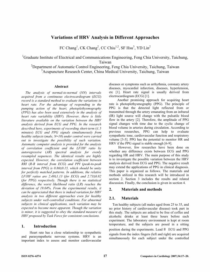
results as depicted in Table 1, in which A, B, C and D
denote the areas corresponding to the Tp, Fp, Fn and Tn
results under certain threshold value. For highly
approximated probability distributions as those in Figure
3, the likelihood ratio (LR) can be used to indicate the
resemblance between them. The LR can be derived
according to the following equation
Figure 3 The depiction for the calculation of likelihood
ratio (LR).
Table 1 Test results under certain decision threshold.
HRECG HRPPG
Test (+)
Test (-)
A (Tp)
C (Fn)
B (Fp)
D (Tn)
DB
BCA
A
LR
+
+=………………………………………(3)
If the two distributions are the same, then A=B and
C=D for any threshold value and a unity LR can thus be
derived. Therefore, the value of (1 LR) is used to
evaluate the deviation between the distributions of LF/HF
ratio derived from HRECG and HRPPG in this research.
Describe your methods here.
3. Results
PPI and RRI sequences for all subjects are derived
from the measured raw ECG and PPG signals according
to the methods described in previous section. In essence,
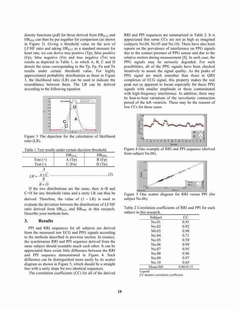
the synchronous RRI and PPI sequence derived from the
same subject should resemble much each other. It can be
appreciated there exists little difference between the RRI
and PPI sequence demonstrated in Figure 4. Such
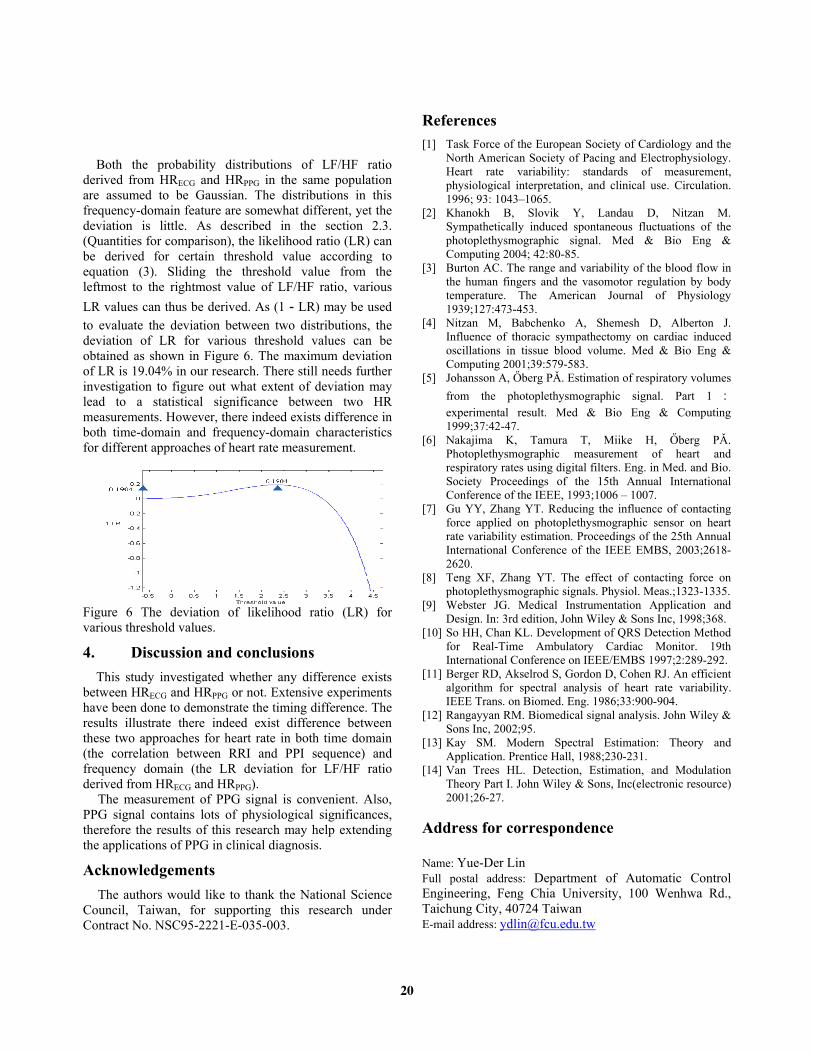
difference can be distinguished more easily by its scatter
diagram as shown in Figure 5, which should be a straight
line with a unity slope for two identical sequences.
The correlation coefficients (CC) for all of the derived
RRI and PPI sequences are summarized in Table 2. It is
appreciated that some CCs are not as high as imagined
(subjects No.04, No.05 and No.10). There have also been
reports on the prevalence of interference on PPG signals
due to the contact pressure of PPG sensor and due to the
relative motion during measurement [8]. In such case, the
PPG signals may be seriously degraded. For such
possibilities, all of the PPG signals have been checked
iteratively to assure the signal quality. As the peaks of
PPG signal are much smoother than those in QRS
complexes of ECG signal, this property makes the real
peak not so apparent to locate especially for those PPG
signals with smaller amplitude or those contaminated
with high-frequency interference. In addition, there may
be beat-to-beat variations of the isovolumic contraction
period of the left ventricle. These may be the reasons of
low CCs for these cases.
Figure 4 One example of RRI and PPI sequence (derived
from subject No.06).
Figure 5 One scatter diagram for RRI versus PPI (for
subject No.06).
Table 2 Correlation coefficients of RRI and PPI for each
subject in this research.
Subject CC
No.01 0.91
No.02 0.92
N0.03 0.98
No.04 0.71
No.05 0.58
No.06 0.99
No.07 0.95
No.08 0.96
No.09 0.97
No.10 0.65 Mean±SD 0.86±0.15
Legend:
CC denotes correlation coefficient.
19
Both the probability distributions of LF/HF ratio
derived from HRECG and HRPPG in the same population
are assumed to be Gaussian. The distributions in this
frequency-domain feature are somewhat different, yet the
deviation is little. As described in the section 2.3.
(Quantities for comparison), the likelihood ratio (LR) can
be derived for certain threshold value according to
equation (3). Sliding the threshold value from the
leftmost to the rightmost value of LF/HF ratio, various
LR values can thus be derived. As (1 LR) may be used
to evaluate the deviation between two distributions, the
deviation of LR for various threshold values can be
obtained as shown in Figure 6. The maximum deviation
of LR is 19.04% in our research. There still needs further
investigation to figure out what extent of deviation may
lead to a statistical significance between two HR
measurements. However, there indeed exists difference in
both time-domain and frequency-domain characteristics
for different approaches of heart rate measurement.
Figure 6 The deviation of likelihood ratio (LR) for
various threshold values.
4. Discussion and conclusions
This study investigated whether any difference exists
between HRECG and HRPPG or not. Extensive experiments
have been done to demonstrate the timing difference. The
results illustrate there indeed exist difference between
these two approaches for heart rate in both time domain
(the correlation between RRI and PPI sequence) and
frequency domain (the LR deviation for LF/HF ratio
derived from HRECG and HRPPG).
The measurement of PPG signal is convenient. Also,
PPG signal contains lots of physiological significances,
therefore the results of this research may help extending
the applications of PPG in clinical diagnosis.
Acknowledgements
The authors would like to thank the National Science
Council, Taiwan, for supporting this research under
Contract No. NSC95-2221-E-035-003.
References
[1] Task Force of the European Society of Cardiology and the
North American Society of Pacing and Electrophysiology.
Heart rate variability: standards of measurement,
physiological interpretation, and clinical use. Circulation.
1996; 93: 1043–1065.
[2] Khanokh B, Slovik Y, Landau D, Nitzan M.
Sympathetically induced spontaneous fluctuations of the
photoplethysmographic signal. Med & Bio Eng &
Computing 2004; 42:80-85.
[3] Burton AC. The range and variability of the blood flow in
the human fingers and the vasomotor regulation by body
temperature. The American Journal of Physiology
1939;127:473-453.
[4] Nitzan M, Babchenko A, Shemesh D, Alberton J.
Influence of thoracic sympathectomy on cardiac induced
oscillations in tissue blood volume. Med & Bio Eng &
Computing 2001;39:579-583.
[5] Johansson A, Pberg P謁. Estimation of respiratory volumes
from the photoplethysmographic signal. Part 1
experimental result. Med & Bio Eng & Computing
1999;37:42-47.
[6] Nakajima K, Tamura T, Miike H, Pberg P謁.
Photoplethysmographic measurement of heart and
respiratory rates using digital filters. Eng. in Med. and Bio.
Society Proceedings of the 15th Annual International
Conference of the IEEE, 1993;1006 – 1007.
[7] Gu YY, Zhang YT. Reducing the influence of contacting
force applied on photoplethysmographic sensor on heart
rate variability estimation. Proceedings of the 25th Annual
International Conference of the IEEE EMBS, 2003;2618-
2620.
[8] Teng XF, Zhang YT. The effect of contacting force on
photoplethysmographic signals. Physiol. Meas.;1323-1335.
[9] Webster JG. Medical Instrumentation Application and
Design. In: 3rd edition, John Wiley & Sons Inc, 1998;368.
[10] So HH, Chan KL. Development of QRS Detection Method
for Real-Time Ambulatory Cardiac Monitor. 19th
International Conference on IEEE/EMBS 1997;2:289-292.
[11] Berger RD, Akselrod S, Gordon D, Cohen RJ. An efficient
algorithm for spectral analysis of heart rate variability.
IEEE Trans. on Biomed. Eng. 1986;33:900-904.
[12] Rangayyan RM. Biomedical signal analysis. John Wiley &
Sons Inc, 2002;95.
[13] Kay SM. Modern Spectral Estimation: Theory and
Application. Prentice Hall, 1988;230-231.
[14] Van Trees HL. Detection, Estimation, and Modulation
Theory Part I. John Wiley & Sons, Inc(electronic resource)
2001;26-27.
Address for correspondence
Name: Yue-Der Lin
Full postal address: Department of Automatic Control
Engineering, Feng Chia University, 100 Wenhwa Rd.,
Taichung City, 40724 Taiwan
E-mail address: [email protected]
20