variation of protein corona composition of gold ... · pdf filevariation of protein corona...
TRANSCRIPT
Variation of Protein Corona Composition of Gold NanoparticlesFollowing Plasmonic HeatingMorteza Mahmoudi,*,†,‡ Samuel E. Lohse,† Catherine J. Murphy,† Arman Fathizadeh,§ Abbas Montazeri,∥
and Kenneth S. Suslick*,†
†Department of Chemistry, University of Illinois at Urbana−Champaign, 600 S. Mathews Avenue, Urbana, Illinois 61801, UnitedStates‡Department of Nanotechnology and Nanotechnology Research Center, Faculty of Pharmacy, Tehran University of Medical Sciences,Tehran, Iran§School of Physics, Institute for Research in Fundamental Sciences (IPM), Tehran, Iran∥Faculty of Mechanical Engineering, K.N. Toosi University of Technology, Tehran, Iran
*S Supporting Information
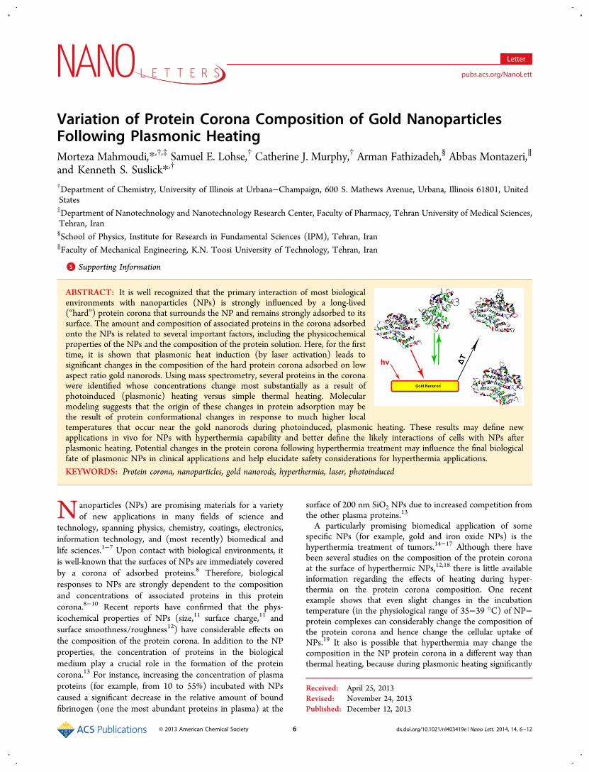
ABSTRACT: It is well recognized that the primary interaction of most biologicalenvironments with nanoparticles (NPs) is strongly influenced by a long-lived(“hard”) protein corona that surrounds the NP and remains strongly adsorbed to itssurface. The amount and composition of associated proteins in the corona adsorbedonto the NPs is related to several important factors, including the physicochemicalproperties of the NPs and the composition of the protein solution. Here, for the firsttime, it is shown that plasmonic heat induction (by laser activation) leads tosignificant changes in the composition of the hard protein corona adsorbed on lowaspect ratio gold nanorods. Using mass spectrometry, several proteins in the coronawere identified whose concentrations change most substantially as a result ofphotoinduced (plasmonic) heating versus simple thermal heating. Molecularmodeling suggests that the origin of these changes in protein adsorption may bethe result of protein conformational changes in response to much higher localtemperatures that occur near the gold nanorods during photoinduced, plasmonic heating. These results may define newapplications in vivo for NPs with hyperthermia capability and better define the likely interactions of cells with NPs afterplasmonic heating. Potential changes in the protein corona following hyperthermia treatment may influence the final biologicalfate of plasmonic NPs in clinical applications and help elucidate safety considerations for hyperthermia applications.
KEYWORDS: Protein corona, nanoparticles, gold nanorods, hyperthermia, laser, photoinduced
Nanoparticles (NPs) are promising materials for a varietyof new applications in many fields of science and
technology, spanning physics, chemistry, coatings, electronics,information technology, and (most recently) biomedical andlife sciences.1−7 Upon contact with biological environments, itis well-known that the surfaces of NPs are immediately coveredby a corona of adsorbed proteins.8 Therefore, biologicalresponses to NPs are strongly dependent to the compositionand concentrations of associated proteins in this proteincorona.8−10 Recent reports have confirmed that the phys-icochemical properties of NPs (size,11 surface charge,11 andsurface smoothness/roughness12) have considerable effects onthe composition of the protein corona. In addition to the NPproperties, the concentration of proteins in the biologicalmedium play a crucial role in the formation of the proteincorona.13 For instance, increasing the concentration of plasmaproteins (for example, from 10 to 55%) incubated with NPscaused a significant decrease in the relative amount of boundfibrinogen (one the most abundant proteins in plasma) at the
surface of 200 nm SiO2 NPs due to increased competition fromthe other plasma proteins.13
A particularly promising biomedical application of somespecific NPs (for example, gold and iron oxide NPs) is thehyperthermia treatment of tumors.14−17 Although there havebeen several studies on the composition of the protein coronaat the surface of hyperthermic NPs,12,18 there is little availableinformation regarding the effects of heating during hyper-thermia on the protein corona composition. One recentexample shows that even slight changes in the incubationtemperature (in the physiological range of 35−39 °C) of NP−protein complexes can considerably change the composition ofthe protein corona and hence change the cellular uptake ofNPs.19 It also is possible that hyperthermia may change thecomposition in the NP protein corona in a different way thanthermal heating, because during plasmonic heating significantly
Received: April 25, 2013Revised: November 24, 2013Published: December 12, 2013
Letter
pubs.acs.org/NanoLett
© 2013 American Chemical Society 6 dx.doi.org/10.1021/nl403419e | Nano Lett. 2014, 14, 6−12

higher temperatures will occur at the NP surface compared tothe average solution temperature.20,21 Therefore, understandingthe effect of local heat (for example, during laser activation ofgold nanorods for hyperthermia) on the composition of theprotein corona at the surface of gold NPs is critical to ourunderstanding of biological fate and transport of hyperthermicNPs.Among the many classes of functionalized NPs that have
been used as biomedical therapeutic or diagnostic agents,functionalized gold nanorods (AuNRs) have become one of themost widely studied. In addition to being composed of arelatively bioinert core material, gold nanorods possess strongabsorbance and light scattering properties that can be preciselycontrolled through the shape of the metal core.22,23 Forinstance, AuNRs with an aspect ratio of 3.5 or greater possess astrong longitudinal surface plasmon resonance in the near-IRregion of the spectrum where light can easily penetratebiological tissue to depths of up to 1.0 cm.22,24 In addition,gold nanorods are potentially appealing hyperthermia agents,due to the variety of functionalization strategies that exist tocontrol the surface chemistry of these AuNRs. Such surfacefunctionalization can enhance both the biocompatibility andtargeting opportunities of AuNRs both in vivo and in vitro.22
All of these properties make functionalized AuNRs appealingtheranostic materials, combining in one structure the ability tosimultaneously image biological targets with therapeuticproperties.12 Indeed, functionalized AuNRs have shown greatpromise in biomedical applications ranging from molecular(drug) delivery to hyperthermia cancer treatments.20,25,26
In order to study the effect of plasmonic heating on the hardprotein corona composition of AuNRs, we immersedcetyltrimethylammonium bromide (CTAB)-stabilized goldnanorods in fetal bovine serum (FBS) solutions and probedthe protein corona composition at the surface of nanorodsbefore and after plasmonic heating induced by continuous laserirradiation for various activation times. We studied the effect ofplasmonic heating on AuNR−protein complexes formed at twodifferent overall protein concentrations: 10 and 100% FBS.These fetal bovine serum concentrations were chosen becausethey simulate two different types of biological media: 10% FBSsimulates an in vitro milieu and 100% FBS simulates an in vivomilieu. FBS solutions, which consist of a mixture ofapproximately 3700 proteins,8 are also suitable models of invivo complexity. The AuNR−protein complexes were charac-terized by UV−vis absorbance spectroscopy, transmissionelectron microscopy, ζ-potential, and LC-MS/MS analysis, inorder to determine changes in the protein corona compositionafter photoinduced heating.The protein−AuNR complexes used in our study were
prepared by incubating CTAB-AuNRs (twice purified andconcentrated by centrifugation to a concentration of 3.2 nM)for 20 min in FBS solution. The AuNRs used in this study wereprepared by a silver-assisted seeded growth procedure and hadan aspect ratio of approximately 3.5 with a longitudinalplasmon band λmax of 756 nm (Figure 1 and Figure S1, S2 ofthe Supporting Information).22 Following purification, AuNR−protein complexes were then prepared for heating studies byincubating the AuNRs in either 10 or 100% FBS solutions.After incubation in the FBS solutions, the AuNR longitudinalSPR λmax blue shifted to 736 nm. This SPR blue shift indicateda change in the dielectric environment immediately surround-ing the AuNRs,27 consistent with protein corona formation.Our absorbance data indicated no significant AuNR aggregation
during protein corona formation at either FBS concentration(AuNP aggregation has been reported for some previousstudies of protein corona formation).18,28 Then 500 μL of theprotein−AuNR complex solutions was then irradiated with a 48mW laser (λ = 785 nm) with a spot size of ∼1.5 mm2 withoutstirring. Laser exposure continued for either 27.5 or 55 min,which should increase the temperature at the surface of theAuNRs up to ∼45 °C at least for short times.25 A 55 minirradiation time was chosen because we have previously shownthat under similar irradiation conditions, this period of time issufficient to achieve an overall solution temperature ofapproximately 39 °C.25 The average temperature of theirradiated solution was monitored during heating by placing athermocouple in the solution at 27.5 min and again at 55 min(Supporting Information). For the photoinduced heatedsamples, the solution temperature after 27.5 min wasapproximately 36 °C, while after 55 min, the temperature wasapproximately 39 °C. This corresponds to an increase in theambient temperature of nearly 7 °C during the course of thesetrials.Following irradiation, the hard corona protein−AuNR
complexes were separated from excess serum by centrifugation,and changes in the hard corona composition were examined byLC-MS/MS and ζ-potential analysis (Table 1; see Supporting
Figure 1. UV−vis absorption spectra and transmission electronmicrographs of AuNR−protein complexes before and after laserirradiation or thermal treatment at 45 °C for 55 min. (A) UV−visspectra for AuNRs after 10% FBS exposure, followed by hyperthermiatreatment. (B) UV−vis spectra for AuNRs after 100% FBS exposure,followed by hyperthermia treatment. (C,D) TEM images of CTAB-AuNRs. (E) TEM image of a protein−AuNR complex (10% FBS). (F)TEM image of protein−AuNR complex (100% FBS).
Nano Letters Letter
dx.doi.org/10.1021/nl403419e | Nano Lett. 2014, 14, 6−127

Information for preparation and procedures). Possible changesto the AuNR core following irradiation and heating wereexamined by UV−vis absorbance spectroscopy and trans-mission electron microscopy (TEM). The composition of thehard protein corona around the AuNRs was determined forfour different treatment options: incubation at 37 °C, thermalheating at 45 °C, and photoinduced heating induced bycontinuous laser exposures (27.5 and 55 min, see Materials andMethods section of Supporting Information for details). It isimportant to note that although the solution temperature isuniform over the course of either thermal or photoinducedheating experiments, we expect the process of heating to bedifferent in the two situations, because heating duringplasmonic activation begins at the AuNR surface (heatingfrom the “inside out”), while thermal heating occurs from the“outside in”. We found that CTAB-stabilized AuNRs that hadnot been exposed to FBS solution showed no significantchanges in shape or surface chemistry over the course of thetreatment time either during the purely thermal heating orphotoinduced heating (see Supporting Information for details).ζ-Potential analysis of the AuNR−protein complexes (Table
1) indicates a minor change in the overall AuNR−proteincorona surface charge following either plasmonic heating orthermal incubation. The modest changes in ζ-potential may beconsistent with some compositional change in the hard proteincorona following heating. We found that the decrease in theoverall surface charge was slightly greater for the 100% FBStreated AuNR than the 10% FBS sample (Table 1), regardlessof the type of heating the AuNR−protein complexesexperienced. Although again, we note that the overall changein the ζ-potential is relatively modest following heating and isnot a statistically significant change for the 10% FBS samples.Furthermore, the length of the irradiation period (27.5 versus55 min.) did not appear to have a significant effect on theoverall charge of the protein-AuNP complexes. UV−vis spectraof the AuNR−protein complexes following heating revealed nosignificant changes in the shape or size of the AuNRs followingirradiation (Figure 1, Supporting Information Table S6 andFigure S2). Interestingly, there is a peak in the ultraviolet regionof the spectrum that appeared post-treatment for the laser-irradiated samples at ∼260 nm (regardless of exposure time);this suggests that proteins with a higher concentration ofaromatic residues (e.g., tryptophan, tyrosine, or phenylalanine)may be present in the corona of AuNR−protein complexesfollowing photoinduced heating, perhaps due to changes inbound apolipoproteins.29−32
To measure the protein adsorption profile of the hardprotein corona at the surface of AuNRs following the thermalor photoinduced heating treatments, liquid chromatographymass spectrometry/mass spectrometry (LC MS/MS) techni-ques were employed. A semiquantitative assessment of theprotein amounts present in the corona was conducted throughapplication of a mass spectral counting (SpC) method13 (seeSupporting Information for details). Normalized SpC (NSpC)values of the predominant corona proteins for different samplesincubated with 10 and 100% protein solutions are presented inFigure 2 and Supporting Information Tables S1 and S2.Complete NSpC results for all detected proteins are presentedin Supporting Information, Tables S3 and S4.The LC-MS results indicate that the relative concentrations
of several biologically significant proteins in the corona changeas a result of the thermal and photoinduced treatments.Interestingly, the composition of the protein corona bound to
Table 1. ζ-Potential Analysis of AuNR-Protein Complexesbefore and after Laser Irradiation As a Function ofTreatments and Protein Concentration
sample ζ-potential (mV)
FBS control (no Au nanorods) −24.5 ± 2.1CTAB-AuNR control +23.7 ± 3.2100% FBS-AuNR, 37 °C −18.9 ± 1.4100% FBS-AuNR, 45 °C −13.5 ± 3.1100% FBS-AuNR, 27 min laser −12.7 ± 0.5100% FBS-AuNR, 55 min laser −12.8 ± 2.110% FBS-AuNR, 37 °C −19.8 ± 1.610% FBS-AuNR, 45 °C −16.9 ± 2.410% FBS-AuNR, 27 min laser −17.1 ± 1.310% FBS-AuNR, 55 min laser −16.3 ± 2.1
Figure 2. Graphs showing the variations of representative proteins(i.e., serum albumin α-1-antiproteinase precursor, α-2-HS-glycoproteinprecursor, apolipoprotein A-I precursor, hemoglobin fetal subunit betahemoglobin, apolipoprotein A-II precursor, and apolipoprotein C−IIIprecursor) in the composition of protein corona around initiallycationic gold nanorods after incubation with 10 and 100% FBSsolutions after different treatments (i.e., incubation at 37 °C, 45 °C,and continuous laser irradiation) as determined by LC MS/MS.
Nano Letters Letter
dx.doi.org/10.1021/nl403419e | Nano Lett. 2014, 14, 6−128

the AuNR is different following photoinduced heating,compared either to thermal heating at 45 °C or to incubationunder standard physiological temperatures (37 °C). As anexample, note the changes in concentration of apolipoproteinA-II precursor in the AuNR sample incubated in 100% FBS.The apolipoprotein A-II precursor concentration was substan-tially decreased after plasmonic or thermal heating of theAuNRs (e.g., decreased by 68% following 55 min ofirradiation). The final protein corona composition, as expected,also depends on the initial bulk FBS concentration (i.e., 10 vs100%) during treatment. Indeed distinctly different changes inthe protein corona composition occur in the 10% FBS AuNRsample, in which the content of apolipoprotein A II precursorincreased slightly for the 10% FBS sample after heat treatmentscompared to the samples incubated at 37 °C with a substantialincrease in the apolipoprotein A II precursor observed in thesample following photothermal irradiation for 55 min.Several other biologically important proteins present in the
hard corona also show significant changes in relativeconcentrations following laser-activated plasmonic heating.For the in vitro milieu model (i.e., 10% FBS solution), theamounts of several important proteins (e.g., serum albumin, α-2-HS-glycoprotein precursor, apolipoprotein A-II precursor,and apolipoprotein C−III precursor) in the protein coronawere significantly increased, by continuous laser activation ofAuNRs (versus incubation at 37 °C). In contrast, a few proteins(e.g., apolipoprotein A-I precursor) are decreased from the hardcorona following irradiation. There are a few significantdifferences in the corona changes for an in vivo model (100%FBS) compared to the in vitro one; for example, the amount ofα-1-antiproteinase precursor, hemoglobin fetal subunit beta,hemoglobin, and apolipoprotein A-II precursor in the coronacomposition after laser activation, were decreased.We also compared the composition of the protein corona for
both the 10% FBS and 100% FBS samples following heating asa function of molecular weight. According to the NSpCamounts in Figure 3 and Table S5 of Supporting Information, itappears that either plasmon-induced heating or purely thermalheating can decrease the relative amounts of low molecularweight proteins (<50 kDa) in the corona for an in vivo model(100% FBS). There are considerable differences in the proteinswith molecular weight in range of <30 kDa, between 10 and100% hard coronas. For instance, none of the heat treatmentsappear to significantly increase the amount of associatedproteins (<30 kDa) in corona for the 10% FBS samples. Incontrast, the amounts of these same proteins were significantlydecreased in the protein corona in the 100% FBS samplesfollowing photoinduced heating but not thermal heating.Interestingly, in the 100% FBS samples no heat treatmentappears to have a significant influence on the amount ofproteins (>30 kDa) present in the protein corona. Thevariation in the observed protein corona at 10 versus 100% FBSconcentrations must arise from both kinetic and thermody-namic factors: the high surface energy of NPs is lowered by theadsorption of proteins, and the essential irreversibility ofadsorption by some proteins implies that the selection andexchange processes on short time scales will depend on therates of diffusion of the multitude of proteins present in FBSthat diffuse to the surface of AuNRs.13
Very recently, we showed that the degree of protein coverageon NP surfaces, as well as the protein corona composition,depend on the incubating temperature (for simple thermalheating) at which the protein corona is formed;19 in addition,
we found that the cellular uptake of NPs is significantly affectedby the temperature at which protein corona formation occurs.19
By extension, small variations in protein corona compositionmay have a significant difference on cellular uptake in vitro.19
Because the structures of small proteins are very sensitive to theincubation temperature, variations of only a few degrees intemperature may influence the protein folding (as we havepreviously shown both experimentally or computationally foramyloid beta proteins).33 Contrary to conventional thermalheating of NP−protein complexes, however, plasmonic heatinginduces a substantial temperature gradient that starts at theAuNR surface and decays with distance. For instance, thetemperature at the AuNR−solution interface has previouslybeen shown to be as high as 45 °C during photoinducedactivation (depending on [AuNR]), but decreases to ∼36 °Cover just a few millimeters.21
In order to model the gradient temperature decay from thesurface of laser activated nanorods, we have employed ananalytical simulation, as described in the SupportingInformation. This simulation assumes a single laser-activatedgold nanorod (40.0 nm × 7.0 nm) with a constant temperatureat the AuNR−PBS interface of 45 °C. The PBS solution isinitially assumed to have a constant temperature of 37 °C. Laseractivation of the AuNR PBS for 10 ns results in a temperaturegradient between 45 and 38 °C that extends up to 100 nm fromthe AuNR surface. After 540 ns, the same temperature gradientnow extends up to 500 nm from the AuNR surface. Thetemperature gradient from the nanorod surface to themaximum affected PBS zone for these two cases has beenillustrated in Figure 4 and Figure S4 of Supporting Information.
Figure 3. Normalized spectral counts (NSpC) of proteins of variousmolecular weights contained in the hard corona incubated in 10 and100% FBS solutions after different treatments (incubation at 37 °C, 45°C, and continuous laser irradiation) as determined by LC MS/MS.
Nano Letters Letter
dx.doi.org/10.1021/nl403419e | Nano Lett. 2014, 14, 6−129

These simulation results demonstrate that during photoinducedheating, unlike thermal heating, the local temperature at thesurface of the laser-activated AuNRs can be substantially higherthan the temperature of the remainder of the solution (>6 °Cacross the gradient). Previous studies have suggested that undersimilar experimental conditions, the global solution may rise by∼10 °C in the first 4 min of photoinduced heating, and theglobal solution temperature will be essentially homogeneousafter ∼20 min (which is roughly consistent with our owntemperature measurements). Local heating zones with highertemperature near the AuNR will likely persist, however, overthe full course of our photoinduced thermal activationperiod.20,21 Accordingly, even from very early heating times,the proteins adsorbed to the AuNR surface may experience verydifferent solution temperatures than free proteins duringphotoinduced thermal treatments.Because of the thermal gradient in solution that results from
photoinduced heating of the AuNRs, any given protein (e.g.,apolipoproteins) may have different conformations depending
on whether the proteins are adsorbed on the AuNR surface orare located a few hundred nanometers from the surface of laseractivated nanorods. Such conformational differences as afunction of position relative to the AuNR surface will changeprotein-particle adsorption−desorption rates that ultimatelyinfluence the composition of the hard corona; different proteinspresent in the protein corona may show very differentconformational responses to photoinduced heating.To demonstrate the effect of slight temperature variation on
the protein conformations, molecular dynamic (MD) simu-lations were employed on apolipoprotein C III andapolipoprotein A I (see Supporting Information for detailedprocedures used in the simulations). To quantitatively probehow much the structure of the apolipoprotein C III andapolipoprotein A I proteins are affected by the temperature,separate simulations were performed on both proteins at 5different temperatures. The simulations for individual temper-ature/proteins (which were performed twice and the root-mean-square deviations (RMSD) and the radius of gyration(Rg) of the proteins by averaging over the simulations at eachtemperature) are shown in Figure 5.
For both proteins, it can be seen that the RMSD and Rg aresensitive to the variations of the temperature. The differences inthe RMSD and Rg values at different temperatures are in theorder of few angstroms, which is sufficient to change the chargedistribution of the protein and thus change their interactionwith the NPs. Previously, it has been shown that some proteins(e.g., serum albumin) have a similar susceptibility to temper-ature variation, whereas other proteins (such as serotransferrin)showed no significant conformational variation over the sametemperature range.19 Interestingly, our experimental data was ingood agreement with the performed simulation results. Theapolipoprotein C III, apolipoprotein A I, and serum albumin allshowed large variations in the hard corona composition of theilluminated AuNRs compared to the conventional thermalheating control samples, while there was no difference in therelative serotransferrin content in the protein coronas of theilluminated AuNRs versus the thermally treated samples (see
Figure 4. Temperature changes from the nanorod surface to themaximum radius of the heated fluid of (A) 100 nm in the case of 10 nsheating and (B) 500 nm for 540 ns heating.
Figure 5. Results of the molecular dynamics simulation onapolipoprotein C−III (green circles) and apoliporotein A-I (cyansquares). The RMSD and Rg and the corresponding error bars arecalculated by performing two separate simulations at each temperature.The results show that the structures of these proteins are sensitive tothe slight variation of temperature.
Nano Letters Letter
dx.doi.org/10.1021/nl403419e | Nano Lett. 2014, 14, 6−1210

Tables S3 and S4 of Supporting Information). It should benoted that the MD simulations indicate that certain proteinconformations are susceptible to temperature changes; theseconformational changes occur on a variety of time scales (fromps to ms). All the experimental characterization we haveperformed on the protein−AuNR complexes, however, requiretime delays of at least several minutes after photoinducedheating had been completed. As a result, fast local conforma-tional changes must influence protein−protein or protein−AuNR interactions that lead to more permanent changes in thecomposition of the protein corona.The changes observed in the protein corona composition
may have implications for the biological fate and transport ofAuNRs (or other hyperthermic NPs) following heating. Onemay speculate, for instance, that the observed increase of theapolipoproteins in the corona may promote prolongedcirculation time in the bloodstream, based on prior literatureof apolipoprotein effects on drug delivery.27 One might alsosuspect that the changes plasmonic heating induced in theapolipoprotein A-II precursor concentration in the AuNRprotein corona could make AuNRs less likely to cross specificbiological barriers (e.g., blood brain barrier31) followinghyperthermia treatment. As shown previously through MRItests on a brain capillary endothelial cell in the presence ofmagnetic NPs with apolipoproteins in their corona composi-tion,31 apolipoproteins are important in mediating NPtransport through the blood brain barrier.Previous studies of AuNRs in laser-activated hyperthermia
and similar laser powers indicate that the overall solutiontemperature during AuNR hyperthermia is 37−45 °C (depend-ing on AuNR concentration, time of heating, and initialtemperature).24 Interestingly, our results show that the type ofheating the AuNRs undergo has a significant influence on thecomposition of the protein corona. As seen in Figures 2, 3, andSupporting Information Tables S1−S4, there are significantdifferences in the composition of the protein corona followingphotoinduced versus conventional thermal heating, under bothin vitro and in vivo simulated conditions. The low molecularweight proteins (especially in the range of <30 kDa) appear tobe present in the hard corona in higher proportions at 45 °Ccompared to the laser activated conditions. For proteins in therange of 30−70 kDa, the amounts of proteins present in thehard corona at 45 °C are lower but represent a higherproportion of the total protein profile compared to the laseractivated conditions at 10 and 100% FBS, respectively; this mayrelated to the various corona thickness and composition atdifferent protein concentrations, as shown before.13
In summary, we have examined the effect of laser-inducedheating of gold nanorods on the composition of the hardprotein corona associated with CTAB-AuNRs immersed in 10and 100% solutions of FBS serum. Using experimental,analytical, and molecular dynamic simulation evaluations, wefound that hyperthermia treatments had minimal effects on theoverall surface charge of the protein corona associated with thegold nanorods following irradiation, but there were significantchanges in the composition of the hard protein coronafollowing both irradiation and thermal heating. Moreinterestingly, plasmonic heating (i.e., photoinduced) andconventional thermal heating produced distinct changes onthe composition of the AuNR protein corona. The composi-tional changes observed in the hard corona that are inducedspecifically by the laser irradiation may reflect relatively highlocalized temperature gradients right at the AuNR surface
during laser irradiation. The variation in the protein coronacomposition, following photoinduced local heating, may changethe biological fate of NPs, and further understanding of thechanging corona may have predictive value for NP therapy andfor the study of nanotoxins.
■ ASSOCIATED CONTENT*S Supporting InformationFull experimental details, protein corona compositions, andcharacterization of gold nanorods. This material is available freeof charge via the Internet at http://pubs.acs.org.
■ AUTHOR INFORMATIONCorresponding Authors*E-mail: (M.M.) [email protected].*E-mail: (K.S.S.) [email protected] authors declare no competing financial interest.
■ ACKNOWLEDGMENTSFinancial support from the National Science Foundation (CJM,CHE-1011980; KSS, DMR-1206355) is gratefully acknowl-edged. The authors also would like to thank Professor PeterYau, director of Protein Sciences Facility at the University ofIllinois at Urbana−Champaign for performing LC MS/MSexperiments.
■ REFERENCES(1) Zhang, D. Y.; Seelig, G. Nat. Chem. 2011, 3, 103−113.(2) Astruc, D. Nat. Chem. 2012, 4, 255−267.(3) Benson, O. Nature 2011, 480, 193−199.(4) Linic, S.; Christopher, P.; Ingram, D. B. Nat. Mater. 2011, 10,911−921.(5) Loh, O. Y.; Espinosa, H. D. Nat. Nanotechnol. 2012, 7, 283−295.(6) Weber, C.; Noels, H. Nat. Med. 2011, 17, 1410−1422.(7) Williams, E. Nature 2011, 479, 354−358.(8) Mahmoudi, M.; Lynch, I.; Ejtehadi, R.; Monopoli, M. P.;Bombelli, F. B.; Laurent, S. Chem. Rev. 2011, 111, 5610−5637.(9) Cedervall, T.; Lynch, I.; Foy, M.; Berggard, T.; Donnelly, S. C.;Cagney, G.; Linse, S.; Dawson, K. A. Angew. Chem., Int. Ed. 2007, 46,5754−5756.(10) Mahmoudi, M.; Hofmann, M.; Rothen-Rutishauser, B.; Fink, A.Chem. Rev. 2012, 112, 2323−2333.(11) Cedervall, T.; Lynch, I.; Lindman, S.; Berggard, T.; Thulin, E.;Nilsson, H.; Dawson, K. A.; Linse, S. Proc. Nat. Acad. Sci.U.S.A. 2007,104, 2050−2055.(12) Mahmoudi, M.; Serpooshan, V. J. Phys. Chem. C 2011, 115,18275−18283.(13) Monopoli, M. P.; Walczyk, D.; Campbell, A.; Elia, G.; Lynch, I.;Baldelli Bombelli, F.; Dawson, K. A. J. Am. Chem. Soc. 2011, 133,2525−2534.(14) Laurent, S.; Dutz, S.; Hafeli, U. O.; Mahmoudi, M. Adv. ColloidsInterface Sci. 2011, 166, 8−23.(15) Andre, M.; Gobin, D.; O’Neal, P.; Halas, N. J.; Drezek, R.; West,J. L. Lasers Surg. Med. 2005, 37, 123−129.(16) Su, Y.; Wei, X.; Peng, F.; Zhong, Y.; Lu, Y.; Su, S.; Xu, T.; Lee,S.-T.; He, Y. Nano Lett. 2012, 12, 1845−1850.(17) Gobin, A. M.; O’Neal, D. P.; Watkins, D. M.; Halas, N. J.;Drezek, R. A.; West, J. L. Lasers Surg. Med. 2005, 37, 123−129.(18) Casals, E.; Pfaller, T.; Duschl, A.; Oostingh, G. J.; Puntes, V.ACS Nano 2010, 4, 3623−3632.(19) Mahmoudi, M.; Abdelmonem, A. M.; Behzadi, S.; Clement, J.H.; Dutz, S.; Ejtehadi, M. R.; Hartmann, R.; Kantner, K.; Linne, U.;Maffre, P.; Metzler, S.; Moghadam, M. K.; Pfeiffer, C.; Rezaei, M.;Ruiz-Lozano, P.; Serpooshan, V.; Shokrgozar, M. A.; Nienhaus, G. U.;Parak, W. J. ACS Nano 2013, 7, 6555−6562.
Nano Letters Letter
dx.doi.org/10.1021/nl403419e | Nano Lett. 2014, 14, 6−1211

(20) Huang, J.; Jackson, K. S.; Murphy, C. J. Nano Lett. 2012, 12,2982−2987.(21) Huang, H.-C.; Rege, K.; Heys, J. J. ACS Nano 2010, 4, 2892−2900.(22) Alkilany, A. M.; Lohse, S. E.; Murphy, C. J. Acc. Chem. Res. 2013,46, 650−661.(23) Dreaden, E. C.; Alkilany, A. M.; Huang, X.; Murphy, C. J.; El-Sayed, M. A. Chem. Soc. Rev. 2012, 41, 2740−2779.(24) Orendorff, C. J.; Murphy, C. J. J. Phys. Chem. B 2006, 110,3990−3994.(25) Hu, K.-W.; Liu, T.-M.; Chung, K.-Y.; Huang, K.-S.; Hsieh, C.-T.;Sun, C.-K.; Yeh, C.-S. J. Am. Chem. Soc. 2009, 131, 14186−14187.(26) Huang, H.-C.; Barua, S.; Kay, D. B.; Rege, K. ACS Nano 2009, 3,2941−2952.(27) Furumoto, K.; Ogawara, K.-i.; Nagayama, S.; Takakura, Y.;Hashida, M.; Higaki, K.; Kimura, T. J. Controlled Release 2002, 83, 89−96.(28) Lacerda, S. H. D. P.; Park, J. J.; Meuse, C.; Pristinski, D.; Becker,M. L.; Karim, A.; Douglas, J. F. ACS Nano 2009, 4, 365−379.(29) Yang, C. Y.; Pownall, H. J.; Gotto, A. M., Jr Anal. Biochem. 1985,145, 67−72.(30) Baker, H. N.; Delahunty, T.; Gotto, A. M., Jr; Jackson, R. L.Proc. Nat. Acad. Sci. U.S.A. 1974, 71, 3631−3634.(31) Krol, S.; Macrez, R.; Docagne, F.; Defer, G.; Laurent, S.;Rahman, M.; Hajipour, M. J.; Kehoe, P. G.; Mahmoudi, M. Chem. Rev.2013, 113, 1877−1903.(32) Delahunty, T.; Baker, H. N.; Gotto, A. M., Jr; Jackson, R. L. J.Biol. Chem. 1975, 250, 2718−2724.(33) Ghavami, M.; Rezaei, M.; Ejtehadi, R.; Lotfi, M.; Shokrgozar, M.A.; Abd Emamy, B.; Raush, J.; Mahmoudi, M. ACS Chem. Neurosci.2012, 4, 375−378.
Nano Letters Letter
dx.doi.org/10.1021/nl403419e | Nano Lett. 2014, 14, 6−1212