variant analysis with hope · panther -2.93 phd-snp disease 2. hope report r287w contacts the...
TRANSCRIPT
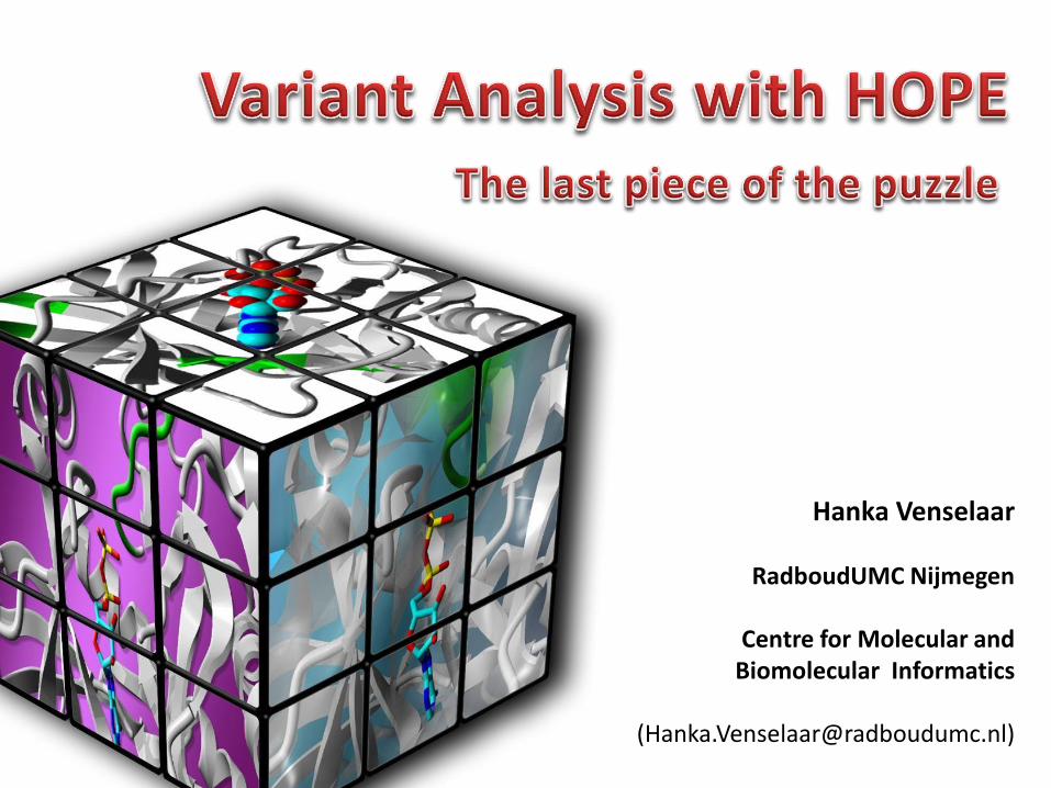
Hanka Venselaar
RadboudUMC Nijmegen
Centre for Molecular and Biomolecular Informatics

Outline
• Structural bioinformatics at Radboud UMC
• Project HOPE, the technical details
• Practical work, how to use HOPE?

Academic hospital RadboudUMCFocus on curing patients + understanding disease
KKIALSDARSMKHALREIKIIRRL
DHDNIVKVYEVLGPKGTDLQGELF
KFSVAYIVQEYMETDLARLLEQGT
LAEEHAKLFMYQLLRGLKYIHSAN
VLHRDLPANIFISTEDLVLKIGDF
GLARIVDQHYSHKGYLSEGLVTKW
YRSPRLLLSPNNYTKAIDMWAAGC
ILAEMLTGRMLFAGAHELEQMQLL
ETIPVIREEDKDELLRVMPSFVSS
?
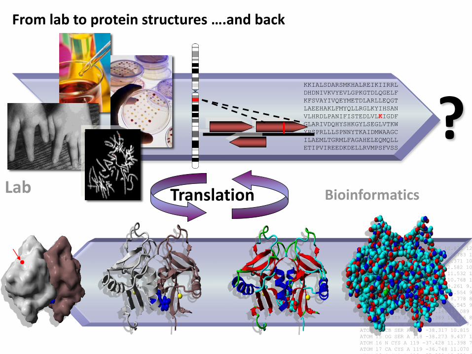
From lab to protein structures ….and back
Lab Translation Bioinformatics
ATOM 1 N GLN A 117 -42.882 10.838 12.153 1.00 58.09 N
ATOM 2 CA GLN A 117 -42.770 10.783 10.668 1.00 58.36 C
ATOM 3 C GLN A 117 -41.435 11.371 10.185 1.00 57.07 C
ATOM 4 O GLN A 117 -41.264 12.582 10.210 1.00 57.81 O
ATOM 5 CB GLN A 117 -43.966 11.532 10.028 1.00 59.40 C
ATOM 6 CG GLN A 117 -45.344 10.768 10.084 1.00 62.58 C
ATOM 7 CD GLN A 117 -45.254 9.261 9.651 1.00 67.37 C
ATOM 8 OE1 GLN A 117 -44.260 8.554 9.948 1.00 68.20 O
ATOM 9 NE2 GLN A 117 -46.304 8.778 8.955 1.00 67.47 N
ATOM 10 N SER A 118 -40.488 10.545 9.741 1.00 54.71 N
ATOM 11 CA SER A 118 -39.144 11.089 9.506 1.00 52.44 C
ATOM 12 C SER A 118 -38.389 10.616 8.251 1.00 50.58 C
ATOM 13 O SER A 118 -38.692 9.566 7.734 1.00 50.83 O
ATOM 14 CB SER A 118 -38.317 10.815 10.736 1.00 52.75 C
ATOM 15 OG SER A 118 -38.273 9.437 10.917 1.00 53.04 O
ATOM 16 N CYS A 119 -37.428 11.398 7.755 1.00 48.00 N
ATOM 17 CA CYS A 119 -36.748 11.070 6.507 1.00 46.41 C
ATOM 18 C CYS A 119 -35.339 10.829 6.835 1.00 45.44 C
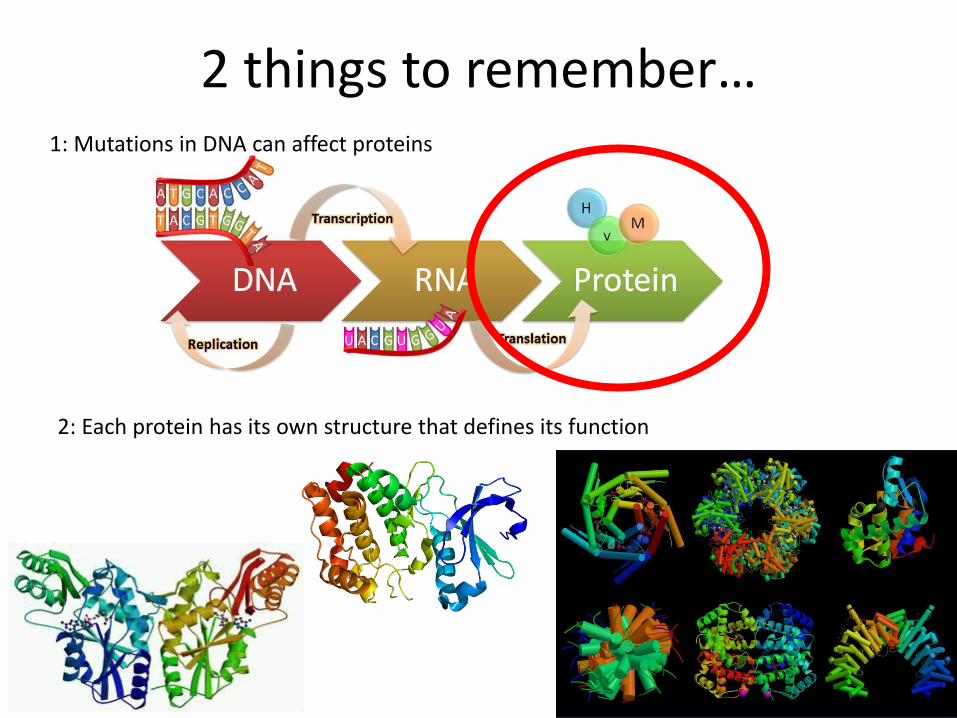
2 things to remember…1: Mutations in DNA can affect proteins
2: Each protein has its own structure that defines its function

Hidden secrets in protein structures
For example:
• Active sites
• Ligands / co-factors
• Surface interactions with other molecules
• Stabilizing interactions
• Transmembrane regions
• Flexible domains
• Etc…etc..etc…
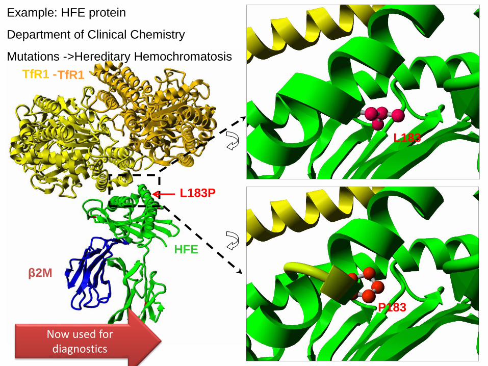
L183P
TfR1
β2M
HFE
TfR1 -
L183
P183
Example: HFE protein
Department of Clinical Chemistry
Mutations ->Hereditary Hemochromatosis
Published in Blood, Cells,
Molecules and Disease
Now used fordiagnostics
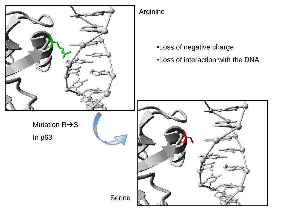
Arginine
Serine
Mutation RS
In p63
•Loss of negative charge
•Loss of interaction with the DNA
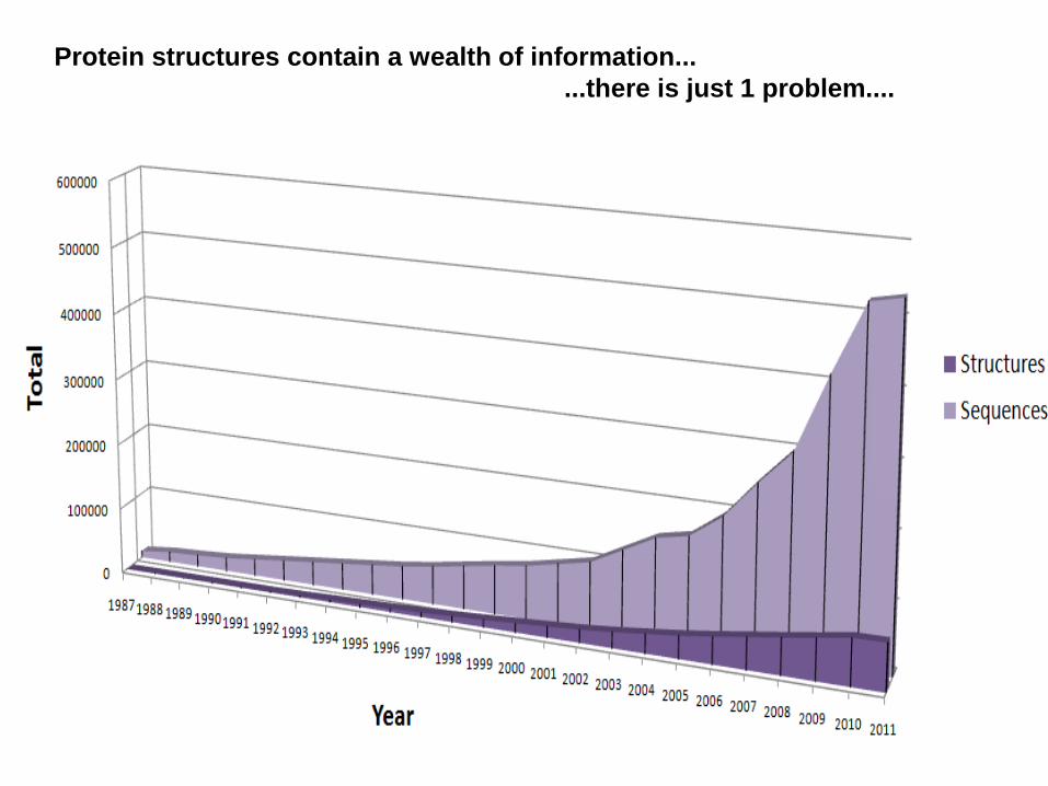
Protein structures contain a wealth of information...
...there is just 1 problem....
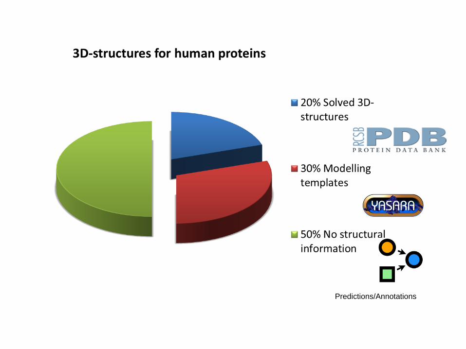
Predictions/Annotations

MSASTQTNEFLSPEVFQHIWDFLEQPICSVQPIDLNFVDEPSEDGATNKIEISMDCIRMQDSDLSDMWPQYTNLGLLNSMDQQIQNGSSSTSPYNTDHAQNSVTAPSPYAQPSSTFDALSPSPAIPSNTDYPGPHSFDVSFQQSSTAKSATWTYSTELKKLYCQIAKTCPIQIKVMTPPPQGAVIRAMPVYKKAEHVTEVVKRCPNHELSREFNEGQIAPPSHLIRVEGNSHAQYVEDPITGRQSVLVPYEPPQVGTEFTTVLYNFMCNSSCVGGMNRRPILIIVTLETRDGQVLGRRCFEARICACPGRDRKADEDSIRKQQVSDSTKNGDGTKRPFRQNTHGIQMTSIKKRRSPDDELLYLPVRGRETYEMLLKIKESLELMQYLPQHTIETYRRVIDAVRFTLRQTISFPPRDEWNDFNFDMDARRNKQQRIKEEGE
BLAST against the PDBSPAIPSNTDYPGPHSFDVSFQQSSTAKSATW
SSSVPSQKTYQGSYGFRLGFLHSGTAKSVTC
GAVIRAMPVYKKAEHVTEVVKRCPNHELSRE
GTRVRAMAIYKQSQHMTEVVRRCPHHERCSD
Known 3D structureMSASTQTNEFLSPEVFQHIWDFLEQPICSVQPIDLNFVDEPSEDGATNKIEISMDCIRMQDSDLSDMWPQYTNLGLLNSMDQQIQNGSSSTSPYNTDHAQNSVTAPSPYAQPSSTFDALSPSPAIPSNTDYPGPHSFDVSFQQSSTAKSATWTYSTELKKLYCQIAKTCPIQIKVMTPPPQGAVIRAMPVYKKAEHVTEVVKRCPNHELSREFNEGQIAPPSHLIRVEGNSHAQYVEDPITGRQSVLVPYEPPQVGTEFTTVLYNFMCNSSCVGGMNRRPILIIVTLETRDGQVLGRRCFEARICACPGRDRKADEDSIRKQQVSDSTKNGDGTKRPFRQNTHGIQMTSIKKRRSPDDELLYLPVRGRETYEMLLKIKESLELMQYLPQHTIETYRRVIDAVRFTLRQTISFPPRDEWNDFNFDMDARRNKQQRIKEEGE
BLAST against the PDB
SPAIPSNTDYPGPHSFDVSFQQSSTAKSATW
--------EFLKSSRLTVDS---VDAKATPF
GAVIRAMPVYKKAEHVTEVVKRCPNHELSRE
ALKMRAMP-----EFLCMNWLNSDDMELS--
Unknown 3D structure
Homology modelHomologous 3D structure
Use the homologous structure to predict the unknown 3D structure
Homology modelling…..

Predictions/Annotations
This is what HOPE will use…

The Molecular Puzzle: from patient to medicine
ExperimentsPatient in
hospitalNew
experiments
Drug
development
Bioinformatics
GEIALWSLVV
LAIERYVVVC
KPMSNFRFGE
NHAIMGVAFT
WVMALARAAP
PLVGWSRYIP
EGMQCSCGID
YYTPHEETNN
Yesteryear vs Nowadays

So, we developed HOPE….
Is easy to use for (bio)medical scientists & Provides structural explanation of point mutations
HOPE, an automatic web server that:
URL: www.cmbi.ru.nl/hope

Input page: Sequence + Mutation
Report page: Extensive explanation of effects on molecular level
Data collection & combination is hidden for user

Data storage (in database) and combination/prioritization
Data collection
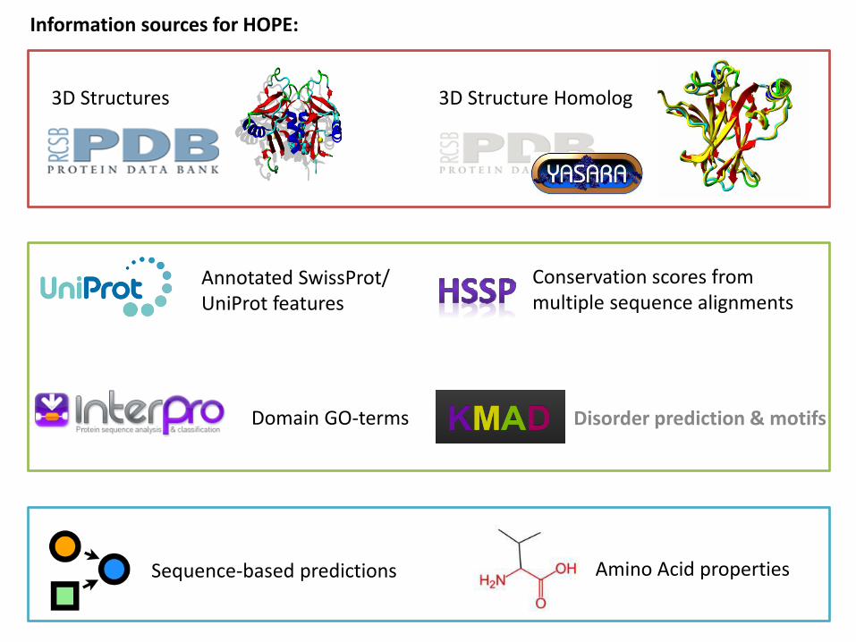
3D Structures 3D Structure Homolog
Annotated SwissProt/ UniProt features
Sequence-based predictions
Information sources for HOPE:
Conservation scores from multiple sequence alignments
Disorder prediction & motifsDomain GO-terms
Amino Acid properties
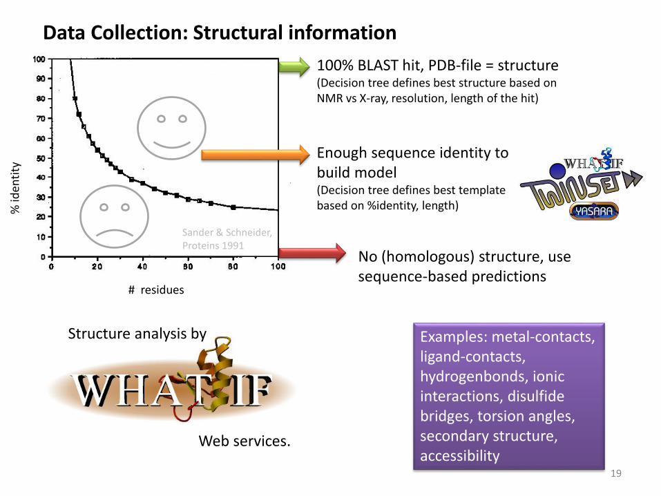
Structure analysis by Examples: metal-contacts, ligand-contacts, hydrogenbonds, ionic interactions, disulfide bridges, torsion angles, secondary structure, accessibility
Data Collection: Structural information
# residues
% id
enti
ty
Sander & Schneider, Proteins 1991
100% BLAST hit, PDB-file = structure(Decision tree defines best structure based on NMR vs X-ray, resolution, length of the hit)
Enough sequence identity to build model(Decision tree defines best template based on %identity, length)
No (homologous) structure, use sequence-based predictions
Web services.
19

• Collect the sequence features in the SwissProt record• Information is annotated, checked and sometimes cross linked
Data colelction: SwissProt annotations
20
Example Uniprot-features: Regions, domains, motifs, mutagenesis sites, variants, active site, signal, glycosylation sites, etc

Data collection: Conservation scores
• Calculated from the multiple sequence alignment in HSSP
•Profile gives % occurrence of residue type on that position
• Precalculated (for PDB-files/templates) or newly generated (sequence)
Domain GO-terms

Data collection: Gene Ontology-terms per domain
• InterPro Predicts functional domains
• Annotated with GO-terms, a controlled vocabulary that describes the protein
• Cellular component, molecular function and biological process
• Examples: GO: 0003677 DNA-binding, GO: 0005155 Protein binding,
GO: 003700 sequence-specific DNA binding transcription factor activity

Data collection: Disorder predictions & Motifs
• Knowledge Based Multiple Sequence Alignment for Intrinsically Disordered Proteins
• Predicts % disorder of the protein, and whether the mutation of interest is located in a disordered region
• Uses Short Linear Motifs from ELM to make alignments and therefore it can also predict whether a mutation will disturb such a motif
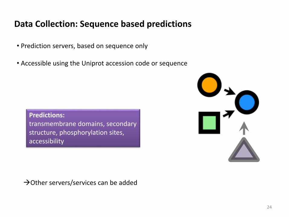
• Prediction servers, based on sequence only
• Accessible using the Uniprot accession code or sequence
Predictions:transmembrane domains, secondary structure, phosphorylation sites, accessibility
Data Collection: Sequence based predictions
Other servers/services can be added
24
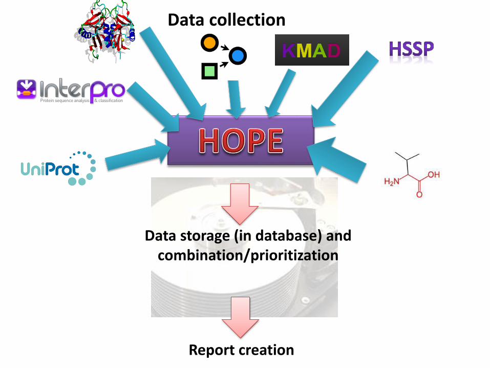
Data storage (in database) and combination/prioritization
Report creation
Data collection

URL for HOPE www.cmbi.ru.nl/hope

The gateway page

The report
Explains effect of the mutation on a molecular level
Contains pictures and moving gifs
Contains crosslinks to other databases
Consists of several paragraphs, each one focusing on 1 aspect.
Information as easy and understandable as possible.
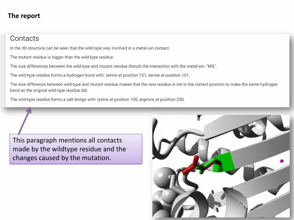
The report
This paragraph mentions all contacts made by the wildtype residue and the changes caused by the mutation.
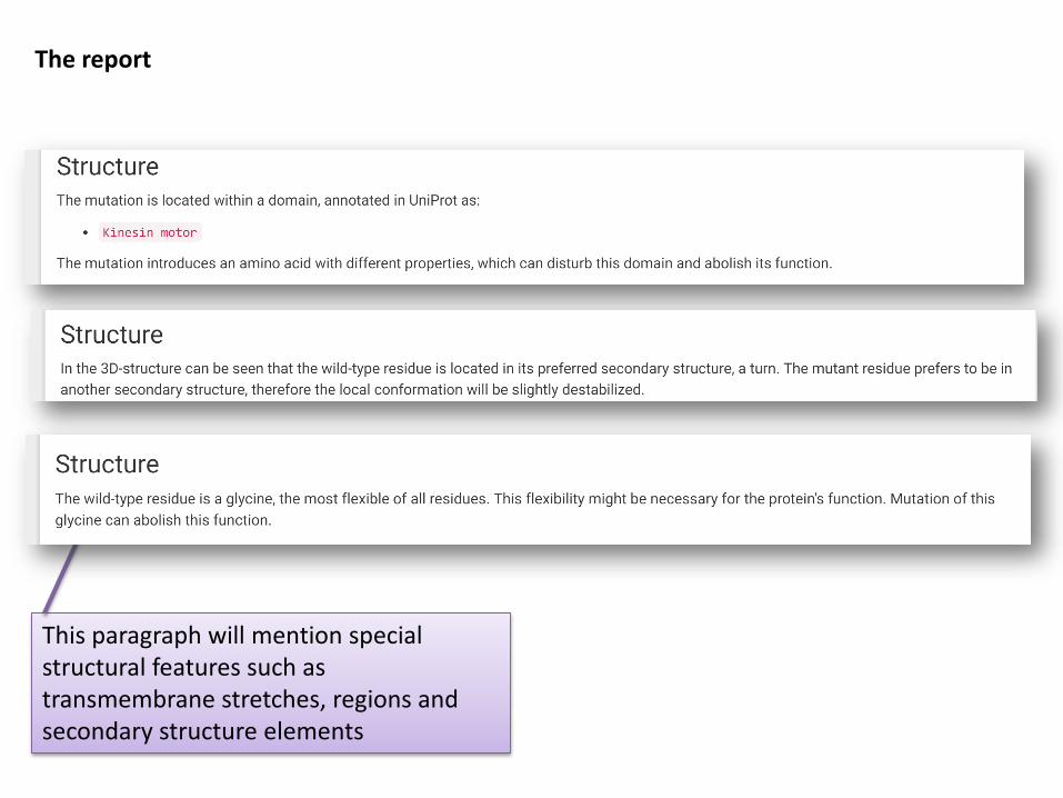
The report
This paragraph will mention special structural features such as transmembrane stretches, regions and secondary structure elements

Predicts whether (and how much of) the protein is disordered and whether the mutation occurs in such a disordered region.
Indicates whether the residue is very conserved (=important) or more variable.
The reportPredicts whether (and how much of) the protein is disordered and whether the mutation occurs in such a disordered region.
Indicates whether the residue is very conserved (=important) or more variable.
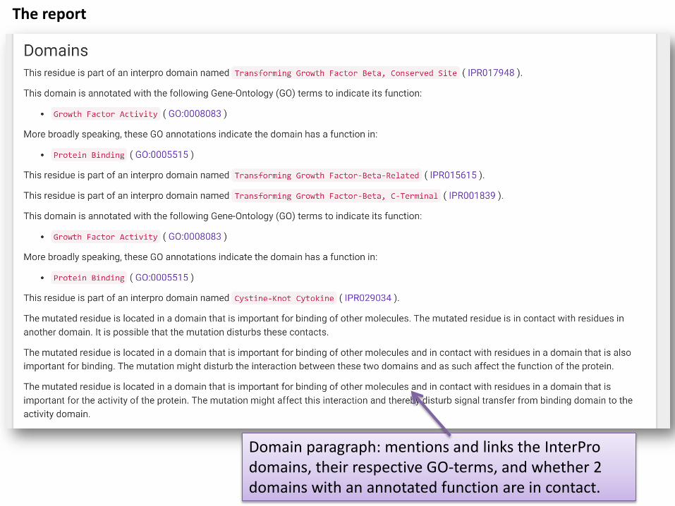
Domain paragraph: mentions and links the InterProdomains, their respective GO-terms, and whether 2 domains with an annotated function are in contact.
The report

Predicts whether the residue was located in an annotated motif (taken from ELM database) and whether this mutation changes that motif
Last remark: the amino acid compared on their properties such as size, hydrophobicity and charge. Accessibility (if availble) is used here.
The report

This will tell you whether the mutation is annotated in dbSNP and Swissprot (Disease vs Polymorphism)
This paragraph will list the annotated/predicted modifications at this position.
The report

Pictures and animated gifs
The report
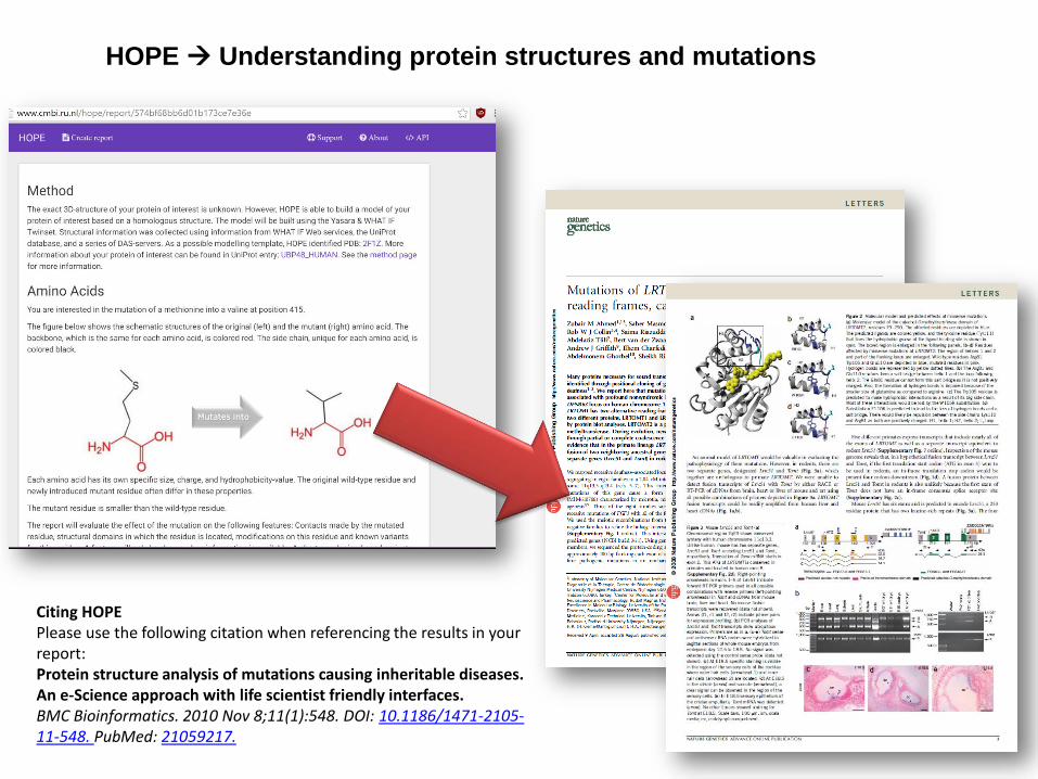
HOPE Understanding protein structures and mutations
Citing HOPEPlease use the following citation when referencing the results in your report:Protein structure analysis of mutations causing inheritable diseases. An e-Science approach with life scientist friendly interfaces.BMC Bioinformatics. 2010 Nov 8;11(1):548. DOI: 10.1186/1471-2105-11-548. PubMed: 21059217.
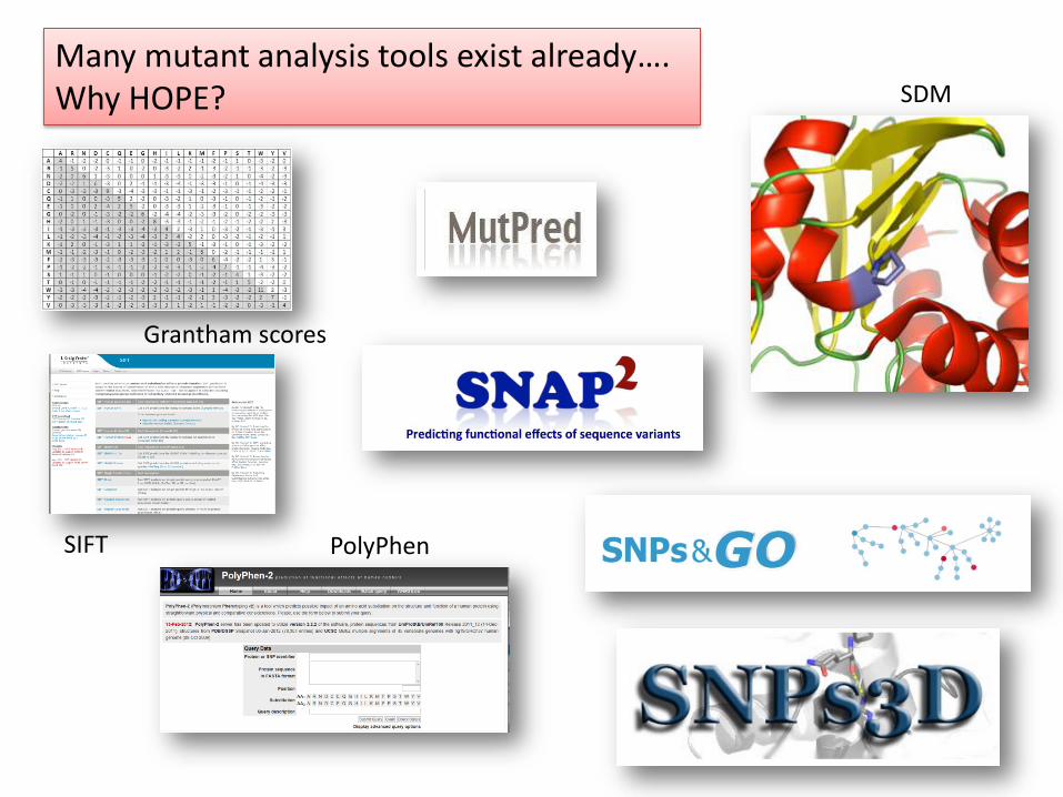
Many mutant analysis tools exist already….Why HOPE?
Grantham scores
SIFT PolyPhen
SDM
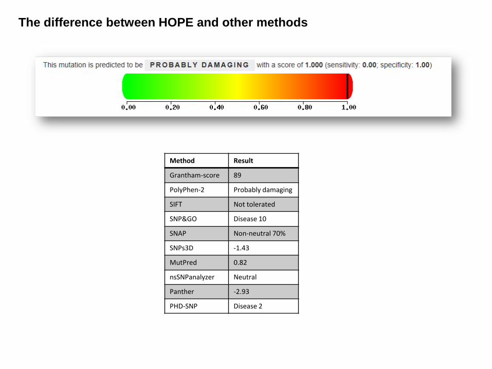
The difference between HOPE and other methods
Method Result
Grantham-score 89
PolyPhen-2 Probably damaging
SIFT Not tolerated
SNP&GO Disease 10
SNAP Non-neutral 70%
SNPs3D -1.43
MutPred 0.82
nsSNPanalyzer Neutral
Panther -2.93
PHD-SNP Disease 2
HOPE report R287W
ContactsThe wildtype residue forms a hydrogen bond with the Leucine onposition 271 and with the Tyrosine on position 237. The size differencebetween wild-type and mutant residue makes that the new residue isnot in the correct position to make the same hydrogenbond as theoriginal wild-type residue did. The difference in hydrophobicity willaffect hydrogenbond formation. The difference in charge will disturbthe ionic interaction made by the original, wild-type residue.ConservationOnly this residue type was found at this position. Mutation of a 100%conserved residue is usually damaging for the protein.DomainsThe mutated residue is buried in a domain that is important for bindingof other molecules. The differences between the wild-type and mutantresidue might disturb the core structure of this domain and therebyaffect the binding properties.Amino acid propertiesThere is a difference in charge between the wild-type and mutantamino acid. The charge of the buried wild-type residue is lost by thismutation. The wild-type and mutant amino acids differ in size. Themutant residue is bigger than the wild-type residue. The wild-typeresidue was buried in the core of the protein. The mutant residue isbigger and probably will not fit.
Method Result
Grantham-score 101
PolyPhen-2 Probably damaging
SIFT Not tolerated
SNP&GO Disease 10
SNAP Non-neutral 93%
SNPs3D -2,35
MutPred 0.918
nsSNPanalyzer Disease
Panther -6,95
PHD-SNP Disease 9

Grantham scores
Analysis of these variants by 4 different methods. Results were compared to our own manual analyses.
SIFT
PolyPhen-2
Amino Acid / Sequence based Structure based
Validation experiment: Grantham scores, SIFT, PolyPhen and HOPE
. . .
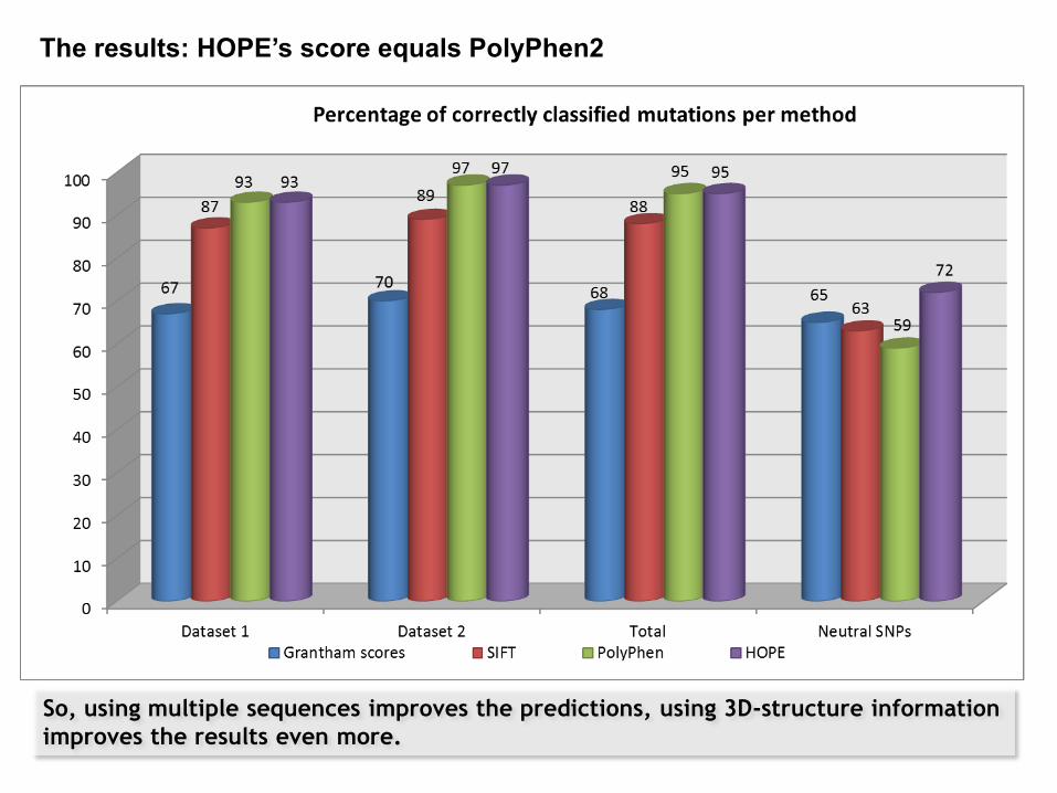
So, using multiple sequences improves the predictions, using 3D-structure information
improves the results even more.
The results: HOPE’s score equals PolyPhen2

Take home messages….
• HOPE can analyze the effect of point mutations on molecular level.
• Use HOPE for those mutations you have identified as possibly harmful, and for which you are missing the last piece of the puzzle to understand structural effects.
• HOPE’s performance is comparable to PolyPhen (and probably to other tools that use structural information).
• HOPE is not a classifier! HOPE provides insight and understanding of the structure and effect of the mutations.

Thanks to…
• Tim te Beek• Remko Kuipers• Maarten Hekkelman• Coos Baakman• Elmar Krieger• Jules Kersenmakers• Jon Black• Joanna Lange
• Students: Annika Borman, Franscesca Camilli, Shimah Golizadeh, Marlou Snelleman• All the HOPE users, every collaborator, and every scientist in the world who added something to the numerous databases available on the web

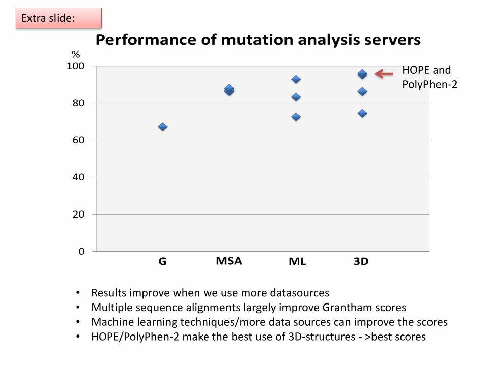
• Results improve when we use more datasources• Multiple sequence alignments largely improve Grantham scores• Machine learning techniques/more data sources can improve the scores• HOPE/PolyPhen-2 make the best use of 3D-structures - >best scores
HOPE andPolyPhen-2
Extra slide:

Validation of HOPE, an experiment
• Dataset of >200 mutations (181 damaging/46 benign) collected from literature
• Mutations were analyzed using 10 online servers and HOPE
MethodDamagingmutations
BenignSNPs
Grantham score 67,4% 65,2%
PhD-SNP 85,6% 73,9%
Panther 86,5% 35,1%
SIFT 87,8% 64,4%
SNPs&GO 72,5% 77,8%
SNAP 83,4% 56,5%
MutPred 92,8% 85,7%
nsSNPanalyzer 74,5% 67,6%
SNPs3D 86,3% 62,8%
PolyPhen-2 95,0% 58,6%
HOPE 96,1% 76,1%
G = differences between amino acids
MSA = Conservation scores from multiple sequence aligments, and amino acid differences
ML = Conservation scores and GO-terms or predictions based on sequence, amino acid differences
3D = 3D-Structural features, Swissprot annotations, conservation scores, amino acid differences
Extra slide:

HOPE demo: practical work
The following examples will guide you through HOPE’s report pages.
You can simply copy and paste the sequence we provided.
We have added some questions, but in general you should try to answer the question “What does this mutation do to the structure and function of the protein?”
Sometimes you might want to use another server to find more information for the protein of interest, for this you can use the accession codes.
Your are free to try other random mutations besides those provided here….
If you have questions, let me know!
Sequence:
MAAADAEAVP ARGEPQQDCC VKTELLGEET PMAADEGSAE KQAGEAHMAA DGETNGSCEN
SDASSHANAA KHTQDSARVN PQDGTNTLTR IAENGVSERD SEAAKQNHVT ADDFVQTSVI
GSNGYILNKP ALQAQPLRTT STLASSLPGH AAKTLPGGAG KGRTPSAFPQ TPAAPPATLG
EGSADTEDRK LPAPGADVKV HRARKTMPKS VVGLHAASKD PREVREARDH KEPKEEINKN
ISDFGRQQLL PPFPSLHQSL PQNQCYMATT KSQTACLPFV LAAAVSRKKK RRMGTYSLVP
KKKTKVLKQR TVIEMFKSIT HSTVGSKGEK DLGASSLHVN GESLEMDSDE DDSEELEEDD
GHGAEQAAAF PTEDSRTSKE SMSEADRAQK MDGESEEEQE SVDTGEEEEG GDESDLSSES
SIKKKFLKRK GKTDSPWIKP ARKRRRRSRK KPSGALGSES YKSSAGSAEQ TAPGDSTGYM
EVSLDSLDLR VKGILSSQAE GLANGPDVLE TDGLQEVPLC SCRMETPKSR EITTLANNQC
MATESVDHEL GRCTNSVVKY ELMRPSNKAP LLVLCEDHRG RMVKHQCCPG CGYFCTAGNF
MECQPESSIS HRFHKDCASR VNNASYCPHC GEESSKAKEV TIAKADTTST VTPVPGQEKG
SALEGRADTT TGSAAGPPLS EDDKLQGAAS HVPEGFDPTG PAGLGRPTPG LSQGPGKETL
ESALIALDSE KPKKLRFHPK QLYFSARQGE LQKVLLMLVD GIDPNFKMEH QNKRSPLHAA
AEAGHVDICH MLVQAGANID TCSEDQRTPL MEAAENNHLE AVKYLIKAGA LVDPKDAEGS
TCLHLAAKKG HYEVVQYLLS NGQMDVNCQD DGGWTPMIWA TEYKHVDLVK LLLSKGSDIN
IRDNEENICL HWAAFSGCVD IAEILLAAKC DLHAVNIHGD SPLHIAAREN RYDCVVLFLS
RDSDVTLKNK EGETPLQCAS LNSQVWSALQ MSKALQDSAP DRPSPVERIV SRDIARGYER
IPIPCVNAVD SEPCPSNYKY VSQNCVTSPM NIDRNITHLQ YCVCIDDCSS SNCMCGQLSM
RCWYDKDGRL LPEFNMAEPP LIFECNHACS CWRNCRNRVV QNGLRARLQL YRTRDMGWGV
RSLQDIPPGT FVCEYVGELI SDSEADVREE DSYLFDLDNK DGEVYCIDAR FYGNVSRFIN
HHCEPNLVPV RVFMAHQDLR FPRIAFFSTR LIEAGEQLGF DYGERFWDIK GKLFSCRCGS
PKCRHSSAAL AQRQASAAQE AQEDGLPDTS SAAAADPL
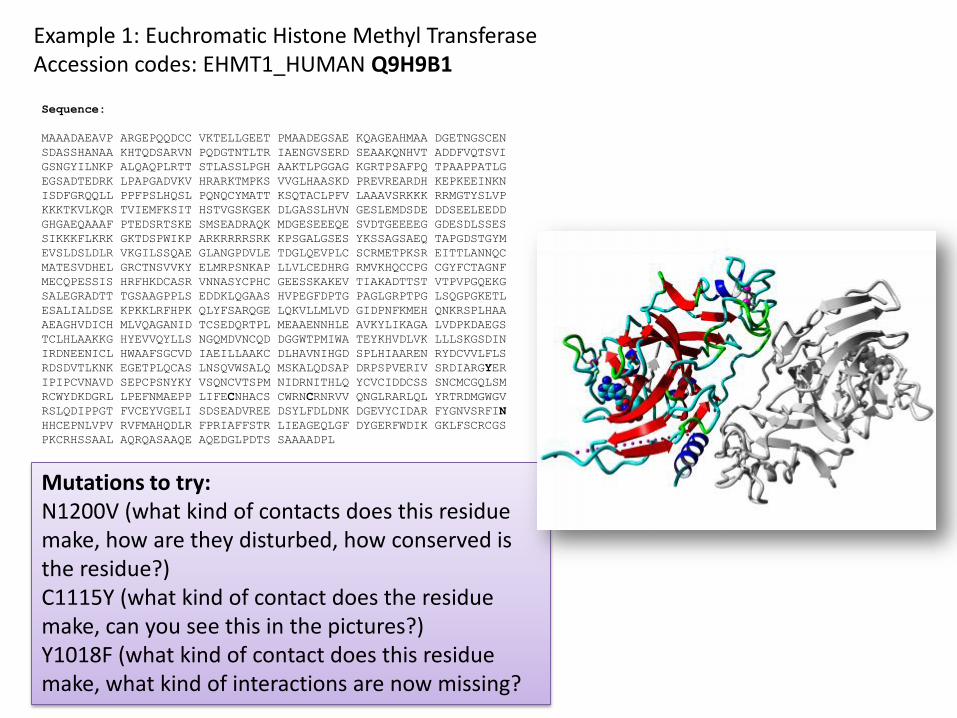
Example 1: Euchromatic Histone Methyl TransferaseAccession codes: EHMT1_HUMAN Q9H9B1
Mutations to try: N1200V (what kind of contacts does this residue make, how are they disturbed, how conserved is the residue?) C1115Y (what kind of contact does the residue make, can you see this in the pictures?)Y1018F (what kind of contact does this residue make, what kind of interactions are now missing?

Mutation N1200V, important notes
First of all: The mutant residue is smaller and more hydrophobic (cannot make hydrogenbonds)
The wildtype made contacts with a ligand (click on the link), and it made hydrogenbonds (which the mutant cannot make). PISA identifies contacts with another molecule, in this case that is the ligand again.
The wildtype and its neighbor are 100% conserved, so they are important! (for ligand binding, something you can also see in the pictures)
Everything tells you that the residue is located in the ligand binding domain and important for interactions with the molecule. Mutation will cause loss of these interactions and therefore cause loss of function.

Mutation Y1018F, important notes
The wildtype and mutant residue differ slightly, the mutant is smaller and missing the –OH, so therefore it cannot make the same hydrogenbonds
The wildtype residue is located on the surface, where it makes a hydrogenbond. The pictures, and PISA, indicate that this is a hydrogenbond to the other monomer. Losing this bond might mean loss of dimerisation.
However! Conservation scores indicate that the mutant residue was seen at this position in other structures. This makes our conclusion less clear and you might need experimental backup to underline your conclusions.

Mutation C1115A, important notes
The wildtype is a S, often involved in disulfide bonds. Mutant A is smaller and cannot make these bonds.
However, the contacts section doesn’t mention a disulfid bond, but a metal contact instead. The pictures show the metal-interaction too.
Conservation scores indicate a damaging mutation too… The mutation will cause loss of stability of the SET-domain because A cannot bind metal similar to S.
Sequence:
MAAADAEAVP ARGEPQQDCC VKTELLGEET PMAADEGSAE KQAGEAHMAA DGETNGSCEN
SDASSHANAA KHTQDSARVN PQDGTNTLTR IAENGVSERD SEAAKQNHVT ADDFVQTSVI
GSNGYILNKP ALQAQPLRTT STLASSLPGH AAKTLPGGAG KGRTPSAFPQ TPAAPPATLG
EGSADTEDRK LPAPGADVKV HRARKTMPKS VVGLHAASKD PREVREARDH KEPKEEINKN
ISDFGRQQLL PPFPSLHQSL PQNQCYMATT KSQTACLPFV LAAAVSRKKK RRMGTYSLVP
KKKTKVLKQR TVIEMFKSIT HSTVGSKGEK DLGASSLHVN GESLEMDSDE DDSEELEEDD
GHGAEQAAAF PTEDSRTSKE SMSEADRAQK MDGESEEEQE SVDTGEEEEG GDESDLSSES
SIKKKFLKRK GKTDSPWIKP ARKRRRRSRK KPSGALGSES YKSSAGSAEQ TAPGDSTGYM
EVSLDSLDLR VKGILSSQAE GLANGPDVLE TDGLQEVPLC SCRMETPKSR EITTLANNQC
MATESVDHEL GRCTNSVVKY ELMRPSNKAP LLVLCEDHRG RMVKHQCCPG CGYFCTAGNF
MECQPESSIS HRFHKDCASR VNNASYCPHC GEESSKAKEV TIAKADTTST VTPVPGQEKG
SALEGRADTT TGSAAGPPLS EDDKLQGAAS HVPEGFDPTG PAGLGRPTPG LSQGPGKETL
ESALIALDSE KPKKLRFHPK QLYFSARQGE LQKVLLMLVD GIDPNFKMEH QNKRSPLHAA
AEAGHVDICH MLVQAGANID TCSEDQRTPL MEAAENNHLE AVKYLIKAGA LVDPKDAEGS
TCLHLAAKKG HYEVVQYLLS NGQMDVNCQD DGGWTPMIWA TEYKHVDLVK LLLSKGSDIN
IRDNEENICL HWAAFSGCVD IAEILLAAKC DLHAVNIHGD SPLHIAAREN RYDCVVLFLS
RDSDVTLKNK EGETPLQCAS LNSQVWSALQ MSKALQDSAP DRPSPVERIV SRDIARGYER
IPIPCVNAVD SEPCPSNYKY VSQNCVTSPM NIDRNITHLQ YCVCIDDCSS SNCMCGQLSM
RCWYDKDGRL LPEFNMAEPP LIFECNHACS CWRNCRNRVV QNGLRARLQL YRTRDMGWGV
RSLQDIPPGT FVCEYVGELI SDSEADVREE DSYLFDLDNK DGEVYCIDAR FYGNVSRFIN
HHCEPNLVPV RVFMAHQDLR FPRIAFFSTR LIEAGEQLGF DYGERFWDIK GKLFSCRCGS
PKCRHSSAAL AQRQASAAQE AQEDGLPDTS SAAAADPL
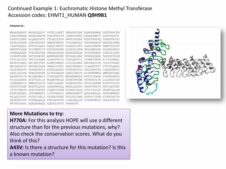
Continued Example 1: Euchromatic Histone Methyl TransferaseAccession codes: EHMT1_HUMAN Q9H9B1
More Mutations to try: H770A: For this analysis HOPE will use a different structure than for the previous mutations, why? Also check the conservation scores. What do you think of this?A43V: Is there a structure for this mutation? Is this a known mutation?
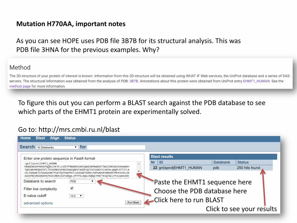
Mutation H770AA, important notes
To figure this out you can perform a BLAST search against the PDB database to see which parts of the EHMT1 protein are experimentally solved.
Go to: http://mrs.cmbi.ru.nl/blast
Paste the EHMT1 sequence hereChoose the PDB database hereClick here to run BLAST
Click to see your results
As you can see HOPE uses PDB file 3B7B for its structural analysis. This was PDB file 3HNA for the previous examples. Why?
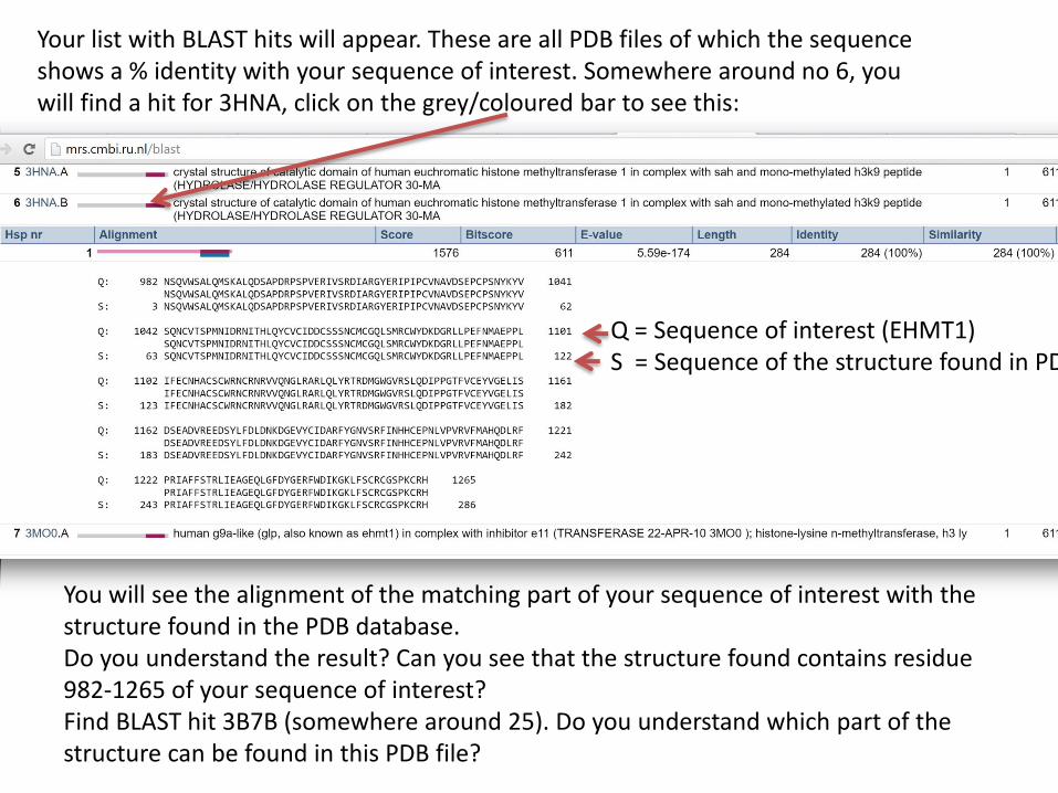
Your list with BLAST hits will appear. These are all PDB files of which the sequence shows a % identity with your sequence of interest. Somewhere around no 6, you will find a hit for 3HNA, click on the grey/coloured bar to see this:
You will see the alignment of the matching part of your sequence of interest with the structure found in the PDB database. Do you understand the result? Can you see that the structure found contains residue 982-1265 of your sequence of interest?Find BLAST hit 3B7B (somewhere around 25). Do you understand which part of the structure can be found in this PDB file?
Q = Sequence of interest (EHMT1)S = Sequence of the structure found in PDB
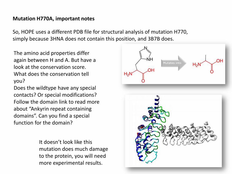
Mutation H770A, important notes
So, HOPE uses a different PDB file for structural analysis of mutation H770, simply because 3HNA does not contain this position, and 3B7B does.
The amino acid properties differ again between H and A. But have a look at the conservation score. What does the conservation tell you? Does the wildtype have any special contacts? Or special modifications?Follow the domain link to read more about “Ankyrin repeat containing domains”. Can you find a special function for the domain?
It doesn’t look like this mutation does much damage to the protein, you will need more experimental results.

Mutation A43V, important notes
As you can see HOPE cannot find a source of structural information. You can use the BLAST results from the previous example to see if there is indeed no structure known for this part of the protein.
It doesn’t look like there is anything known about the function of the wildtype residue. And the two residue types only differ in size.
However, the mutation was found before and is known in the Expasy database
You can follow the link to read more about this variant, would you classify this as a damaging mutation?
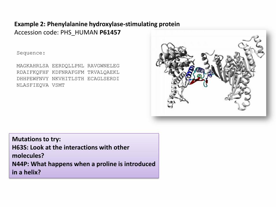
Sequence:
MAGKAHRLSA EERDQLLPNL RAVGWNELEG
RDAIFKQFHF KDFNRAFGFM TRVALQAEKL
DHHPEWFNVY NKVHITLSTH ECAGLSERDI
NLASFIEQVA VSMT
Example 2: Phenylalanine hydroxylase-stimulating proteinAccession code: PHS_HUMAN P61457
Mutations to try: H63S: Look at the interactions with other molecules?N44P: What happens when a proline is introduced in a helix?

Mutation H63S, important notes
H and S are very different residues and conservation is high.
Read the information about contacts indicated by PISA and look at the pictures. Do you agree that this residue is in contact with other molecules?
Does the domain information agree with the fact that this protein needs to function in a complex?
Look at the moving animations, how many mutations can you see? Why? In what kind of complex does this protein function? If you want you can read more information about the protein here:http://mrs.cmbi.ru.nl/entry?db=sprot&nr=300243&q=phs_human

Mutation N44P, important notes
N an P have a few differences in amino acid properties. You can see that due to size and hydrophobicity changes a few contacts will be lost.
More important for this protein is the fact that a proline will be introduced in a helix structure, this will cause a kink and destabilization of the structure. The pictures show that the helix is important for interactions with the other subunits in the complex.
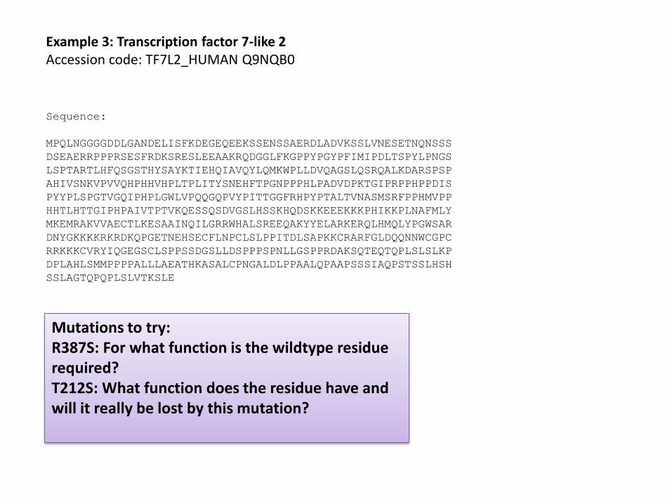
Sequence:
MPQLNGGGGDDLGANDELISFKDEGEQEEKSSENSSAERDLADVKSSLVNESETNQNSSS
DSEAERRPPPRSESFRDKSRESLEEAAKRQDGGLFKGPPYPGYPFIMIPDLTSPYLPNGS
LSPTARTLHFQSGSTHYSAYKTIEHQIAVQYLQMKWPLLDVQAGSLQSRQALKDARSPSP
AHIVSNKVPVVQHPHHVHPLTPLITYSNEHFTPGNPPPHLPADVDPKTGIPRPPHPPDIS
PYYPLSPGTVGQIPHPLGWLVPQQGQPVYPITTGGFRHPYPTALTVNASMSRFPPHMVPP
HHTLHTTGIPHPAIVTPTVKQESSQSDVGSLHSSKHQDSKKEEEKKKPHIKKPLNAFMLY
MKEMRAKVVAECTLKESAAINQILGRRWHALSREEQAKYYELARKERQLHMQLYPGWSAR
DNYGKKKKRKRDKQPGETNEHSECFLNPCLSLPPITDLSAPKKCRARFGLDQQNNWCGPC
RRKKKCVRYIQGEGSCLSPPSSDGSLLDSPPPSPNLLGSPPRDAKSQTEQTQPLSLSLKP
DPLAHLSMMPPPPALLLAEATHKASALCPNGALDLPPAALQPAAPSSSIAQPSTSSLHSH
SSLAGTQPQPLSLVTKSLE
Example 3: Transcription factor 7-like 2Accession code: TF7L2_HUMAN Q9NQB0
Mutations to try: R387S: For what function is the wildtype residue required?T212S: What function does the residue have and will it really be lost by this mutation?
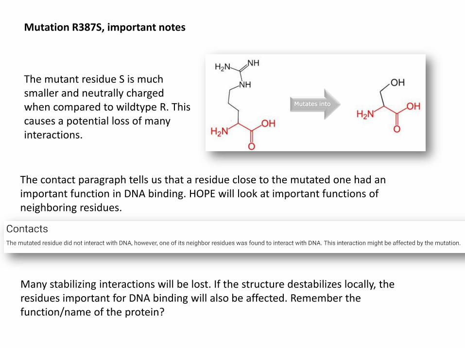
Mutation R387S, important notes
The mutant residue S is much smaller and neutrally charged when compared to wildtype R. This causes a potential loss of many interactions.
The contact paragraph tells us that a residue close to the mutated one had an important function in DNA binding. HOPE will look at important functions of neighboring residues.
Many stabilizing interactions will be lost. If the structure destabilizes locally, the residues important for DNA binding will also be affected. Remember the function/name of the protein?
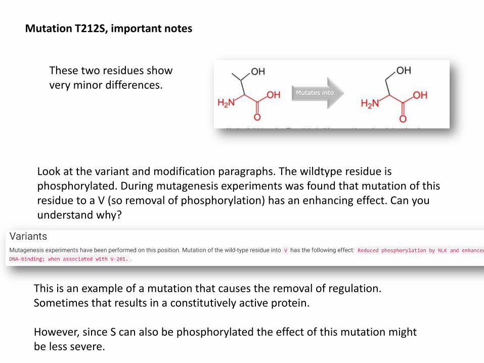
Mutation T212S, important notes
These two residues show very minor differences.
Look at the variant and modification paragraphs. The wildtype residue is phosphorylated. During mutagenesis experiments was found that mutation of this residue to a V (so removal of phosphorylation) has an enhancing effect. Can you understand why?
This is an example of a mutation that causes the removal of regulation. Sometimes that results in a constitutively active protein.
However, since S can also be phosphorylated the effect of this mutation might be less severe.

Your own examples…..
Now it’s time to try your own examples….
1: Find the amino acid sequence of your protein of interest. And submit the sequence in HOPE
Hint: use MRS or Uniprot to search for your protein: http://mrs.cmbi.ru.nl/ or http://www.uniprot.org/
2: Choose a mutation (if you don’t know an interesting mutation, you can try to find an interesting residue in MRS or Uniprot and see whether HOPE finds the same info). Submit this mutation to HOPE.
3: Wait for HOPE to finish the report and read it. See if you understand the results. Write down any questions you have so we can discuss these results.
You can also send any questions to [email protected]