varadi,a.,tsuboi,t.andrutter,g.a.(2005)myosinvatransports...
TRANSCRIPT

Varadi, A., Tsuboi, T. and Rutter, G. A. (2005) Myosin Va transportsdense core secretory vesicles in pancreatic MIN6 -cells. Molecular Bi-
ology of the Cell, 16 (6). pp. 2670-2680. ISSN 1059-1524 Availablefrom: http://eprints.uwe.ac.uk/5792
We recommend you cite the published version.
The publisher’s URL is:
http://dx.doi.org/10.1091/mbc.E04-11-1001
Refereed: Yes
This publication was vital for a successful BBSRC New Investigators Award
and a Wellcome Trust Project Grant recently awarded to Aniko Varadi.
Disclaimer
UWE has obtained warranties from all depositors as to their title in the material
deposited and as to their right to deposit such material.
UWE makes no representation or warranties of commercial utility, title, or fit-
ness for a particular purpose or any other warranty, express or implied in respect
of any material deposited.
UWE makes no representation that the use of the materials will not infringe
any patent, copyright, trademark or other property or proprietary rights.
UWE accepts no liability for any infringement of intellectual property rights
in any material deposited but will remove such material from public view pend-
ing investigation in the event of an allegation of any such infringement.
PLEASE SCROLL DOWN FOR TEXT.
Molecular Biology of the CellVol. 16, 2670–2680, June 2005
Myosin Va Transports Dense Core Secretory Vesicles inPancreatic MIN6 �-Cells□V
Aniko Varadi,*† Takashi Tsuboi,* and Guy A. Rutter*
*Henry Wellcome Laboratories for Integrated Cell Signalling and Department of Biochemistry, School ofMedical Sciences, University of Bristol, Bristol BS8 1TD, United Kingdom; and †Genomics Research Institute,Centre for Research in Biomedicine, University of the West of England, Bristol BS16 1QY, United Kingdom
Submitted November 16, 2004; Revised February 7, 2005; Accepted March 14, 2005Monitoring Editor: Benjamin Glick
The role of unconventional myosins in neuroendocrine cells is not fully understood, with involvement suggested in themovement of both secretory vesicles and mitochondria. Here, we demonstrate colocalization of myosin Va (MyoVa) withinsulin in pancreatic �-cells and show that MyoVa copurifies with insulin in density gradients and with the vesiclemarker phogrin-enhanced green fluorescent protein upon fluorescence-activated sorting of vesicles. By contrast, MyoVaimmunoreactivity was poorly colocalized with mitochondrial or other markers. Demonstrating an important role forMyoVa in the recruitment of secretory vesicles to the cell surface, a reduction of MyoVa protein levels achieved by RNAinterference caused a significant decrease in glucose- or depolarization-stimulated insulin secretion. Similarly, expressionof the dominant-negative–acting globular tail domain of MyoVa decreased by �50% the number of vesicles docked at theplasma membrane and by 87% the number of depolarization-stimulated exocytotic events detected by total internalreflection fluorescence microscopy. We conclude that MyoVa-driven movements of vesicles along the cortical actinnetwork are essential for the terminal stages of regulated exocytosis in �-cells.
INTRODUCTION
Glucose and other stimuli cause large dense core insulin-containing vesicles (LDCVs) to move toward, and eventu-ally fuse reversibly with, the plasma membrane in pancre-atic islet �-cells (Rutter, 2001, 2004). We have recently shownthat insulin-containing vesicles are transported from the cellcenter to the cortex primarily on the microtubule (MT)-based motor protein conventional kinesin, also called kine-sin I (Varadi et al., 2002, 2003). By contrast, the short-rangemovements in the cortical regions of the cell that carryvesicles over the last few hundred nanometers to the cellsurface seem more likely to involve F-actin, which is abun-dant in this region (Nakata and Hirokawa, 1992; Varadi etal., 2003; Tsuboi et al., 2003). Prime candidates for transportalong these filaments are class V myosin motors (Mermall etal., 1998; Rudolf et al., 2003). Myosin Va (MyoVa) is com-posed of two heavy chains that dimerize via a coiled-coilmotif located in the stalk region of the heavy chain (Cheneyet al., 1993). The heavy chain contains an amino-terminal,
actin-binding motor domain (Cheney et al., 1993) followedby a neck region to which up to six regulatory light chainsare bound, and a carboxy-terminal globular domain(Cheney et al., 1993) that is thought to mediate organellebinding specificity (Reck-Peterson et al., 2000).
Insight into the potential functions of MyoVa has comefrom several lines of investigation, including identificationof organelles and proteins that interact with MyoVa (Reck-Peterson et al., 2000; Karcher et al., 2002; Langford, 2002). Themost powerful strategy has been the phenotypic character-ization of lethal alleles of the dilute (MyoVa lethal mutant)mouse and cells derived from them (Mercer et al., 1991). Thedilute mice develop severe seizures and die within 3 wk afterbirth. The primary neuronal defect thought be responsible isthe absence of smooth endoplasmic reticulum within thedendritic spines of Purkinje neurons (Takagishi et al., 1996).Analyses of organelle movement in cultured melanocytes(Provance et al., 1996; Wu et al., 1998), neurons (Bridgman,1999) and macrophages (Al Haddad et al., 2001) from diluteand wild-type mice also have provided evidence for a directrole of MyoVa in both organelle transport and tethering.Moreover, MyoVa has been identified on synaptic vesicles(Evans et al., 1998; Miller and Sheetz, 2000), and inhibition ofmyosin ATPase activity reduces neurotransmitter release inbrain slices (Prekeris and Terrian, 1997). In addition, a recentreport revealed the role of MyoVa in the distribution ofsecretory granules in PC12 cells (Rudolf et al., 2003). Finally,the yeast homologue of MyoV, Myo2p, is involved in secre-tory vesicle (Schott et al., 1999), peroxisome (Hoepfner et al.,2001), and vacuole (Ishikawa et al., 2003) transport.
The role of MyoVa in mitochondrial transport is, however,less clear. In vitro motility assay demonstrated that actin-dependent motor activity is associated with mitochondria(Simon et al., 1995). Immunoelectron microscopy revealedassociation of MyoVa with the mitochondrial membrane in
This article was published online ahead of print in MBC in Press(http://www.molbiolcell.org/cgi/doi/10.1091/mbc.E04–11–1001)on March 23, 2005.□V The online version of this article contains supplemental materialat MBC Online (http://www.molbiolcell.org).
Address correspondence to: Aniko Varadi ([email protected]) or Guy A. Rutter ([email protected]).
Abbreviations used: [Ca2�]i, intracellular free calcium ion concen-tration; ER, endoplasmic reticulum; EGFP, enhanced green fluores-cent protein; FACS, fluorescence activated cell sorting; KRH, Krebs-Ringer-HEPES; LDCV, large dense core vesicle (secretory granule);MGTD, myosin Va globular tail domain; MT, microtubule; MyoVa,myosin Va; NPY, neuropeptide Y; siRNA, small interfering RNA;TIRF, total internal reflection fluorescence.
2670 © 2005 by The American Society for Cell Biology
melanoma cells (Nascimento et al., 1997). Furthermore, arecent study also described the presence of this motor pro-tein on mitochondria in myoblasts and suggested a role forMyoVa in calcium-dependent movements of these or-ganelles (Yi et al., 2004). In contrast, mitochondrial transportwas not affected in cells bearing mutations in either of thegenes for myosins I, II, and V (DePina and Langford, 1999),and a shortening of the lever arm of Myo2p had no effect onmitochondrial morphology, actin organization, or the rate ofmitochondrial movement in the mother cell (Boldogh et al.,1998). On the other hand, Myo2p mutant cells were defectivein the segregation of mitochondria (Boldogh et al., 1998,2004; Itoh et al., 2002).
Myosin Va is recruited to the vesicle surface by membersof the Rab family of G proteins (Fukuda et al., 2002; Hume etal., 2002; Provance et al., 2002, Wu et al., 2002b). This processrequires the presence of a rabphilin-like effector protein thatbridges the indirect interaction between the Rab protein andmyosin Va (Wu et al., 2002a; Fukuda et al., 2002). In mela-nocytes, Rab27a binds to the melanosome first and thenrecruits melanophilin/Slac-2a, a rabphilin-like effector pro-tein, which in turn recruits myosin Va (Fukuda et al., 2002;Wu et al., 2002a,b). In pancreatic �-cells, suppression ofRab27a function impaired insulin exocytosis triggered bysecretagogues (Waselle et al., 2003). The �-cells also expressa Rab27a effector protein granuphilin-a/SLP4-a, which islocalized on the membrane of insulin granules (Wang et al.,1999; Yi et al., 2002; Izumi et al., 2003), and its overexpressioncauses a profound inhibition of depolarization-induced in-sulin secretion (Wang et al., 1999; Coppola et al., 2002; Toriiet al., 2002). Granuphilin directly binds to syntaxin 1a (Toriiet al., 2002, 2004) and Munc 18-1 (Coppola et al., 2002) and isthought to be involved in tethering insulin granules to theplasma membrane (Torii et al., 2004). MyRIP/Slac2c, anotherRab27a effector (Kuroda et al., 2002), is also expressed in�-cells and involved in the regulation of insulin release(Waselle et al., 2003). However, unlike for Rab27 or its effec-tor proteins, the role of MyoVa in insulin-containing vesicletransport and membrane fusion is not known.
Here, we investigated the involvement of MyoVa in thedistribution, motility, and exocytosis of insulin-containingvesicles in single pancreatic �-cells (MIN6). Inhibition ofMyoVa function achieved by RNA interference or overex-pression of a dominant-negative mutant resulted in cluster-ing of secretory granules, a substantial reduction in thenumber of vesicles docked at the plasma membrane, and adecrease in the number of single exocytotic events and inglobal insulin release. These data demonstrate that MyoVa isessential for trafficking secretory organelles in neuroendo-crine cells.
MATERIALS AND METHODS
MaterialsCell culture reagents were from Invitrogen (Life Science Research, Paisley,United Kingdom), and all molecular biologicals were from Roche Diagnostics(Lewes, United Kingdom). Alexa Fluor goat anti-rabbit or anti-guinea pig 488and 568 secondary antibodies and Alexa Fluor 488-phalloidin were fromMolecular Probes (Eugene, OR). Mouse monoclonal anti-�-tubulin was ob-tained from Sigma Chemical (Poole, Dorset, United Kingdom). Rabbit poly-clonal anti-myosin Va antibody, DIL2, was raised against a glutathione S-transferase (GST)-fusion protein containing myosin V heavy chain residues910-1106, which correspond to the first segment of �-helical coiled-coil in thecentral rod domain (Wu et al., 1998).
Cell CultureMIN6 pancreatic �-cells (passages 19–35) were cultured in DMEM supple-mented with 15% (vol/vol) fetal calf serum, 100 U ml�1 penicillin, 0.1 mgml�1 streptomycin, and 2 mM l-glutamine at 37°C in an atmosphere of
humidified air (95%) and CO2 (5%) as described previously (Molnar et al.,1995).
PlasmidsA plasmid encoding mitochondrially targeted Discoideum red fluorescentprotein (Mito.DsRed) was generated as described previously (Varadi et al.,2002). The expression vector myosin Va globular tail domain (MGTD).pEGFPwas described previously (Wu et al., 1998). Plasmid encoding neuropeptide Y(NPY)-targeted monomeric red fluorescent protein (NPY-mRFP) was gener-ated as described previously (Tsuboi et al., 2003).
RNA Preparation, Reverse Transcription, and PCRTotal RNA from mouse brain, spleen, and MIN6 pancreatic �-cells wasprepared as described previously (Varadi et al., 1996). The total RNA (3 �g)was reverse transcribed at 42°C for 60 min in 50 �l of reaction mixturecontaining 1� reverse transcriptase buffer (50 mM Tris-HCl, pH 8.3, 40 mMKCl, 1 mM dithiothreitol [DTT], and 6 mM MgCl2) containing 1.25 �g ofoligo(dT), 0.5 mM each dNTP, 10 mM DTT, 40 U of rRNasin, and 500 U ofMoloney murine leukemia virus reverse transcriptase. One-tenth of the cDNAwas subjected to a PCR in 50-�l reaction mixture containing 1� PCR buffer (50mM KCl, 10 mM Tris-HCl, pH 9.0, and 5 mM DTT), which contained 2 or 4mM MgCl2, 0.1 mM each dNTP, 20 pmol of each primer, and 0.5 U of TaqDNA polymerase (Promega, Madison, WI). PCR was carried out in a pro-grammable thermal controller (PTC-100; MJ Research, Essex, United King-dom) at 95°C for 2 min followed by 30 cycles at 95°C for 1 min, 53°C for 1 min,and 72°C for 1 min. The last cycle was followed by a final extension step at72°C for 10 min. After PCR, the samples were subjected to electrophoresis ina 1.9% (wt/vol) agarose gel containing 0.4 �g of ethidium bromide in 1� TBEbuffer (89 mM Tris base, 89 mM boric acid, and 2 mM EDTA). For DNAsequencing and restriction enzyme digestion, 100-�l PCR samples were pre-cipitated with ethanol and then fractionated by electrophoresis in 1.3% (wt/vol) low melting point agarose gels. The separated bands were isolated, andthe PCR products were extracted from the gel slices by Wizard PCR prepsDNA purification system (Promega). The primers were designed to flank thealternative spliced forms in the C-terminal tail domain of MyoVa (Seperack etal., 1995). The following forward 1) 5�-CGAGCTGAATGAGTTGCGC-3�, nu-cleotides 3700–3718; 2) 5�-CATCTTGAGGTCGCAGCTGG-3�, nucleotides3841–3860; and reverse 3) 5�-GATATGTTCTCCATCTGCC-3�, nucleotides4371–4389; 4) 5�-GCTCATCGATGATCTCTCCTG-3, nucleotides 4392–4412,primers were used. Numbers correspond to MyoVa cDNA accession numberNM_010864. We used these primers in combinations (1–3, 1–4, 2–3, and 2–4),and all PCR reactions resulted in identical alternative spliced forms in allsamples tested. PCR amplification of MyoVa with primers 1 and 3 are shownon Figure 1A. The following controls were used to check for possible ampli-fication of contaminant DNA and RNA by PCR: RNA blanks taken throughthe cDNA step in the absence of reverse transcriptase were used in every PCRreaction and for each set of primers; samples without templates were run forevery primer pair for each PCR experiment.
Live Cell Imaging and ImmunocytochemistryCells were cotransfected with 1 �g of plasmids encoding 1) MGTD.EGFP andNPY.mRFP, 2) MGTD.EGFP and Mito.DsRed; or 3) empty vectors (pAdTrack-CMV, the latter encodes enhanced green fluorescent protein [EGFP]) (He et al.,1998), by using 10 �g ml�1 LipofectAMINE in Opti-MEM I medium (Invitro-gen) for 4 h. For live imaging, a Nipkov disk-based UltraVIEW confocalsystem (PerkinElmer Life and Analytical Sciences, Boston, MA) was used at2–4 Hz for 100 s (200–400 frames in total) in Krebs-Ringer-HEPES (KRH)bicarbonate buffer composed of 140 mM NaCl, 3.6 mM KCl, 0.5 mMNaH2PO4, 0.5 mM MgSO4, 2.0 mM NaHCO3, 16–30 mM glucose, 10 mMHEPES, pH 7.4, and 1.0 mM CaCl2 equilibrated with O2/CO2 [95:5 (vol/vol)]at 37°C. Immunocytochemistry was performed as described previously(Varadi and Rutter, 2002), and then images were captured on an UltraVIEWconfocal microscope (Varadi et al., 2004).
Total Internal Reflection Fluorescence (TIRF) MicroscopyCells were imaged in KRH bicarbonate buffer initially at 3 mM and then at 30mM glucose. Incubations were performed on a thermostatically controlled(37°C) stage of an Olympus IX-70 microscope (Olympus UK, London, UnitedKingdom) fitted with a high numerical aperture objective lens (Apochromatic,100�, 1.65 numerical aperture, infinity corrected; Olympus UK). To assess thedynamics of NPY.mRFP, we used a TIRF microscope (Till Photonics, Munich,Germany) as described previously (Tsuboi et al., 2000; Pinton et al., 2002;Tsuboi and Rutter, 2003). Images were captured at 2 Hz with a cooledcharge-coupled device camera (640 � 480 pixels, IMAGO; Till Photonics)controlled by TillvislON software. Image analysis was performed with Meta-Morph software (Universal Imaging, Downingtown, PA).
Measurement of Intracellular Free Ca2� Concentration[Ca2�]i
Changes in [Ca2�]i were measured at 37°C with entrapped Fura-2 (Grynk-iewicz et al., 1985) by using a Leica DM-IRBI inverted microscope (40�
Myosin Va in Insulin Granule Movement
Vol. 16, June 2005 2671
objective) and a Hamamatsu C4742-995 charged-coupled device cameradriven by OpenLab software (Improvision, Coventry, United Kingdom)(Varadi and Rutter, 2002).
Silencing of Endogenous Myosin Va Expression withSmall Interfering RNAs (siRNAs)Sequences corresponding to the mouse myosin Va cDNA (Strobel et al., 1990)2390–2410, 5�-AAGAGATACCTGTGTATGCAG-3�; and 2507–2528, 5�-AAG-TACTGGCGCATGTATGTG-3�) were used as targets for siRNA. The follow-ing siRNAs were used as controls: 5�-AAGTGTGCAACCATGTGAATG-3�and 5�-AAGTGGTTGGCGATTACCGAT-3�. These sequences showed no sig-nificant homology to any other gene known analyzed using BLAST search.
siRNAs were synthesized by in vitro transcription by using the SilencersiRNA construction kit (Ambion, Austin, TX). MIN6 cells were transfectedwith 60 pmol of siRNA duplex by using 3 �l of Oligofectamine (Invitrogen)in growth medium without antibiotics or serum for 5 h (Varadi et al., 2003).Whole cell lysate was prepared 24, 48, 72, and 96 h after transfection. Celllysate (10 �g) from siRNA and control RNA-transfected cells were separatedon a 9% polyacrylamide gel and then blotted onto Immobilon-P transfermembrane and probed with a rabbit polyclonal anti-myosin Va antibody anda mouse monoclonal anti-tubulin antibody. The blots were scanned andquantified with NIH ImageJ software (http://rsb.info.nih.gov/ij/).
Assay of Insulin ReleaseMIN6 cells were seeded at a density of 4–6 � 105 ml�1 on 24-mm-diameterpoly-l-lysine–coated coverslips and cultured overnight. Cells were trans-fected with siRNAs as described above. The cells were cultured for 48 h, andinsulin release was stimulated and assayed as described previously (Varadi etal., 2002) using Mercodia ultrasensitive mouse insulin enzyme-linked immu-nosorbent assay (ELISA) kit (Mercodia AB, Uppsala, Sweden).
Subcellular Fractionation by Using OptiPrep DensityGradient CentrifugationCells were homogenized in a buffer containing 0.3 M sucrose, 1 mM EDTA, 1mM MgSO4, 10 mM MES-NaOH, pH 6.5, 1 mM phenylmethylsulfonyl fluo-ride (PMSF), 5 �g ml�1 aprotinin, and 5 �g ml�1 leupeptin by using aball-bearing homogenizer and then centrifuged at 500 � g for 10 min. Thepostnuclear supernatant was layered on top of a continuous 8–19% (wt/vol)OptiPrep gradient obtained using a Gradient Master (BioComp Instruments,Fredericton, New Brunswick, Canada) and centrifuged at 16,000 � g for 16 h.Gradient fractions were collected by downward displacement (Gradient sta-tion; BioComp Instruments).
Fluorescence-activated Cell Sorting (FACS) of Vesicles andPrecipitation with Trichloroacetic Acid (TCA)MIN6 cells were infected with the recombinant phogrin.EGFP adenoviralconstruct at a multiplicity of 30–100 viral particles/cell, for 1 h. Cells weresubsequently used 24 h postinfection when �95% of cells were infected. Cellswere scraped into ice-cold buffer containing 10 mM 3-(N-morpholino)pro-panesulfonic acid (MOPS), 260 mM sucrose, pH 6.5, 1 mM PMSF, 5 �g ml�1
aprotinin, and 5 �g ml�1 leupeptin and then homogenized with a Teflonhomogenizer and centrifuged at 500 � g for 5 min. The postnuclear super-natant was resuspended in MOPS buffer to a concentration of 1–2 mg ml�1
and sorted into two fractions: particles labeled with EGFP and unlabeledorganelles. Sorting was carried out on a FACS Vantage sorter (BD Biosciences,San Jose, CA) fitted with a 488-nm argon ion laser. The EGFP fluorescence wasmeasured using a band pass filter at 530/30 nm. Vesicles (7 � 106) wereobtained following sorting. Vesicles (7 � 104) were seeded onto poly-l-lysine–coated coverslips and used for immunocytochemistry. The remaining vesiclesuspension was treated with equal volume of 20% (vol/vol) TCA for 30 minat 4°C and then centrifuged at 13,000 � g for 10 min. The pellet was washedtwice with cold acetone and then dried. The proteins were separated onSDS-gels.
Statistical AnalysisData are presented as the mean � SEM for the number of observations given,and statistical significance was calculated using an unpaired Student’s t test.
RESULTS
Myosin Va Is Expressed in Clonal MIN �-Cells and IsAssociated with Insulin-containing VesiclesInsulin-containing vesicles undergo short (non-Brownian)movements in the F-actin–rich cell cortex in clonal MIN6pancreatic �-cells (Tsuboi et al., 2003). To investigatewhether this movement might involve F-actin–dependenttransport via myosin motors, we investigated the presenceof unconventional MyoVa on insulin-containing LDCVs.
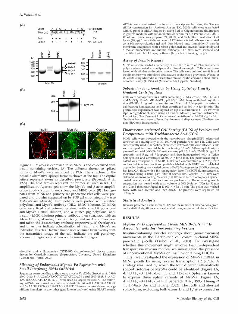
First, we investigated the expression of MyoVa mRNA inMIN6 �-cells by using reverse transcription (RT)-PCR. Astrategy was used by which the four different alternativelyspliced isoforms of MyoVa could be identified (Figure 1A;-B�D�F, -B�D-F, -B-D�F, and �B-D-F). Spleen is knownto express three splice variants of MyoVa (Figure 1A;-B�D�F, -B�D-F, -B-D�F; Seperack et al., 1995; Huang etal., 1998a,b; Au and Huang, 2002). The forth and shortestsplice form, excluding both exons D and F,‘ is expressed in
Figure 1. MyoVa is expressed in MIN6 cells and colocalized withinsulin-containing vesicles. (A) The different alternative splicedforms of MyoVa were amplified by PCR. The structure of thepossible alternative spliced forms is shown at the top. The capitalletters represent exons as described previously (Seperack et al.,1995). The bold arrows represent the primer set used in RT-PCRamplification. Agarose gels show the MyoVa and �-actin amplifi-cation products from brain, spleen, and MIN6 cells. (B) Homoge-nates from MIN6 and primary rat pancreatic islet cells were pre-pared and proteins separated on by SDS gel chromatography (seeMaterials and Methods). Immunoblots were probed with a rabbitpolyclonal anti-MyoVa antibody (DIL2, 1:5000 dilution). (C) MIN6cells were fixed and coimmunostained with a rabbit polyclonalanti-MyoVa (1:1000 dilution) and a guinea pig polyclonal anti-insulin (1:1000 dilution) primary antibody then visualized with anAlexa Fluor goat anti-guinea pig 568 (a) and an Alexa Fluor goatanti-rabbit 488 (b) secondary antibody, respectively. (c) Overlay of aand b. Arrows indicate colocalization of insulin and MyoVa onindividual vesicles. Hatched boundaries obtained from overlay withthe transmitted image of the cell, indicate the cell periphery.Zoomed in regions are shown on the inserted images.
A. Varadi et al.
Molecular Biology of the Cell2672
brain (Figure 1A; �B-D-F; Seperack et al., 1995; Huang et al.,1998a,b ; Au and Huang, 2002). Similar to brain and otherneuroendocrine cells (Seperack et al., 1995; Nagashima et al.,2002; Wu et al., 2002b), MIN6 �-cells expressed only the�B-D-F splice variant (Figure 1A). Immunoblot analysisrevealed that MyoVa is present at the protein level in bothMIN6 �-cells and primary rat islets of Langerhans (Figure1B). By using double-immunofluorescent microscopy,MyoVa was localized principally on insulin-positive struc-tures in both MIN6 cells (Figure 1C, zoomed in regions,arrowheads) and in primary rat �-cells (our unpublisheddata).
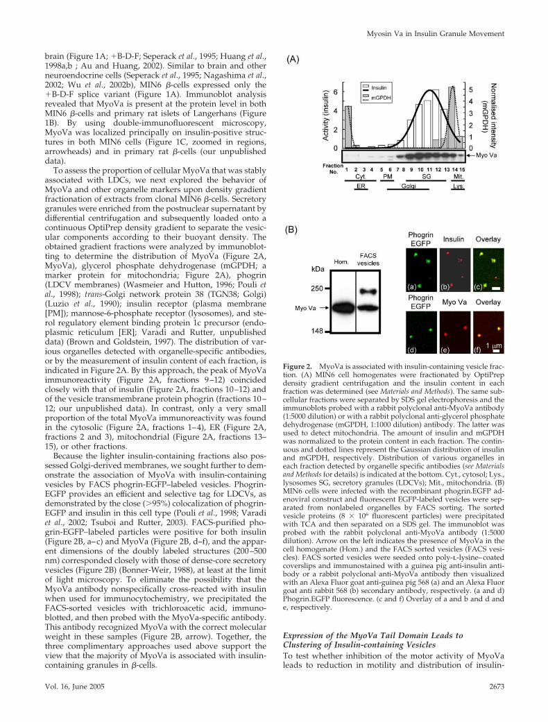
To assess the proportion of cellular MyoVa that was stablyassociated with LDCs, we next explored the behavior ofMyoVa and other organelle markers upon density gradientfractionation of extracts from clonal MIN6 �-cells. Secretorygranules were enriched from the postnuclear supernatant bydifferential centrifugation and subsequently loaded onto acontinuous OptiPrep density gradient to separate the vesic-ular components according to their buoyant density. Theobtained gradient fractions were analyzed by immunoblot-ting to determine the distribution of MyoVa (Figure 2A,MyoVa), glycerol phosphate dehydrogenase (mGPDH; amarker protein for mitochondria; Figure 2A), phogrin(LDCV membranes) (Wasmeier and Hutton, 1996; Pouli etal., 1998); trans-Golgi network protein 38 (TGN38; Golgi)(Luzio et al., 1990); insulin receptor (plasma membrane[PM]); mannose-6-phosphate receptor (lysosomes), and ste-rol regulatory element binding protein 1c precursor (endo-plasmic reticulum [ER]; Varadi and Rutter, unpublisheddata) (Brown and Goldstein, 1997). The distribution of var-ious organelles detected with organelle-specific antibodies,or by the measurement of insulin content of each fraction, isindicated in Figure 2A. By this approach, the peak of MyoVaimmunoreactivity (Figure 2A, fractions 9–12) coincidedclosely with that of insulin (Figure 2A, fractions 10–12) andof the vesicle transmembrane protein phogrin (fractions 10–12; our unpublished data). In contrast, only a very smallproportion of the total MyoVa immunoreactivity was foundin the cytosolic (Figure 2A, fractions 1–4), ER (Figure 2A,fractions 2 and 3), mitochondrial (Figure 2A, fractions 13–15), or other fractions.
Because the lighter insulin-containing fractions also pos-sessed Golgi-derived membranes, we sought further to dem-onstrate the association of MyoVa with insulin-containingvesicles by FACS phogrin-EGFP–labeled vesicles. Phogrin-EGFP provides an efficient and selective tag for LDCVs, asdemonstrated by the close (�95%) colocalization of phogrin-EGFP and insulin in this cell type (Pouli et al., 1998; Varadiet al., 2002; Tsuboi and Rutter, 2003). FACS-purified pho-grin-EGFP–labeled particles were positive for both insulin(Figure 2B, a–c) and MyoVa (Figure 2B, d–f), and the appar-ent dimensions of the doubly labeled structures (200–500nm) corresponded closely with those of dense-core secretoryvesicles (Figure 2B) (Bonner-Weir, 1988), at least at the limitof light microscopy. To eliminate the possibility that theMyoVa antibody nonspecifically cross-reacted with insulinwhen used for immunocytochemistry, we precipitated theFACS-sorted vesicles with trichloroacetic acid, immuno-blotted, and then probed with the MyoVa-specific antibody.This antibody recognized MyoVa with the correct molecularweight in these samples (Figure 2B, arrow). Together, thethree complimentary approaches used above support theview that the majority of MyoVa is associated with insulin-containing granules in �-cells.
Expression of the MyoVa Tail Domain Leads toClustering of Insulin-containing VesiclesTo test whether inhibition of the motor activity of MyoValeads to reduction in motility and distribution of insulin-
Figure 2. MyoVa is associated with insulin-containing vesicle frac-tion. (A) MIN6 cell homogenates were fractionated by OptiPrepdensity gradient centrifugation and the insulin content in eachfraction was determined (see Materials and Methods). The same sub-cellular fractions were separated by SDS gel electrophoresis and theimmunoblots probed with a rabbit polyclonal anti-MyoVa antibody(1:5000 dilution) or with a rabbit polyclonal anti-glycerol phosphatedehydrogenase (mGPDH, 1:1000 dilution) antibody. The latter wasused to detect mitochondria. The amount of insulin and mGPDHwas normalized to the protein content in each fraction. The contin-uous and dotted lines represent the Gaussian distribution of insulinand mGPDH, respectively. Distribution of various organelles ineach fraction detected by organelle specific antibodies (see Materialsand Methods for details) is indicated at the bottom. Cyt., cytosol; Lys.,lysosomes SG, secretory granules (LDCVs); Mit., mitochondria. (B)MIN6 cells were infected with the recombinant phogrin.EGFP ad-enoviral construct and fluorescent EGFP-labeled vesicles were sep-arated from nonlabeled organelles by FACS sorting. The sortedvesicle proteins (8 � 106 fluorescent particles) were precipitatedwith TCA and then separated on a SDS gel. The immunoblot wasprobed with the rabbit polyclonal anti-MyoVa antibody (1:5000dilution). Arrow on the left indicates the presence of MyoVa in thecell homogenate (Hom.) and the FACS sorted vesicles (FACS vesi-cles). FACS sorted vesicles were seeded onto poly-l-lysine–coatedcoverslips and immunostained with a guinea pig anti-insulin anti-body or a rabbit polyclonal anti-MyoVa antibody then visualizedwith an Alexa Fluor goat anti-guinea pig 568 (a) and an Alexa Fluorgoat anti rabbit 568 (b) secondary antibody, respectively. (a and d)Phogrin.EGFP fluorescence. (c and f) Overlay of a and b and d ande, respectively.
Myosin Va in Insulin Granule Movement
Vol. 16, June 2005 2673
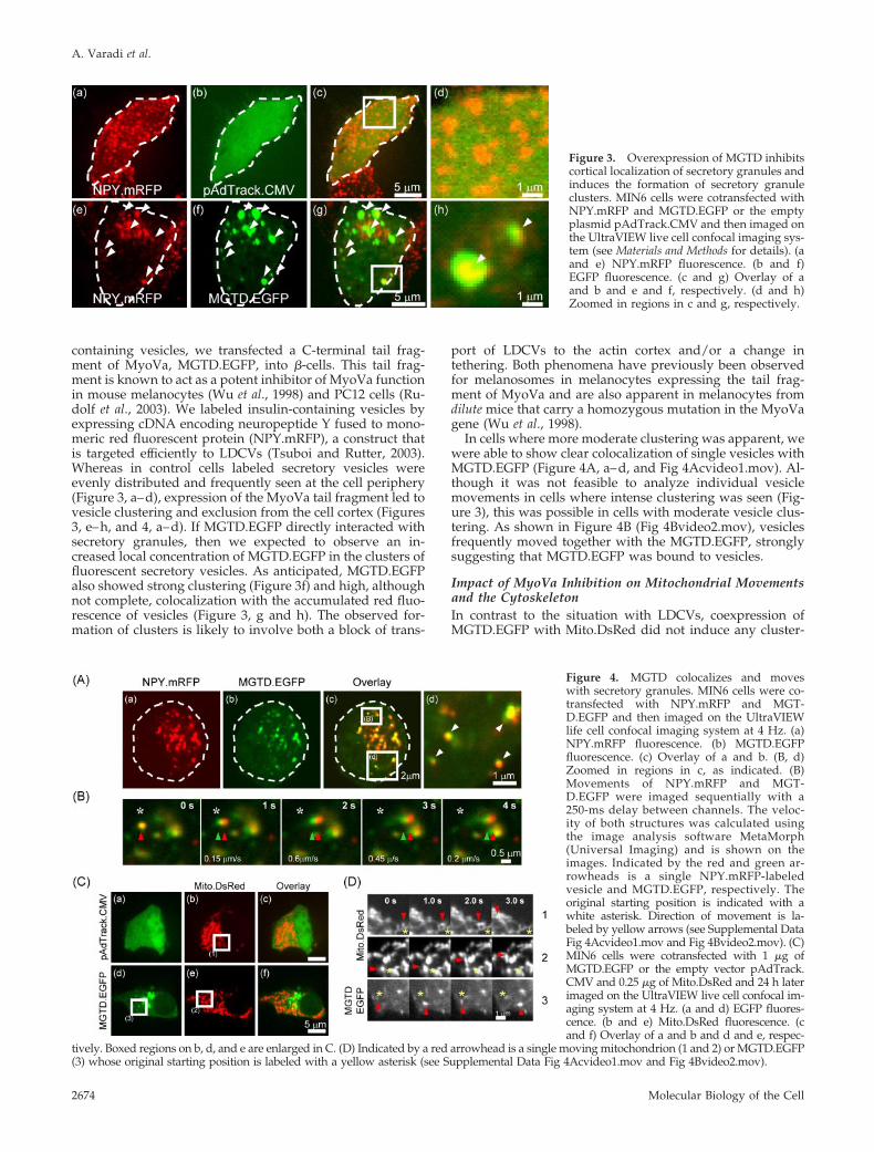
containing vesicles, we transfected a C-terminal tail frag-ment of MyoVa, MGTD.EGFP, into �-cells. This tail frag-ment is known to act as a potent inhibitor of MyoVa functionin mouse melanocytes (Wu et al., 1998) and PC12 cells (Ru-dolf et al., 2003). We labeled insulin-containing vesicles byexpressing cDNA encoding neuropeptide Y fused to mono-meric red fluorescent protein (NPY.mRFP), a construct thatis targeted efficiently to LDCVs (Tsuboi and Rutter, 2003).Whereas in control cells labeled secretory vesicles wereevenly distributed and frequently seen at the cell periphery(Figure 3, a–d), expression of the MyoVa tail fragment led tovesicle clustering and exclusion from the cell cortex (Figures3, e–h, and 4, a–d). If MGTD.EGFP directly interacted withsecretory granules, then we expected to observe an in-creased local concentration of MGTD.EGFP in the clusters offluorescent secretory vesicles. As anticipated, MGTD.EGFPalso showed strong clustering (Figure 3f) and high, althoughnot complete, colocalization with the accumulated red fluo-rescence of vesicles (Figure 3, g and h). The observed for-mation of clusters is likely to involve both a block of trans-
port of LDCVs to the actin cortex and/or a change intethering. Both phenomena have previously been observedfor melanosomes in melanocytes expressing the tail frag-ment of MyoVa and are also apparent in melanocytes fromdilute mice that carry a homozygous mutation in the MyoVagene (Wu et al., 1998).
In cells where more moderate clustering was apparent, wewere able to show clear colocalization of single vesicles withMGTD.EGFP (Figure 4A, a–d, and Fig 4Acvideo1.mov). Al-though it was not feasible to analyze individual vesiclemovements in cells where intense clustering was seen (Fig-ure 3), this was possible in cells with moderate vesicle clus-tering. As shown in Figure 4B (Fig 4Bvideo2.mov), vesiclesfrequently moved together with the MGTD.EGFP, stronglysuggesting that MGTD.EGFP was bound to vesicles.
Impact of MyoVa Inhibition on Mitochondrial Movementsand the CytoskeletonIn contrast to the situation with LDCVs, coexpression ofMGTD.EGFP with Mito.DsRed did not induce any cluster-
Figure 4. MGTD colocalizes and moveswith secretory granules. MIN6 cells were co-transfected with NPY.mRFP and MGT-D.EGFP and then imaged on the UltraVIEWlife cell confocal imaging system at 4 Hz. (a)NPY.mRFP fluorescence. (b) MGTD.EGFPfluorescence. (c) Overlay of a and b. (B, d)Zoomed in regions in c, as indicated. (B)Movements of NPY.mRFP and MGT-D.EGFP were imaged sequentially with a250-ms delay between channels. The veloc-ity of both structures was calculated usingthe image analysis software MetaMorph(Universal Imaging) and is shown on theimages. Indicated by the red and green ar-rowheads is a single NPY.mRFP-labeledvesicle and MGTD.EGFP, respectively. Theoriginal starting position is indicated with awhite asterisk. Direction of movement is la-beled by yellow arrows (see Supplemental DataFig 4Acvideo1.mov and Fig 4Bvideo2.mov). (C)MIN6 cells were cotransfected with 1 �g ofMGTD.EGFP or the empty vector pAdTrack.CMV and 0.25 �g of Mito.DsRed and 24 h laterimaged on the UltraVIEW live cell confocal im-aging system at 4 Hz. (a and d) EGFP fluores-cence. (b and e) Mito.DsRed fluorescence. (cand f) Overlay of a and b and d and e, respec-
tively. Boxed regions on b, d, and e are enlarged in C. (D) Indicated by a red arrowhead is a single moving mitochondrion (1 and 2) or MGTD.EGFP(3) whose original starting position is labeled with a yellow asterisk (see Supplemental Data Fig 4Acvideo1.mov and Fig 4Bvideo2.mov).
Figure 3. Overexpression of MGTD inhibitscortical localization of secretory granules andinduces the formation of secretory granuleclusters. MIN6 cells were cotransfected withNPY.mRFP and MGTD.EGFP or the emptyplasmid pAdTrack.CMV and then imaged onthe UltraVIEW live cell confocal imaging sys-tem (see Materials and Methods for details). (aand e) NPY.mRFP fluorescence. (b and f)EGFP fluorescence. (c and g) Overlay of aand b and e and f, respectively. (d and h)Zoomed in regions in c and g, respectively.
A. Varadi et al.
Molecular Biology of the Cell2674
ing of mitochondria (Figure 4C) and the majority of theexpressed MGTD.EGFP failed to localize with these or-ganelles (Figure 4C, d–f). Moreover, the movement of indi-vidual mitochondria was not affected by expression of MGT-D.EGFP (Figure 4, D and C). Thus, rapid movements ofMGTD.EGFP were observed that did not coincide with thoseof the separately labeled mitochondria (Figure 4D).
To eliminate the possibility that the clustering of LDCVsmay be due to altered organization of actin filaments and/ormicrotubules, we visualized these cytoskeletal structures incontrol and MGTD.EGFP-expressing cells. As anticipatedgiven the normal mitochondrial distribution and motility,no apparent effect of MGTD.EGFP was observed on thestructure and organization of actin and tubulin networks(our unpublished data). In MIN6 cells, strong phalloidinstaining was observed predominantly at the cortical regions,whereas tubulin extended from the perinuclear region to thecortex (our unpublished data). As expected, a small propor-tion of vesicles, probably those that were docked at theplasma membrane, clearly localized close to the phalloidin-positive F-actin cortex (our unpublished data). The above-mentioned data suggest that the expression of tail fragmentof MyoVa does not inhibit cellular membrane traffic in gen-eral but is selective for MyoVa-associated organelles andinterferes with their F-actin–dependent transport in the cor-tex.
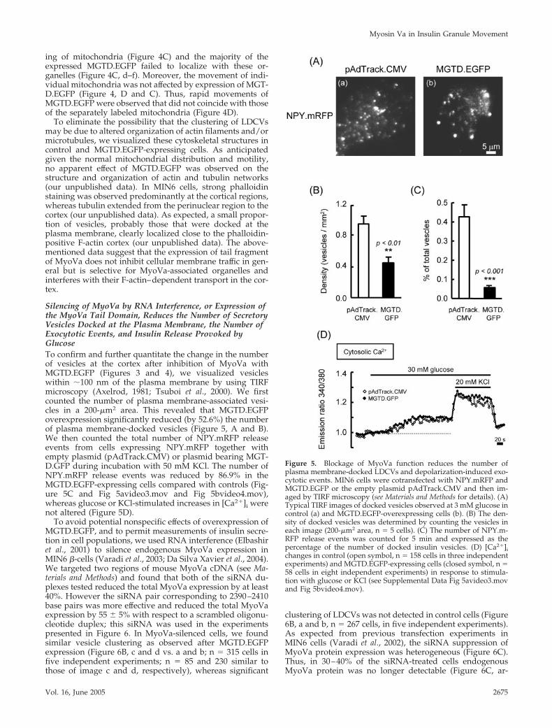
Silencing of MyoVa by RNA Interference, or Expression ofthe MyoVa Tail Domain, Reduces the Number of SecretoryVesicles Docked at the Plasma Membrane, the Number ofExocytotic Events, and Insulin Release Provoked byGlucoseTo confirm and further quantitate the change in the numberof vesicles at the cortex after inhibition of MyoVa withMGTD.EGFP (Figures 3 and 4), we visualized vesicleswithin �100 nm of the plasma membrane by using TIRFmicroscopy (Axelrod, 1981; Tsuboi et al., 2000). We firstcounted the number of plasma membrane-associated vesi-cles in a 200-�m2 area. This revealed that MGTD.EGFPoverexpression significantly reduced (by 52.6%) the numberof plasma membrane-docked vesicles (Figure 5, A and B).We then counted the total number of NPY.mRFP releaseevents from cells expressing NPY.mRFP together withempty plasmid (pAdTrack.CMV) or plasmid bearing MGT-D.GFP during incubation with 50 mM KCl. The number ofNPY.mRFP release events was reduced by 86.9% in theMGTD.EGFP-expressing cells compared with controls (Fig-ure 5C and Fig 5avideo3.mov and Fig 5bvideo4.mov),whereas glucose or KCl-stimulated increases in [Ca2�]i werenot altered (Figure 5D).
To avoid potential nonspecific effects of overexpression ofMGTD.EGFP, and to permit measurements of insulin secre-tion in cell populations, we used RNA interference (Elbashiret al., 2001) to silence endogenous MyoVa expression inMIN6 �-cells (Varadi et al., 2003; Da Silva Xavier et al., 2004).We targeted two regions of mouse MyoVa cDNA (see Ma-terials and Methods) and found that both of the siRNA du-plexes tested reduced the total MyoVa expression by at least40%. However the siRNA pair corresponding to 2390–2410base pairs was more effective and reduced the total MyoVaexpression by 55 � 5% with respect to a scrambled oligonu-cleotide duplex; this siRNA was used in the experimentspresented in Figure 6. In MyoVa-silenced cells, we foundsimilar vesicle clustering as observed after MGTD.EGFPexpression (Figure 6B, c and d vs. a and b; n � 315 cells infive independent experiments; n � 85 and 230 similar tothose of image c and d, respectively), whereas significant
clustering of LDCVs was not detected in control cells (Figure6B, a and b, n � 267 cells, in five independent experiments).As expected from previous transfection experiments inMIN6 cells (Varadi et al., 2002), the siRNA suppression ofMyoVa protein expression was heterogeneous (Figure 6C).Thus, in 30–40% of the siRNA-treated cells endogenousMyoVa protein was no longer detectable (Figure 6C, ar-
Figure 5. Blockage of MyoVa function reduces the number ofplasma membrane-docked LDCVs and depolarization-induced exo-cytotic events. MIN6 cells were cotransfected with NPY.mRFP andMGTD.EGFP or the empty plasmid pAdTrack.CMV and then im-aged by TIRF microscopy (see Materials and Methods for details). (A)Typical TIRF images of docked vesicles observed at 3 mM glucose incontrol (a) and MGTD.EGFP-overexpressing cells (b). (B) The den-sity of docked vesicles was determined by counting the vesicles ineach image (200-�m2 area, n � 5 cells). (C) The number of NPY.m-RFP release events was counted for 5 min and expressed as thepercentage of the number of docked insulin vesicles. (D) [Ca2�]ichanges in control (open symbol, n � 158 cells in three independentexperiments) and MGTD.EGFP-expressing cells (closed symbol, n �58 cells in eight independent experiments) in response to stimula-tion with glucose or KCl (see Supplemental Data Fig 5avideo3.movand Fig 5bvideo4.mov).
Myosin Va in Insulin Granule Movement
Vol. 16, June 2005 2675
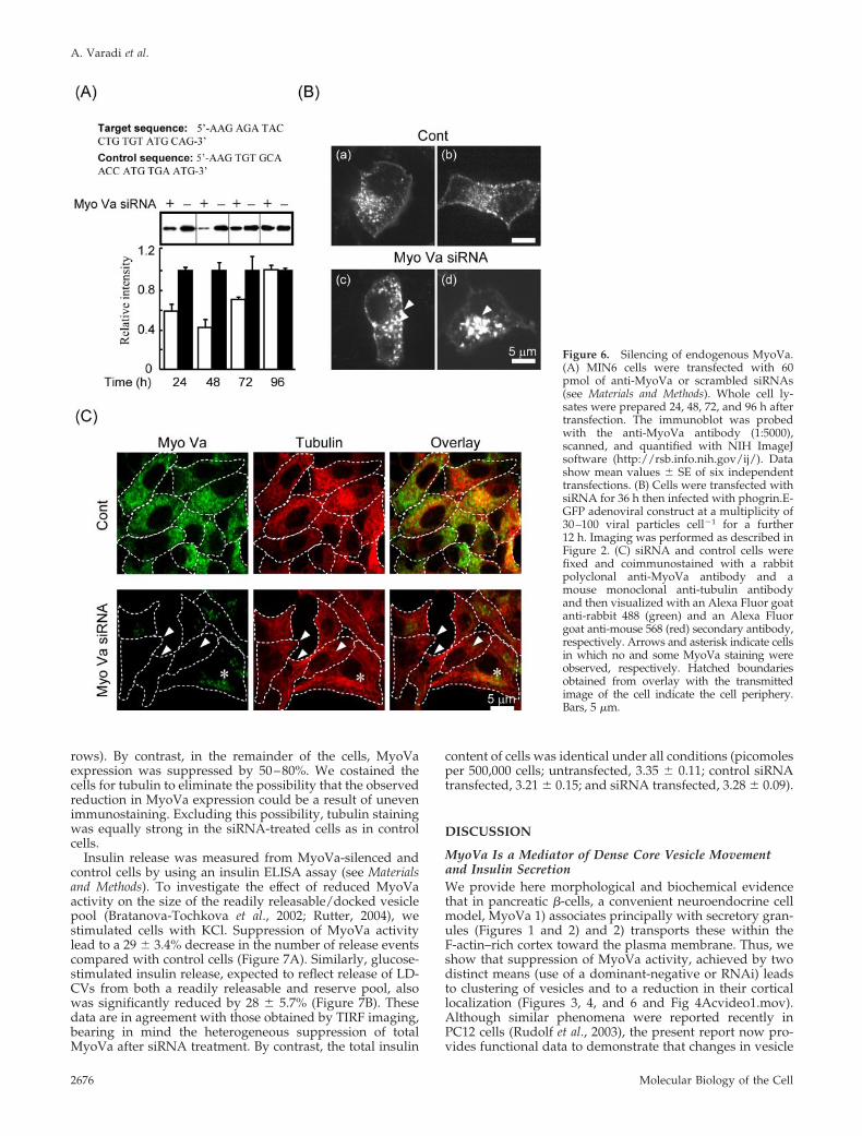
rows). By contrast, in the remainder of the cells, MyoVaexpression was suppressed by 50–80%. We costained thecells for tubulin to eliminate the possibility that the observedreduction in MyoVa expression could be a result of unevenimmunostaining. Excluding this possibility, tubulin stainingwas equally strong in the siRNA-treated cells as in controlcells.
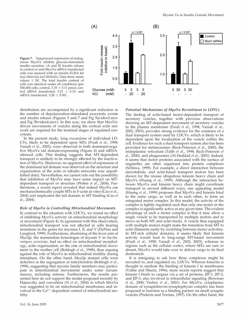
Insulin release was measured from MyoVa-silenced andcontrol cells by using an insulin ELISA assay (see Materialsand Methods). To investigate the effect of reduced MyoVaactivity on the size of the readily releasable/docked vesiclepool (Bratanova-Tochkova et al., 2002; Rutter, 2004), westimulated cells with KCl. Suppression of MyoVa activitylead to a 29 � 3.4% decrease in the number of release eventscompared with control cells (Figure 7A). Similarly, glucose-stimulated insulin release, expected to reflect release of LD-CVs from both a readily releasable and reserve pool, alsowas significantly reduced by 28 � 5.7% (Figure 7B). Thesedata are in agreement with those obtained by TIRF imaging,bearing in mind the heterogeneous suppression of totalMyoVa after siRNA treatment. By contrast, the total insulin
content of cells was identical under all conditions (picomolesper 500,000 cells; untransfected, 3.35 � 0.11; control siRNAtransfected, 3.21 � 0.15; and siRNA transfected, 3.28 � 0.09).
DISCUSSION
MyoVa Is a Mediator of Dense Core Vesicle Movementand Insulin SecretionWe provide here morphological and biochemical evidencethat in pancreatic �-cells, a convenient neuroendocrine cellmodel, MyoVa 1) associates principally with secretory gran-ules (Figures 1 and 2) and 2) transports these within theF-actin–rich cortex toward the plasma membrane. Thus, weshow that suppression of MyoVa activity, achieved by twodistinct means (use of a dominant-negative or RNAi) leadsto clustering of vesicles and to a reduction in their corticallocalization (Figures 3, 4, and 6 and Fig 4Acvideo1.mov).Although similar phenomena were reported recently inPC12 cells (Rudolf et al., 2003), the present report now pro-vides functional data to demonstrate that changes in vesicle
Figure 6. Silencing of endogenous MyoVa.(A) MIN6 cells were transfected with 60pmol of anti-MyoVa or scrambled siRNAs(see Materials and Methods). Whole cell ly-sates were prepared 24, 48, 72, and 96 h aftertransfection. The immunoblot was probedwith the anti-MyoVa antibody (1:5000),scanned, and quantified with NIH ImageJsoftware (http://rsb.info.nih.gov/ij/). Datashow mean values � SE of six independenttransfections. (B) Cells were transfected withsiRNA for 36 h then infected with phogrin.E-GFP adenoviral construct at a multiplicity of30–100 viral particles cell�1 for a further12 h. Imaging was performed as described inFigure 2. (C) siRNA and control cells werefixed and coimmunostained with a rabbitpolyclonal anti-MyoVa antibody and amouse monoclonal anti-tubulin antibodyand then visualized with an Alexa Fluor goatanti-rabbit 488 (green) and an Alexa Fluorgoat anti-mouse 568 (red) secondary antibody,respectively. Arrows and asterisk indicate cellsin which no and some MyoVa staining wereobserved, respectively. Hatched boundariesobtained from overlay with the transmittedimage of the cell indicate the cell periphery.Bars, 5 �m.
A. Varadi et al.
Molecular Biology of the Cell2676
distribution are accompanied by a significant reduction inthe number of depolarization-stimulated exocytotic eventsand insulin release (Figures 5 and 7 and Fig 5avideo3.movand Fig 5bvideo4.mov). In this way, we show that MyoVa-driven movements of vesicles along the cortical actin net-work are required for the terminal stages of regulated exo-cytosis.
In the present study, long excursions of individual LD-CVs, likely to be dependent upon MTs (Pouli et al., 1998;Varadi et al., 2003), were observed in both dominant-nega-tive MyoVa tail domain-expressing (Figure 4) and siRNA-silenced cells. This finding suggests that MT-dependenttransport is unlikely to be strongly affected by the inactiva-tion of MyoVa. Moreover, no apparent effect of expression ofthe dominant tail domain was observed on the structure andorganization of the actin or tubulin networks (our unpub-lished data). Nevertheless, we cannot rule out the possibilitythat inhibition of MyoVa may have some impact on MT-dependent transport or structure (Weber et al., 2004). Fur-thermore, a recent report revealed that indeed MyoVa canmechanochemically couple MTs to F-actin in vitro (Cao et al.,2004) and implicated the tail domain in MT binding (Cao etal., 2004).
Role of MyoVa in Controlling Mitochondrial MovementsIn contrast to the situation with LDCVs, we found no effectof inhibiting MyoVa activity on mitochondrial morphologyor movement (Figure 4). In agreement with this observation,mitochondrial transport was not affected in cells bearingmutations in the genes for myosins I, II, and V (DePina andLangford, 1999). Furthermore, shortening of the lever arm ofMyo2p, the mammalian homologue of myosin V in Saccha-romyces cerevisiae, had no effect on mitochondrial morphol-ogy, actin organization, or the rate of mitochondrial move-ment in the mother cell (Boldogh et al., 1998), thus arguingagainst the role of MyoVa in mitochondrial motility duringinterphase. On the other hand, Myo2p mutant cells weredefective in the segregation of mitochondria (Boldogh et al.,1998), suggesting that myosin family members may partici-pate in mitochondrial movements under some circum-stances, including mitosis. Furthermore, the results pre-sented here do not support the model proposed recently byHajnoczky and coworkers (Yi et al., 2004) in which MyoVawas suggested to be on mitochondrial membranes and in-volved in the Ca2�-dependent control of mitochondrial mo-tility.
Potential Mechanisms of MyoVa Recruitment to LDVCsThe finding of actin-based motor-dependent transport ofsecretory vesicles, together with previous observationsshowing an MT-dependent movement of secretory vesiclesto the plasma membrane (Pouli et al., 1998; Varadi et al.,2002, 2003), provides strong evidence for the existence of adual transport system used by LDCVs, which is likely to bedependent upon the localization of the vesicle within thecell. Evidence for such a dual transport system also has beenprovided for melanosomes (Reck-Peterson et al., 2000), theendoplasmic reticulum (Tabb et al., 1998; Reck-Peterson etal., 2000), and phagosomes (Al Haddad et al., 2001). Indeed,it seems that motor proteins associated with the surface oforganelles are often organized into protein complexes(Schliwa, 1999). For example, a direct interaction betweenmicrotubule- and actin-based transport motors has beenshown for the mouse ubiquitous kinesin heavy chain andMyoVa (Huang et al., 1999). Although the interaction be-tween MyoVa and kinesin heavy chain might coordinatetransport in several different ways, one appealing model(Huang et al., 1999) proposes that MyoVa and kinesin bindto the same cargo, as well as to each other, to form anintegrated motor complex. In this model, the activity of thecomplex is tightly regulated such that only one motor in thecomplex is significantly active at any given time. The evidentadvantage of such a motor complex is that it may allow asingle vesicle to be transported by multiple motors and tomove on both MT and actin tracks. A vesicle thus equippedwith multiple motors might make the transition from MT toactin filaments easily by switching between motor activities.In MT-rich cellular domains, it seems likely that kinesinactivity would lead to long-range MT-based movement(Pouli et al., 1998; Varadi et al., 2002, 2003), whereas inregions such as the cellular cortex, where MTs are rare orabsent, MyoVa would take over to deliver cargo to its finaldestination.
It is intriguing to ask how these complexes might berecruited to, and regulated on, LDCVs. Whereas kinectin isthought to mediate the binding of kinesin I to membranes(Vallee and Sheetz, 1996), more recent reports suggest thatkinesin I binds to cargoes via a set of proteins, JIP-1, JIP-2,and JIP-3, also involved in intracellular signaling (Bowmanet al., 2000; Verhey et al., 2001). For MyoVa, cytoplasmicdomain of synaptobrevin-synaptophysin complex has beenproposed to function as a binding partner on small synapticvesicles (Prekeris and Terrian, 1997). On the other hand, the
Figure 7. Suppressed expression of endog-enous MyoVa inhibits glucose-stimulatedinsulin secretion. (A and B) Insulin releasein control or anti-MyoVa siRNA-transfectedcells was assayed with an insulin ELISA kit(see Materials and Methods). Data show meanvalues � SE. The total insulin content ofcells was identical under all conditions (per500,000 cells; control, 3.35 � 0.11 pmol; con-trol siRNA transfected, 3.21 � 0.15; andsiRNA transfected, 3.28 � 0.09).
Myosin Va in Insulin Granule Movement
Vol. 16, June 2005 2677
small monomeric G protein family member Rab27a has beenimplicated in the interaction between melenosomes andMyoVa (Deacon and Gelfand, 2001) in a receptor-motorcomplex including the Rab27a binding protein melanophi-lin, a bridging protein with strong similarity to the Rab3aeffector protein rabphilin (Matesic et al., 2001).
The interaction between melanophilin and MyoVa hasbeen shown to be regulated by a melanocyte-specific alter-native splicing in the tail domain of MyoVa. In MyoVa,alternative splicing takes place between exons A and G(Figure 1A) with four alternatively spliced isoforms contain-ing different combinations of exons B, D, and F being iden-tified (Huang et al., 1998a,b; Seperack et al., 1995). The pres-ence of exon F, an alternatively spliced exon expressed inmelanocytes but not in neuroendocrine cells, is required forMyoVa to localize to melanosomes (Seperack et al., 1995;Nagashima et al., 2002; Wu et al., 2002b). In addition, theglobular tail domain is required for melanophilin–MyoVainteraction (Wu et al., 2002a). In PC12 cells, overexpressionof a tail fragment containing exon F did not target to vesic-ular structures. However, the expression of exon B wasrequired for the localization of MyoVa to vesicle structuresin this neuroendocrine cell (Au and Huang, 2002). In agree-ment with these results, we found that the neuronal splicevariant containing exon B but missing exons D and F isexpressed in �-cells.
Given the above, it seems likely that exon B and theglobular tail domain of MyoVa are involved in the interac-tion with a Rab effector protein in �-cells as described inmelanosomes. However, the composition of the organellereceptor complex may well be different in the two cell types.Thus, Rab27a and MyRIP have been implicated in the reg-ulation of exocytosis in pancreatic �-cells and neuroendo-crine PC12 cells (Waselle et al., 2003, Desnos et al., 2003).However, the Rab27/MyRIP protein complex does not seemto require recruitment of myosin on the secretory granulesfor function (Waselle et al., 2003). The association of myosinVa and Rab27/MyRIP has not been demonstrated in vivo,although it contributes to the cytoplasmic distribution ofsecretory granules in PC12 cells (Rudolf et al., 2003). Mightother members of the Rab family be involved in bindingMyoVa to vesicles? Rab3a is known to be specifically asso-ciated with small synaptic vesicles and insulin granules(Regazzi et al., 1996) and thus may have a role, a viewsupported by the fact that Rab3a�/� mice develop of hyper-glycemia and insulin secretory deficiencies (Yaekura et al.,2003). Further studies will be necessary to distinguish be-tween these possibilities and to explore the mechanismswhereby increases in glucose concentration might lead tothe recruitment of MyoVa to LDCVs in insulin-secretingcells.
ACKNOWLEDGMENTS
This study was supported by Wellcome Trust Programme Grant 067081/Z/02/Z and a Juvenile Diabetes Research Foundation Postdoctoral Fellowship(to T. T.). We thank Dr. Mark Jepson and Alan Leard (Bristol MRC ImagingFacility, University of Bristol, Bristol, United Kingdom) for technical assis-tance, Professor Peter Cullen for the use of the UltraVIEW confocal micro-scope, and Dr. Andrew Herman for the FACS of vesicles. We are grateful toProf. V. Gelfand (University of Illinois, Urbana-Champaign, IL) for providingthe MGTD.EGFP construct and DIL2 rabbit polyclonal anti-myosin V anti-body. G.A.R. is a Wellcome Trust Research Leave Fellow.
REFERENCES
Al Haddad, A., et al. (2001). Myosin Va bound to phagosomes binds to F-actinand delays microtubule-dependent motility. Mol. Biol. Cell 12, 2742–2755.
Au, J. S., and Huang, J. D. (2002). A tissue-specific exon of myosin Va isresponsible for selective cargo binding in melanocytes. Cell Motil. Cytoskel-eton 53, 89–102.
Axelrod, D. (1981). Cell-substrate contacts illuminated by total internal reflec-tion fluorescence. J. Cell Biol. 89, 141–145.
Boldogh, I., Vojtov, N., Karmon, S., and Pon, L. A. (1998). Interaction betweenmitochondria and the actin cytoskeleton in budding yeast requires two inte-gral mitochondrial outer membrane proteins, Mmm1p and Mdm10p. J. CellBiol. 141, 1371–1381.
Boldogh, I. R., Ramcharan, S. L., Yang, H. C., and Pon, L. A. (2004). A type Vmyosin (Myo2p) and a Rab-like G-protein (Ypt11p) are required for retentionof newly inherited mitochondria in yeast cells during cell division. Mol. Biol.Cell 15, 3994–4002.
Bonner-Weir, S. (1988). Morphological evidence for pancreatic polarity of betacells within islets of Langerhans. Diabetes 37, 616–621.
Bowman, A. B., Kamal, A., Ritchings, B. W., Philp, A. V., McGrail, M.,Gindhart, J. G., and Goldstein, L. S. (2000). Kinesin-dependent axonal trans-port is mediated by the Sunday driver (SYD) protein. Cell 103, 583–594.
Bratanova-Tochkova, T. K., Cheng, H., Daniel, S., Gunawardana, S., Liu, Y. J.,Mulvaney-Musa, J., Schermerhorn, T., Straub, S. G., Yajima, H., and Sharp,G. W. (2002). Triggering and augmentation mechanisms, granule pools, andbiphasic insulin secretion. Diabetes 51 (suppl 1), S83–S90, S83–S90.
Bridgman, P. C. (1999). Myosin Va movements in normal and dilute-lethalaxons provide support for a dual filament motor complex. J. Cell Biol. 146,1045–1060.
Brown, M. S., and Goldstein, J. L. (1997). The SREBP pathway: regulation ofcholesterol metabolism by proteolysis of a membrane-bound transcriptionfactor. Cell 89, 331–340.
Cao, T. T., Chang, W., Masters, S. E., and Mooseker, M. S. (2004). Myosin-Vabinds to and mechanochemically couples microtubules to actin filaments.Mol. Biol. Cell 15, 151–161.
Cheney, R. E., O’Shea, M. K., Heuser, J. E., Coelho, M. V., Wolenski, J. S.,Espreafico, E. M., Forscher, P., Larson, R. E., and Mooseker, M. S. (1993). Brainmyosin-V is a two-headed unconventional myosin with motor activity. Cell75, 13–23.
Coppola, T., Frantz, C., Perret-Menoud, V., Gattesco, S., Hirling, H., andRegazzi, R. (2002). Pancreatic beta-cell protein granuphilin binds Rab3 andMunc-18 and controls exocytosis. Mol. Biol. Cell 13, 1906–1915.
Da Silva Xavier, G., Qian, Q., Cullen, P. J., and Rutter, G. A. (2004). Distinctroles for insulin and insulin-like growth factor-1 receptors in pancreaticbeta-cell glucose sensing revealed by RNA silencing. Biochem. J. 377, 149–158.
Deacon, S. W., and Gelfand, V. I. (2001). Of yeast, mice, and men. Rab proteinsand organelle transport. J. Cell Biol. 152, F21–F24.
DePina, A. S., and Langford, G. M. (1999). Vesicle transport: the role of actinfilaments and myosin motors. Microsc. Res. Tech. 472, 93–106.
Desnos, C., et al. (2003). Rab27A and its effector MyRIP link secretory granulesto F-actin and control their motion towards release sites. J. Cell Biol. 163,559–570.
Elbashir, S. M., Harborth, J., Lendeckel, W., Yalcin, A., Weber, K., and Tuschl,T. (2001). Duplexes of 21-nucleotide RNAs mediate RNA interference incultured mammalian cells. Nature 411, 494–498.
Evans, L. L., Lee, A. J., Bridgman, P. C., and Mooseker, M. S. (1998). Vesicle-associated brain myosin-V can be activated to catalyze actin-based transport.J. Cell Sci. 111, 2055–2066.
Fukuda, M., Kuroda, T. S., and Mikoshiba, K. (2002). Slac2-a/melanophilin,the missing link between Rab27 and myosin Va: implications of a tripartiteprotein complex for melanosome transport. J. Biol. Chem. 277, 12432–12436.
Grynkiewicz, G., Poenie, M., and Tsien, R. Y. (1985). A new generation ofCa2� indicators with greatly improved fluorescence properties. J. Biol. Chem.260, 3440–3450.
He, T. C., Zhou, S., da Costa, L. T., Yu, J., Kinzler, K. W., and Vogelstein, B.(1998). A simplified system for generating recombinant adenoviruses. Proc.Natl. Acad. Sci. USA 95, 2509–2514.
Hoepfner, D., van den Berg, M., Philippsen, P., Tabak, H. F., and Hettema,E. H. (2001). A role for Vps1p, actin, and the Myo2p motor in peroxisomeabundance and inheritance in Saccharomyces cerevisiae. J. Cell Biol. 155, 979–990.
Huang, J. D., Cope, M. J., Mermall, V., Strobel, M. C., Kendrick-Jones, J.,Russell, L. B., Mooseker, M. S., Copeland, N. G., and Jenkins, N. A. (1998a).Molecular genetic dissection of mouse unconventional myosin-VA: headregion mutations. Genetics 148, 1951–1961.
A. Varadi et al.
Molecular Biology of the Cell2678
Huang, J. D., Mermall, V., Strobel, M. C., Russell, L. B., Mooseker, M. S.,Copeland, N. G., and Jenkins, N. A. (1998b). Molecular genetic dissection ofmouse unconventional myosin-VA: tail region mutations. Genetics 148, 1963–1972.
Huang, J. D., Brady, S. T., Richards, B. W., Stenolen, D., Resau, J. H., Cope-land, N. G., and Jenkins, N. A. (1999). Direct interaction of microtubule- andactin-based transport motors. Nature 397, 267–270.
Hume, A. N., Collinson, L. M., Hopkins, C. R., Strom, M., Barral, D. C., Bossi,G., Griffiths, G. M., and Seabra, M. C. (2002). The leaden gene product isrequired with Rab27a to recruit myosin Va to melanosomes in melanocytes.Traffic 3, 193–202.
Ishikawa, K., Catlett, N. L., Novak, J. L., Tang, F., Nau, J. J., and Weisman, L. S.(2003). Identification of an organelle-specific myosin V receptor. J. Cell Biol.160, 887–897.
Itoh, T., Watabe, A., Toh, E., and Matsui, Y. (2002). Complex formation withYpt11p, a rab-type small GTPase, is essential to facilitate the function ofMyo2p, a class V myosin, in mitochondrial distribution in Saccharomycescerevisiae. Mol. Cell. Biol. 22, 7744–7757.
Izumi, T., Gomi, H., Kasai, K., Mizutani, S., and Torii, S. (2003). The roles ofRab27 and its effectors in the regulated secretory pathways. Cell Struct. Funct.28, 465–474.
Karcher, R. L., Deacon, S. W., and Gelfand, V. I. (2002). Motor-cargo interac-tions: the key to transport specificity. Trends Cell Biol. 12, 21–27.
Kuroda, T. S., Fukuda, M., Ariga, H., and Mikoshiba, K. (2002). The Slphomology domain of synaptotagmin-like proteins 1–4 and Slac2 functions asa novel Rab27A binding domain. J. Biol. Chem. 277, 9212–9218.
Langford, G. M. (2002). Myosin-V, a versatile motor for short-range vesicletransport. Traffic 3, 859–865.
Luzio, J. P., Brake, B., Banting, G., Howell, K. E., Braghetta, P., and Stanley,K. K. (1990). Identification, sequencing and expression of an integral mem-brane protein of the trans-Golgi network (TGN38). Biochem. J. 270, 97–102.
Matesic, L. E., Yip, R., Reuss, A. E., Swing, D. A., O’Sullivan, T. N., Fletcher,C. F., Copeland, N. G., and Jenkins, N. A. (2001). Mutations in Mlph, encodinga member of the Rab effector family, cause the melanosome transport defectsobserved in leaden mice. Proc. Natl. Acad. Sci. USA 98, 10238–10243.
Mercer, J. A., Seperack, P. K., Strobel, M. C., Copeland, N. G., and Jenkins,N. A. (1991). Novel myosin heavy chain encoded by murine dilute coat colourlocus. Nature 349, 709–713.
Mermall, V., Post, P. L., and Mooseker, M. S. (1998). Unconventional myosinsin cell movement, membrane traffic, and signal transduction. Science 279,527–533.
Miller, K. E., and Sheetz, M. P. (2000). Characterization of myosin V bindingto brain vesicles. J. Biol. Chem. 275, 2598–2606.
Molnar, E., Varadi, A., McIlhinney, R.A.J., and Ashcroft, S.J.H. (1995). Iden-tification of functional ionotropic glutamate receptor proteins in pancreaticbeta-cells and in islets of Langerhans. FEBS Lett. 371, 253–257.
Nagashima, K., Torii, S., Yi, Z., Igarashi, M., Okamoto, K., Takeuchi, T., andIzumi, T. (2002). Melanophilin directly links Rab27a and myosin Va throughits distinct coiled-coil regions. FEBS Lett. 517, 233–238.
Nakata, T., and Hirokawa, N. (1992). Organization of cortical cytoskeleton ofcultured chromaffin cells and involvement in secretion as revealed by quick-freeze, deep-etching, and double-label immunoelectron microscopy. J. Neu-rosci. 12, 2186–2197.
Nascimento, A. A., Amaral, R. G., Bizario, J. C., Larson, R. E., and Espreafico,E. M. (1997). Subcellular localization of myosin-V in the B16 melanoma cells,a wild-type cell line for the dilute gene. Mol. Biol. Cell 10, 1971–1988.
Pinton, P., Tsuboi, T., Ainscow, E. K., Pozzan, T., Rizzuto, R., and Rutter, G. A.(2002). Dynamics of glucose-induced recruitment of protein kinase C �II inliving pancreatic islet �-cells. J. Biol. Chem. 277, 37702–37710.
Pouli, A. E., Emmanouilidou, E., Zhao, C., Wasmeier, C., Hutton, J. C., andRutter, G. A. (1998). Secretory granule dynamics visualised in vivo with aphogrin-green fluorescent protein chimaera. Biochem. J. 333, 193–199.
Prekeris, R., and Terrian, D. M. (1997). Brain myosin V is a synaptic vesicle-associated motor protein: evidence for a Ca2�-dependent interaction with thesynaptobrevin-synaptophysin complex. J. Cell Biol. 137, 1589–1601.
Provance, D. W., Jr., Wei, M., Ipe, V., and Mercer, J. A. (1996). Culturedmelanocytes from dilute mutant mice exhibit dendritic morphology andaltered melanosome distribution. Proc. Natl. Acad. Sci. USA 93, 14554–14558.
Provance, D. W., James, T. L., and Mercer, J. A. (2002). Melanophilin, theproduct of the leaden locus, is required for targeting of myosin-Va to mela-nosomes. Traffic 2, 124–132.
Reck-Peterson, S. L., Provance, D. W., Jr., Mooseker, M. S., and Mercer, J. A.(2000). Class V myosins. Biochim. Biophys. Acta 1496, 36–51.
Regazzi, R., Ravazzola, M., Lezzi, M., Lang, J. C., Zahraoui, A., Andereggen,E., Morel, P., Takai, Y., and Wollheim, C. B. (1996). Expression, localizationand functional role of small GTPases of the Rab3 family in insulin-secretingcells. J. Cell Sci. 109, 2265–2273.
Rudolf, R., Kogel, T., Kuznetsov, S. A., Salm, T., Schlicker, O., Hellwig, A.,Hammer, J. A., 3rd, and Gerdes, H. H. (2003). Myosin Va facilitates thedistribution of secretory granules in the F-actin rich cortex of PC12 cells. J. CellSci. 116, 1339–1348.
Rutter, G. A. (2001). Nutrient-secretion coupling in the pancreatic islet �-cell:recent advances. Mol. Asp. Med. 22, 247–284.
Rutter, G. A. (2004) Visualising Insulin Secretion. The Minkowski Lecture2004. Diabetologia 47, 1861–1872.
Schliwa, M. (1999). Molecular motors join forces. Nature 397, 204–205.
Schott, D., Ho, J., Pruyne, D., and Bretscher, A. (1999). The COOH-terminaldomain of Myo2p, a yeast myosin V, has a direct role in secretory vesicletargeting. J. Cell Biol. 147, 791–808.
Seperack, P. K., Mercer, J. A., Strobel, M. C., Copeland, N. G., and Jenkins,N. A. (1995). Retroviral sequences located within an intron of the dilute genealter dilute expression in a tissue-specific manner. EMBO J. 14, 2326–2332.
Simon, V. R., Swayne, T. C., and Pon, L. A. (1995). Actin-dependent mito-chondrial motility in mitotic yeast and cell-free systems: identification of amotor activity on the mitochondrial surface. J. Cell Biol. 130, 345–354.
Strobel, M. C., Seperack, P. K., Copeland, N. G., and Jenkins, N. A. (1990).Molecular analysis of two mouse dilute locus deletion mutations: spontane-ous dilute lethal20J and radiation-induced dilute prenatal lethal Aa2 alleles.Mol. Cell. Biol. 10, 501–509.
Tabb, J. S., Molyneaux, B. J., Cohen, D. L., Kuznetsov, S. A., and Langford,G. M. (1998). Transport of ER vesicles on actin filaments in neurons by myosinV. J. Cell Sci. 111, 3221–3234.
Takagishi, Y., Oda, S., Hayasaka, S., Dekker-Ohno, K., Shikata, T., Inouye, M.,and Yamamura, H. (1996). The dilute-lethal (dl) gene attacks a Ca2� store inthe dendritic spine of Purkinje cells in mice. Neurosci. Lett. 215, 169–172.
Torii, S., Takeuchi, T., Nagamatsu, S., and Izumi, T. (2004). Rab27 effectorgranuphilin promotes the plasma membrane targeting of insulin granules viainteraction with syntaxin 1a. J. Biol. Chem. 279, 22532–22538.
Torii, S., Zhao, S., Yi, Z., Takeuchi, T., and Izumi, T. (2002). Granuphilinmodulates the exocytosis of secretory granules through interaction with syn-taxin 1a. Mol. Cell. Biol. 22, 5518–5526.
Tsuboi, T., Da Silva Xavier, G., Leclerc, I., and Rutter, G. A. (2003). 5�AMP-activated protein kinase controls insulin-containing secretory vesicledynamics. J. Biol. Chem. 278, 52042–52051.
Tsuboi, T., and Rutter, G. A. (2003) Multiple forms of kiss and run exocytosisrevealed by evanescent wave microscopy. Curr. Biol. 13, 563–567.
Tsuboi, T., Zhao, C., Terakawa, S., and Rutter, G. A. (2000). Simultaneousevanescent wave imaging of insulin vesicle membrane and cargo during asingle exocytotic event. Curr. Biol. 10, 1307–1310.
Vallee, R. B., and Sheetz, M. P. (1996). Targeting of motor proteins. Science271, 1539–1544.
Varadi, A., Ainscow, E. K., Allan, V. J., and Rutter, G. A. (2002). Involvementof conventional kinesin in glucose-stimulated secretory-granule movementsand exocytosis in clonal pancreatic �-cells. J. Cell Sci. 115, 4177–4189.
Varadi, A., Molnar, E., Ostenson, C. G., and Ashcroft, S. J. (1996). Isoforms ofendoplasmic reticulum Ca(2�)-ATPase are differentially expressed in normaland diabetic islets of Langerhans. Biochem. J. 319, 521–527.
Varadi, A., Johnson-Cadwell, L. I., Cirulli, V., Yoon, Y., Allan, V. J., andRutter, G. A. (2004). Cytoplasmic dynein regulates the subcellular distributionof mitochondria by controlling the recruitment of the fission factor dynamin-related protein-1. J. Cell Sci. 117, 4389–4400.
Varadi, A., and Rutter, G. A. (2002). Dynamic imaging of endoplasmic retic-ulum [Ca2�] in MIN6 �-cells using recombinant chameleons: roles of SERCA2and ryanodine receptors. Diabetes 51, S190–S201.
Varadi, A., Tsuboi, T., Johnson-Cadwell, L. I., Allan, V. J., and Rutter, G. A.(2003). Kinesin I and cytoplasmic dynein orchestrate glucose-stimulated in-sulin-containing vesicle movements in clonal MIN6 beta-cells. Biochem. Bio-phys. Res. Commun. 311, 272–282.
Verhey, K. J., Meyer, D., Deehan, R., Blenis, J., Schnapp, B. J., Rapoport, T. A.,and Margolis, B. (2001). Cargo of kinesin identified as JIP scaffolding proteinsand associated signaling molecules. J. Cell Biol. 152, 959–970.
Myosin Va in Insulin Granule Movement
Vol. 16, June 2005 2679
Wang, J., Takeuchi, T., Yokota, H., and Izumi, T. (1999). Novel rabphilin-3-likeprotein associates with insulin-containing granules in pancreatic beta cells.J. Biol. Chem. 274, 28542–28548.
Waselle, L., Coppola, T., Fukuda, M., Iezzi, M., El Amraoui, A., Petit, C., andRegazzi, R. (2003). Involvement of the Rab27 binding protein Slac2c/MyRIPin insulin exocytosis. Mol. Biol. Cell 14, 4103–4113.
Wasmeier, C., and Hutton, J. C. (1996). Molecular cloning of phogrin, aprotein-tyrosine phosphatase homologue localized to insulin secretory gran-ule membranes. J. Biol. Chem. 271, 18161–18170.
Weber, K. L., Sokac, A. M., Berg, J. S., Cheney, R. E., and Bement, W. M. (2004).A microtubule-binding myosin required for nuclear anchoring and spindleassembly. Nature 431, 325–329.
Wu, X., Bowers, B., Rao, K., Wei, Q., and Hammer, J. A. (1998). Visualizationof melanosome dynamics within wild-type and dilute melanocytes suggests aparadigm for myosin V function in vivo. J. Cell Biol. 143, 1899–1918.
Wu, X. S., Rao, K., Zhang, H., Wang, F., Sellers, J. R., Matesic, L. E., Copeland,N. G., Jenkins, N. A., and Hammer, J. A., 3rd. (2002a) Identification of anorganelle receptor for myosin-Va. Nat. Cell Biol. 4, 271–278.
Wu, X., Wang, F., Rao, K., Sellers, J. R., and Hammer, J. A., 3rd. (2002b)Rab27a is an essential component of melanosome receptor for myosin Va.Mol. Biol. Cell 5, 1735–1749.
Yaekura, K., et al. (2003). Insulin secretory deficiency and glucose intolerancein Rab3A null mice. J. Biol. Chem. 278, 9715–9721.
Yi, M., Weaver, D., and Hajnoczky, G. (2004). Control of mitochondrialmotility and distribution by the calcium signal: a homeostatic circuit. J. CellBiol. 167, 661–672.
Yi, Z., Yokota, H., Torii, S., Aoki, T., Hosaka, M., Zhao, S., Takata, K.,Takeuchi, T., and Izumi, T. (2002). The Rab27a/granuphilin complex regu-lates the exocytosis of insulin-containing dense-core granules. Mol. Cell. Biol.22, 1858–1867.
A. Varadi et al.
Molecular Biology of the Cell2680