value of antegrade ureteral dilation for late ureter obstruction in renal transplants
TRANSCRIPT

Transplant Int (1989) 2: 33-35 International
0 Springer-Verlag 1989
Value of antegrade ureteral dilation for late ureter obstruction in renal transplants G. Benoit', P. Icard', H. Bensadoun', B.Charpentier2, M. Moukarzel', A. Jardin', and D. Fries2
Department of Urology and * Department of.Nephrology, HBpital Universitaire de Bicitre, 78, rue du Gtntral Leclerc, F-94275 Le Kremlin Bicitre Cedex, France
Abstract. We report on eight kidney-allografted pa- tients treated for delayed ureteral obstruction be- tween January 1986 and January 1987. In all cases, standard endourological dilation was performed using a balloon catheter, and this was followed by in- sertion of a pigtail stent. All eight cases showed im- provement 1 month after dilation (decrease in crea- tinine and caliceal dilation). At 6 months, renal function had deteriorated in six patients but re- mained good in two. One of the six patients was redi- lated with apparently good results. The remaining five were operated on using their own ureter. We conclude that while internal drainage helps in distin- guishing between obstruction and other causes of creatinine increase, antegrade dilation is the treat- ment of choice for delayed ureteral obstruction.
Key words: Ureteral obstruction in renal transplanta- tion - Antegrade ureteral dilation.
The viability of the ureter in renal transplants de- pends particularly upon its blood supply from the side of the kidney, so extreme care must be taken to preserve the blood supply during organ procure- ment.
The occurrence of urological complications after renal transplantation can be reduced by using a short, well-vascularized ureter [2]. Yet, even after this precaution has been taken, the risk of ureteral ste- nosis remains high and even increases with time: 4.6% after 1 year, 7.7% after 2 years, and 9.1 Yo after 5 years [lo, 151. The overall incidence of ureteral ste- nosis at our institution is 2.9% (1100 transplants).
Endourological techniques have assumed an im- portant role in the management of early urological
Oflprinr requests 10: G. Benoit
complications in renal transplantation [l, 3, 6, 7, 9, 161. However, treatment procedures for delayed ureteral stenosis are not unequivocal. In some cases, when there is no loss of graft function, a simple fol- low-up might be recommended. In other cases, sur- gical repair is indicated.
We report here on the use of antegrade ureteral dilation for the treatment of delayed ureteral ste- nosis.
Patients and methods
Between 1986 and 1987. eight kidney-allografted patients were treated for ureteral obstruction. In all cases there was a uretero- vesical implantation performed according to the Leadbetter Poli- tan0 technique using a 2 cm submucosal tunnel. The epigastric vessels were always cut and ligated.
The mean time from renal transplantation to diagnosis of ureteral obstruction was 21.9f 13 months. Two of the eight pa- tients had weak bladder compliance, but no true functional blad- der lesion was present. The mean creatinine level was 220+ 133 pmol/l (range: 100-400 pnol/l).
The diagnosis of ureteral obstruction was made using echog- raphy, intravenous pyelogram (IVP; Fig. l), and wmTc-diethylene triaminepenta-acetic acid renography with furosemide injection. A Whitaker test was never used.
An 18-gauge Chiba needle was inserted into the pelvis under echographic and fluoroscopic control, followed by localization of the stenotic level and insertion of a guide wire through the ureteral stenosis into the bladder. A balloon catheter (10 cni long and 7 mm in diameter) was then inserted over the guide wire. The bal- loon was inflated two times within 5 minutes and withdrawn after checking that the stenosis had disappeared after dilation. An 8 Fr double pigtail stent was inserted and left in place for 1 month. Prophylactic antibiotherapy during dilation consisted of ampicil- lin, oxacillin, and gentamicin; this was replaced by trimethopnm sulfamethoxazole after 2 days. If the first attempt to pass through the stenosis was unsuccessful (one case), a small modified Am- platz sheath (10 Fr) was introduced percutaneously into the pelvis. A flexible ureteroscope was then inserted, allowing for localiza- tion, catheterization. and dilation of the stenosis.
34
i
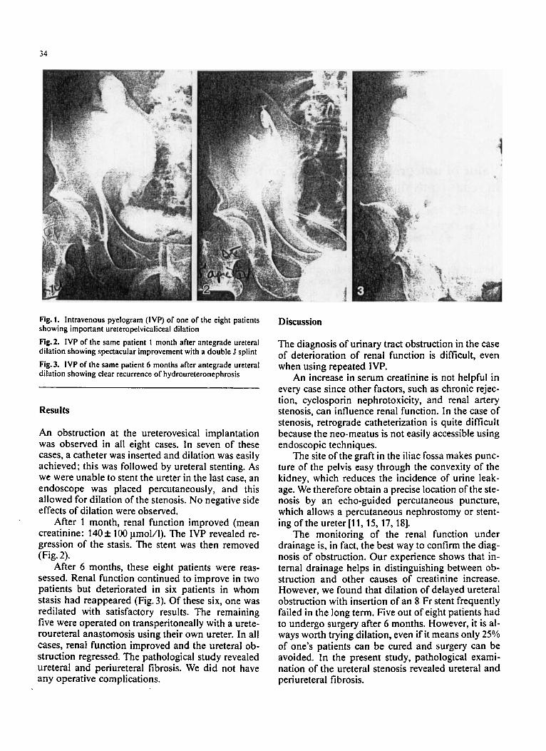
Fig. 1. Intravenous pyelogram (IVP) of one of the eight patients showing important ureteropelvicaliceal dilation Fig.2. IVP of the same patient 1 month after antegrade ureteral dilation showing spectacular improvement with a double J splint Fig.3. IVP of the same patient 6 months after antegrade ureteral dilation showing clear recurrence of hydroureteronephrosis
Results
An obstruction at the ureterovesical implantation was observed in all eight cases. In seven of these cases, a catheter was inserted and dilation was easily achieved; this was followed by ureteral stenting. As we were unable to stent the ureter in the last case, an endoscope was placed percutaneously, and this allowed for dilation of the stenosis. No negative side effects of dilation were observed.
After 1 month, renal function improved (mean creatinine: 140+ 100 pmol/l). The IVP revealed re- gression of the stasis. The stent was then removed (Fig. 2).
After 6 months, these eight patients were reas- sessed. Renal function continued to improve in two patients but deteriorated in six patients in whom stasis had reappeared (Fig. 3). Of these six, one was redilated with satisfactory results. The remaining five were operated on transperitoneally with a urete- roureteral anastomosis using their own ureter. In all cases, renal function improved and the ureteral ob- struction regressed. The pathological study revealed ureteral and periureteral fibrosis. We did not have any operative complications.
Discussion
The diagnosis of urinary tract obstruction in the case of deterioration of renal function is difficult, even when using repeated IVP.
An increase in serum creatinine is not helpful in every case since other factors, such as chronic rejec- tion, cyclosporin ncphrotoxicity, and renal artery stenosis, can influence renal function. In the case of stenosis, retrograde catheterization is quite difficult because the neo-meatus is not easily accessible using endoscopic techniques.
The site of the graft in the iliac fossa makes punc- ture of the pelvis easy through the convexity of the kidney, which reduces the incidence of urine leak- age. We therefore obtain a precise location of the ste- nosis by an echo-guided percutaneous puncture, which allows a percutaneous nephrostomy or stent- ing of the ureter [ l l , 15, 17, 181.
The monitoring of the renal function under drainage is, in fact, the best way to confirm the diag- nosis of obstruction. Our experience shows that in- ternal drainage helps in distinguishing between ob- struction and other causes of creatinine increase. However, we found that dilation of delayed ureteral obstruction with insertion of an 8 Fr stent frequently failed in the long term. Five out of eight patients had to undergo surgery after 6 months. However, it is al- ways worth trying dilation, even if it means only 25% of one's patients can be cured and surgery can be avoided. In the present study, pathological exami- nation of the ureteral stenosis revealed ureteral and periureteral fibrosis.

35
Liebermann et al. reported a case of a stenosis di- lated percutaneously with good results after 7 months [12]. Streem et al. treated two stenoses with good results [18]. Van Gansbeke treated six stenoses percutaneously with good results in. three [4], and Warner treated four, all of which relapsed [19]. These results confirm that the percutaneous approach to transplanted kidneys is easy and allows for dilation of ureteral stenosis. Failures of this method are generally due to periureteral fibrosis, which is a char- acteristic feature of delayed ureteral stenosis.
Ureteral stenosis occurs frequently after renal transplantation. When loss of renal function occurs in association with urinary tract dilation, the percu- taneous approach to the transplanted kidney, in combination with stenting, is a good method for monitoring the function of the graft and for confirm- ing obstruction as the possible cause of deterioration of the renal function. Although antegrade dilation shows less than 30% favorable results, it is the preferred procedure for the initial treatment of ureteral stenosis.
References
1. Barbaric ZL, Thomson KR (1978) Percutaneous nephrostomy in the management of obstructed renal transplant. Radiology
2. Benoit G, Benarbia S, Bellamy J, Charpentier B, Schrameck E, Fries D (1985) Complication urologique de la transplantation renale. Ann Urol 19 [Suppl3]: 165-171
3. Berger RE, Ansell JS, Tremann JA, Hen JH. Rattauo LC, Marchioro TL (1980) The use of self retained ureteral stents in the management of urologic complications in renal transplant recipients. J Urol 124: 781-782
4. Gansbeke D van, Matos C, Genevois P, Pauw L de, Struyven J, Kinnaert P (1987) Percutaneous interventional procedures in the management of urologic complications of renal trans- plantation. Transplant Proc 19 [Suppl 11: 2205
5. Glass NR, Crummy AB, Fischer DT, Vehling DT, Lieberman R, Belzer FO (1982) Management of ureteral obstruction after transplantation by percutaneous antegrade pyelography and pyeloureterostomy. Urology 20: 15- 19
126: 639-642
6. Goldstein I , Cho SI, Olsson CA (1981) Nephrostomy drainage for renal transplant complications. J Urol 126: 159-163
7. Himter DW, Castaneda-Zuruga WR, Coleman CC, Herrera M, Amplatz K (1983) Percutaneous techniques in the manage- ment of urological complications in renal transplant patients. Radiology 148: 407-412
8. Icard P, Lumbroso J, Hiesse C, Charpentier B, Fries D, Ham- moudi Y, Jardin A, Benoit G (1987) Valeur de la scintigraphie au DTPA aprks injection de FurosCmide dans le diagnostic d'obstacle en transplantation rknale. Ann Urol 21 [Suppl 51:
9. Jarowenko MV, Flechner SM. Sandler CM, Buren CT van, Kahan BD (1985) Salvage of difficult transplant complica- tions by percutaneous techniques. J Urol 133: 840-843
10. Kinnaert P, Hall M, Janssen T, Vergerstraeten P, Toussaint G, Geertruyden JV (1985) Ureteral stenosis after kidney trans- plantation: true incidence and long-term follow-up after surgi- cal correction. J Urol 133: 17-20
11. Lieberman SF, Keller FS, Barry JM, Rosch J (1982) Percuta- neous antegrade transluminal ureteroplasty for renal allograft ureteral stenosis. J Urol 128: 122-123
12. Liebemann RP, Crummy AB. Glass NR, Belzer FO (1981) Fine needle antegrade pyelography in the renal transplant. J Urol 126: 155-158
13. MacGregor RJ, Konnakf JW, Thrall JM, Campbell DA, Koff SA (1983) Diuretic radionuclide urography in the diagnosis of suspected ureteral obstruction following renal transplantation. J Urol 129: 708-710
14. Mitchell A, Fellows GJ, Wright FW, Moms PJ (1981) Hy- dronephrosis in a transplanted kidney. The use of pressure- flow studies to exclude ureteric obstruction. Transplantation
15.Mundy AL, Podesta ML, Bewick M, Eudge CJ, Ellis FG (1981) The urological complications of lo00 renal transplants. Br J Urol53 [Suppl5]: 397-402
16. Schiff M Jr, McGuire EJ, Weiss RM, Lytton B (1976) Manage- ment of urinary fistula after renal transplantation. J Urol 115:
17. Schiff M. Rosenfield AT, McGuire EJ (1979) The use of percu- taneous antegrade renal perfusion in kidney transplant re- cipients. J Urol 122: 246-248
18.Streem SB. Novick AC, Steinmuller DR, Musselman PW (1986) Percutaneous techniques for the management of uro- logical renal transplant recipients. J Urol 135: 456-459
19. Warner JJ, Maralon TAS, Rabin DN. Patel SK. Sensik sc, Merkel FK (1987) Percutaneous interventional radiologic pro- cedures for diagnosis and treatment of urological complica- tions in renal transplant patients. Transplant Proc 19 [Suppl 11:
370-374
32: 152-153
251-256
2203-2204