uva-dare (digital academic repository) strategies to ... · chapterr portallveinligationisa...
TRANSCRIPT

UvA-DARE is a service provided by the library of the University of Amsterdam (http://dare.uva.nl)
UvA-DARE (Digital Academic Repository)
Strategies to improve outcome after partial liver resection
Dinant, S.
Link to publication
Citation for published version (APA):Dinant, S. (2006). Strategies to improve outcome after partial liver resection.
General rightsIt is not permitted to download or to forward/distribute the text or part of it without the consent of the author(s) and/or copyright holder(s),other than for strictly personal, individual use, unless the work is under an open content license (like Creative Commons).
Disclaimer/Complaints regulationsIf you believe that digital publication of certain material infringes any of your rights or (privacy) interests, please let the Library know, statingyour reasons. In case of a legitimate complaint, the Library will make the material inaccessible and/or remove it from the website. Please Askthe Library: https://uba.uva.nl/en/contact, or a letter to: Library of the University of Amsterdam, Secretariat, Singel 425, 1012 WP Amsterdam,The Netherlands. You will be contacted as soon as possible.
Download date: 11 Aug 2020
C h a p t er r
Portall vein ligation is as effective ass sequential portal vein and hepaticc artery ligation in inducing contralaterall liver hypertrophy
Reetaa L. Vetelainen, Sander Dinant, Arlène K. van Vliet and Thomas M. van Gulik
Dept.. of Surgery, Surgical Laboratory, Academic Medical Center, Amsterdam, Thee Netherlands
Chapterr 7
Abstract t
Purpose.. Dual embolization of hepatic artery and portal vein has been proposed to enhance
contralaterall liver regeneration prior to resection. The aim of this study was to evaluate
thee effect of portal vein ligation compared to dual ligation, either simultaneously or with
sequentiall approach, on regeneration, proinflammatory response and liver damage.
Methods.. Single hepatic artery, portal vein ligation (PVL) of 70% or dual ligation of hepatic
arteryy and portal vein of 70 % cither simultaneously or sequentially with a 48h interval was
performedd in a rat model. Liver regeneration, proinflammatory mediators, hepatocellular
syntheticc function and injury, histopathology and apoptosis were assessed up to 14 days after
surgery. .
Results.. Sequential dual ligation resulted in faster increase in hepatocyte proliferation at
24hh without additional increase in liver mass compared to PVL after 14 days. Both the dual
ligationss significantly increased proinflammatory response in plasma and in the regenerating
liverr compared to PVL alone. 14 days after PVL, the hepatic parenchyma was completely
restoredd compared to fibromecrosis in sequentially dual ligated and complete necrosis in
simultaneouslyy ligated groups. Increased apoptosis in the regenerating liver and prolonged
hepaticc dysfunction was observed after both dual ligations.
Conclusions.. PVL is as effective as dual ligation in inducing liver regeneration. No
additionall benefit of arterial ligation was observed.
100 0
Thee effect of portal vein and/or hepatic artery ligation
Introductio n n
Severee postoperative complications and mortality after extensive resection are directly related
too the size and function of the remnant liver i : . Especially patients with parenchymal liver
diseasee have an increased risk of postoperative liver dysfunction due to already impaired
preoperativee function combined with an impaired regenerative capacity leading to slower
recoveryy of liver mass after resection '~\ Portal vein embolization (PVE) was introduced to
enablee more extensive liver resections by inducing compensatory hypertrophy in the non-
embolizedd future remnant liver and atrophy in the embolized lobe planned for resection 44 r>. With PVE, the future remnant liver volume can be increased up to 40%, consequently
decreasingg liver-dysfunction related complications caused by insufficient remnant liver 6"8.
Duall embolization of hepatic artery and portal vein has been suggested to induce sufficiënt liver
regenerationn in patients with potentially impaired liver regeneration. The obvious advantage
off dual embolization compared to PVE is the complete occlusion of both portal and arterial
bloodd supply to the tumor bearing liver segments 9,1°. However, if PVE and transarterial
embolizationn are combined simultaneously, the total occlusion of blood supply potentially
causess hepatic infarction and massive hepatocellular necrosis consequently triggering
systemicc proinflammatory cytokine response. This might as a result lead to acute liver failure
impairingg hepatic synthetic and metabolic functions 9. Therefore, a sequential embolization
withh 48h interval has been suggested to prevent these life-threatening complications y i i .
However,, no studies are available assessing the potential systemic or local effect directly
afterr sequential embolization and the impact of sequentially ligated liver lobes on adjacent
regeneratingg lobes is unclear. Currently used approaches to evaluate hepatocellular injury
suchh as histopathology of the resected liver earliest after 4-8 weeks after embolization
orr plasma aminotransferases, give only an estimation of the actual response and give no
informationn of the state of the regenerating lobes 9 12.
Thee purpose of this study was to compare portal vein ligation as surrogate PVE, with
simultaneouss or sequential dual ligation of hepatic artery and portal vein. The effect of
ligationn on liver proliferation and hepatocellular damage and function was assessed along
withh evaluation of local and systemic proinflammatory cytokine response.
Material ss and methods
Animals s
Malee Wistar rats (250-300g) were obtained (Harlan CPB, Zeist, Netherlands). The animals
weree housed at constant 24 °C with 12 h light- dark cycle and were fed a standard rodent
choww (Hope farms, Woerden, The Netherlands) and water ad libitum. Rats were allowed to
acclimatizee 7 days to laboratory conditions before surgery. During all procedures the animals
weree treated according to the guidelines of the Dutch legislation and international standards
forr animal care and handling. The protocol was approved by the Animal Ethics Committee
off University of Amsterdam, The Netherlands.
101 1
Chapterr 7
Exper imentall des ign and surgical procedures
Surgeryy was performed under inhalation anesthesia of a mixture of 0.,/N.,0 (1:1 V/V, 2 1 /
min)) and isoflurane (1-2 % Florene, Abbott laboratories Ltd, Queensborough, UK) and pain
medicationn (Temgesic i,v. 0,033 mg/ 0,1 kg). Ligation of the portal vein and/or the hepatic
arteryy to the median and left lateral liver lobes was performed, occluding perfusion of 70%
off total liver mass ]i . Rats were divided into 5 groups (n=6); only mobilization of the liver
(SHAM),, only arteria hepatica ligation (AHL), only portal vein ligation (PVL), simultaneous
ahll and pvl (DUALO) and subsequent ahl and pvl after 48h (DUAL48). After operation,
alll animals were allowed to recover in a warm environment with free access to water and
food.. At 6 h. 72 h and 7 days postoperatively blood was collected via tail vein puncture under
generall anaesthesia and a maximum of 10 % of total blood volume per rat was collected.
Underr above mentioned general anaesthesia animals were sacrificed (n=6 per time point) after
244 h and 48 h for evaluation of early response and after 14 days for the end point analysis.
Bloodd was collected by heart puncture, centrifuged {10 min, 3,000 RPM. 4°C) and plasma
wass stored at -80 °C until analysis. The liver lobes were removed, weighed and thin slices
weree immersed in 10% formalin for light microscopy (H& E and Sirius red staining) and for
immunohistochemistry. .
Assessmentt of l iver regenerat ion
Thee weight of the non-ligated liver lobes divided by the total liver weight was used as a
parameterr to evaluate the regenerative capacity of the liver. For assessment of hepatic
proliferation,, MIB-5, a rat equivalent of Ki-67 antibody was used which detects all active
partss of the cell cycle. The MIB-5 index has a strong positive correlation with proliferating
antigenn expression, bromodeoxyuridine incorporation and thymidine incorporation 14. Briefly,
44 um sections were deparaffinized, preheated and boiled (citric acid pH 6.0, 2 bar,120°C, 20
min)) in a pressure cooker. Sections were incubated with a MIB-5 antibody (dilution 1:50, 60
min,, DAKOCytomation, Glostrup, Denmark). After incubation with a secondary antibody
(dilutionn 1:1, Poly-HRP, Invitrogen, Carlsbad. US) 3.3-diaminobezidine (Sigma chemical,
Munich,, Germany) was used to visualise the peroxidase complexes together with haematoxylin
eountcrstaining.. The proliferative index was determined in 30 high power fields (HPF) at 40X
magnificationn and expressed as the percentage of positive cells per 1000 hepatocytes.
Hepatocel lu larr damage and hepat ic synthet ic funct io n
Plasmaa was analysed for aspartate aminotransferase (AST), alanine aminotransferase (ALT)
andd total bilirubin (T-Bil) and for albumin and prothrombin time using in the Department of
Clinicall Chemistry (AMC) using standard laboratory methods.
Pro in f lammator yy cy tok in e response
Liverr samples were homogenized in buffer (PBS, pH 6.0), centrifuged (10.000 xg, 4 °C, 10
min)) and supernatant was used for analysis of IL-lp1, IL-6 and TNF-a, the most important
acutee phase response cytokines produced by hepatic macrophages. Plasma and hepatic
concentrationss were measured using an enzyme-linked immunosorbent assay (Quantikine
Ratt TNF- a, Duoset Rat IL-l b and IL-6, RnD Systems Europe Ltd, England) according to
102 2
Thee effect of portal vein and/or hepatic artery ligation
manufacturer'ss instructions. Al l samples were measured in duplicate in a 96-well microtitre
platee and the concentrations were calculated from a standard curve. The hepatic protein
concentrationn was measured with a BCA Protein Assay kit (Pierce, Rockford, US) and the
hepaticc cytokine concentrations were expressed in pg/ mg protein.
Hepaticc neutrophil activity
Forr neutrophil activation, hepatic myeloperoxidase (MPO) activity was measured by
ass described by Krawisz et al '5. Briefly, tissue samples were homogenized (PBS, pH 6.0),
centrifugedd for lOmin at 10.000 rpm at 4 "C.The pellet was homogenized in HTAB-EDTA
bufferr (0.5% hexadecyltrimethyl ammonium bromide, lOmM EDTA in PBS, pH 6.0, Sigma
Chemicals,, Munich, Germany) sonicated and centrifuged. The supernatant was incubated
(2h,, 60°C) and the MPO activity was measured spectrophotometrically after addition of
o-dianisidee hydrochloride (Sigma Chemical, Munich, Germany). The MPO activity was
expressedd as units/mg protein and one unit was defined as the amount of enzyme necessary
too produce a change in absorbance of 1.0 per minute.
Apoptosis s
Forr primary and secondary antibody, a cleaved caspase- 3 (dilution 1:200, Cell Signaling
Technology,, Frankfurt, Germany) and Poly-HRP (dilution 1:1, Invitrogen, Carlsbad, US)
respectivelyy were used. The apoptotic index was determined at 40X magnification in 30 HPF
andd expressed as the amount of positive cells per 1000 hepatocytes.
Histopathology y
Paraffin-embeddedd liver samples were routinely stained with haematoxylin-eosin (H&E)
andd Sirius red (0,1% Fast red in picric acid, Immunotech, The Netherlands). Histology of
thee ligated and non-ligated lobes was observed by light microscopy. Examination was
performedd by two independent investigators blinded to the treatment groups. Necrosis was
expressedd as percentage of necrotic tissue: 0= no necrosis, 1= < 25 %, 2= 25-50%, 3= 50-75 %,
4== >75 % necrosis. Inflammatory activity was determineted as follows: 1= focal collections
off mononuclear cells, 2= diffuse infiltrates of mononuclear cells, 3=focal collections
off polymorphonuclear cells in addition to mononuclear cells, 4= diffuse infiltrates of
polymorphonuclearr cells.
Statisticall analysis
Dataa analysis was performed with GraphPad Prism 3.02 for Windows (GraphPad Software
Inc.,, San Diego, US).The results are presented as mean SEM. Significant differences between
groupss were tested using Kruskal-Wallis one-way analysis and Mann-Whitney's U-test. P
valuess less than 0.05 were considered significant.
103 3
Chapterr 7
Results s
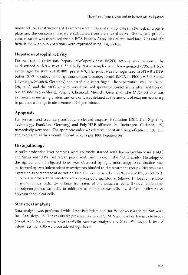
Hepaticc proliferation, regeneration and apoptosis
Bothh the regeneration ratio and hepatocyte proliferation index, measured by MIB-5 positive
cellss in the nonligated lobes, were increased in the PVL and both dual ligation groups at
alll time points compared to the SHAM and the AHL groups (Fig. IA, B, respectively).
Furthermore,, the regeneration ratio in the DUALO group was lower compared to the PVL
andd the DUAL48 groups at all time points (p<0.05). At 24h, the regeneration ratio was
significantlyy increased in the DUAL48 group compared to the PVL group. At 24h, the
hepatocytee proliferation index was significantly lower in the PVL group compared to the
DUALOO and DUAL48 groups (p<0.05). The percentage of caspase-3 positive cells was higher
inn both the dual groups compared to the other groups and in the DUALO compared to
DUAL488 group at 24 h and 48h after surgery (p<0.05) (Fig.lC).
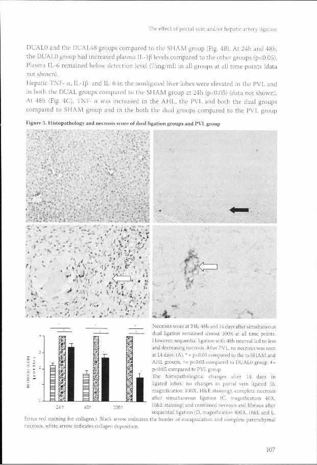
Figuree 1. Regeneration ratio, MIB- 5 positive cellss and caspase-3 positive cells
SHAM M AHL L PVL L DUALO O
II DUAL48
144 days
50 0 45540 0
30 0 26 6 20 0 15 5 10--5 5
0 0 I I
11 3-8 8
II r^ i ES3 F^l m JÈS.JÈS. V
Theree were no changes after 14 days in the liver regeneration ratioratio (A) between PVL and sequential dual ligation. The proliferationn index (B) of MIB-5 positive hepatocytes at 24hh was increased in sequential ligation group, however noo changes were seen at 48h. Both regeneration ration andd MIB-5 index were decreased after simultaneous dual ligation.. The apoptotic index (C) was increased in the non-ligatedd liver lobes of the both dual ligated groups. * == p<0.05 compared to the to SHAM and AHL groups, t= p<0.055 compared to DUALO group.
104 4
Thee effect of portal vein and/or hepatic artery ligation
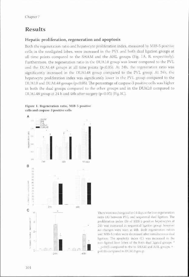
Hepatocellularr damage
Att all time points, no differences were observed in hepatocellular damage between the
SHAMM and the AHL groups. The PVL and both the dual groups had significantly increased
ASTT levels at 24h, 48h and 72h compared to SHAM group (p<0.05). In the DUALO group,
plasmaa AST was increased already at 6h compared to the all other groups and remained
increasedd until 72h (p<0.05)(Fig.2A). In the DUAL48 group, AST was also elevated already
att 6h compared to the SHAM and PVL groups. In the PVL group, AST was increased at 48h
andd 72h compared to the DUAL48 group (p<0.05). ALT followed the pattern of AST with the
exceptionn that there were no differences between both dual groups at 6h postoperatively (Fig.
2B).. Plasma bilirubin followed the pattern of AST and ALT in all groups (data not shown).
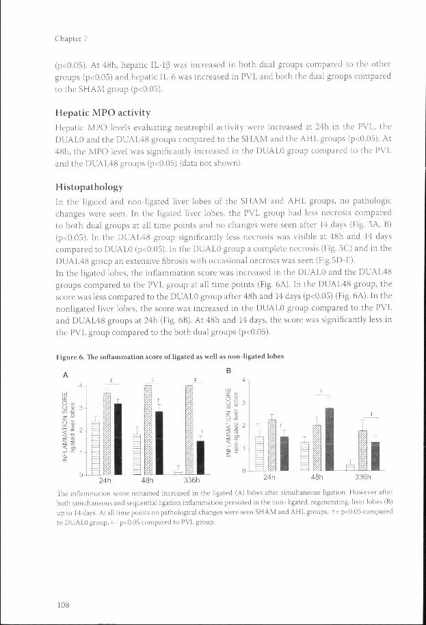
Figuree 2. Plasma levels of hepatocellular damage markerss AST (A) and ALT (B)
A A 51000 0
41000 0
31000 0
21000 0
11000 0
1200 0
1000 0
800 0
600 0
200 0
0 0
I
ml ml postopeff atrve hours
iJilriKK II I I 77 days
BB 10000
20000 -1000»--
800 0
600 0
400 0
2000 -
00 c "- -' 0 0
itóii oJI nJ oostopefal^'ee hours
-- L _ Theree was a significant increase at 6h and 24h in AST andd ALT after dual ligation groups compared to PVL groupss in which peak was seen at 48h. * = p<0.05 comparedd to the to SHAM and AHL groups, t= p<0.05 comparedd to PVL group.
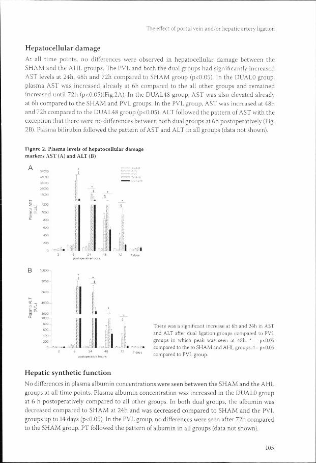
Hepaticc synthetic function
Noo differences in plasma albumin concentrations were seen between the SHAM and the AHL
groupss at all time points. Plasma albumin concentration was increased in the DUALO group
att 6 h postoperatively compared to all other groups. In both dual groups, the albumin was
decreasedd compared to SHAM at 24h and was decreased compared to SHAM and the PVL
groupss up to 14 days (p<0.05). In the PVL group, no differences were seen after 72h compared
too the SHAM group. PT followed the pattern of albumin in all groups (data not shown).
105 5
Chapterr 7
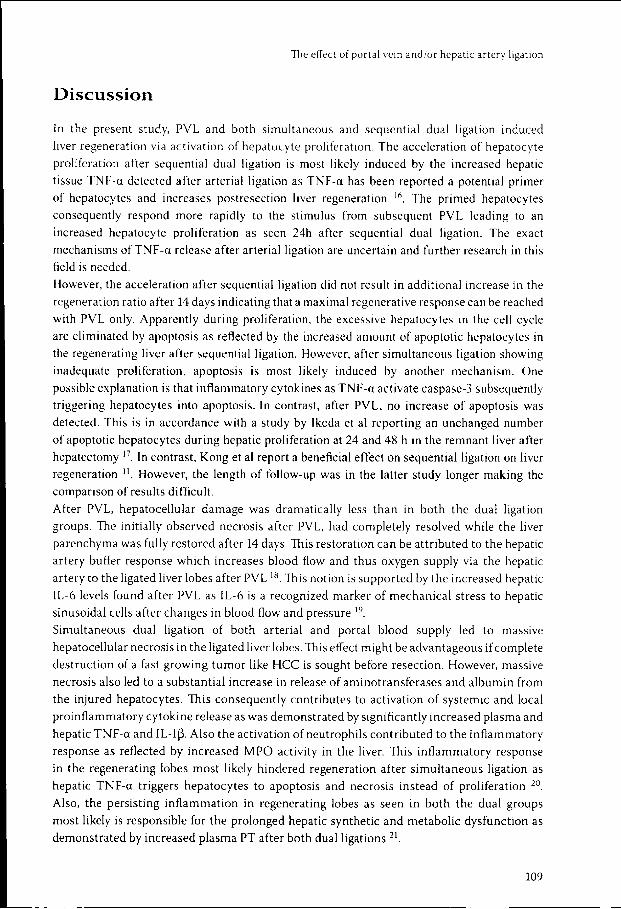
Figuree 3. Hepatocellular synthetic function evaluated byy plasma albumin level
50-, , ! Z ^^ SHAM CZ1AHL L CZUPVL L EH33 DUALO
II DUAL48
00 6 24 48 postoperativee hours
77 days 14 days
AA prolonged dysfunction in both dual ligation groups iss visible. * = p<0.05 compared to the to SHAM and AHLL groups, t= p<0.05 compared to DUALO group, t== p<0.05 compared to PVL group and § = p <0.05 comparedd to DUAL.48 group.
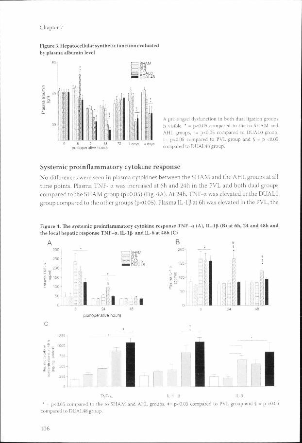
Systemicc proinflammatory cytokine response
Noo differences were seen in plasma cytokines between the SHAM and the AHL groups at all
timee points. Plasma TNF- a was increased at 6h and 24h in the PVL and both dual groups
comparedd to the SHAM group (p<0.05) (Fig. 4A). At 24h, TNF-a was elevated in the DUALO
groupp compared to the other groups (p<0.05). Plasma IL-1J3 at 6h was elevated in the PVL, the
Figuree 4. thee local
A A
Thee systemic proinflammator y cytokine response TNF-a (A), IL-1B (B) at 6h, 24 and 48h and hepaticc response TNF-a, IL-i p and 1L-6 at 48h (C)
3 0 0 --
2 5 0 --
uLL 200 -
Ê ! ! | gg 150-i/ii —' ra ra
Q-- 100-
50 0
0--
SHAM M II AHL ,, PVL '' DUALO 11 DUAL48
I I postoperativee hours
0)) ? ~ 1000 .ïï x a 22 "> ö && 5 S- ' 5 0
li tt M0
SS 250
_! ! I I TNF-aa IL-1 |i IL-6
** = p<0.05 compared to the to SHAM and AHL groups, *= p<0.05 compared to PVL group and § comparedd to DUAL48 group.
pp <0.05
106 6
Thee effect of portal vein and/or hepatic artery ligation
DUALOO and the DUAL48 groups compared to the SHAM group (Fig. 4B). At 24h and 48h,
thee DUALO group had increased plasma IL-1|3 levels compared to the other groups (p<0.05).
Plasmaa IL-6 remained below detection level (75ng/ml) in all groups at all time points (data
nott shown).
Hepaticc TNF- a, IL-1J3 and IL-6 in the nonligated liver lobes were elevated in the PVL and
inn both the DUAL groups compared to the SHAM group at 24h (p<0.05) (data not shown).
Att 48h (Fig. 4C), TNF- a was increased in the AHL , the PVL and both the dual groups
comparedd to SHAM group and in the both the dual groups compared to the PVL group
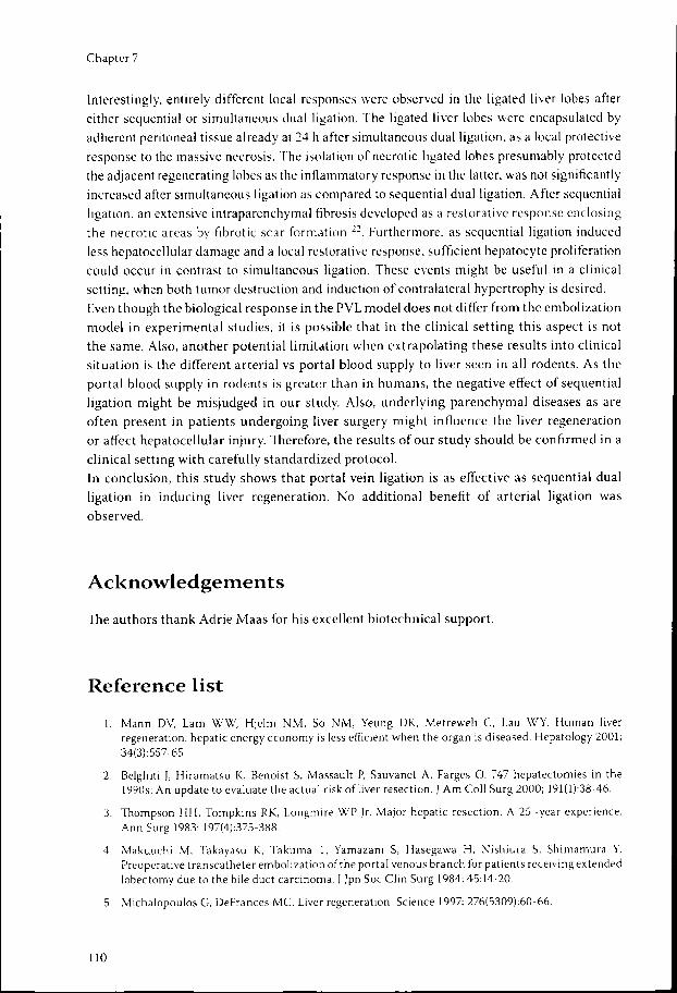
Figuree 5. Histopathology and necrosis score of dual ligation groups and PVL group
.. ""
ii ...
** * t
«*,, • 1 ' - >>
'' • ! ; ' '-,',- V< ' ; C=] ]
5 5 I I I I 1 1 1 1
:-- -
JL JL
Necrosiss score at 24h, 48h and 14 days after simultaneous duall ligation remained almost 100% at all time points. However,, sequential ligation with 48h interval led to less andd decreasing necrosis. After PVL, no necrosis was seen att 14 days. (A). * = p<0.05 compared to the to SHAM and AHLL groups, t= p<0.05 compared to DUALO group, t= p<0.055 compared to PVL group.
Thee histopathological changes after 14 days in ligatedd lobes; no changes in portal vein ligated (B, magnificationn 100X, H&E staining), complete necrosis afterr simultaneous ligation (C, magnification 40X, H&EE staining) and combined necrosis and fibrosis after sequentiall ligation (D, magnification 400X, H&E and E,
Siriuss red staining for collagen,). Black arrow indicates the border of encapsulation and complete parenchymal necrosis,, white arrow indicates collagen deposition.
m m
ii I 1 1 mm • - :
107 7
Chapterr 7
(p<0.05).. At 48h, hepatic IL-lG was increased in both dual groups compared to the other
groupss (p<0.05) and hepatic IL-6 was increased in PVL and both the dual groups compared
too the SHAM group (p<0.05).
Hepaticc MPO activity
Hepaticc MPO levels evaluating neutrophil activity were increased at 24h in the PVL, the
DUALOO and the DUAL48 groups compared to the SHAM and the AHL groups (p<0.05). At
48h,, the MPO level was significantly increased in the DUALO group compared to the PVL
andd the DUAL48 groups (p<0.05) (data not shown).
Histopathology y
Inn the ligated and non-ligated liver lobes of the SHAM and AHL groups, no pathologic
changess were seen. In the ligated liver lobes, the PVL group had less necrosis compared
too both dual groups at all time points and no changes were seen after 14 days (Fig. 5A, B)
(p<0.05).. In the DUAL48 group significantly less necrosis was visible at 48h and 14 days
comparedd to DUALO (p<0.05). In the DUALO group a complete necrosis (Fig. 5C) and in the
DUAL488 group an extensive fibrosis with occasional necrosis was seen (Fig.5D-E).
Inn the ligated lobes, the inflammation score was increased in the DUALO and the DUAL48
groupss compared to the PVL group at all time points (Fig. 6A). In the DUAL48 group, the
scoree was less compared to the DUALO group after 48h and 14 days (p<0.05) (Fig. 6A). In the
nonligatedd liver lobes, the score was increased in the DUALO group compared to the PVL
andd DUAL48 groups at 24h (Fig. 6B). At 48h and 14 days, the score was significantly less in
thee PVL group compared to the both dual groups (p<0.05).
Figuree 6. The inflammation score of ligated as well as non-ligated lobes
Thee inflammation score remained increased in the ligated (A) lobes after simultaneous ligation. However after bothh simultaneous and sequential ligation inflammation persisted in the non- ligated, regenerating, liver lobes (B) upp to 14 days. At all time points no pathological changes were seen SHAM and AHL groups. t= p<0.05 compared too DUALO group, t= p<0.05 compared to PVL group.
108 8
Thee effect of portal vein and/or hepatic artery ligation
Discussion n
Inn the present study, PVL and both simultaneous and sequential dual ligation induced
liverr regeneration via activation of hepatucyte proliferation. The acceleration of hepatocyte
proliferationn after sequential dual ligation is most likely induced by the increased hepatic
tissuee TNF-a detected after arterial ligation as TNF-ct has been reported a potential primer
off hepatocytes and increases postresection liver regeneration 16. The primed hepatocytes
consequentlyy respond more rapidly to the stimulus from subsequent PVL leading to an
increasedd hepatocyte proliferation as seen 24h after sequential dual ligation. The exact
mechanismss of TNF-a release after arterial ligation are uncertain and further research in this
fieldd is needed.
However,, the acceleration after sequential ligation did not result in additional increase in the
regenerationn ratio after 14 days indicating that a maximal regenerative response can be reached
withh PVL only. Apparently during proliferation, the excessive hepatocytes in the cell cycle
aree eliminated by apoptosis as reflected by the increased amount of apoptotic hepatocytes in
thee regenerating liver after sequential ligation. However, after simultaneous ligation showing
inadequatee proliferation, apoptosis is most likely induced by another mechanism. One
possiblee explanation is that inflammatory cytokines as TNF-a activate caspase-3 subsequently
triggeringg hepatocytes into apoptosis. In contrast, after PVL, no increase of apoptosis was
detected.. This is in accordance with a study by Ikeda et al reporting an unchanged number
off apoptotic hepatocytes during hepatic proliferation at 24 and 48 h in the remnant liver after
hepatectomyy 17. In contrast, Kong et al report a beneficial effect on sequential ligation on liver
regenerationn ". However, the length of follow-up was in the latter study longer making the
comparisonn of results difficult.
Afterr PVL, hepatocellular damage was dramatically less than in both the dual ligation
groups.. The initially observed necrosis after PVL, had completely resolved while the liver
parenchymaa was fully restored after 14 days. This restoration can be attributed to the hepatic
arteryy buffer response which increases blood flow and thus oxygen supply via the hepatic
arteryy to the ligated liver lobes after PVL 18. This notion is supported by the increased hepatic
IL-66 levels found after PVL as IL-6 is a recognized marker of mechanical stress to hepatic
sinusoidall cells after changes in blood flow and pressure 19.
Simultaneouss dual ligation of both arterial and portal blood supply led to massive
hepatocellularr necrosis in the ligated liver lobes. This effect might be advantageous if complete
destructionn of a fast growing tumor like HCC is sought before resection. However, massive
necrosiss also led to a substantial increase in release of aminotransferases and albumin from
thee injured hepatocytes. This consequently contributes to activation of systemic and local
proinflammatoryy cytokine release as was demonstrated by significantly increased plasma and
hepaticc TNF-a and IL-lp1. Also the activation of neutrophils contributed to the inflammatory
responsee as reflected by increased MPO activity in the liver. This inflammatory response
inn the regenerating lobes most likely hindered regeneration after simultaneous ligation as
hepaticc TNF-a triggers hepatocytes to apoptosis and necrosis instead of proliferation 20.
Also,, the persisting inflammation in regenerating lobes as seen in both the dual groups
mostt likely is responsible for the prolonged hepatic synthetic and metabolic dysfunction as
demonstratedd by increased plasma PT after both dual ligations 21.
109 9
Chapterr 7
Interestingly,, entirely different local responses were observed in the ligated liver lobes after
eitherr sequential or simultaneous dual ligation. The ligated liver lobes were encapsulated by
adherentt peritoneal tissue already at 24 h after simultaneous dual ligation, as a local protective
responsee to the massive necrosis. The isolation of necrotic ligated lobes presumably protected
thee adjacent regenerating lobes as the inflammatory response in the latter, was not significantly
increasedd after simultaneous ligation as compared to sequential dual ligation. After sequential
ligation,, an extensive intraparenchymal fibrosis developed as a restorative response enclosing
thee necrotic areas by fibrotic scar formation 22. Furthermore, as sequential ligation induced
lesss hepatocellular damage and a local restorative response, sufficient hepatocyte proliferation
couldd occur in contrast to simultaneous ligation. These events might be useful in a clinical
setting,, when both tumor destruction and induction of contralateral hypertrophy is desired.
Evenn though the biological response in the PVL model does not differ from the embolization
modell in experimental studies, it is possible that in the clinical setting this aspect is not
thee same. Also, another potential limitation when extrapolating these results into clinical
situationn is the different arterial vs portal blood supply to liver seen in all rodents. As the
portall blood supply in rodents is greater than in humans, the negative effect of sequential
ligationn might be misjudged in our study. Also, underlying parenchymal diseases as are
oftenn present in patients undergoing liver surgery might influence the liver regeneration
orr affect hepatocellular injury. Therefore, the results of our study should be confirmed in a
clinicall setting with carefully standardized protocol.
Inn conclusion, this study shows that portal vein ligation is as effective as sequential dual
ligationn in inducing liver regeneration. No additional benefit of arterial ligation was
observed. .
Acknowledgements s
Thee authors thank Adrie Maas for his excellent biotechnical support.
Referencee list
1.. Mann DV, Lam WW, Hjelm NM, So NM, Yeung DK, Metreweli C, Lau WY. Human liver regeneration:: hepatic energy economy is less efficient when the organ is diseased. Hepatology 2001; 34(3):557-65. .
2.. Belghiti I, Hiramatsu K, Benoist S, Massault P, Sauvanet A, Farges O. 747 hepatectomies in the 1990s:: An update to evaluate the actual risk of liver resection.} Am Coll Surg 2000; 191(l):38-46.
3.. Thompson HH, Tompkins RK, Longmire WP Jr. Major hepatic resection. A 25 -year experience. Annn Surg 1983: 197(4):375-388.
4.. Makuuchi M, Takayasu K, Takuma T, Yamazani S, Hasegawa H, Nishiura S, Shimamura Y. Preoperativee transcatheter embolization of the portal venous branch for patients receiving extended lobectomyy due to the bile duct carcinoma. J Jpn Soc Clin Surg 1984; 45:14-20.
5.. Michalopoulos G, DeFrances MC. Liver regeneration. Science 1997; 276(5309):60-66.
110 0
Thee effect of portal vein and/or hepatic artery ligation
6.. Nagino M, Ando M, Kamiya J, Uesaka K, Sano T, Nimura Y. Liver regeneration after major hepatectomyy for biliary cancer. Br J Surg 2001; 88(8):1084-1091.
7.. Azoulay D, Castaing D, Smail A, Adam R, Cailliez V, Laurent A, Lemoine A, Bismuth H. Resection of nonresectablee liver metastases from colorectal cancer after percutaneous portal vein embolization. Annn Surg 2000; 231(4):480-486.
8.. Kubota K, Makuuchi M, Kusaka K, Kobayashi T, Miki K, Hasegawa K, Harihara Y, Takayama T. Measurementt of liver volume and hepatic functional reserve as a guide to decision-making in resectionall surgery for hepatic tumors. Hepatology 1997; 26(5):1176-1181.
9.. Nakao N, Miura K, Takahashi H, Ohnishi M, Miura T, Okamoto E, Ishikawa Y. Hepatocellular carcinoma:: combined hepatic, arterial and portal venous embolization. Radiology 1986; 161(2):303-330. .
10.. Aoki T, Imamura H, Hasegawa K, Matsukura A, Sano K, Sugawara Y, Kokudo N, Makuuchi M. Sequentiall preoperative arterial and portal venous embolizations in patients with hepatocellular carcinoma.. Arc Surg 2005; 139(7)766-774.
11.. KongD, Kusano M, AraseT, NishinoN, JinZ, KameyamaS, KatoH, NiiyaT, FujiokaT, Murakami M, Itohh Y. Liver regeneration after portal vein plus hepatic artery ligation performed heterochronously inn rats. J Hepatobiliary Pancreat Surg 2002; 9(l):86-92.
12.. Ogasawara K, Uchino J, Une Y, Fujioka Y, Selective portal vein embolization with absolute ethanol inducess hepatic hypertrophy and makes more extensive hepatectomy possible. Hepatology 1996; 23(2):338-345. .
13.. Rozga J, Jeppson B, Bengmark S. Portal branch ligation in the rat. Re-evaluation of the model. Am J Pathh 1986; 125(2):300-308.
14.. Gerlach C, Sakkab DY, Scholzen T, Dassler R, Alison MR, Gerdes J. Ki-67 expression during rat liverr regeneration after partial hepatectomy. Hepatology 1997; 26(3):573-578.
15.. Krawisz JE, Sharon P, Stenson WF. Quantitative assay for acute intestinal inflammation based on myeloperoxidasee activity. Gastroenterology 1984; 87(6):1344-1350.
16.. Webber EM, Bruix J, Pierce RH, Fausto N. Tumor necrosis factor primes hepatocytes for DNA replicationn a the rat. Hepatology 1998; 28(5):1226-1234.
17.. Ikeda K, Kinoshita H, Hirohashi K, Kubo S, Kaneda K. The ultrastructure,kinetics and intralobular distributionn of apoptotic hepatocytes after portal branch ligation with special reference to their relationshipp to necrotic hepatocytes. Arch Histol Cytol 1995; 58(2):171-184.
18.. Suzuki S, Nakamura S, Sakaguchi T, Ochiai H, Konno H, Baba S, Baba S. Alteration of reticuloendotheliall phagocytic function and tumor necrosis factor-alpha production after total hepaticc ischemia. Transplantation 1997; 64(6):821-827.
19.. Kawai M, Naruse K, Komatsu S, Kobayashi S, Nagino M, Nimura Y, Sokabe M. Mechanical stress-dependantt secretion of interleukin 6 by endothelial cells after portal vein embolization; clinical and experimentall studies. J Hepatol 2002; 37(2):240-246.
20.. Diehl AM. Cytokine regulation of liver injury and repair. Immunol Rev 2000; 174:160-171.
21.. Wieser W. Cost of growth in cells and organisms and comparative aspects. Biol Rev Camb Philos Socc 1994;68{l):l-33
22.. Kaplowitz N. Biochemical and cellular mechanisms of toxic liver injury. Sem Liv Dis 2002; 22(2):: 137-144.
I l l l