uv stimulation of dna-mediated transformation of human cells
TRANSCRIPT

MOLECULAR AND CELLULAR BIOLOGY, Apr. 1985, p. 734-741 Vol. 5, No. 40270-7306/85/040734-08$02.00/0Copyright C 1985, American Society for Microbiology
UV Stimulation of DNA-Mediated Transformation of Human CellsM. VAN DUIN, A. WESTERVELD, AND J. H. J. HOEIJMAKERS*
Department of Cell Biology and Genetics, Erasmus University, 3000 DR Rotterdam, The Netherlands
Received 3 October 1984/Accepted 8 January 1985
Irradiation of dominant marker DNA with UV light (150 to 1,000 J/m4) was found to stimulate thetransformation of human cells by this marker from two- to more than fourfold. This phenomenon is alsodisplayed by xeroderma pigmentosum cells (complementation groups A and F), which are deficient in theexcision repair of UV-induced pyrimidine dimers in the DNA. Also, exposure to UV of the transfected(xeroderma pigmentosum) cells enhanced the transfection efficiency. Removal of the pyrimidine dimers fromthe DNA by photoreactivating enzyme before transfection completely abolished the stimulatory effect,indicating that dimer lesions are mainly responsible for the observed enhancement. A similar stimulation of thetransformation efficiency is exerted by 2-acetoxy-2-acetylaminofiuorene modification of the DNA. No stimula-tion was found after damaging vector DNA by treatment with DNase or -y rays. These findings suggest thatlesions which are targets for the excision repair pathway induce the increase in transformation frequency. Thestimulation was found to be independent of sequence homology between the irradiated DNA and the hostchromosomal DNA. Therefore, the increase of the transformation frequency is not caused by a mechanisminducing homologous recombination between these two DNAs. UV treatment of DNA before transfection didnot have a significant effect on the amount of DNA integrated into the xeroderma pigmentosum genome.
UV light induces a variety of lesions into DNA, such aspyrimidine dimers (the main product), thymidine glycols, 6-4photoproducts, and DNA protein cross-links. Deformationof the DNA double-helix structure by these lesions disturbsessential processes, such as replication and transcription,and ultimately can result in mutagenesis and lethality. Themajor repair system operating on many of these lesions is theexcision repair pathway (see references 19, 22, and 29 forreviews). Irradiation of prokaryotic and eukaryotic cellswith UV light and in some cases transfection with DNAexposed to UV light induces a variety of processes, such asstimulation of recombination (13, 17, 21, 23), enhancementof survival of UV-damaged viruses (host cell reactivation; 6,8, 9, 37) or bacteriophages (Weigle reactivation; 46), en-hanced capacity to repair potentially lethal damage (45), andincreased mutagenesis (7-9, 12, 37). In Escherichia colimany of these phenomena are part of the pleiotropic SOSresponse (reviewed in reference 30). In eukaryotes severalof these features are expressed concomitantly, and it islikely that they are at least in part related to each other.However, the nature of the inducing signal(s) is still un-known. Here we report that UV irradiation has a stimulatoryeffect on the genetic transformation of human cells. Thisphenomenon was encountered in the course of experimentsaimed at the cloning of a human DNA repair gene comple-menting the excision repair defect of xeroderma pigmen-tosum (XP) cells. The rational of this approach was to rescuea UV-damaged dominant marker when cotransfected withthe XP-correcting wild-type repair gene present in carrierDNA. It appeared that UV irradiation of the dominantmarker did not exercise a deleterious but rather a stimula-tory effect on the transformation frequency of XP andnormal cells. This finding, which confirms recently reportedobservations of Spivak et al. (41), is studied in more detail inthe present paper. Evidence is presented that pyrimidinedimers are the responsible UV lesions. A similar effect isexerted by modification of DNA by n-acetyl-aminofluorene(AAF). This carcinogen preferentially interacts with the C-8
* Corresponding author.
position of guanine, resulting in guanine-AAF (27, 33),which is like pyrimidine dimers removed by excision repair(16). Finally, we demonstrate that homologous recombina-tion between transfected DNA and the genome of therecipient cells is not responsible for UV stimulation of thetransformation.
MATERIALS AND METHODS
Cell lines and culture conditions. XP12ROSV40 (10) andXP2YO(SV) (49) are simian virus 40 (SV40)-transformedexcision repair-deficient XP fibroblasts of complementationgroups A and F, respectively. CHO12RO is an excisionrepair deficient-Chinese hamster ovary (CHO) cell line (43).SV40wtA, an SV40-transformed human fibroblast, a wild-type CHO cell line, and HeLa-S3 cells were used as repair-proficient cell lines.The cell lines were grown in F10 medium (without thymi-
dine) and Dulbecco minimal essential medium (1:1) supple-mented with antibiotics and 3% fetal and 7% newborn calfserum.
Isolation of DNA. High-molecular-weight DNA of HeLa-S3 cells was isolated as described (24). Plasmid DNA wasisolated by using the alkaline lysis procedure (5).
Induction ofDNA damage. UV irradiation of plasmid DNA(and, where specified, HeLa DNA) was carried out in amaximum volume of 250 ,ul on a watch-glass by using a UVsource at a fluence of 0.6 J/m2. For high UV doses thedistance to the lamp was decreased. The DNA concentrationduring irradiation ranged from 0.1 to 0.4 mg/ml. UV-irradi-ated DNA (and nonirradiated control DNA) was ethanolprecipitated before use for transfection or analysis on alka-line gels.X irradiation of plasmid DNA was done at a DNA
concentration of 4 mg/ml in a closed Eppendorf tube byusing a Philips X-ray machine (10 mA; 175 rads/min).
Modification of plasmid DNA with AAF was performed asdescribed by Landegent et al. (28).
Transfection and selection. The transfection protocol wasas described (18, 48), with some modifications. One daybefore transfection 10 petri dishes were seeded with 2 x 10⁵
734
on Novem
ber 24, 2018 by guesthttp://m
cb.asm.org/
Dow
nloaded from
UV-STIMULATED TRANSFORMATION OF HUMAN CELLS
to 5 x 10⁵ cells per dish. DNA (2 ,ug of plasmid DNA and 8to 10 p,g of HeLa carrier DNA) was added to the cells as acalcium phosphate precipitate. After exposure to the DNA(5 h for human and 12 h for CHO cells) cells were subjectedto a dimethyl sulfoxide shock (10%; 30 min) and subse-quently cultured in nonselective medium for 48 h, afterwhich selection was started. Usually 10 to 14 days latercolonies were fixed and stained. Only colonies of more than20 cells were counted. Unless indicated otherwise fourplates were used to determine the average number of colo-nies per plate (±+ standard error of the mean [SEM]).Dominant markers harboring the TnS neomycine resistancegene were pSV2neo (40) and the cosmid vectors pTCF (20),pMCS (20), and pMCS-Alu (see also Fig. 4). pMCS isidentical to pTCF except for the fact that it carries an SV40ori region in front of the thymidine kinase (TK) promotor.pMCS-alu contains one copy of a human Alu repeat clonedin the unique BamHI site of pMCS. The dominant markerpSV3gptH (47) contains the eco-gpt gene that gives resist-ance to mycophenolic acid.
Cells transfected with pSV3gptH were selected in myco-phenolic acid medium as specified (47). For selection of cellstransfected with dominant markers carrying the TnSneomycin resistance gene, the normal medium was supple-mented with G-418 (300 ,ug/ml; Schering Corp.). The selec-tion medium was refreshed every 2 to 3 days.UV irradiation of cells. To determine the transformation
frequency after UV exposure of the recipient cells, cellswere irradiated immediately after the dimethyl sulfoxideshock by using a UV lamp at a fluence of 0.3 J/m2 per s.Parallel to the transfection experiment the survival at thesame UV dose was determined. After irradiation the cellswere trypsinized, and 2,000 cells were seeded onto 10-cmpetri dishes. After 11 days the number of clones wasdetermined and compared with that of nonirradiated controlcells. The cloning efficiency was -25%.Enzyme treatments of DNA. (i) Restriction endonucleases.
Digestions of DNA with restriction enzymes and DNase Iwere done as recommended by the suppliers (Boehringer;New England Biolabs).
(ii) T4 endonuclease V. Plasmid DNA (UV irradiated) wasincubated with purified T4 endonuclease (a generous gift of
(0
-4)0
"O. 11(
0.0E
C
800r
400
0 14 140
B
I I lI I _I0 14 140
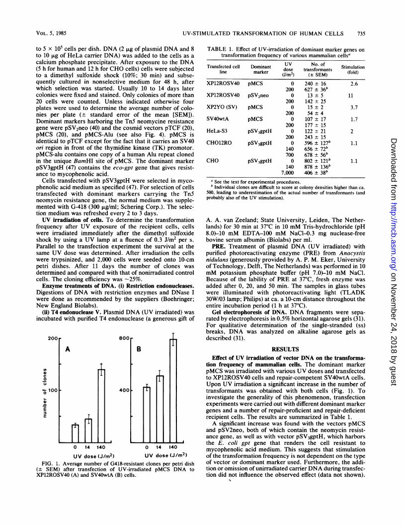
UV dose (J/m2) UV dose (J/m2)FIG. 1. Average number of G418-resistant clones per petri dish
(+ SEM) after transfection of UV-irradiated pMCS DNA toXP12ROSV40 (A) and SV40wtA (B) cells.
TABLE 1. Effect of UV-irradiation of dominant marker genes ontransformation frequency of various mammalian cellsa
Transfected cell Dominant UV No. of Stmlioline marker ~~dose transformants Stmlioliansfle.tecell marker (J/m2) (± SEM) (fold)
XP12ROSV40 pMCS 0 240 ± 16 2.6200 627 ± 36b
XP12ROSV40 pSV2neo 0 13 ± 5 11200 142 25
XP2YO (SV) pMCS 0 15 + 2 3.7200 54 ± 4
SV40wtA pMCS 0 107 ± 17 1.7200 177 ± 15
HeLa-S3 pSV3gptH 0 122 ± 21 2200 243 ± 15
CHO12RO pSV3gptH 0 596 ± 127b 1.1140 656 ± 72b700 678 ± 56b
CHO pSV3gptH 0 802 + 121b 1.1140 878 ± 136b
7,000 406 ± 38ba See the text for experimental procedures.b Individual clones are difficult to score at colony densities higher than ca.
500, leading to underestimation of the actual number of transformants (andprobably also of the UV stimulation).
A. A. van Zeeland; State University, Leiden, The Nether-lands) for 30 min at 37°C in 10 mM Tris-hydrochloride (pH8.0)-10 mM EDTA-100 mM NaCI-0.3 mg nuclease-freebovine serum albumin (Biolabs) per ml.PRE. Treatment of plasmid DNA (UV irradiated) with
purified photoreactivating enzyme (PRE) from Anacystisnidulans (generously provided by A. P. M. Eker, Universityof Technology, Delft, The Netherlands) was performed in 10mM potassium phosphate buffer (pH 7.0)-10 mM NaCl.Because of the lability of PRE at 37°C, fresh enzyme wasadded after 0, 20, and 50 min. The samples in glass tubeswere illuminated with photoreactivating light (TLADK30W/03 lamp; Philips) at ca. a 10-cm distance throughout theentire incubation period (1 h at 37°C).
Gel electrophoresis of DNA. DNA fragments were sepa-rated by electrophoresis in 0.5% horizontal agarose gels (31).For qualitative determination of the single-stranded (ss)breaks, DNA was analyzed on alkaline agarose gels asdescribed (31).
RESULTSEffect of UV irradiation of vector DNA on the transforma-
tion frequency of mammalian cells. The dominant markerpMCS was irradiated with various UV doses and transfectedto XP12ROSV40 cells and repair-competent SV40wtA cells.Upon UV irradiation a significant increase in the number oftransformants was obtained with both cells (Fig. 1). Toinvestigate the generality of this phenomenon, transfectionexperiments were carried out with different dominant markergenes and a number of repair-proficient and repair-deficientrecipient cells. The results are summarized in Table 1.A significant increase was found with the vectors pMCS
and pSV2neo, both of which contain the neomycin resist-ance gene, as well as with vector pSV3gptH, which harborsthe E. coli gpt gene that renders the cell resistant tomycophenolic acid medium. This suggests that stimulationof the transformation frequency is not dependent on the typeof vector or dominant marker used. Furthermore, the addi-tion or omission of unirradiated carrierDNA during transfec-tion did not influence the observed effect (data not shown).
VOL. 5, 1985 735
on Novem
ber 24, 2018 by guesthttp://m
cb.asm.org/
Dow
nloaded from
736 VAN DUIN, WESTERVELD, AND HOEIJMAKERS
4OOr
U,
0u
0
a)
-cE
c
B
200p
FT+
1T+1I1
- + PREn vnn liVrwna- (.lm2)V SV VUVUUbe J / 111- )
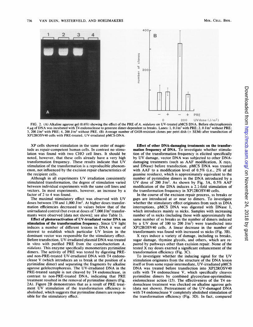
FIG. 2. (A) Alkaline agarose gel (0.6%) showing the effect of the PRE of A. nidulans on UV-treated pMCS DNA. Before electrophoresis4 pug ofDNA was incubated with T4 endonuclease to generate dimer-dependent ss breaks. Lanes: 1, 0 J/m2 with PRE; 2, 0 J/m2 without PRE;3, 200 J/m2 with PRE; 4, 200 J/m2 without PRE. (B) Average number of G418-resistant clones per petri dish (± SEM) after transfection ofXP12ROSV40 cells with PRE-treated, UV-irradiated pMCS-DNA.
XP cells showed stimulation in the same order of magni-tude as repair-competent human cells. In contrast no stimu-lation was found with two CHO cell lines. It should benoted, however, that these cells already have a very hightransformation frequency. These results indicate that UVstimulation of the transformation is a reproducible phenom-enon, not influenced by the excision repair characteristics ofthe recipient cells.Although in all experiments UV irradiation consistently
stimulated transformation, the degree of stimulation variedbetween individual experiments with the same cell lines andvectors. In most experiments, however, an increase by afactor of 2 to 4 was found.The maximal stimulatory effect was observed with UV
doses between 150 and 1,000 J/m2. At higher doses transfor-mation efficiencies decreased to values below that of theunirradiated control (but even at doses of 7,000 J/m2 transfor-mants were observed [data not shown]; see also Table 1).
Effect of photoreactivation of UV-irradiated vector DNA onstimulation of the transformation frequency. Since UV lightinduces a number of different lesions in DNA it was ofinterest to establish which particular UV lesion in thedominant vector was responsible for the stimulatory effect.Before transfection, UV-irradiated plasmid DNA was treatedin vitro with purified PRE from the cyanobacterium A.nidulans. This enzyme specifically monomerizes pyrimidinedimers. The activity of PRE was tested by digesting PRE-and non-PRE-treated UV-irradiated DNA with T4 endonu-clease V (which introduces an ss break at the position of apyrimidine dimer) and separating the fragments by alkalineagarose gelelectrophoresis. The UV-irradiated DNA in thePRE-treated sample is not cleaved by T4 endonuclease, incontrast to non-PRE-treated DNA, indicating that PREtreatment resulted in the removal of pyrimidine dimers (Fig.2A). Figure 2B demonstrates that as a result of PRE treat-ment UV stimulation of the transformation efficiency isabolished, which suggests that pyrimidine dimers are respon-sible for the stimulatory effect.
Effect of other DNA-damaging treatments on the transfor-mation frequency of DNA. To investigate whether stimula-tion of the transformation frequency is elicited specificallyby UV damage, vector DNA was subjected to other DNA-damaging treatments (such as AAF modification, X rays,and DNase) before transfection. pMCS DNA was treatedwith AAF to a modification level of 0.5% (i.e., 2% of allguanine residues), which is approximately equivalent to thenumber of pyrimidine dimers in the DNA introduced by aUV dose of 200 J/m2. As shown by Fig. 3A, 0.5% AAFmodification of the DNA induces a 2.1-fold stimulation ofthe transformation frequency in XP12ROSV40 cells.
In the course of the excision repair process, ss breaks orgaps are introduced at or near to dimers. To investigatewhether the stimulatory effect originates from such ss DNAinterruptions, pMCS DNA was digested with DNase I,which introduces mainly ss nicks. Samples with increasingnumber of ss nicks (including those with approximately thesame number of ss breaks as the number of dimers inducedby a UV dose of 100 to 200 J/m2) were transfected intoXP12ROSV40 cells. A linear decrease in the number oftransformants was found with increased ss nicks (Fig. 3B).X rays induce a variety of damage, including ss breaks,
sugar damage, thymine glycols, and others, which are re-paired by pathways other than excision repair. None of thetested X ray doses exerted a significant enhancement of thetransformation efficiency (Fig. 3C).To investigate whether the inducing signal for the UV
stimulation originates from the structure of the DNA lesionitself or from some repair intermediate, UV-irradiated pMCSDNA was treated before transfection into XP12ROSV40cells with T4 endonuclease V, which specifically cleavespyrimidine dimers by combined glycosylase-apyrimidinicendonuclease action (15). The effectiveness of the T4 en-donuclease treatment was checked on alkaline agarose gels(data not shown). Pretreatment of the UV-damaged DNAwith T4 endonuclease V completely abolished stimulation ofthe transformation efficiency (Fig. 3D). In fact, compared
1 2 3 41
MOL. CELL. BIOL.
on Novem
ber 24, 2018 by guesthttp://m
cb.asm.org/
Dow
nloaded from

UV-STIMULATED TRANSFORMATION OF HUMAN CELLS
with nonirradiated pMCS, a drastic decrease in the numberof transformants was even observed, which resembled theeffect of DNase.
Effect of UV irradiation of recipient cells on the transfor-mation frequency of dominant marker genes. To investigatewhether UV irradiation of the host cell also induces astimulatory effect on the transformation efficiency,XP12ROSV40 and repair-proficient SV40-transformed hu-man fibroblasts were transfected with pMCS DNA and UVirradiated immediately after transfection (0.5 J/m2 for XPcells; 0.5 and 5 J/m2 for SV40wtA cells). Irradiation of cellsdid not result in stimulation comparable to that found afterUV irradiation of vector DNA (Table 2). However, takinginto account the survival ofXP and wild-type cells at the UVdoses tested, considerable stimulation was found, particu-larly in the case of XP cells.
Role of homologous recombination between exogenous andhost cellular DNA in UV-stimulated transformation fre-quency. To investigate whether DNA sequence homologybetween the UV-damaged dominant vector and genomic
0-
0
Ec
B
400-
200
- + AAF
4,
0 2 10
TABLE 2. Effect of UV irradiation of recipient cells on thetransformation frequency of pMCS
Cell line UV dose No. of clonesCellline (Ji~Qm2) (±SEM)-
XP12ROSV40 0 341 ± 200.5 404 ± 30 (2,020)
SV40wtA 0 703 ± 280.5 897 ± 655.0 863 ± 42 (1,233)
a Values in parentheses represent the number of clones after correction forcell survival. At a dose of 0.5 J/m2 XP12ROSV40 cells displayed a survivalrate of ca. 20%, as determined in a parallel experiment. SV40wtA cells had asurvival rate of 70 to 80%o after UV irradiation with 5 jI/m2.
DNA of recipient cells plays a role in the enhancement oftransformation, three types of experiments were carried out.Two pMCS derivatives, one without SV40 (ori-) sequences(pTCF) and one containing a human Alu sequence (pMCS-alu) (see Fig. 4, upper part), were UV irradiated (200 J/m2)and transfected to XP12ROSV40 cells. The results (Fig. 4,lower part) demonstrate that irradiation of the vector DNAin all cases induces approximately the same relative stimu-lation. UV irradiation of pMCS-alu, which possesses homol-ogy with ca. 4 x 10⁵ Alu copies in the human genome, givesthe same relative increase in transformation efficiency as UVirradiation of vector pMCS, which has only SV40 sequencehomology, and vector pTCF, which has no homology at allwith the XP12ROSV40 genome. Furthermore, this experi-
E cos S agpt Ptk EA pTCF
B P. SV40 cos SB pMCS i
20 DNAse(min)
agpt Ptk B4IIIII-m7
CMCS
B P.SV40 cos S agptB k aluB
-alu 11111
8001
C
400
0
v-200
0
E
600
200
0 22 168 448 kRAD - + + UVdose(200J/m2)- - + T4 endo
FIG. 3. Average number of G418-resistant clones ofXP12ROSV40 cells per petri dish (+ SEM) after transfection withpMCS DNA treated with different DNA-damaging agents beforetransfection. (A) AAF modification (0.5%); (B) partial DNase diges-tion (incubation times indicated); (C) X irradiation; (D) UV plus T4endonuclease incubation.
0
'.-400 -
0
Ec 200
-+ -+ -+ UVdoseA B C (200 J/m2)
FIG. 4. UV stimulation of the transformation of XP12ROSV40cells with different UV-irradiated plasmids harboring the G418resistance gene. P, and Ptk represent early promotor from the SV40ori-enhancer fragment and herpes TK promotor, respectively. agpt,Aminoglycosylphosphoribosyltransferase gene, encoding resistanceagainst G418; E, EcoRI; B, BamHI; S, Sall; cos, cos sequence ofbacteriophage lambda. Transfection was with pTCF (A), pMCS(B),and pMCS-alu (C).
VOL. 5, 1985 737
on Novem
ber 24, 2018 by guesthttp://m
cb.asm.org/
Dow
nloaded from

738 VAN DUIN, WESTERVELD, AND HOE1JMAKERS
0
400
+ - + - + ligation- + + + + UV / HeLa DNA
-+ - - - - + + Uvi pMCSFIG. 5. G418-resistant clones after transfection of XP12ROSV40
cells with BamHI-digested pMCS DNA ligated to BamHI-cleavedHeLa DNA. When indicated, DNAs were irradiated with a UV doseof 200 J/m2. The first two lanes represent control experiments withundigested pMCS DNA.
ment shows that potential extra-chromosomal replication ofvector pMCS, due to the SV40 origin, is not involved in theprocess that leads to stimulation of the transformationfrequency. It is evident that deletion of the SV40 ori-regionreduces the transformation frequency more than 30-fold.
In the second experiment UV-irradiated and nonirradiatedhuman DNA fragments (obtained after digestion of HeLaDNA with restriction endonuclease BamHI) were ligated invitro to BamHI-linearized pMCS and transfected intoXP12ROSV40 cells. The results (Fig. 5) demonstrate thatlinearization of pMCS by BamHI, which separates the SV40ori-enhancer from the TK promotor of the neomycin resis-tance gene (Fig. 4, upper part), drastically reduces thetransformation frequency (Fig. 5, cf. columns 1 and 3). Thiswas not the case when the vector was linearized on othersites (e.g., at the unique Sall site) (Fig. 4, upper part),leaving the SV40 ori in front of the TK promoter (unpub-lished data). When the cleaved vector was ligated in vitrowith BamHI-cleaved HeLa DNA (in a 1:2 molar ratio), thetransformation efficiency was partly restored (Fig. 5, cf.columns 3 and 4). This can be explained by the fact that theTK promotor of part of the pMCS molecules is religated toan SV40 ori-enhancer from its own or another vector mole-cule. These findings are in agreement with the idea that theSV40 ori fragment provides an enhancer function to the TKpromotor of the neomycine resistance gene and are inconcordance with results reported by others (4). Ligation ofUV-irradiated HeLa DNA to the dominant vector did notresult in enhancement of the transformation frequency (Fig.5, cf. columns 4 and 6).To exclude the possibility that sequence homology plays a
role when UV-induced lesions are present in both thetransfected DNA and the host genome, we tested whetherUV irradiation of pMCS-alu gave stronger stimulation of thetransformation frequency than UV-treated pMCS when therecipient XP12ROSV40 cells were also UV irradiated. Nosignificant difference in the transformation efficiencies wasobserved between the two vectors (data not shown). Thedata presented above rule out the possibility that stimulationof the transformation frequency is caused by enhancedhomologous recombination between (damaged) vector andhost DNA.
Effect of UV irradiation on integration of vector DNA. It isof interest to know whether UV irradiation has an effect on
the number of vector copies or on the amount of unirradiatedcotransferred carrier DNA that integrate in the genome ofthe recipient cell. Southern blot analysis was done onXP12ROSV40 cells transfected with (UV-irradiated) pMCS.After transfection and selection, DNA was extracted fromthe total transformed cell population, originating from petridishes with 300 to 600 independent clones. This DNA wasdigested with restriction endonucleases, and the fragmentswere size fractionated by agarose gel electrophoresis. South-ern blots of the gel were hybridized with 32P-labeled pMCSDNA. The autoradiogram of the hybridized filter shown inFig. 6 demonstrates that there is no notable difference in theintensity of vector fragments between the irradiated andnonirradiated DNA samples. Since the DNA is derived from300 to 600 independent clones, the relative intensity of theinternal vector fragments gives the average number of inte-grated copies per cell. From the intensity of coelectropho-
UV dose J/mr2
o 0o 0
0 a 0 (Nm r---- -
9.5-
6.6- 0 t
4.4- 40
2.3-2D-
A B
FIG. 6. Southern blot analysis of G418-resistant XP12ROSV40-pMCS transformants with a 32P-labeled pMCS probe. Transfectionwas carried out fivefold with nonirradiated and irradiated (200 J/m2)pMCS DNA. UV stimulation of the transformation frequency wasdetermined by scoring the number of clones on three plates. Theother two plates, containing + 300 clones per plate (nonirradiated)or + 600 clones per plate (200 J/m2) were trypsinized, and cells weregrown into mass culture under selective conditions for DNA isola-tion. DNA (15 pLg) was digested with EcoRI (A) or EcoRI-SalI (B).The first lane shows the hybridization signal of one copy ofEcoRI-digested pMCS DNA per genome. The two bands indicatedby arrows are derived from the SV40 copy in the host genome, towhich the SV40 ori part of the probe hybridizes. Numbers on the leftside represent molecular weight markers.
MOL. CELL. BIOL.
on Novem
ber 24, 2018 by guesthttp://m
cb.asm.org/
Dow
nloaded from
UV-STIMULATED TRANSFORMATION OF HUMAN CELLS
resed vector DNA in amounts equivalent to one copy pergenome on the same blot, it appears that on the average onlya few pMCS vector copies are integrated into theXP12ROSV40 genome. We conclude, therefore, that UVdamage in vector DNA does not drastically influence th,amount of DNA integrated into the genome of the trans-formed cell.
DISCUSSIONThis study demonstrates that UV-induced lesions in vec-
tor DNA stimulate its transformation efficiency. The UVdose range which gives an optimal stimulatory effect induces20 to 150 dimers per pMCS dominant marker molecule, ofwhich ca. 1/5 is estimated to be within the selectable geneitself. Evidently the deleterious consequences of this dam-age on the integrity and expression of the transfected geneare outweighed by enhancement of a step(s) in the transfor-mation process. Moreover, the UV-induced lesions appearto be equally harmful in excision repair-deficient XP cellsand wild-type cells, as judged from the fact that the trans-formation stimulation in XP-A cells is at least as high as innormal cells. Similar observations have recently been re-ported by Spivak et al. (41). These authors showed that UVirradiation of pSV2neo or pSV2gpt results in enhancedtransformation frequency of human cells. The reported dosedependency and stimulation factor correspond well with ourresults.The finding that similar stimulation is found with XP and
wild-type cells contrasts with the survival of UV-damagedvirus in XP cells, which is considerably lower than in normalcells (1, 2). The absence of an effect of the XP mutation onthe transformation frequency of UV-irradiated vector DNAcan be explained in different ways.
(i) The more deleterious effects of the UV lesions in XPcells are compensated by stronger stimulation of the step(s)in the transformation process which is influenced by the UVdamage in the transfected DNA (e.g., because in XP cellsthe lesions exist for a longer period of time).
(ii) The XP-A cells repair the lesions in the exogenousDNA. Mortelmans et al. (34) and Kano and Fujiwara (25)have reported that extracts from various XP complementa-tion groups (including XP-A and XP-F) are capable ofexcising pyrimidine dimers from purified DNA and fromheterologous (but not homologous) chromatin. If XP cellscan remove the dimers in the introduced DNA, this couldexplain the absence of a difference in UV stimulation of thetransformation frequency. Further experiments are requiredto decide between these possibilities.Which step in the transformation process is influenced by
UV damage in DNA? The finding that initially unlinkedDNA molecules eventually are integrated as a large concat-enate into the host cell genome indicates that before orduring incorporation frequent ligation and recombinationevents occur among transfected DNA molecules (32). Instable transformants the donor DNA is inserted at one (or afew) site(s) in the host chromosomal DNA (36), whichsuggests that the integration event might be one of thelimiting steps in the transformation process. Since UVirradiation of cells, as well as transfection of cells withUV-irradiated DNA, enhance recombination (13, 21), it ispossible that the UV stimulation of transformation is aconsequence of the effect on recombination. UV light mightstimulate recombination of exogenous DNA molecules witheach other to form a concatenate and with the host DNA forstable integration. The following observations are consistantwith this interpretation. (i) Stimulation of recombination is
also displayed by excision deficient XP cells (21). (ii) Theenhancement of the recombination frequency by UV light isin the same order of magnitude as that of transformation andrequires comparable doses of UV light (21). (iii) The exper-iments presented in Table 2 suggest that the presence of UVdamage in the genome of the recipient cells also enhance thetransformation frequency. This would indicate that UVdamage in either one of the recombining DNAs results instimulation of transformation. (The observation that XP cellsexhibit higher stimulation than wild-type cells, after correc-tion for cell survival, might be due to longer persistance ofthe UV lesions in the XP genome.)Two types of recombination take place in the cell after
transfection. The occurrence of homologous recombinationbetween appropriately constructed exogenous DNA mole-cules is well documented (11, 14, 26, 35, 38, 39, 44). Therecombination frequency is directly related to the size of thehomologous segment between the transfected molecules (35,39). Nonhomologous recombination between transfectedDNAs has also been observed (3, 44). In this case smallregions of partial homology between the donor moleculesmay play a role in the recombination process (3). In bothtypes of recombination, replication of the transfected DNAis not required (44). As far as the interaction of exogenousDNA and host genome is concerned, it is known thatintegration can occur in any of a large number of sites in thehost chromosomes (36). The lack of site specificity-evenwhen ribosomal DNA segments, which have ca. 200 homol-ogous gene copies in the mammalian genome (42), aretransfected-strongly argues against homologous recombi-nation as a predominant mode of integration. In our exper-iments the amount of homology between dominant markerand host genome has no effect on the level of UV stimulationof the transformation process. Therefore, if UV light exertsits effect on the transformation via enhancement of recom-bination between exogenous and host DNA, the latter mustbe of the nonhomologous type.
Southern blot analysis of DNA of XP12ROSV40 transfor-mants obtained with dominant markers indicates that UVirradiation of the vector does not have a significant effect onthe average number of marker molecules, nor on the amountof unirradiated cotransfected DNA (unpublished data) incor-porated into the genome. The transformed XP12ROSV40cells appear to have incorporated on the average one to twocopies of the dominant marker. Compared with transfectionswith Ltk- cells, which can integrate in the order of 7,000kilobases (36), the amount of integrated DNA inXP12ROSV40 cells is extremely low. We are currentlyinvestigating whether the low amount of exogenous DNAstably inserted into the XP12ROSV40 genome is a uniquefeature of this cell line.What is the inducing signal to the increase of transforma-
tion frequency? The experiments with pretreatment of UV-irradiated DNA with PRE, which acts specifically on dimers,indicate that pyrimidine dimers are responsible for thisphenomenon, although the contribution of other, quantita-tively less important UV lesions is not excluded. The findingthat AAF mimics UV, whereas DNase and y rays (at thedoses tested) do not, suggests that stimulation of transfor-mation is a property of lesions on which the excision repairpathway is operative. It is possible that the structure of thelesion itself (local deformation of the helix) produces stimu-lation of the transformation efficiency, e.g., by provokingrecombination at those sites. Alternatively, UV-like lesionsmight induce some kind of SOS response in the transfectedcell, one of the consequences of which is enhancement of
VOL. 5, 1985 739
on Novem
ber 24, 2018 by guesthttp://m
cb.asm.org/
Dow
nloaded from

740 VAN DUIN, WESTERVELD, AND HOEIJMAKERS
recombination and integration into the host genome. Thefact that this occurs in XP-A and XP-F cells would indicatethat these cells behave normally with respect to this part ofthe response and that a functional excision repair pathway isnot required.
ACKNOWLEDGMENTS
We are very grateful to A. P. M. Eker (University of Technology,Delft, The Netherlands) and A. A. van Zeeland (State University,Leiden, The Netherlands) for the generous gifts of photoreactivatingenzyme and T4 endonuclease, respectively. We thank H. Odijk andJ. de Wit for help in some of the experiments, D. Bootsma, G. C.Grosveld, and N. G. J. Jaspers for stimulating discussions, R. J.Boucke and C. J. M. Meijerink-Clerkx for typing the manuscript,and T. van Os for photography.
This work was supported by the Commission of the EuropeanCommunity contract BIO-E-404-NL and FUNGO, Foundation ofMedical Scientific Research in The Netherlands.
LITERATURE CITED1. Aaronson, S. A., and C. D. Lytle. 1970. Decreased host cell
reactivation of irradiated SV40 virus in xeroderma pigmen-tosum. Nature (London) 228:359-361.
2. Abrahams, P. J., and A. J. van der Eb. 1976. Host-cell reacti-vation of ultraviolet-irradiated SV40 DNA in five complemen-tation groups of xeroderma pigmentosum. Mutat. Res. 35:13-22.
3. Anderson, R. A., S. Kato, and R. D, Camerini-Otero. 1984. Apattern of partially homologous recombination in mouse L-cells.Proc. Natl. Acad. Sci. U.S.A. 81:206-210.
4. Berg, P. A., and W. F. Anderson. 1984. Correlation of geneexpression and transformation frequency with the presence ofan enhancing sequence in the transforming DNA. Mol. Cell.Biol. 4:368-370.
5. Birnboim, H. C., and J. Doly. 1979. A rapid alkaline extractionprocedure for screening recombinant plasmid DNA. NucleicAcids Res. 7:1513-1523.
6. Bochstahler, L. E. 1981. Induction and enhanced reactivation ofmammalian viruses by light. Prog. Nucleic Acid Res. Mol. Biol.26:303-313.
7. Bourre, F., and A. Sarasin. 1983. Targeted mutagenesis of SV40DNA induced by UV-light. Nature (London) 305:68-70.
8. Cornelis, J. J., Z. Z. Su, and J. Rommelaere. 1982. Direct andindirect effects of ultraviolet light on the mutagenesis of parvo-virus fi-1 in human cells. EMBO J. 6:693-699.
9. DasGupta, U. B., and W. C. Summers. 1978. Ultraviolet reacti-vation of herpes simplex virus is mutagenic and inducible inmammalian cells. Proc. Natl. Acad. Sci. U.S.A. 75:2378-2381.
10. de Jonge, A. J. R., W. Vermeulen, B. Klein, and J. H. J,HoeUmakers. 1983. Microinjection of human cell extracts cor-rects xeroderma pigmentosum defect. EMBO J. 2:637-641.
11. De Saint Vincent, B. R., and G. Wahl. 1983. Homologousrecombination in mammalian cells mediates formation of afunctional gene from two overlapping gene fragments. Proc.Natl. Acad. Sci. U.S.A. 80:2002-2006.
12. Dinsart, C., J. J. Cornelis, B. Klein, A. J. van der Eb, and J.Rommelaere. 1984. Transfection with extracellularly UV-dam-aged DNA induces human and rat cells to express a mutatorphenotype towards parvovirus H-1. Mol. Cell. Biol. 4:324-328.
13. Dubbs, D. R., M. Rachmeler, and S. Kit. 1974. Recombinationbetween temperature-sensitive mutants of Simian virus 40.Virology 57:161-174.
14. Folger, K. R., E. A. Wong, G. Wahl, and M. R. Capecchi. 1982.Pattern of integration of DNA microinjected into cultured cells:evidence for homologous recombination between injectedplasmid DNA molecules. Mol. Cell. Biol. 2:1372-1387.
15. Friedberg, E. C., T. Bonura, E. H. Radamy, and J. D. Love.1981. Enzymes that incise damaged DNA, p. 251-279. In P. D.Boyer (ed.), The enzymes, vol. 14. Academic Press, Inc., NewYork.
16. Fuchs, R. P. P., and E. Seeberg. 1984. pBR322 plasmid DNA
modified with 2-acetylaminofluorene derivatives: transformingactivity and in vitro strand cleavage by the Escherichia coliuvrABC endonuclease. EMBO J. 3:757-760.
17. Golub, E. I., and K. B. Low. 1983. Indirect stimulation ofgenetic recombination. Proc. Natl. Acad. Sci. U.S.A.80:1401-1405.
18. Graham, F. L., and A. J. van der Eb. 1973. A new technique forthe assay of infectivity of human adenovirus 5 DNA. Virology52:456-467.
19. Grossman, L. 1981. Enzymes involved in the repair of damagedDNA. Arch. Biochem. Biophys. 211:511-522.
20. Grosveld, F. H., T. Lund, E. J. Murray, A. L. Mellar, H. M. M.Dahl, and R. A. Flavell. 1982. The construction of cosmidlibraries which can be used to transform eukaryotic cells.Nucleic Acids Res. 10:6715-6732.
21. Hall, J. D., J. D. Featherston, and R. E. Almy. 1980. Evidencefor repair of ultraviolet light-damaged Herpes virus in humanfibroblasts by a recombination mechanism. Virology 105:490-500.
22. Hanawalt, P. C., P. K. Cooper, A. K. Ganesan, and C. A. Smith.1979. DNA repair in bacteria and mammalian cells. Annu. Rev.Biochem. 48:783-836.
23. Jacob, F., and E. C. Wollman. 1955. Etude genetique d'unbacteriophage tempere d'Escherichia coli III-effect du rayon-nement ultraviolet sur la recombinaison genetique. Ann. Inst.Pasteur (Paris) 88:724-749.
24. Jeffreys, A. J., and R. A. Flavell. 1977. A physical map of theDNA regions flanking the rabbit ,-globin gene. Cell 12:429-439.
25. Kano, Y., and Y. Fujiwara. 1983. Thymine dimer excisioncatalyzed by cell-free extracts from xeroderma pigmentosumchromatin. Carcinogenesis 4:1419-1424.
26. Kretschner, P. J., A. H. Bowinan, M. H. Huberman, L. Sand-ers-Hargh, L. Killos, and W. F. Anderson. 1981. Recovery ofbacterial plasmids from E. coli transformed with DNA frommicroinjected mouse cells. Nucleic Acids Res. 9:6199-6217.
27. Kriek, E., J. A. Miller, U. Juhl, and E. C. Miller. 1967.8-(N-2-fluorenylacetamide) guanosine an arylamidation reactionproduct of guanosine and the carcinogen N-acetoxy-N-2-fluorenylacetamide in neutral solution. Biochemistry 6:177-182.
28. Landegent, J. E., N. Jansen in de Wal, R. A. Baan, J. H. J.Hoeijmakers, and M. van de Ploeg. 1984. 2-Acetylamino fluorenemodified probes for the immuno-hybridochemical detection ofspecific nucleic acid sequences. Exp. Cell Res. 153:61-72.
29. Lehman, A. R. 1982. Xeroderma pigmentosum, Cockayne syn-drome and ataxia telangiectasia: disorders relating DNA repairto carcinogenesis. Cancer Surveys 1:93-118.
30. Little, J. W., and D. W. Mount. 1982. The SOS regulatorysystem of Escherichia coli. Cell 29:11-22.
31. Maniatis, T., E. F. Fritsch, and J. Sambrock. 1982. Molecularcloning. Cold Spring Harbor Laboratory. Cold Spring Harbor,N.Y.
32. Miller, C. K., and H. M. Temin. 1983. High-efficiency ligationand recombination of DNA fragments by vertebrate cells.Science 220:606-609.
33. Miller, E. C., U. Juhl, and J. A. Miller. 1966. Nucleic acidguanine: reaction with the carcinogen N-acetoxy-2-ace-tylaminofluorene. Science 153:1125-1127.
34. Mortelmans, K., E. C. Friedberg, H. Slor, G. Thomas, and J. E.Cleaver. 1976. Defective thymine dimer excision cell-free ex-tracts of xeroderma pigmentosum cells. Proc. Natl. Acad. Sci.U.S.A. 73:2757-2761.
35. Pomerantz, B. J., M. Naujokas, and J. A. Hassell. 1983. Homol-ogous recombination between transfected DNAs. Mol. Cell.Biol. 3:1680-1685.
36. Robins, D. M., S. Ripley, A. S. Henderson, and R. Axel. 1981.Transforming DNA integrates into the host chromosome. Cell23:29-39.
37. Sarasin, A., F. Bourre, and A. Benoit. 1982. Error-prone repli-cation of ultraviolet-irradiated Simian virus 40 in carcinogen-treated monkey kidney cells. Biochimie 64:815-821.
38. Shapira, G., J. L. Stachelek, A. Letson, L. K. Soodak, and R. M.Liskay. 1983. Novel use of synthetic oligonucleotide insertionmutations for the study of homologous recombination in mam-
MOL. CELL. BIOL.
on Novem
ber 24, 2018 by guesthttp://m
cb.asm.org/
Dow
nloaded from
UV-STIMULATED TRANSFORMATION OF HUMAN CELLS
malian cells. Proc. Natl. Acad. Sci. U.S.A. 80:4827-4831.39. Small, J., and G. Scangos. 1983. Recombination during gene
transfer into mouse cells can restore the function of deletedgenes. Science 219:174-176.
40. Southern, P. J., and P. Berg. 1982. Transformation of mamma-lian cells to antibiotic resistance with a bacterial gene undercontrol of the SV40 early region promotor. J. Mol. Appl. Genet.1:327-341.
41. Spivak, G., A. K. Ganesan, and P. C. Hanawalt. 1984. Enhancedtransformation of human cells by UV-irradiated pSV2 plasmids.Mol. Cell Biol. 4:1169-1171.
42. Steele, R. E., A. H. Bakken, and R. H. Reeder. 1984. Plasmidscontaining mouse rDNA do not recombine with cellularribosomal genes when introduced into cultured mouse cells.Mol. Cell. Biol. 4:576-582.
43. Stefanini, M., A. Reuser, and D. Bootsma. 1982. Isolation ofChinese hamster ovary cells with reduced unscheduled DNAsynthesis after UV-irradiation. Somatic Cell Genet. 8:635-642.
44. Subramani, S., and P. Berg. 1983. Homologous and nornho-mologous recombination in monkey cells. Mol. Cell. Biol.
3:1040-1052.45. Tyrrell, R. M. 1984. Exposure of nondividing populations of
primary human fibroblasts to UV (254 nm) radiation induces a
transient enhancement in capacity to repair potentially lethalcellular damage. Proc. Natl. Acad. Sci. U.S.A. 81:781-784.
46. Weigle, J. J. 1953. Induction of mutation in a bacterial virus.Proc. Natl. Acad. Sci. U.S.A. 39:628-636.
47. Westerveld, A., J. H. J. Hoeijmakers, M. van Duin, J. de Wit, H.Odijk, A. Pastink, R. D. Wood, and D. Bootsma. 1984. Molec-ular cloning of a human DNA repair gene. Nature (London)310:425-429.
48. Wigler, M. R., R. Sweet, G. K. Sim, B. Wjold, A. Pellicar, E.Lacy, T. Maniatis, S. Silverstein, and R. Axel. 1979. Transfor-mation of mammalian cells with genes from prokaryotes andeukaryotes. Cell 16:777-785.
49. Yagi, T., and H. Takebe. 1983. Establishment by SV40 trans-formation and characteristics of a cell line of xeroderma pig-mentosum belonging to complementation group F. Mutat. Res.112:59-66.
VOL. 5, 1985 741
on Novem
ber 24, 2018 by guesthttp://m
cb.asm.org/
Dow
nloaded from