uterine myometrial preischemia: yet another physiological
TRANSCRIPT

Uterine Myometrial Preischemia: Yet AnotherPhysiological Change of Pregnancy!Kusum Jashnani1 Meherrituja Palve2
1Department of Pathology, TNMC & BYL Nair Ch. Hospital, Mumbai,Maharashtra, India
2Department of Pathology, KJSMC and Research Centre, Mumbai,Maharashtra, India
J Lab Physicians
Address for correspondence Meherrituja Palve, MBBS, MD,Department of Pathology, KJSMC and Research Centre, Mumbai400022, Maharashtra, India (e-mail: [email protected]).
Key Messages
Understanding physiological changes has always givenanswers to the complex questions of the pathology ofvarious conditions.
In this study, we have analyzed the uteri for variousphysiological changes of pregnancy and puerperium.We found some novel physiological changes like preische-mia and positive correlation between vaginal delivery andcervical wall hemorrhages.
Keywords
► pregnancy► myometrial
hypertrophy► preischemia► vascular remodeling► cervical wall
hemorrhages
Abstract Background The uterus shows tremendous increase in size during pregnancy tonurture the fetus within it. It may show a spectrum of physiological changes orpathological lesions that may affect the pregnancy favorably or adversely. The mainpurpose of our study was to analyze the physiological changes in the uterus duringpregnancy and the postpartum period, thereby gaining deeper knowledge.Materials and Methods We studied a total of 152 uterine specimens obtained fromobstetric hysterectomies andmaternal autopsies for the presence or absence of normalphysiological changes. As a control group, an equal number of surgical uterinespecimens received for abnormal uterine bleeding were studied.Results Decidual change was observed from 6 weeks of gestation to 16 dayspostpartum. It was partially deficient to absent in four cases of placenta accreta.Myometrial hypertrophy was seen in 150 cases (98.68%) from 8 weeks of gestation till30 days postpartum period. Vascular remodeling was partially deficient to absent ineight cases of pregnancy-induced hypertension. Preischemia of myometrial fibers wasan unexpected finding noticed from 20 weeks of gestation to 16 days postpartum in131 cases (86.18%). Cervical wall hemorrhages were seen in 84.84% cases of vaginaldelivery and in only 17.64% cases of lower segment cesarean section.Conclusion Ours is the first study to describe the duration of the routine physiologicaluterine changes during pregnancy. The relationship between cervical wall hemor-rhages and vaginal delivery as well as betweenmyometrial preischemia and gestationalage, both being normal physiologic findings, was found to be statistically significant.
DOI https://doi.org/10.1055/s-0041-1734016.ISSN 0974-2727.
© 2021. The Indian Association of Laboratory Physicians. All rightsreserved.This is an open access article published by Thieme under the terms of the
Creative Commons Attribution-NonDerivative-NonCommercial-License,
permitting copying and reproduction so long as the original work is given
appropriate credit. Contents may not be used for commercial purposes, or
adapted, remixed, transformed or built upon. (https://creativecommons.org/
licenses/by-nc-nd/4.0/)
Thieme Medical and Scientific Publishers Pvt. Ltd., A-12, 2nd Floor,Sector 2, Noida-201301 UP, India
THIEME
Original Article
Published online: 2021-09-08

A pathologist should be aware of these physiologicalchanges lest it may be mistaken to be pathological. Also,further ultrastructural studies may help in unveiling theexact mechanism of the adaptive powers of the uteri thatmay possibly have therapeutic applications someday.
Introduction
The female genital tract, especially uterus, undergoes exten-sive physiological and structural adaptations to pregnancybecause it has to contain and nurture the growing fetus. Itincreases from the size of a small pear in its nonpregnantstate to accommodate a full-term baby at 40 weeks ofgestation (►Fig. 1A, B). The increase in uterine size isachieved by uterine smooth muscle hypertrophy and hyper-plasia. Deep placentation in the uterus requires physiologictransformation of the spiral arteries into uteroplacentalvessels. Absent or inadequate physiological changes of preg-nancy underlie the defective fetomaternal interactionsresulting in preeclampsia, fetal intrauterine growth retarda-tion, antepartum, and postpartum hemorrhage.
In the majority of abnormal pregnancies, for obviousreasons, the uterus is not available for histopathological
study so that deductions about the disorder in questionmust be made from the delivered placenta and, in cases ofperinatal death, from the autopsy studies of the neonate. Theuterus involutes postpartum and any pregnancy-associateddefect on the maternal side of the placenta disappears in theprocess and may not recur in subsequent pregnancies. Thetechnique of placental bed biopsy and autopsies in cases ofmaternal death can provide some knowledge; however, theemphasis is more upon the cause of death and less upon theelucidation of the pathology of the condition leading to thefatality.1–4
Uterine specimens obtained in the cases of obstetrichysterectomies (OHs) are also a useful source for studyingthe adaptations of pregnant state and their disorders. How-ever, majority of the studies on the OHs focus more on theindications, management, and complications of the proce-dure than on the pathophysiological changes in the uterus.
The uterus may showa spectrum of physiological changesor pathological lesions that may affect the pregnancy favor-ably or adversely. These lesions may be symptomatic or justincidental findings. The main purpose of our study is toanalyze the physiological changes in the uterus duringpregnancy and the postpartum period, thereby gainingdeeper knowledge.
Materials and Methods
This was a retrospective and prospective observational 3-year study. Institutional Ethics Committee approval wasobtained. One-hundred fifty-two consecutive uteri receivedin the form of OHs and uteri examined/removed duringautopsies performed on maternal deaths were included inthe study.Medicolegal autopsies onmaternal death and uteriremoved after 42 days of delivery were excluded from thestudy. Adnexa and placental findings were not included.
One-hundred fifty-two uteri of nonpregnant femalesreceived in surgical pathology laboratory in the form ofhysterectomies performed due to abnormal uterine bleed-ing were taken as control group. Cases of uterine or cervicalcancers were excluded in this group. Relevant clinical data,gross examination findings of the uterine specimens, etc.were taken from the records. Hematoxylin and eosin-stained slides of the specimens were reviewed for themicroscopic findings. Physiological changes sought for inthe specimens on histopathological examination were asfollows:
• Decidual changes in the endometrium.• Hypertrophy of the smooth muscle fibers.• Preischemic changes of the muscle fibers—this was a
novel finding noticed.• Trophoblastic invasion of decidua and myometrium.• Transformation of the spiral arteries into uteroplacental
vessels by remodeling.• Areas of hemorrhage and congested blood vessels in the
cervical wall.• Absence or inadequacy of the above physiological changes
was looked for.
Fig. 1 Normal uterus with slit like endometrial cavity. (A) Full-termpostpartum uterus with marked increase in size showing dilatedendometrial cavity. (B) Myometrium in nonpregnant uterus showingnormal sized smooth muscle fibers with basophilic elongated nuclei.(C) 400�, H&E; Mild increase in smooth muscle fiber size andpreserved basophilic elongated nuclei in a case of 15-week gestationalage—hypertrophy only. (D) 400�, H&E; Increase in size of smoothmuscle fibers with inconspicuous to absent nuclei in 28 (E) and 32weeks (F) of gestational age—hypertrophy with preischemia;400�, H&E.
Journal of Laboratory Physicians © 2021. The Indian Association of Laboratory Physicians. All rights reserved.
Uterine Myometrial Preischemia Jashnani, Palve
Statistical TestQualitative data was represented in the form of frequencyand percentage.
Association between qualitative variableswas assessed bychi-squared test for all 2�2 tables and Fisher’s exact test for all2�2 tables where p-value of chi-squared test is not valid dueto small counts.
Results
Minimumageat presentation in the studygroupwas18yearsand maximum age at presentation was 40 years. About 71%of cases (n¼108) belonged to 21 to 30 years age group. The152 uterine specimens were obtained from OHs (n¼37) andfrom maternal postmortems (n¼115). Most patients in thecontrol group were in the age group of 31 to 40 years withminimum age at presentation being 29 years and maximumage at presentation being 45 years.
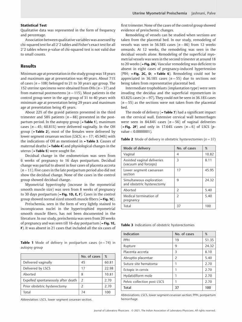
About 22% of the gravid patients presented in the thirdtrimester and 58% patients (n¼88) presented in the post-partum period. In the autopsy group (►Table 1), maximumcases (n¼45; 60.81%) were delivered vaginally. In the OHgroup (►Table 2), most of the females were delivered bylower segment cesarean section (LSCS; n¼17; 45.94%) withthe indications of OH as mentioned in ►Table 3. Causes ofmaternal deaths (►Table 4) and physiological changes in theuterus (►Table 5) were sought for.
Decidual change in the endometrium was seen from6 weeks of pregnancy to 16 days postpartum. Decidualchangewas partial to absent in four cases of placenta accreta(n¼11). Five cases in the late postpartum period also did notshow the decidual change. None of the cases in the controlgroup showed decidual change.
Myometrial hypertrophy (increase in the myometrialsmooth muscle size) was seen from 8 weeks of pregnancyto 30 days postpartum (►Fig. 1D, E, F). Cases in the controlgroup showed normal sized smoothmusclefibers (►Fig. 1C).
Preischemia, seen in the form of very lightly stained toinconspicuous nuclei in the hypertrophied myometrialsmooth muscle fibers, has not been documented in theliterature. In our study, preischemiawas seen from 20weeksof pregnancy andwas seen till 16-day postpartum (►Fig. 1E,
F). It was absent in 21 cases that included all the six cases of
first trimester. None of the cases of the control group showedevidence of preischemic changes.
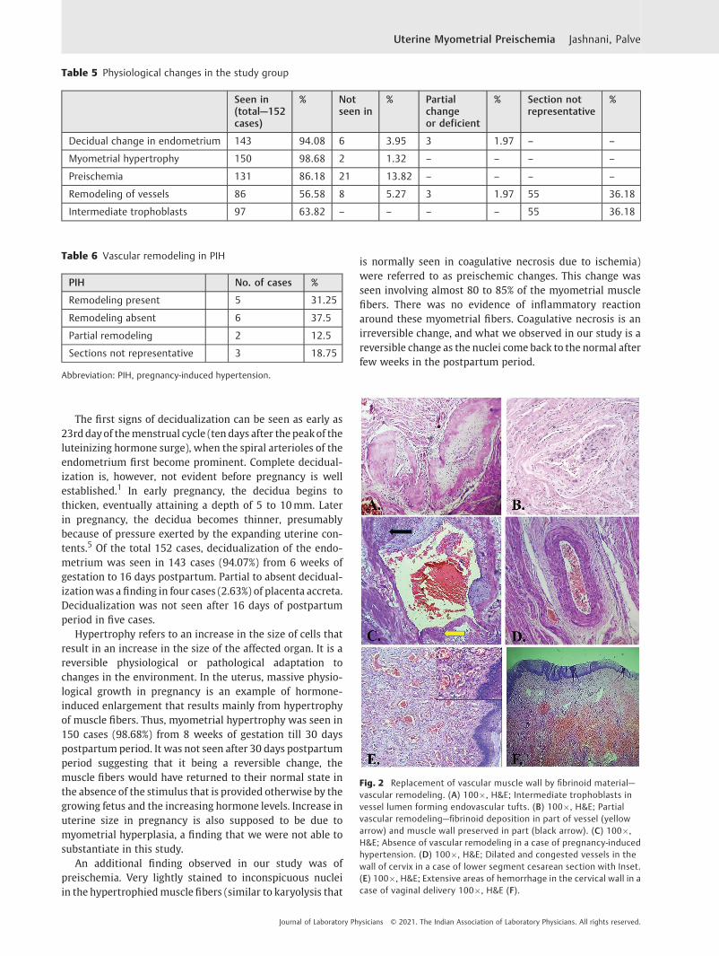
Remodeling of vessels can be studied when sections aretaken from the placental bed. In our study, remodeling ofvessels was seen in 56.58% cases (n¼86) from 12 weeksonwards. At 12 weeks, the remodeling was seen in thedecidual vessels alone. Remodeling of the superficial myo-metrial vesselswas seen in the second trimester at around 18to 20 weeks (►Fig. 2A). Vascular remodeling was deficient toabsent in eight cases of pregnancy-induced hypertension(PIH; ►Fig. 2C, D; ►Table 6). Remodeling could not beappreciated in 36.18% cases (n¼55) due to sections notbeing taken from representative placental bed.
Intermediate trophoblasts (implantation type) were seeninvading the decidua and the superficial myometrium in63.82% cases (n¼97). They could not be seen in 36.18% cases(n¼55) as the sections were not taken from the placentalbed.
The mode of delivery (►Table 7) had a significant impacton the cervical wall. Extensive cervical wall hemorrhageswere seen in 84.84% cases (n¼56) of vaginal deliveries(►Fig. 2F) and only in 17.64% cases (n¼6) of LSCS (p-value<0.0000001).
Table 1 Mode of delivery in postpartum cases (n¼74) inautopsy group
No. of cases %
Delivered vaginally 45 60.81
Delivered by LSCS 17 22.98
Aborted 8 10.81
Expelled spontaneously after death 2 2.70
Prior obstetric hysterectomy 2 2.70
Total 74 100
Abbreviation: LSCS, lower segment cesarean section.
Table 2 Mode of delivery in obstetric hysterectomies (n¼ 37)
Mode of delivery No. of cases %
Vaginal 4 10.82
Assisted vaginal deliveries(vacuum and forceps)
3 8.11
Lower segment caesareansection
17 45.95
Simultaneous explorationand obstetric hysterectomy
9 24.32
Aborted 2 5.40
Medical termination ofpregnancy
2 5.40
Total 37 100
Table 3 Indications of obstetric hysterectomies
Indication No. of cases %
PPH 19 51.35
Rupture 9 24.32
Placenta accreta 3 8.10
Abruptio placentae 2 5.40
Suture site hematoma 1 2.70
Ectopic in cervix 1 2.70
Hydatidiform mole 1 2.70
Pelvic collection post LSCS 1 2.70
Total 37 100
Abbreviations: LSCS, lower segment cesarean section; PPH, postpartumhemorrhage.
Journal of Laboratory Physicians © 2021. The Indian Association of Laboratory Physicians. All rights reserved.
Uterine Myometrial Preischemia Jashnani, Palve

Discussion
In this study, we have examined the uteri in detail forphysiologic changes during pregnancy and postpartum pe-riod. The uterine specimens included those removed during
maternal death autopsies and those received as OHs. Pres-ence or absence of normal physiologic uterine wall changesof decidualization, uterine myometrial hypertrophy, andpreischemia as well as vascular remodeling was studied ineach and every case.
Table 4 Final causes of death in autopsy cases
No. of cases %
PIH related 12 10.42
Direct causes of maternaldeaths
Puerperal sepsis 3 2.60
(n¼25, 21.74%) DIC 3 2.60
PPH 2 1.73
Cortical venous sinus thrombosis 2 1.73
Acute fatty liver of pregnancy 1 0.86
Abruptio placentae 1 0.86
Rupture 1 0.86
Indirect 1. Infection 38 33.04
Causes of maternal deaths(n¼90,78.26%)
I) Hepatitis E 20 17.39
II) Hepatitis B 1 0.86
III) TB 10 8.69
IV) Dengue 3 2.60
V) H1N1 2 1.73
VI) Malaria 1 0.86
VII) Neurocysticercosis 1 0.86
2. Pulmonary 16 13.91
I) Bronchopneumonia 7 5.90
II) ARDS 5 4.33
III) Pulmonary edema 3 2.59
IV) Aspiration pneumonitis 1 0.86
3. Cardiovascular 15 13.04
I) Rheumatic heart disease 9 7.82
II) Dilated cardiomyopathy 5 4.34
III) Acute coronary insufficiency with IHD 1 0.86
4. Liver 8 6.95
I) Hepatic necrosis—cause unknown 6 5.21
II) Cirrhosis 1 0.86
III) Acute hepatitis 1 0.86
5. Septicemia 6 5.21
6. Bilateral renal cortical necrosis 2 1.73
7. Hepatorenal failure 1 0.86
8. Metabolic 1 0.86
9. Subarachnoid hemorrhage 1 0.86
10. Anemia 1 0.86
11. GI bleeding 1 0.86
Abbreviations: ARDS, acute respiratory distress syndrome; DIC, disseminated intravascular coagulation; GI, gastrointestinal; IHD, ischemic heartdisease; PIH, pregnancy-induced hypertension; PPH, postpartum hemorrhage; TB, tuberculosis.
Journal of Laboratory Physicians © 2021. The Indian Association of Laboratory Physicians. All rights reserved.
Uterine Myometrial Preischemia Jashnani, Palve
The first signs of decidualization can be seen as early as23rddayof themenstrual cycle (ten days after the peakof theluteinizing hormone surge), when the spiral arterioles of theendometrium first become prominent. Complete decidual-ization is, however, not evident before pregnancy is wellestablished.1 In early pregnancy, the decidua begins tothicken, eventually attaining a depth of 5 to 10mm. Laterin pregnancy, the decidua becomes thinner, presumablybecause of pressure exerted by the expanding uterine con-tents.5 Of the total 152 cases, decidualization of the endo-metrium was seen in 143 cases (94.07%) from 6 weeks ofgestation to 16 days postpartum. Partial to absent decidual-izationwas a finding in four cases (2.63%) of placenta accreta.Decidualization was not seen after 16 days of postpartumperiod in five cases.
Hypertrophy refers to an increase in the size of cells thatresult in an increase in the size of the affected organ. It is areversible physiological or pathological adaptation tochanges in the environment. In the uterus, massive physio-logical growth in pregnancy is an example of hormone-induced enlargement that results mainly from hypertrophyof muscle fibers. Thus, myometrial hypertrophy was seen in150 cases (98.68%) from 8 weeks of gestation till 30 dayspostpartum period. It was not seen after 30 days postpartumperiod suggesting that it being a reversible change, themuscle fibers would have returned to their normal state inthe absence of the stimulus that is provided otherwise by thegrowing fetus and the increasing hormone levels. Increase inuterine size in pregnancy is also supposed to be due tomyometrial hyperplasia, a finding that we were not able tosubstantiate in this study.
An additional finding observed in our study was ofpreischemia. Very lightly stained to inconspicuous nucleiin the hypertrophiedmusclefibers (similar to karyolysis that
is normally seen in coagulative necrosis due to ischemia)were referred to as preischemic changes. This change wasseen involving almost 80 to 85% of the myometrial musclefibers. There was no evidence of inflammatory reactionaround these myometrial fibers. Coagulative necrosis is anirreversible change, and what we observed in our study is areversible change as the nuclei come back to the normal afterfew weeks in the postpartum period.
Table 5 Physiological changes in the study group
Seen in(total—152cases)
% Notseen in
% Partialchangeor deficient
% Section notrepresentative
%
Decidual change in endometrium 143 94.08 6 3.95 3 1.97 – –
Myometrial hypertrophy 150 98.68 2 1.32 – – – –
Preischemia 131 86.18 21 13.82 – – – –
Remodeling of vessels 86 56.58 8 5.27 3 1.97 55 36.18
Intermediate trophoblasts 97 63.82 – – – – 55 36.18
Table 6 Vascular remodeling in PIH
PIH No. of cases %
Remodeling present 5 31.25
Remodeling absent 6 37.5
Partial remodeling 2 12.5
Sections not representative 3 18.75
Abbreviation: PIH, pregnancy-induced hypertension.
Fig. 2 Replacement of vascular muscle wall by fibrinoid material—vascular remodeling. (A) 100�, H&E; Intermediate trophoblasts invessel lumen forming endovascular tufts. (B) 100�, H&E; Partialvascular remodeling—fibrinoid deposition in part of vessel (yellowarrow) and muscle wall preserved in part (black arrow). (C) 100�,H&E; Absence of vascular remodeling in a case of pregnancy-inducedhypertension. (D) 100�, H&E; Dilated and congested vessels in thewall of cervix in a case of lower segment cesarean section with Inset.(E) 100�, H&E; Extensive areas of hemorrhage in the cervical wall in acase of vaginal delivery 100�, H&E (F).
Journal of Laboratory Physicians © 2021. The Indian Association of Laboratory Physicians. All rights reserved.
Uterine Myometrial Preischemia Jashnani, Palve

Hence, we termed these changes as “Preischemia,” as webelieve it may occur due to the ischemia caused by thepressure effects of the growing fetus that compresses theuterine spiral, radial, and arcuate arteries and reduces myo-metrial blood flow, thus inducing starvation conditions foruterine myocytes. Whether the preischemic changes foundin our study are due to an underlying autophagic process canonly be confirmed by further immunohistochemical andelectron microscopic studies.
In our study, preischemia was seen in 131 cases (86.18%)from 20 weeks of gestation to 16 days postpartum. Relation-ship between preischemia and gestational age was found tobe statistically significant, p<0.0000001. To the best of ourknowledge, it has not been documented before in the litera-ture. In pregnancy, uterus is responding to the mechanicalstrain caused by the enlarging fetus, placenta, and amnioticfluid. The effect of all this is not only increase in size of theuterus as evident by myometrial hypertrophy but also theseso called preischemic changes in the myometrial smoothmuscle fibers. All these efforts are taken by the human bodyto allow for appropriate growth of the fetus with placentawithin the uterus during pregnancy. It is important toidentify the nuclear changes of preischemia as a naïvepathologist may mistake it with coagulative necrosis.Flake et al6 have used a term inanosis to denote a conditionof cellular inanition in uterine leiomyomas resulting fromgradual nutritional deprivation. This is a process of severeatrophy (as against hypertrophy in our study), eventuating incell death by inanosis, where blood flow is diminished byprogressive stenosis of vascular lumens, resulting from theintratumoral vascular smooth muscle proliferation and fi-brosis. In these cells, there is loss of nuclear staining withbarely visible nuclei, and these are assumed to be nonviable.These cells are regarded asmyocyte tombstones, and theyarethe histologic hallmark and end stage of the atrophic processis designated as inanosis.
The placental bed contains the uterine spiral arteries thatsupply oxygenated blood to the growing placenta and fetus.Extravillous trophoblast (EVT) cells proliferate from anchor-ing chorionic villi and invade the decidualized endometriumby two pathways: interstitial EVT cells invade the decidual-ized endometrium and inner myometrium and endovascularEVT cells invade the lumen of the spiral arteries. Some EVTbecome incorporated into the spiral arterywall as intramuraltrophoblast. The endothelium, vascular smooth muscle, and
elastic lamina are destroyed and replaced by fibrinoid. Byterm, maternal vascular repair occurs with reendothelializa-tion.7 EVT cell invasion is thought to be instrumental in thetransformation of the muscular spiral arteries into dis-tended, thin-walled flaccid vessels. Remodeling is necessaryfor a successful pregnancy.8
In our study, remodeling was seen in 56.68% cases (n¼86)and absent in 5.26% cases (n¼8) of the total sample size of152 cases. Partial remodeling was seen in 1.97% cases (n¼3).In few cases, intermediate trophoblasts were present asvascular intraluminal tufts (►Fig. 2B). Placental bed wasnot included in 36.18% cases (n¼55) as it is difficult toidentify in postpartum uteri, as already mentioned in previ-ous studies also9,10
Preeclampsia, defined by hypertension and proteinuria,which affects 3 to 7% of pregnancies, is amajor contributor tomaternal and fetal morbidity and mortality.11 The origin ofpreeclampsia is not understood but is strongly associatedwith failure or partial failure of spiral artery remodelling.7
Unmodified/partially modified vessels can supply high orintermittent pulses of pressure flow and so damage to theplacenta results from hydrostatic stress or from changes inoxygen delivery.12,13
In our study, we had 16 cases of PIH of which vascularremodeling was present in five cases (►Table 4). Thesechanges were absent to deficient in eight cases, which isan expected finding in PIH.8,14–16 However, in remainingthree cases we could not comment on these changes as thesections were not taken from the representative placentalbed. It is difficult to identify the placental bed in postpartumuteri. The relationship between partial/absent remodelingand PIH was statistically significant (p<0.0000001).
The possible reason for sections from five cases showingnormal physiological changes in the vessels may be due tothe fact that lack of physiological change in uterine spiralarteries is not an all-or-none process, with some abnormalcases showing apparently normal remodeling. This mayrelate to the fact that not all cases of preeclampsia and fetalgrowth retardation are attributable to failed spiral arteryremodelling.13
The mode of delivery had a significant effect on thecervical wall changes. Of the 66 cases that were deliveredvaginally, 75.75% cases (n¼50) showed congested vessels inthe wall of cervix and 84.84% cases (n¼56) showed hemor-rhages in the wall. Of the total 34 cases of LSCS, cervical
Table 7 Mode of delivery versus cervical wall status
Mode of delivery Total cases Hemorrhage inthe cervix
% Congestedblood vessels
%
Vaginal 66 56 84.84 50 75.75
LSCS 34 6 17.64 16 47.05
Not delivered 41 3 7.31 25 60.97
Simultaneous LSCS andhysterectomy
11 3 27.27 2 18.18
Abbreviation: LSCS, lower segment cesarean section.
Journal of Laboratory Physicians © 2021. The Indian Association of Laboratory Physicians. All rights reserved.
Uterine Myometrial Preischemia Jashnani, Palve

congested vessels were seen in 47.05% cases(n¼16; ►Fig. 2E) and cervical wall hemorrhages in only17.64% cases (n¼6). Thus, cervical wall hemorrhages weremaximally seen in vaginally delivered cases. The gross spec-imen showed markedly congested to hemorrhagic cervicalwall. The relationship between vaginal delivery and cervicalwall hemorrhages was found to be statistically significant(p<0.0000001). These hemorrhages seen in vaginal deliver-ies may be due to the tearing of the cervical wall vesselsduring the process of expulsion of the fetus. A naïve pathol-ogist may consider the hemorrhages to be a sign of dissemi-nated intravascular coagulation (DIC). However, we believethat the finding of hemorrhages in the cervical wall in case ofvaginal deliveries should be considered a normal finding andnot a sign of DIC. We would like to end by saying that thisstudy is a morphological analysis. Further molecular andultrastructural studies are required that may help in betterunderstanding the mechanisms of uterine changes in preg-nancy and postpartum.
The present study has been undertaken to gain in-depthknowledge of uterine physiology in pregnancy and immedi-ate postpartum period. Literature does not provide anyinformation on the exact duration of the normal physiologi-cal changes in pregnancy. Preischemic changes in the uterinemyometrium as well as cervical wall hemorrhages are notdocumented in literature; therefore, these study findings arenovel findings with significant implications if not alreadyknown to a naïve pathologist.
Authors’ ContributionKJ substantially contributed to the conception and designof thework, definition of intellectual content, and analysisand interpretation of data for thework. KJ also drafted thework and revised it critically for important intellectualcontent. KJ was involved in final approval of the version tobe published. KJ agreed to be accountable for all aspects ofthe work in ensuring that questions related to the accura-cy or integrity of any part of the work are appropriatelyinvestigated and resolved.MP substantially contributed to the literature searchand the acquisition, analysis, or interpretation of datafor the work. MP also drafted the work or revised itcritically for important intellectual content. MP wasinvolved in final approval of the version to be published.MP agreed to be accountable for all aspects of the workin ensuring that questions related to the accuracy orintegrity of any part of the work are appropriatelyinvestigated and resolved.
Conflict of InterestNone.
AcknowledgementA part of this study has been presented as an oral paper inthe state level pathology conference-MAPCON in Septem-ber 2016 in Mumbai, India.
References1 Haines M, Taylor CW, Fox H, Wells M. Haines and Taylor Obstetri-
cal and Gynaecological Pathology. 5th edition. Edinburgh:Churchill Livingstone; 2003. Chapter 39, Pathology of pregnantuterus; pp. 1327–1357
2 Hustin J, Foidart JM, Lambotte R. Maternal vascular lesions in pre-eclampsia and intrauterine growth retardation: light microscopyand immunofluorescence. Placenta 1983;4(Spec No):489–498
3 Robertson WB, Khong TY, Brosens I, De Wolf F, Sheppard BL,Bonnar J. The placental bed biopsy: review from three Europeancenters. Am J Obstet Gynecol 1986;155(02):401–412
4 Rushton DI, Dawson IM. Thematernal autopsy. J Clin Pathol 1982;35(09):909–921
5 Cunningham F, Leveno K, Bloom S, Hauth J, Rouse D. WilliamsObstetrics. 23 rd edition. New York: McGraw- Hill Medical; 2010.Chapter 3, Implantation, Embryogenesis and Placental develop-ment; pp. 36–77
6 Flake GP, Moore AB, Sutton D, et al. The natural history of uterineleiomyomas: light and electron microscopic studies of fibroidphases, interstitial ischemia, inanosis, and reclamation. ObstetGynecol Int 2013;2013:528376. Doi: 10.1155/2013/528376
7 Pijnenborg R, Vercruysse L, Hanssens M. The uterine spiral arter-ies in human pregnancy: facts and controversies. Placenta 2006;27(9-10):939–958
8 Lyall F, Robson SC, Bulmer JN. Spiral artery remodeling andtrophoblast invasion in preeclampsia and fetal growth restric-tion: relationship to clinical outcome. Hypertension 2013;62(06):1046–1054
9 Jashnani KD, Rupani AB, Wani RJ. Maternal mortality: an autopsyaudit. J Postgrad Med 2009;55(01):12–16
10 Jashnani KD, Chandekar SA, Pawar A, Dalal AR, Wani RJ. HepatitisE infection and pregnancy: a fatal combination. Int J Basic ApplMed Sci. 2015;5(02):264–269
11 Redman CW, Sargent IL. Latest advances in understanding pre-eclampsia. Science 2005;308(5728):1592–1594
12 Burton GJ, Woods AW, Jauniaux E, Kingdom JC. Rheological andphysiological consequences of conversion of the maternal spiralarteries for uteroplacental blood flow during human pregnancy.Placenta 2009;30(06):473–482
13 Burton GJ, Yung HW, Cindrova-Davies T, Charnock-Jones DS. Pla-cental endoplasmic reticulum stress and oxidative stress in thepathophysiology of unexplained intrauterine growth restrictionand early onset preeclampsia. Placenta 2009;30(Suppl A):S43–S48
14 Meekins JW, Pijnenborg R, HanssensM,McFadyen IR, vanAsshe A.A study of placental bed spiral arteries and trophoblast invasionin normal and severe pre-eclamptic pregnancies. Br J ObstetGynaecol 1994;101(08):669–674
15 Pijnenborg R, Anthony J, Davey DA, et al. Placental bed spiralarteries in the hypertensive disorders of pregnancy. Br J ObstetGynaecol 1991;98(07):648–655
16 Avagliano L, Bulfamante GP, Morabito A, Marconi AM. Abnormalspiral artery remodelling in the decidual segment during preg-nancy: fromhistology to clinical correlation. J Clin Pathol 2011;64(12):1064–1068
Journal of Laboratory Physicians © 2021. The Indian Association of Laboratory Physicians. All rights reserved.
Uterine Myometrial Preischemia Jashnani, Palve