usefulness of postmortem biochemistry in identification of
TRANSCRIPT
Legal Medicine 22 (2016) 23–29
Contents lists available at ScienceDirect
Legal Medicine
journal homepage: www.elsevier .com/locate / legalmed
Case Report
Usefulness of postmortem biochemistry in identification of ketosis:Diagnosis of ketoacidosis at the onset of autoimmune type 1 diabetesin an autopsy case with cold exposure and malnutrition
http://dx.doi.org/10.1016/j.legalmed.2016.07.0061344-6223/� 2016 Elsevier Ireland Ltd. All rights reserved.
⇑ Corresponding author at: Department of Legal Medicine, Osaka City UniversityMedical School, Asahi-machi 1-4-3, Abeno, 545-8585 Osaka, Japan.
E-mail address: [email protected] (T. Ishikawa).
Naoto Tani a,b, Tomomi Michiue a,b, Jian-Hua Chen a,b, Shigeki Oritani a, Takaki Ishikawa a,b,⇑aDepartment of Legal Medicine, Osaka City University Medical School, Asahi-machi 1-4-3, Abeno, 545-8585 Osaka, Japanb Forensic Autopsy Section, Medico-legal Consultation and Postmortem Investigation Support Center, c/o Department of Legal Medicine, Osaka City University Medical School,Asahi-machi 1-4-3, Abeno, 545-8585 Osaka, Japan
a r t i c l e i n f o a b s t r a c t
Article history:Received 12 February 2016Received in revised form 4 July 2016Accepted 25 July 2016Available online 26 July 2016
Keywords:Autoimmune type 1 diabetesPostmortem biochemistryImmunohistochemistryKetone bodyGlucose
A severely malnourished, Japanese female in her twenties was found dead in her apartment. On autopsy,most of the findings from the internal examination were suggestive of hypothermia. Postmortem bio-chemistry, however, showed severely increased levels of glycated hemoglobin (HbA1c) and blood andurine glucose levels. Levels of acetone, 3-hydroxybutyric acid, and acetoacetate in various body fluidswere also highly increased, indicating ketosis. The serum insulin and c-peptide levels were severelylow, and subsequent testing was positive for anti-GAD antibodies. Immunohistochemical examinationof the pancreatic islet cells revealed few insulin-positive cells but many glucagon-positive cells on stain-ing. Furthermore, slight invasion of CD8-positive lymphocytes in the pancreatic islets of Langerhans wasobserved. Results of immunostaining of the pancreatic and bronchial epithelial tissues were partlypositive for the Influenza A virus. We concluded that severe ketoacidosis associated with rapid-onsethyperglycemia due to autoimmune type 1 diabetes (AT1D) had occurred shortly before death.However, the ketosis was accompanied by hypothermia and malnutrition as well as diabetic ketoacidosis(DKA). Therefore, we retrospectively collected biochemical data on cases of hypothermia andmalnutrition and compared them with the present case. Serum glucose, acetone, 3-hydroxybutyric acid,and acetoacetic acid can be used for screening and diagnosis to distinguish DKA from ketosis due tohypothermia and malnutrition. Therefore, in the present case, we diagnosed that the natural cause ofdeath was due to AT1D. In conclusion, screening investigations for relevant biochemical markers canprovide essential information for the diagnosis of metabolic disturbances, which fail to demonstratecharacteristic autopsy findings.
� 2016 Elsevier Ireland Ltd. All rights reserved.
1. Introduction
Diabetes mellitus (DM) may cause severe metabolic distur-bances, which that can be fatal. These can be difficult to diagnosebased on macroscopic and histological findings alone at autopsy[1–3]. In such cases, postmortem biochemical markers can providesupporting information [1]. When investigating on metabolic andendocrin disorders, a number of postmortem biochemical labora-tory procedures have been reported to be useful for autopsy diag-noses [1,4,5]. Particularly in the postmortem diagnosis of DM, acommon metabolic disease, the main evidence for the diagnosiscan be derived from postmortem biochemical parameters [3,6,7].
Diabetic ketoacidosis (DKA) at the onset of autoimmunetype 1 diabetes (AT1D) is a complication of DM characterizedby extreme and rapid progression of hyperglycemia andketoacidosis due to the destruction of the pancreatic b cells[8,9]. The clinical diagnosis of DKA at the onset of AT1D canbe made when the following biochemical parameters are found:(1) elevation of urinary and/or serum ketone bodies; (2) highplasma and urinary glucose levels (>250 mg/dL and >100 mg/dL,respectively); (3) an anion gap of >12 mEq/L and arterial bloodpH of <7.3; and (4) elevation of plasma blood urea nitrogen(BUN) and creatinine levels [8]. Patients diagnosed with DKAat the onset of AT1D are likely to die within 24 h of the onsetof hyperglycemia unless they receive immediate treatment forDKA [8]. In forensics, DKA at the onset of AT1D has becomemore widely recognized as a cause of sudden death [2,10].However, methods for the postmortem diagnosis of DKA at the
24 N. Tani et al. / Legal Medicine 22 (2016) 23–29
onset of AT1D have not yet been fully established because thereare no characteristic anatomical findings.
Here, we report on a postmortem biochemical diagnosis of DKAat the onset of AT1D, which was made in spite of considerableautopsy findings that were suggestive of hypothermia as beingthe cause of death. We compared these findings to postmortemcases of other ketotic conditions, namely hypothermia and malnu-trition. We also considered whether it was possible to use the post-mortem biochemical profile for DKA diagnosis and compare it toother ketotic conditions, such as hypothermia and malnutrition.
2. Case report
2.1. Case history
A Japanese female in her twenties was found dead in her apart-ment bathroom by her boyfriend at 7:30 a.m. in early Februarymorning.
Five days earlier, she had had a bicycle accident and received ablow to the right inguinal region, after which she experiencednausea and general malaise. She was absent from work for thefollowing 3 days. On the 4th day, she was found dead in herapartment bathroom. She was nude from the waist down andhad fallen while taking a cold-water shower. A gas safety devicewas present, so that hot water would not flow out of the showerand its temperature reduced. The gas safety device seems to workin around five hours. According to her colleagues, she had reportedlosing weight approximately 2 months prior to her death.However, she did not consult a doctor and therefore she was notevaluated for diabetes including the serum glucose and HbA1clevels. In addition, her medical history was unremarkable.
Her room did not have a heater except for a foot-warmer frame(known as kotatsu in Japan). Her rectal temperature at 4:30 p.m. onthe day she was found was 13 �C, and the room temperature was10.5 �C.
A forensic autopsy was performed approximately 1 day afterdeath.
2.2. Postmortem imaging
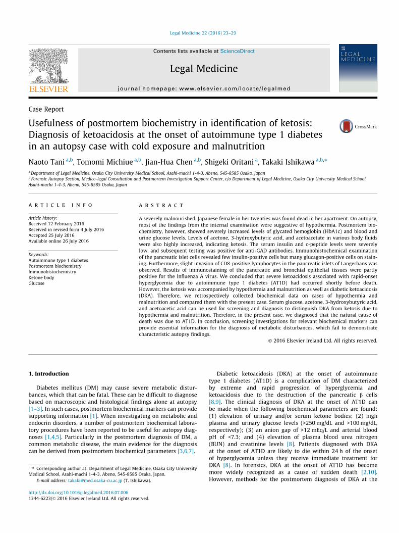
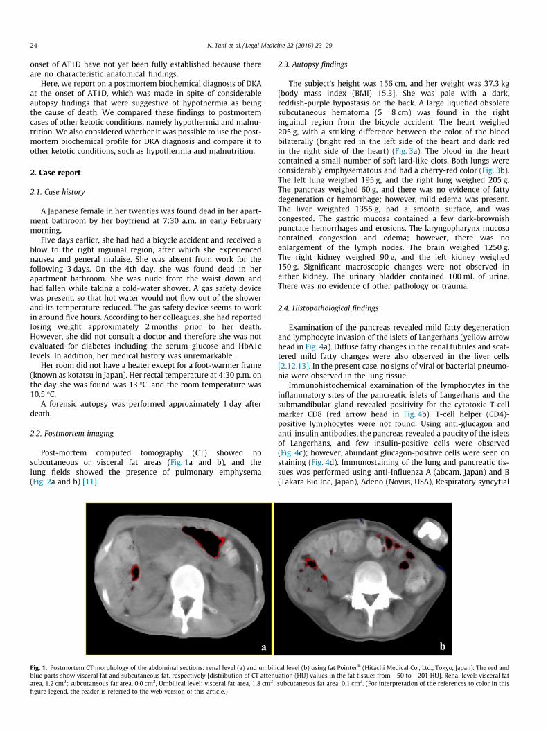
Post-mortem computed tomography (CT) showed nosubcutaneous or visceral fat areas (Fig. 1a and b), and thelung fields showed the presence of pulmonary emphysema(Fig. 2a and b) [11].
a
Fig. 1. Postmortem CT morphology of the abdominal sections: renal level (a) and umbiliblue parts show visceral fat and subcutaneous fat, respectively [distribution of CT attenuarea, 1.2 cm2; subcutaneous fat area, 0.0 cm2, Umbilical level: visceral fat area, 1.8 cm2;figure legend, the reader is referred to the web version of this article.)
2.3. Autopsy findings
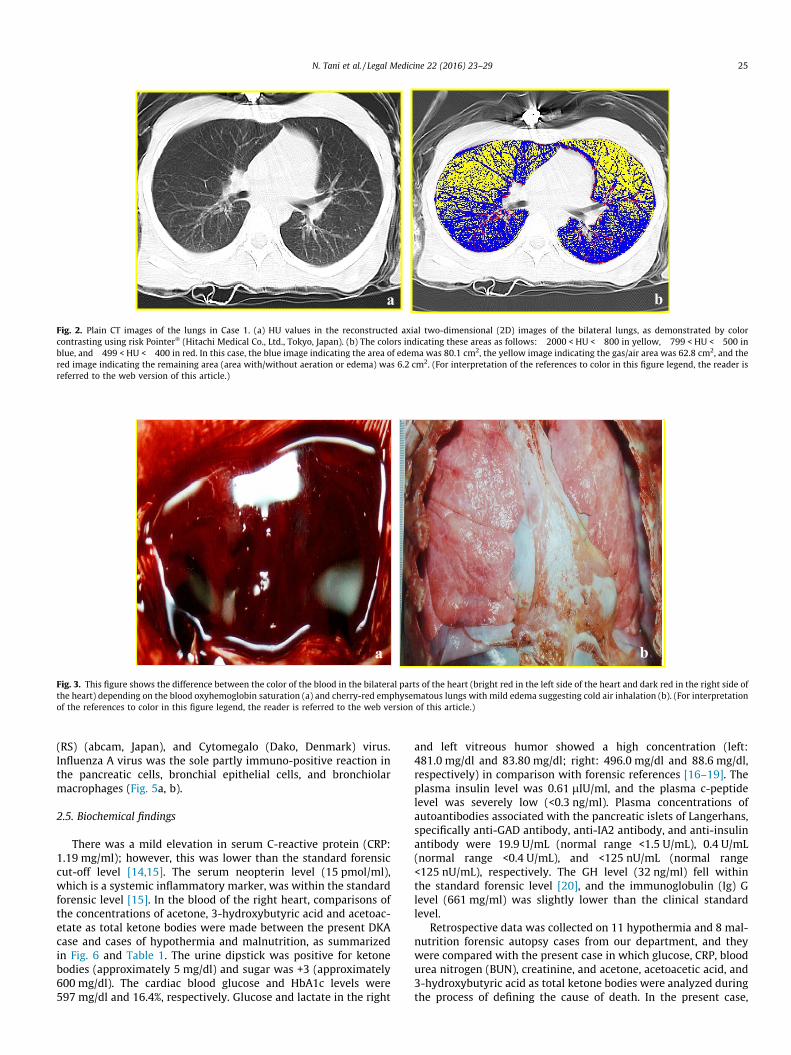
The subject’s height was 156 cm, and her weight was 37.3 kg[body mass index (BMI) 15.3]. She was pale with a dark,reddish-purple hypostasis on the back. A large liquefied obsoletesubcutaneous hematoma (5 � 8 cm) was found in the rightinguinal region from the bicycle accident. The heart weighed205 g, with a striking difference between the color of the bloodbilaterally (bright red in the left side of the heart and dark redin the right side of the heart) (Fig. 3a). The blood in the heartcontained a small number of soft lard-like clots. Both lungs wereconsiderably emphysematous and had a cherry-red color (Fig. 3b).The left lung weighed 195 g, and the right lung weighed 205 g.The pancreas weighed 60 g, and there was no evidence of fattydegeneration or hemorrhage; however, mild edema was present.The liver weighted 1355 g, had a smooth surface, and wascongested. The gastric mucosa contained a few dark-brownishpunctate hemorrhages and erosions. The laryngopharynx mucosacontained congestion and edema; however, there was noenlargement of the lymph nodes. The brain weighed 1250 g.The right kidney weighed 90 g, and the left kidney weighed150 g. Significant macroscopic changes were not observed ineither kidney. The urinary bladder contained 100 mL of urine.There was no evidence of other pathology or trauma.
2.4. Histopathological findings
Examination of the pancreas revealed mild fatty degenerationand lymphocyte invasion of the islets of Langerhans (yellow arrowhead in Fig. 4a). Diffuse fatty changes in the renal tubules and scat-tered mild fatty changes were also observed in the liver cells[2,12,13]. In the present case, no signs of viral or bacterial pneumo-nia were observed in the lung tissue.
Immunohistochemical examination of the lymphocytes in theinflammatory sites of the pancreatic islets of Langerhans and thesubmandibular gland revealed positivity for the cytotoxic T-cellmarker CD8 (red arrow head in Fig. 4b). T-cell helper (CD4)-positive lymphocytes were not found. Using anti-glucagon andanti-insulin antibodies, the pancreas revealed a paucity of the isletsof Langerhans, and few insulin-positive cells were observed(Fig. 4c); however, abundant glucagon-positive cells were seen onstaining (Fig. 4d). Immunostaining of the lung and pancreatic tis-sues was performed using anti-Influenza A (abcam, Japan) and B(Takara Bio Inc, Japan), Adeno (Novus, USA), Respiratory syncytial
b
cal level (b) using fat Pointer� (Hitachi Medical Co., Ltd., Tokyo, Japan). The red andation (HU) values in the fat tissue: from �50 to �201 HU]. Renal level: visceral fatsubcutaneous fat area, 0.1 cm2. (For interpretation of the references to color in this
a b
Fig. 3. This figure shows the difference between the color of the blood in the bilateral parts of the heart (bright red in the left side of the heart and dark red in the right side ofthe heart) depending on the blood oxyhemoglobin saturation (a) and cherry-red emphysematous lungs with mild edema suggesting cold air inhalation (b). (For interpretationof the references to color in this figure legend, the reader is referred to the web version of this article.)
a b
Fig. 2. Plain CT images of the lungs in Case 1. (a) HU values in the reconstructed axial two-dimensional (2D) images of the bilateral lungs, as demonstrated by colorcontrasting using risk Pointer� (Hitachi Medical Co., Ltd., Tokyo, Japan). (b) The colors indicating these areas as follows: �2000 < HU < �800 in yellow, �799 < HU < �500 inblue, and �499 < HU < �400 in red. In this case, the blue image indicating the area of edema was 80.1 cm2, the yellow image indicating the gas/air area was 62.8 cm2, and thered image indicating the remaining area (area with/without aeration or edema) was 6.2 cm2. (For interpretation of the references to color in this figure legend, the reader isreferred to the web version of this article.)
N. Tani et al. / Legal Medicine 22 (2016) 23–29 25
(RS) (abcam, Japan), and Cytomegalo (Dako, Denmark) virus.Influenza A virus was the sole partly immuno-positive reaction inthe pancreatic cells, bronchial epithelial cells, and bronchiolarmacrophages (Fig. 5a, b).
2.5. Biochemical findings
There was a mild elevation in serum C-reactive protein (CRP:1.19 mg/ml); however, this was lower than the standard forensiccut-off level [14,15]. The serum neopterin level (15 pmol/ml),which is a systemic inflammatory marker, was within the standardforensic level [15]. In the blood of the right heart, comparisons ofthe concentrations of acetone, 3-hydroxybutyric acid and acetoac-etate as total ketone bodies were made between the present DKAcase and cases of hypothermia and malnutrition, as summarizedin Fig. 6 and Table 1. The urine dipstick was positive for ketonebodies (approximately 5 mg/dl) and sugar was +3 (approximately600 mg/dl). The cardiac blood glucose and HbA1c levels were597 mg/dl and 16.4%, respectively. Glucose and lactate in the right
and left vitreous humor showed a high concentration (left:481.0 mg/dl and 83.80 mg/dl; right: 496.0 mg/dl and 88.6 mg/dl,respectively) in comparison with forensic references [16–19]. Theplasma insulin level was 0.61 lIU/ml, and the plasma c-peptidelevel was severely low (<0.3 ng/ml). Plasma concentrations ofautoantibodies associated with the pancreatic islets of Langerhans,specifically anti-GAD antibody, anti-IA2 antibody, and anti-insulinantibody were 19.9 U/mL (normal range <1.5 U/mL), 0.4 U/mL(normal range <0.4 U/mL), and <125 nU/mL (normal range<125 nU/mL), respectively. The GH level (32 ng/ml) fell withinthe standard forensic level [20], and the immunoglobulin (Ig) Glevel (661 mg/ml) was slightly lower than the clinical standardlevel.
Retrospective data was collected on 11 hypothermia and 8 mal-nutrition forensic autopsy cases from our department, and theywere compared with the present case in which glucose, CRP, bloodurea nitrogen (BUN), creatinine, and acetone, acetoacetic acid, and3-hydroxybutyric acid as total ketone bodies were analyzed duringthe process of defining the cause of death. In the present case,

b a
Fig. 5. Immunostaining for Influenza A virus showed positive reaction in pancreatic cells (a), bronchial epithelial cells, and bronchiolar macrophages (b).
a
c d
b
Fig. 4. (a) Histopathological findings in the pancreas (peri-islets): yellow arrow head shows lymphocytic infiltration (hematoxylin eosin staining, magnification �100). (b)Red arrow head shows anti-human CD-8 positive T-cells on immunostaining in the peri-islets (magnification �200). Immunopositivity findings of insulin (c) into islets wasalmost absent, and glucagon (d) was barely present (magnification �200). (For interpretation of the references to color in this figure legend, the reader is referred to the webversion of this article.)
26 N. Tani et al. / Legal Medicine 22 (2016) 23–29
glucose, acetoacetic acid, 3-hydroxybutyric acid, acetone, and totalketone body levels were all extremely elevated compared withthe cases of hypothermia and malnutrition (Fig. 6 and Table 1)[21–24].
2.6. Blood gas analysis
Postmortem left and right blood gases revealed oxygen partialpressures (PO2) of 93.9% and 41.4%, respectively. It is thought thatshe was placed under low temperature exposure as for this result[25].
2.7. Postmortem microbiology
The mitochondrial (mt) DNA 3243 mutation [26] was notdetected in the whole blood.
2.8. Toxicology findings
No blood alcohol was detected in several body fluids. Drugscreening, including blood screening for amphetamines and psy-chotropic drugs using immunoassay and gas chromatography–mass spectrometry (GC–MS), was negative [27].

Table 1Summary of the ketosis-related markers in the present case compared with cases of hypothermia and malnutrition.
Right heart blood Hypothermia (n = 11) (Male: n = 6, Female:n = 5) (BMI: 12.7–25.3 kg/m2, median:19.1 kg/m2)
Malnutrition (n = 8) (Male: n = 3, Female:n = 5) (BMI: 9.8–18.4 kg/m2, median:14.0 kg/m2)
Present case ForensicCut-off value
Reference No.
Glucose 10.0–484.0 mg/dL (median: 111.0 mg/dL) 1.0–120.0 mg/dL (median: 28.5 mg/dL) 597.0 mg/dL 227.5 mg/dL [21]Acetone 0.0–52.8 lg/mL (median: 2.1 lg/mL 0.0–103.6 lg/mL (median: 16.4 lg/mL) 332.4 lg/mL <50 lg/mL [22]Total ketone
bodies86.0–4410.0 lmol/L (median: 465.0 lmol/L) 115.0–3750.0 lmol/L (median: 1027.5 lmol/L) 15269.0 lmol/L 217.5 lmol/L [21]
Acetoacetic acid 2.0–90.0 lmol/L (median: 2.0 lmol/L) 2.0–26.0 lmol/L (median: 3.5 lmol/L) 376.0 lmol/L 12.0 lmol/L No publisheddata
3-Hydroxybutyricacid
86.0–4320.0 lmol/L (median: 449.0 lmol/L) 110.0–3750.0 lmol/L (median: 1014.5 lmol/L) 14893.0 lmol/L 260.0 lmol/L No publisheddata
Blood urea nitrogen(BUN)
11.4–74.3 mg/dL (median: 41.6 mg/dL) 16.0–206.0 mg/dL (median: 66.2 mg/dL) 20.5 mg/dL 38.48 mg/dL [23]
Creatinine 0.5–2.4 mg/dL (median: 0.8 mg/dL 0.09–5.5 mg/dL (median: 2.8 mg/dL 0.7 mg/dL 3.32 mg/dL [23]C-reactive protein
(CRP)0.05–5.72 mg/dL (median: 0.4 mg/dL) 0.02–14.9 mg/dL (median: 9.4 mg/dL) 1.19 mg/dL <0.1 mg/dL [24]
Hypothermia Malnutrition
Glu
cose
(mg/dL)
Hypothermia Malnutrition
Cre
atin
ine
(mg/dL)
Hypothermia Malnutrition
Blo
od u
rea
nitr
ogen
(BU
N)
(mg/dL)
C-r
eact
ive
prot
ein
(CR
P)
Hypothermia Malnutrition
(mg/dL)
Hypothermia Malnutrition
Ace
tone
(µg/mL)
Hypothermia Malnutrition
Ace
toac
etic
aci
d
(µmol/L)
Hypothermia Malnutrition
Tota
lke
tone
bod
y
(µmol/L)
3-H
ydro
xybu
tyri
c ac
id
(µmol/L)
Hypothermia Malnutrition
Forensic cut-off value
Forensic cut-off value
Forensic cut-off value
Forensic cut-off value
Forensic cut-off value
Forensic cut-off value
Forensic cut-off value
Forensic cut-off value
Fig. 6. We collected retrospective data on 11 hypothermia (cold exposure) and 8 malnutrition forensic autopsy cases and compared themwith the present case (red-circle) inwhich glucose, acetone, total ketone bodies, acetoacetic acid, 3-hydroxybutyric acid, BUN, creatinine, and CRP were analyzed during the process of defining the cause of death.Glucose, acetone, total ketone bodies, 3-hydroxybutyric acid, and acetoacetic acid (red color in the graph) may be useful for the screening and diagnosis of diabeticketoacidosis in order to distinguish it from ketosis due to hypothermia and malnutrition. (For interpretation of the references to color in this figure legend, the reader isreferred to the web version of this article.)
N. Tani et al. / Legal Medicine 22 (2016) 23–29 27
3. Discussion
In the present case, most of the macropathological findings oninternal examination suggested possible hypothermia (cold expo-sure) as the cause of death. However, according to the postmortembiochemistry, postmortem HbA1c levels were markedly increased
and the glucose levels in the blood of the right heart and urineglucose levels were extremely elevated. HbA1c levels have beenreported to stabilize within 72 h postmortem [28], while bloodglucose concentrations have been reported to decrease after deathand should not be underestimated [6]. HbA1c is formed whenglucose is non-enzymatically added to hemoglobin. The proportion

28 N. Tani et al. / Legal Medicine 22 (2016) 23–29
of HbA1c compared with total hemoglobin reflects the mean bloodglucose level for the previous 1–2 months, which is the lifespan ofred blood cells. However, in a postmortem context, HbA1c levelsare useful for distinguishing diabetic ketoacidosis (DKA) from alco-holic ketoacidosis (AKA) and for revealing undiagnosed or poorlymanaged diabetes [29–31].
Furthermore, in the present case, glucose and lactate in bothsides of the vitreous humor showed a high concentration incomparison with forensic references. Karlovsek [17,18] comparedseveral biochemical parameters (glycated hemoglobin, glucose,lactate, and combined glucose) in vitreous and cerebrospinal fluidsin 112 forensic cases divided into two diagnostic groups. Theauthor proposed that vitreous glucose levels greater than13 mmol/l (corresponding to 234 mg/dl) or combined glucoseand lactate values in vitreous or cerebrospinal fluids greater thanthe threshold values of 23.7 mmol/l (427 mg/dl) and 23.4 mmol/l(422 mg/dl), respectively, could indicate antemortem hyper-glycemia with a fatal outcome. Therefore, in the present case, takentogether, the Hb1Ac and glucose levels suggested that hyper-glycemia occurred rapidly and shortly before death [21].
We retrospectively collected data on 11 hypothermia (coldexposure) and 8 malnutrition forensic autopsy cases from ourdepartment and compared them with the data on the present case,in which the levels of glucose, acetone, total ketone bodies,3-hydroxybutyric acid, acetoacetic acid, BUN, creatinine, and CRPwere assessed for the determination of the cause of death. Ouranalysis suggests that glucose, acetone, total ketone bodies,3-hydroxybutyric acid, and acetoacetic acid may be useful for thescreening and diagnosis of diabetic ketoacidosis and distinguishingit from ketosis caused by hypothermia and malnutrition. BUN,creatinine, and CRP are not ideal markers of DKA because theseare elevated only in the most severe cases of hypothermia andmalnutrition; however, they and other metabolic markers suchas HbA1c can aid a forensic pathologist in the diagnosis ofdiabetes-related death [32]. An increase in blood glucose levelshas been observed in hypothermia cases, likely caused by theenhanced secretion of adrenaline and corticosterone [33]. Anelevation of BUN, creatinine and CRP was also associated withgastrointestinal bleeding, pneumonia, and fatal hypothermia, inwhich known dehydration/hemoconcentration and elevatedprotein catabolism may have caused the elevation [34–37]. In thepresent case, BUN, creatinine, and CRP of biochemical markerswere belonged to a category of hypothermia and malnutritiondue to protein catabolism, respectively. Therefore, based onprevious reports [7,38,39], the subject must have been in a stateof severe ketoacidosis due to hyperglycemia at the time of death.
Subsequent testing was positive for anti-GAD antibodies in theserum; however, anti-IA2 and anti-insulin antibodies were notdetected. Furthermore, mtDNA 3243 mutations were not detected.Among Japanese patients with diabetes, mtDNA 3243 mutationsare related to the development of diabetes, and these mutationsare associated with not only a decrease in insulin secretion butwith also advanced diabetic microvascular complications [40].Although mtDNA 3243 was negative in this case, this is not unex-pected because the incidence of mtDNA3243 mutation is very low,at approximately 0.5–2.8% [41]. From these postmortem biochem-ical findings, we concluded that severe ketoacidosis associatedwith a rapid onset of hyperglycemia occurred shortly before deathdue to autoimmune type 1 diabetes mellitus [8]. Although the sur-vival period in this case was short, fulminant type 1 diabetes mel-litus was not diagnosed because the findings did not correspondwith the diagnostic criteria for fulminant type 1 diabetes mellitus(such as >8.7% HbA1c and positive anti-GAD antibodies) [42].
In addition, in the present case, CD8-positive T-cells wereobserved in the pancreas and submandibular gland. In type 1diabetes mellitus, it is assumed that CD8-positive T-cells in the
pancreas primarily contribute to selective pancreatic b-celldestruction [43,44]. Only a few previous reports have suggestedthat immunostaining of the pancreas can be useful in forensicautopsy cases in which CD8-positive lymphocytes suggest viralinfection [45]. In the present case, immunopositivity to InfluenzaA virus was observed in the pancreatic and bronchial epithelialcells and bronchiolar macrophages, although there were no clearsigns of viral pneumonia. Therefore, we assume that the InfluenzaA virus infection had induced the development of diabeticketoacidosis.
With respect to the blood biochemistry results in the presentcase, a high HbA1c level, and high levels of serum glucose, acetone,total ketone body, 3-hydroxybutyric acid, and acetoacetic acid maybe useful in distinguishing DKA from the ketosis associated withhypothermia and malnutrition cases. In addition, diabetes-relatedautoantibody was positive. It did not contradict it for the immuno-histochemistry in DKA due to AT1D either. On the basis of ouranalyses, we concluded that in the present case, DKA due toAT1D was the cause of death and that cold exposure and malnutri-tion contributed to the death process; the and manner of deathwas regarded as natural death. These conclusions are supportedby both the autopsy findings and the biochemical results.
The present case can be considered to be a strong example notonly of the usefulness of postmortem biochemical investigationsbut also of the importance of performing these analyses to deter-mine the manner of death.
Conflict of interest statement
The authors declare that they have no proprietary, financial,professional, or other personal interest of any kind in any product,service, and/or company that could be construed as influencingthis current manuscript entitled ‘‘Usefulness of postmortembiochemistry in forensic autopsy: diagnosis of ketoacidosis at theonset of autoimmune type 1 diabetes in an autopsy case with coldexposure and malnutrition.”
References
[1] C. Palmiere, M. Lesta Mdel, S. Sabatasso, P. Mangin, M. Augsburger, F. Sporkert,Usefulness of postmortem biochemistry in forensic pathology: illustrative casereports, Leg. Med. (Tokyo) 14 (2012) 27–35.
[2] T. Mizutani, T. Yoshimoto, R. Kaneko, A. Ishii, Diagnosis of fulminant type 1diabetes mellitus in an autopsy case with postmortem changes, Leg. Med.(Tokyo) 13 (2011) 250–253.
[3] B. Zilg, K. Alkass, S. Berg, H. Druid, Postmortem identification of hyperglycemia,Forensic Sci. Int. 185 (2009) 89–95.
[4] C. Palmiere, P. Mangin, Postmortem chemistry update part I, Int. J. Leg. Med.126 (2012) 187–198.
[5] S.A. Blana, F. Musshoff, T. Hoeller, R. Fimmers, B. Madea, Variations in vitreoushumor chemical values as a result of pre-analytical treatment, Forensic Sci. Int.210 (2011) 263–270.
[6] C. Hess, F. Musshoff, B. Madea, Disorders of glucose metabolism-post mortemanalyses in forensic cases: part 1, Int. J. Leg. Med. 125 (2011) 163–170.
[7] C. Hess, F. Musshoff, B. Madea, Disorders of glucose metabolism-postmortemanalyses in forensic cases: part 2, Int. J. Leg. Med. 125 (2011) 171–180.
[8] T. Hanafusa, A. Imagawa, Fulminant type 1 diabetes: a novel clinical entityrequiring special attention by all medical practitioners, Nat. Clin. Pract.Endocrinol. Metab. 3 (2007) 36–45.
[9] J. Lu, J. Zhou, Y. Bao, T. Chen, Y. Zhang, A. Zhao, Y. Qiu, G. Xie, C. Wang, W. Jia, W.Jia, Serum metabolic signatures of fulminant type 1 diabetes, J. Proteome Res.11 (2012) 4705–4711.
[10] S. Goto, H. Abiru, M. Iino, K. Tamaki, An autopsy of fulminant type 1 diabetesmellitus, Jpn. J. Legal Med. 59 (2005) 201 [in Japanese].
[11] N. Sogawa, T. Michiue, T. Ishikawa, O. Kawamoto, S. Oritani, H. Maeda,Postmortem volumetric CT data analysis of pulmonary air/gas content withregard to the cause of death for investigating terminal respiratory function inforensic autopsy, Forensic Sci. Int. 241 (2014) 112–117.
[12] C. Zhou, L. Moore, A. Yool, A. Jaunzems, R.W. Byard, Renal tubular epithelialvacuoles-a marker for both hyperlipidemia and ketoacidosis at autopsy, J.Forensic Sci. 60 (2015) 638–641.
[13] S. Kodikara, P. Paranitharan, M.S. Pollanen, The role of the Armanni-Ebsteinlesion, hepatic steatosis, biochemical analysis and second generation

N. Tani et al. / Legal Medicine 22 (2016) 23–29 29
anti-psychotic drugs in fatal diabetic ketoacidosis, J. Forensic Leg. Med.(Tokyo) 20 (2013) 108–111.
[14] M.Q. Fujita, B.L. Zhu, K. Ishida, L. Quan, S. Oritani, H. Maeda, Serum C-reactiveprotein levels in postmortem blood – an analysis with special reference to thecause of death and survival time, Forensic Sci. Int. 130 (2002) 160–166.
[15] T. Ishikawa, M. Hamel, B.L. Zhu, D.R. Li, D. Zhao, T. Michiue, H. Maeda,Comparative evaluation of postmortem serum concentrations of neopterinand C-reactive protein, Forensic Sci. Int. 179 (2008) 135–143.
[16] C. Palmiere, P. Mangin, Postmortem chemistry updata part I, Int. J. Legal Med.126 (2012) 187–198.
[17] M.Z. Karlovsek, Diagnostic values of combined glucose and lactate values incerebrospinal fluid and vitreous humour-our experiences, Forensic Sci. Int. 146(2004) S19–S23.
[18] B. Jacob, W. Bonte, Advances in Forensic Sciences: Forensic Criminalistic 2,Verg Dr Kösrner, Berlin, 1995.
[19] V.J. DiMaio, D.D. DiMaio, Forensic Pathology, second ed., CRC Press, New York,2001.
[20] T. Ishikawa, T. Michiue, H. Maeda, Evaluation of postmortem serum andcerebrospinal fluid growth hormone levels in relation to the cause of death inforensic autopsy, Hum. Cell 24 (2011) 74–77.
[21] J.H. Chen, T. Michiue, O. Inamori-Kawamoto, S. Ikeda, T. Ishikawa, H. Maeda,Comprehensive investigation of postmortem glucose levels in blood and bodyfluids with regard to the cause of death in forensic autopsy cases, Leg. Med.(Tokyo) 17 (2015) 475–482.
[22] M. Tominaga, T. Ishikawa, T. Michiue, S. Oritani, I. Koide, Y. Kuramoto, M.Ogawa, H. Maeda, Postmortem analyses of gaseous and volatile substances inpericardial fluid and bone marrow aspirate, J. Anal. Toxicol. 37 (2013) 147–151.
[23] B.L. Zhu, K. Ishida, L. Quan, M. Taniguchi, S. Oritani, D.R. Li, M.Q. Fujita, H.Maeda, Postmortem serum uric acid and creatinine levels in relation to thecauses of death, Forensic Sci. Int. 125 (2002) 59–66.
[24] T. Ishikawa, M. Hamel, B.L. Zhu, D.R. Li, D. Zhao, T. Michiue, H. Maeda,Comparative evaluation of postmortem serum concentrations of neopterinand C-reactive protein, Forensic Sci. Int. 179 (2008) 135–143.
[25] K. Shimizu, H. Mizukami, T. Fukushima, M. Sasaki, H. Shiono, Use of a CO-oximeter for forensic diagnosis of hypothermia, Nihon Hoigaku Zasshi 52(1998) 196–201 (Japanese).
[26] M. Fukuda, S. Nakano, N. Imaizumi, M. Kitazawa, M. Nishizawa, T. Kigoshi, K.Uchida, Mitochondrial DNA mutations are associated with both decreasedinsulin secretion and advanced microvascular complications in Japanesediabetic subjects, J. Diabetes Complications 13 (1999) 277–283.
[27] M. Tominaga, T. Michiue, T. Ishikawa, O. Inamori-Kawamoto, S. Oritani, H.Maeda, Evaluation of postmortem drug concentrations in cerebrospinal fluidcompared with blood and pericardial fluid, Forensic Sci. Int. 254 (2015)118–125.
[28] K. Uemura, K. Shintani-Ishida, K. Saka, M. Nakajima, H. Ikegaya, Y. Kikuchi, K.Yoshida, Biochemical blood markers and sampling sites in forensic autopsy, J.Forensic Leg. Med. 15 (2008) 312–317.
[29] J. Hockenhull, W. Dhillo, R. Andrews, S. Paterson, Investigation of markers toindicate and distinguish death sue to alcoholic ketoacidosis, diabetic
ketoacidosis and hyperosmolar hyperglycemic state using post-mortemsamples, Forensic Sci. Int. 214 (2012) 142–147.
[30] C. Palmiere, D. Bardy, P. Mangin, D. Werner, Postmortem diagnosis ofunsuspected diabetes mellitus, Forensic Sci. Int. 226 (2013) 160–167.
[31] T. Keltanen, A. Sajantila, T. Valonen, T. Vamhala, K. Lindroos, Measuringpostmortem glycated hemoglobin – a comparison of three methods, Leg. Med.(Tokyo) 15 (2013) 72–78.
[32] Keltanen, A. Sajantila, T. Valonen, T. Vanhala, K. Lindroos, Measuringpostmortem glycated hemoglobin – a comparison if three methods, Leg.Med. (Tokyo) 15 (2013) 72–78.
[33] C. Palmiere, P. Mangin, Postmortem biochemical investigations inhypothermia fatalities, Int. J. Legal Med. 127 (2013). 267-31.
[34] D.F. Danzl, J.R. Hedges, R.S. Pozos, Hypothermia outcome score: developmentand implications, Crit. Care Med. 17 (1989) 227–231.
[35] R.H. Danzl, W.W. Lawrence, Surgical Diagnosis and Treatment, ninth ed.,Appleton and Lange, CT, 1991.
[36] B.L. Zhu, K. Ishida, L. Quan, M. Taniguchi, S. Oritani, D.R. Li, M.Q. Fujita, H.Maeda, Postmortem serum uric acid and creatinine levels in relation to thecauses of death, Forensic Sci. Int. 125 (2002) 59–66.
[37] H. Maeda, B.L. Zhu, Bessho, T. Ishikawa, L. Quan, T. Michiue, D. Zhao, D.R. Li, A.Komatsu, Postmortem serum nitrogen compounds and C-reactive proteinlevels with special regard to investigation of fatal hyperthermia, Forensic Sci.Med. Pathol. 4 (2008) 175–180.
[38] J. Kanetake, Y. Kanawaku, S. Mimasaka, J. Sakai, M. Hashiyada, M. Nata, M.Funayama, The relationship of a high level of serum beta-hydroxybutyrate tocause of death, Leg. Med. (Tokyo) 7 (2005) 169–174.
[39] E. Osuna, G. Vivero, J. Conejero, J.M. Abenza, P. Martínez, A. Luna, M.D. Pérez-Cárceles, Postmortem vitreous humor beta-hydroxybutyrate: its utility for thepostmortem interpretation of diabetes mellitus, Forensic Sci. Int. 153 (2005)189–195.
[40] M. Inada, M. Oishi, M. Nishikawa, S. Kurata, H. Imura, Clinical evaluation ofmeasuring glycosylated hemoglobin levels for assessing the long-term bloodglucose control in diabetics, Endocrinol. Jpn. 27 (1980) 411–415.
[41] S. Suzuki, Y. Okada, T. Kadowaki, A. Kanatsuka, T. Kuzuya, M. Kobayashi, T.Sanke, Y. Seino, K. NanjoResearch committee or specific types of diabetesmellitus with gene mutations of the Japan diabetes society, Clinical features ofdiabetes mellitus with the mitochondrial DNA 3243 (A-G) mutation inJapanese: maternal inheritance and mitochondria-related complications,Diabetes Res. Clin. Pract. 59 (2003) 207–211.
[42] S. Shibasaki, A. Imagawa, T. Hanafusa, Fuluminant type 1 diabetes mellitus: anew class of type 1 diabetes, Adv. Exp. Med. Biol. 771 (2012) 20–23.
[43] M. Nagata, H. Moriyama, R. Kotani, H. Yasuda, M. Kishi, M. Kurohara, K. Hara, K.Yokono, Immunological aspects of ‘fulminant type 1 diabetes’, Diabetes Res.Clin. Pract. 77 (2007) S99–S103.
[44] T. Hanafusa, A. Imagawa, Insulitis in human type 1 diabetes, Ann. N. Y. Acad.Sci. 1150 (2008) 297–299.
[45] R.S. Fujinami, M.B. Oldstone, Z. Wroblewska, M.E. Frankel, H. Koprowski,Molecular mimicry in virus infection: crossreaction of measles virusphosphoprotein or of herpes simplex virus protein with human intermediatefilaments, Proc. Natl. Acad. Sci. USA 80 (1983) 2346–2350.