use of mitraclip for mitral valve repair in patients with
TRANSCRIPT

Henry Ford Health System Henry Ford Health System
Henry Ford Health System Scholarly Commons Henry Ford Health System Scholarly Commons
Cardiology Articles Cardiology/Cardiovascular Research
2-18-2021
Use of MitraClip for mitral valve repair in patients with acute Use of MitraClip for mitral valve repair in patients with acute
mitral regurgitation following acute myocardial infarction: Effect mitral regurgitation following acute myocardial infarction: Effect
of cardiogenic shock on outcomes (IREMMI Registry) of cardiogenic shock on outcomes (IREMMI Registry)
Rodrigo Estévez-Loureiro
Mony Shuvy
Maurizio Taramasso
Tomas Benito-Gonzalez
Paolo Denti
See next page for additional authors
Follow this and additional works at: https://scholarlycommons.henryford.com/cardiology_articles

Authors Authors Rodrigo Estévez-Loureiro, Mony Shuvy, Maurizio Taramasso, Tomas Benito-Gonzalez, Paolo Denti, Dabit Arzamendi, Marianna Adamo, Xavier Freixa, Pedro Villablanca, Lian Krivoshei, Neil Fam, Konstantinos Spargias, Andrew Czarnecki, Dan Haberman, Yoram Agmon, Doron Sudarsky, Isaac Pascual, Vlasis Ninios, Salvatore Scianna, Igal Moaraf, Davide Schiavi, Michael Chrissoheris, Ronen Beeri, Arthur Kerner, Estefanía Fernández-Peregrina, Mattia Di Pasquale, Ander Regueiro, Lion Poles, Andres Iñiguez-Romo, Felipe Fernández-Vázquez, and Francesco Maisano

OR I G I N A L S T UD I E S
Use of MitraClip for mitral valve repair in patients with acutemitral regurgitation following acute myocardial infarction:Effect of cardiogenic shock on outcomes (IREMMI Registry)
Rodrigo Estévez-Loureiro MD, PhD1 | Mony Shuvy MD2 |
Maurizio Taramasso MD, PhD3 | Tomas Benito-Gonzalez MD4 |
Paolo Denti MD, PhD5 | Dabit Arzamendi MD, PhD6 | Marianna Adamo MD7 |
Xavier Freixa MD, PhD8 | Pedro Villablanca MD, MSc9 | Lian Krivoshei MD10 |
Neil Fam MD, PhD11 | Konstantinos Spargias MD12 | Andrew Czarnecki MD13 |
Dan Haberman MD14 | Yoram Agmon MD15 | Doron Sudarsky MD16 |
Isaac Pascual MD, PhD17 | Vlasis Ninios MD18 | Salvatore Scianna MD3 |
Igal Moaraf MD10 | Davide Schiavi MD5 | Michael Chrissoheris MD12 |
Ronen Beeri MD2 | Arthur Kerner MD15 | Estefanía Fernández-Peregrina MD6 |
Mattia Di Pasquale MD7 | Ander Regueiro MD, PhD8 | Lion Poles MD14 |
Andres Iñiguez-Romo MD, PhD1 | Felipe Fernández-Vázquez MD, PhD4 |
Francesco Maisano MD3
1Interventional Cardiology Unit, Hospital �Alvaro Cunqueiro, Vigo, Spain
2Heart Institute, Hadassah-Hebrew University Medical Center, Jerusalem, Israel
3Heart Valve Clinic, University Hospital of Zurich, Zurich, Switzerland
4Interventional Cardiology Unit, Complejo Asistencial Universitario de Leon, Leon, Spain
5Department of Cardiovascular Surgery, San Raffaele University Hospital, Milan, Italy
6Interventional Cardiology Unit, Hospital Sant Pau i Santa Creu, Barcelona, Spain
7Cardiac Catheterization Laboratory, Cardiothoracic Department, Spedali Civili Brescia, Brescia, Italy
8Interventional Cardiology Unit, Hospital Clinic, Barcelona, Spain
9Interventional Cardiology, Structural Heart Disease Interventions, Endovascular Interventions, The Center for Structural Heart Disease, Henry Ford Hospital, Detroit, Michigan
10Department of Cardiology, Kantonsspital Baden, Baden, Switzerland
11Division of Cardiology, St. Michael's Hospital, University of Toronto, Toronto, Ontario, Canada
12Department of Transcatheter Heart Valves, HYGEIA Hospital, Athens, Greece
13Division of Cardiology, Sunnybrook Heath Sciences Centre, University of Toronto, Shulich Heart Centre, Tronto, Ontario, Canada
14Heart Center, Kaplan Medical Center, Affiliated to the Hebrew University, Jerusalem, Israel
15Department of Cardiology, Rambam Medical Center, and B. Rappaport Faculty of Medicine, Technion Medical School, Haifa, Israel
16Cardiovascular Institute, Padeh Medical Center, Tiberias, Israel
17Interventional Cardiology Unit, Hospital Universitario Central de Asturias, Oviedo, Spain
18Department of Cardiology, Interbalkan European Medical Center, Thessaloniki, Greece
Abbreviations: AMI, acute myocardial infarction; CS, cardiogenic shock; LVEF, left ventricular ejection fraction; MR, mitral regurgitation; MVARC, Mitral Valve Academic Research Consortium;
NYHA, New York Heart Association; PMVR, percutaneous mitral valve repair; SPAP, systolic pulmonary arterial pressure.
Rodrigo Estévez-Loureiro and Mony Shuvy contributed equally to this study.
Received: 24 July 2020 Revised: 19 January 2021 Accepted: 1 February 2021
DOI: 10.1002/ccd.29552
Catheter Cardiovasc Interv. 2021;1–9. wileyonlinelibrary.com/journal/ccd © 2021 Wiley Periodicals LLC. 1

Correspondence
Rodrigo Estévez-Loureiro MD PhD FESC,
Interventional Cardiology Unit, Department of
Cardiology University Hospital Alvaro
Cunqueiro, Vigo, Spain c/ Clara Campoamor
341, 36312 Vigo.
Email: [email protected]
Abstract
Objectives: To assess outcomes in patients with acute mitral regurgitation
(MR) following acute myocardial infarction (AMI) who received percutaneous mitral
valve repair (PMVR) with the MitraClip device and to compare outcomes of patients
who developed cardiogenic shock (CS) to those who did not (non-CS).
Background: Acute MR after AMI may lead to CS and is associated with high
mortality.
Methods: This registry analyzed patients with MR after AMI who were treated
with MitraClip at 18 centers within eight countries between January 2016 and
February 2020. Patients were stratified into CS and non-CS groups. Primary out-
comes were mortality and rehospitalization due to heart failure. Secondary out-
comes were acute procedural success, functional improvement, and MR reduction.
Multivariable Cox regression analysis evaluated association of CS with clinical
outcomes.
Results: Among 93 patients analyzed (age 70.3 ± 10.2 years), 50 patients (53.8%)
experienced CS before PMVR. Mortality at 30 days (10% CS vs. 2.3% non-CS;
p = .212) did not differ between groups. After median follow-up of 7 months (IQR
2.5–17 months), the combined event mortality/re-hospitalization was similar (28%
CS vs. 25.6% non-CS; p = .793). Likewise, immediate procedural success (90% CS
vs. 93% non-CS; p = .793) and need for reintervention (CS 6% vs. non-CS 2.3%,
p = .621) or re-admission due to HF (CS 13% vs. NCS 23%, p = .253) at 3 months
did not differ. CS was not independently associated with the combined end-point
(hazard ratio 1.1; 95% CI, 0.3–4.6; p = .889).
Conclusions: Patients found to have significant MR during their index hospitaliza-
tion for AMI had similar clinical outcomes with PMVR whether they presented in or
out of cardiogenic shock, provided initial hemodynamic stabilization was first
achieved before PMVR.
K E YWORD S
acute myocardial infarction, cardiogenic shock, MitraClip, mitral regurgitation, percutaneousmitral valve repair
1 | INTRODUCTION
Acute mitral regurgitation (MR) may develop in the setting of an
acute myocardial infarction (AMI) as a result of papillary muscle dys-
function or rupture. Acute MR is a severe complication that may
occur in up to 3% of AMI patients, is more prevalent in patients pre-
senting with hemodynamic instability, and has been linked to a
worse prognosis even in the modern era of transcatheter reperfu-
sion.1-4 Acute MR without papillary muscular rupture may induce
severe MR due to leaflet tethering produced by the sudden onset of
regional or global left ventricular dysfunction and can lead to pulmo-
nary edema or cardiogenic shock (CS) during the acute or subacute
phase of the MI.5 Thus, more commonly patients who present with
AMI may also be found to have severe functional mitral regurgita-
tion. Although percutaneous revascularization may improve the
degree of MR,4 in some cases, MR remains unaltered or worsens,
potentially leading to clinical deterioration that may prompt
intervention.
Until recently, the only intervention for correcting MR was sur-
gery; however, surgery is associated with high rates of morbidity and
increased mortality nearing 20–25%.6 Additionally, patients treated
solely with medical therapy have the highest mortality rates.7 There-
fore, the development of less invasive interventions for repairing MR
would be beneficial. The method of percutaneous mitral valve repair
(PMVR) with the MitraClip device (Abbot Vascular, Santa Clara,
California) has been shown to be a safe and effective technique for
reducing MR in patients who are at high risk for open-heart surgery,8
and this method can improve symptoms, quality of life, and prognosis
in patients with functional MR.9-12 Whereas most MR cases are in
patients who are in a stable clinical situation with advanced functional
2 ESTÉVEZ-LOUREIRO ET AL.

class and chronic MR, patients with acute MR are underrepresented
in registries and randomized trials.
Although experiences with MitraClip for correcting MR following
AMI have been reported,13-17 data on the effect of this treatment in
patients who develop CS during the event are lacking, and the issue
remains understudied. The aim of this registry-based study was to
assess procedural, clinical, and echocardiographic outcomes of
patients with acute MR after AMI who were treated by PMVR with
the MitraClip device and to assess whether patients who developed
CS had different outcomes from those who did not (non-CS).
2 | METHODS
We performed a registry-based record review study by assessing all
consecutive patients who were found to have severe MR when pre-
senting with AMI and who underwent PMVR with the MitraClip
device in 18 centers worldwide between January 2016 and January
2020. A list of participating centers is shown in Table S1.
3 | PATIENT POPULATION
3.1 | Inclusion criteria
Patients were included in the study who had an AMI in the prior
4 weeks and experienced symptomatic severe MR that was diag-
nosed by transthoracic echocardiogram or transesophageal echocar-
diogram following current guidelines and recommendations.18,19
Symptoms of MR were different from heart failure to CS. All patients
were considered by a heart team to be at high risk for conventional
surgery.
3.2 | Exclusion criteria
Patients were excluded if their anatomy was not suitable for MitraClip
implantation.
All patients were cared for following current practice guidelines
for both AMI and acute heart failure.6,20 Administration of intrave-
nous diuretics and inotropes/vasopressors and indication for mechani-
cal ventilation or circulatory support before PMVR were conducted
based on attending team criteria. Patients with CS included those
patients who fulfilled the CS definition of the Society for Cardiovascu-
lar Angiography and Intervention Stage C-E.21 The timing of shock
evaluation to classify patients was the time of MitraClip strategy
decision.
3.3 | Procedure
All procedures were performed in a cardiac catheterization laboratory
or in a hybrid room, and patients were under general anesthesia using
transesophageal echocardiogram and fluoroscopic guidance. Prep-
rocedural transthoracic and transesophageal echocardiography were
performed in all patients for semiquantitative MR analysis and to
assess morphologic suitability for MitraClip implantation.
3.4 | Study end points
Baseline and echocardiographic features and immediate procedural out-
comes were collected. Procedural and clinical adverse events during
follow-up were defined according to the Mitral Valve Academic
Research Consortium.22 Technical success was defined as correct
implantation of at least one clip and the absence of procedural mortality
or emergent cardiovascular intervention related to the device or the
access site. Immediate procedural success was defined as technical suc-
cess associated with a reduction of MR to ≤2+. Procedural success at
30-days follow-up was defined as device success in the absence of life-
threatening bleeding, major vascular or structural complications, MI,
severe acute kidney injury, or hemodynamic instability. Total mortality
and admission due to heart failure were the main clinical end-points.
Secondary outcomes were acute procedural success, periprocedural
complications, functional improvement, and MR reduction.
Echocardiographic and clinical follow-up were carried out per
New York Heart Association functional classification. Patients were
contacted by phone if necessary. Data collection was approved by the
local ethics committee of every institution and written informed con-
sent was obtained.
3.5 | Statistical analysis
Continuous variables were summarized as mean ± SD or as median
and interquartile range (IQR). Comparisons were made using unpaired
Student's t-test or the nonparametric Wilcoxon rank sum test if data
did not follow a normal distribution. Distribution normality was
assessed with the Shapiro–Wilk test. Categorical variables were
described as percentages and were compared using Chi-square test
for frequencies greater than 5 and Fisher exact test for frequencies
less than 5. Survival curves for time-to-event were constructed using
all available follow-up data with Kaplan–Meier estimates. Compari-
sons between CS and non-CS patients were performed using the log-
rank test. A Cox-regression analysis adjusted by age, EuroScore II, and
acute procedural success was performed to evaluate the independent
effect of CS on clinical events. A p-value <.05 was considered statisti-
cally significant. Statistical analyses were performed using SPSS ver-
sion 25.0. (IBM Corp. Released 2017. IBM SPSS Statistics for
Windows, Version 25.0. Armonk, NY: IBM Corp.).
4 | RESULTS
A total of 93 records in the IREMMI registry of patients who had
mitral valve repair with the MitraClip device between January 2016
ESTÉVEZ-LOUREIRO ET AL. 3
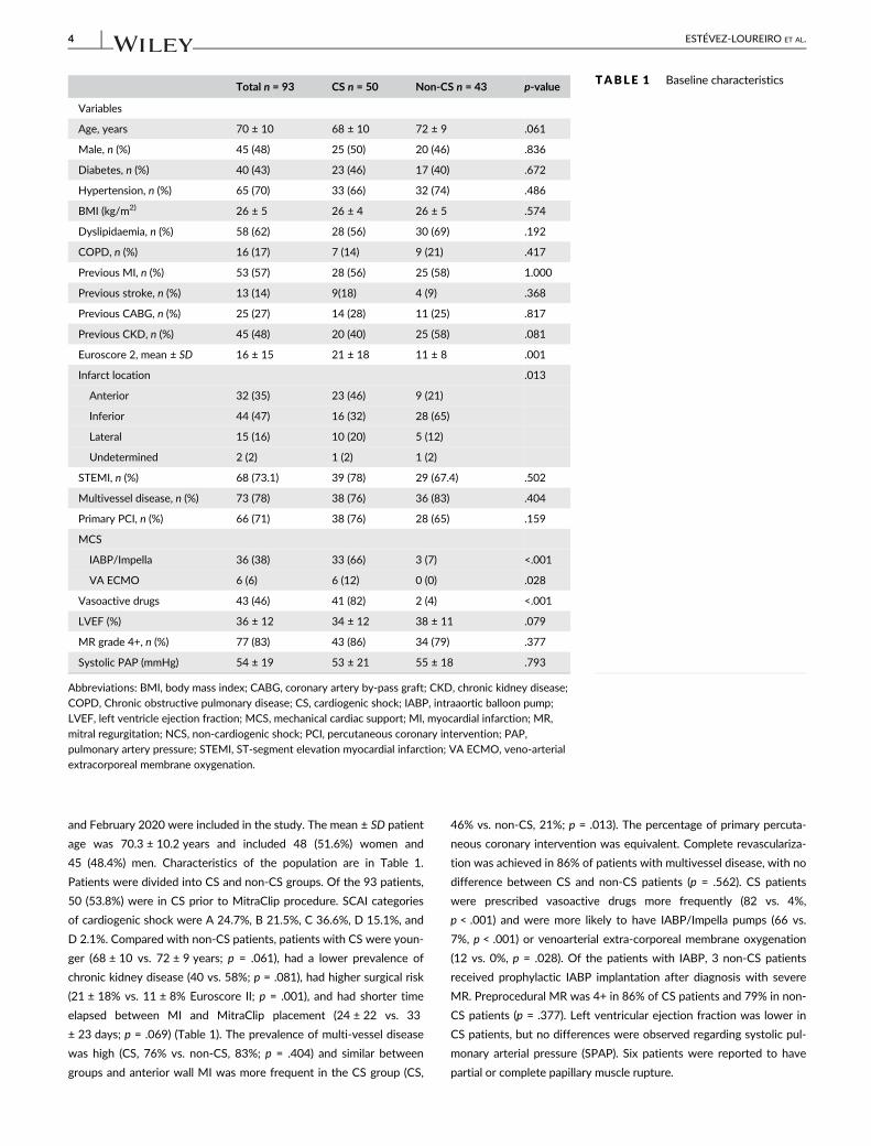
and February 2020 were included in the study. The mean ± SD patient
age was 70.3 ± 10.2 years and included 48 (51.6%) women and
45 (48.4%) men. Characteristics of the population are in Table 1.
Patients were divided into CS and non-CS groups. Of the 93 patients,
50 (53.8%) were in CS prior to MitraClip procedure. SCAI categories
of cardiogenic shock were A 24.7%, B 21.5%, C 36.6%, D 15.1%, and
D 2.1%. Compared with non-CS patients, patients with CS were youn-
ger (68 ± 10 vs. 72 ± 9 years; p = .061), had a lower prevalence of
chronic kidney disease (40 vs. 58%; p = .081), had higher surgical risk
(21 ± 18% vs. 11 ± 8% Euroscore II; p = .001), and had shorter time
elapsed between MI and MitraClip placement (24 ± 22 vs. 33
± 23 days; p = .069) (Table 1). The prevalence of multi-vessel disease
was high (CS, 76% vs. non-CS, 83%; p = .404) and similar between
groups and anterior wall MI was more frequent in the CS group (CS,
46% vs. non-CS, 21%; p = .013). The percentage of primary percuta-
neous coronary intervention was equivalent. Complete revasculariza-
tion was achieved in 86% of patients with multivessel disease, with no
difference between CS and non-CS patients (p = .562). CS patients
were prescribed vasoactive drugs more frequently (82 vs. 4%,
p < .001) and were more likely to have IABP/Impella pumps (66 vs.
7%, p < .001) or venoarterial extra-corporeal membrane oxygenation
(12 vs. 0%, p = .028). Of the patients with IABP, 3 non-CS patients
received prophylactic IABP implantation after diagnosis with severe
MR. Preprocedural MR was 4+ in 86% of CS patients and 79% in non-
CS patients (p = .377). Left ventricular ejection fraction was lower in
CS patients, but no differences were observed regarding systolic pul-
monary arterial pressure (SPAP). Six patients were reported to have
partial or complete papillary muscle rupture.
TABLE 1 Baseline characteristicsTotal n = 93 CS n = 50 Non-CS n = 43 p-value
Variables
Age, years 70 ± 10 68 ± 10 72 ± 9 .061
Male, n (%) 45 (48) 25 (50) 20 (46) .836
Diabetes, n (%) 40 (43) 23 (46) 17 (40) .672
Hypertension, n (%) 65 (70) 33 (66) 32 (74) .486
BMI (kg/m2) 26 ± 5 26 ± 4 26 ± 5 .574
Dyslipidaemia, n (%) 58 (62) 28 (56) 30 (69) .192
COPD, n (%) 16 (17) 7 (14) 9 (21) .417
Previous MI, n (%) 53 (57) 28 (56) 25 (58) 1.000
Previous stroke, n (%) 13 (14) 9(18) 4 (9) .368
Previous CABG, n (%) 25 (27) 14 (28) 11 (25) .817
Previous CKD, n (%) 45 (48) 20 (40) 25 (58) .081
Euroscore 2, mean ± SD 16 ± 15 21 ± 18 11 ± 8 .001
Infarct location .013
Anterior 32 (35) 23 (46) 9 (21)
Inferior 44 (47) 16 (32) 28 (65)
Lateral 15 (16) 10 (20) 5 (12)
Undetermined 2 (2) 1 (2) 1 (2)
STEMI, n (%) 68 (73.1) 39 (78) 29 (67.4) .502
Multivessel disease, n (%) 73 (78) 38 (76) 36 (83) .404
Primary PCI, n (%) 66 (71) 38 (76) 28 (65) .159
MCS
IABP/Impella 36 (38) 33 (66) 3 (7) <.001
VA ECMO 6 (6) 6 (12) 0 (0) .028
Vasoactive drugs 43 (46) 41 (82) 2 (4) <.001
LVEF (%) 36 ± 12 34 ± 12 38 ± 11 .079
MR grade 4+, n (%) 77 (83) 43 (86) 34 (79) .377
Systolic PAP (mmHg) 54 ± 19 53 ± 21 55 ± 18 .793
Abbreviations: BMI, body mass index; CABG, coronary artery by-pass graft; CKD, chronic kidney disease;
COPD, Chronic obstructive pulmonary disease; CS, cardiogenic shock; IABP, intraaortic balloon pump;
LVEF, left ventricle ejection fraction; MCS, mechanical cardiac support; MI, myocardial infarction; MR,
mitral regurgitation; NCS, non-cardiogenic shock; PCI, percutaneous coronary intervention; PAP,
pulmonary artery pressure; STEMI, ST-segment elevation myocardial infarction; VA ECMO, veno-arterial
extracorporeal membrane oxygenation.
4 ESTÉVEZ-LOUREIRO ET AL.

4.1 | Procedural characteristics
Technical success was achieved in 100% of patients. MitraClip NT and
NTR devices were used in 80 patients, XTR devices were used in
10 patients, and a combination of clips was used in 3 patients. More
than 1 clip was used in 58.1% of patients. Immediate procedural suc-
cess was high, with no difference between groups (CS, 90% vs. non-CS,
93%, p = .793). However, mean procedure length was longer in patients
with CS than in non-CS patients (143 ± 113 vs. 82 ± 44 min; p = .003).
The percentage of in-hospital major complications (including partial
clip detachment, air embolism, MI, stroke, vascular injury, pericardial
effusion, and bleeding events) was low after procedure and did not dif-
fer between groups (CS, 4% vs. non-CS, 7%, p = .659). Overall, mean
SPAP was significantly decreased after the procedure (before, 54
± 19 mmHg vs. after, 44 ± 20 mmHg; p < .001), with no difference
between groups. The mean mitral valve gradient increased significantly
after procedure (before, 1.7 ± 0.9 mmHg vs. after, 3.3 ± 1.6 mmHg;
p < .001). After PMVR, the gradient did not differ between CS and non-
CS patients (CS, 3.7 ± 1.9 mmHg vs. non-CS, 3.6 ± 1.7 mmHg; p = .741).
4.2 | Clinical follow-up
At 30-day follow-up, mortality was higher in the CS group (10 vs.
2.3%), but the difference was not significant (p = .207). Procedural
success at 30 days was lower in patients with CS, but the difference
was not significant (CS, 59% vs. non-CS, 74%; p = .136).
At 3-month follow-up, there were no differences in the percent-
age of re-admissions due to heart failure (CS, 13% vs. non-CS, 23%,
p = .253) or repeated MitraClip intervention or surgery (CS, 6%
vs. non-CS, 2.3%; p = .621). New York Heart Association functional
classifications improved significantly compared with baseline, but no
differences were observed between groups (Figure 1). Likewise, echo-
cardiographic evaluation during this period showed a marked reduc-
tion in MR with no difference between groups (Figure 2). Reduction in
SPAP persisted, and no differences were observed between groups
(CS, 40 ± 13 mmHg vs. non-CS, 44 ± 19 mmHg; p = .441).
After a median follow-up of 7 months (range 0–81 months, IQR
2.7–17), overall mortality did not differ between groups (CS, 16% vs.
NCS, 9.3%; p = .377), and the combined event mortality/
rehospitalization due to heart failure was similar (CS, 28% vs. NCS,
26%; p = .793). Survival curves for both end-points are shown in
Figure 3.
In a Cox-regression analysis adjusted by age, Euroscore II, and
procedural success, CS was not independently associated with the
combined end-point (hazard ratio [HR], 1.1; 95% CI, 0.3–4.6;
p = .889). The only variable independently associated with the com-
bined end-point was the immediate procedural success (HR, 0.1; 95%
CI, 0.01–0.6; p = .012). Univariate and multivariate analyses are
shown in Table 2.
5 | DISCUSSION
To our knowledge, this is the largest registry-based study evaluating
the outcomes of patients who developed acute MR after AMI and
who received PMVR with the MitraClip device. Our data suggest that
this procedure may be a safe and effective strategy for reducing MR
and improving prognosis in such high-risk populations. Also, we
observed no differences in outcomes after PMVR in patients with CS,
suggesting that CS may not be an important factor for precluding
implementation of the therapy, providing that clinical stabilization
could be achieved to receive PMVR.
MR after AMI is a serious complication that occurs in roughly 3%
of cases. Complete papillary muscle rupture is uncommon (0.25% of
MIs in the percutaneous coronary intervention era) and is often fatal.23
However, acute MR due to leaflet tethering, partial papillary muscle
F IGURE 1 New York Heart Association functional class at3 months [Color figure can be viewed at wileyonlinelibrary.com]
F IGURE 2 MR reduction postprocedural (panel a) and at3 months (panel b) [Color figure can be viewed atwileyonlinelibrary.com]
ESTÉVEZ-LOUREIRO ET AL. 5

rupture, or new onset of left ventricular dysfunction may be more fre-
quent, with a possible prevalence of 35%, according to a study of a
series of MI treated by primary percutaneous coronary intervention.4 In
a cohort of surgical patients, complete papillary muscle rupture after
MI-related MR was responsible for 45% of cases,24 which means that
severe functional MR after MI is common and may cause deterioration
in a patient's condition enough to justify intervention.
Regarding treatment of MR (excluding complete papillary muscle
rupture), it has been reported that revascularization by means of pri-
mary percutaneous coronary intervention can significantly improve
the degree of MR and therefore should be a first line treatment.4 In
our study, primary percutaneous coronary intervention was carried
out in almost 72% of cases. However, even after successful percuta-
neous revascularization, the degree of MR may worsen, and further
treatment may be required. When CS develops within the context of
MR, mortality rises significantly, and stabilization through pharmaco-
logical afterload reduction and/or mechanical circulatory support may
be lifesaving. However, the use of this advanced support should not
be a destination therapy, but rather a bridge to a subsequent interven-
tional therapy focused in correcting MR.1,6,25
Until recently, cardiac surgery was the only option available for
the treatment of MR, and a systematic review revealed a pooled
30-day mortality rate of 19%, with some studies showing mortality
rates as high as 39%.7 Notwithstanding, in recent years PMVR has
been extensively developed, and MitraClip is the device that has been
used for PMVR most extensively so far.
MitraClip has been shown to be safer than conventional surgery,
although less effective.26 In real-world registries of patients with
both degenerative and functional MR and who are at high-risk for
surgery, MitraClip use is associated with clinical improvements and a
significant reduction of MR.10,11,27 However, most MitraClip inter-
ventions performed to date have been in patients who were in a sta-
ble clinical situation with advanced functional class and chronic (not
acute) MR. Our data suggest that MitraClip could be a safe and
effective alternative to surgical intervention in clinically unstable
patients who have a high risk of 30-day mortality, and this therapy
may even be safe for the 10% of patients who develop CS, which is
encouraging.
In our study, we saw that clinical results of PMVR did not differ
significantly between CS and non-CS patients, despite the poorer
F IGURE 3 Kaplan–Meier survival curves comparing CS and non-CS groups. Panel a: Survival free from death. Panel b: Survival free from
death and rehospitalization due to heart failure [Color figure can be viewed at wileyonlinelibrary.com]
TABLE 2 Univariate and multivariatepredictors of combined death/rehospitalization due to heart failureduring follow-up
Univariate Multivariate
HR 95% CI p HR 95% CI p
Age 0.99 0.95–1.03 .651 1.05 0.97–1.13 .227
CKD 1.11 0.48–2.60 .810
DM 1.90 0.81–4.46 .140
EuroScore II 1.02 0.99–1.05 .087 1.02 0.99–1.06 .154
Pre IHD 0.98 0.38–2.56 .979
LVEF 0.99 0.95–1.03 .592
Cardiogenic shock 0.97 0.42–2.24 .936 1.1 0.3–4.6 .889
Procedural success 0.18 0.06–0.57 .004 0.10 0.02–0.60 .012
MCS 0.60 0.23–1.54 .288
Abbreviations: CI, confidence interval; CKD, chronic kidney disease; DM, Diabetes Mellitus; HR, hazard
ratio; IHD, ischemic heart disease; LVEF, left ventricular ejection fraction; MCS, mechanical cardiac
support.
6 ESTÉVEZ-LOUREIRO ET AL.

clinical situation and lower left ventricular ejection fraction in patients
with CS. This underscores the fact that in a CS scenario, the main
component responsible for clinical deterioration is the MR and its del-
eterious hemodynamic effects rather than the role of pump failure.
This could explain the difference in mortality observed in our series
compared with classic series of CS following MI28 or in those series of
PMVR in cardiogenic shock patients where patients were in a clinical
unstable condition but after mainly a long-standing ischemic or non-
ischemic cardiomyopathy in an end-stage situation.29-31 On the other
hand, the time from MI to MR treatment was several weeks in our
patient sample, and this could be related to the intention to stabilize
the clinical condition with the belief medical therapy was the only
option for the MR, since patients were not surgical candidates and the
MitraClip approach was rather new in this setting. However, treating
the MR quickly produces such positive clinical benefits that the inter-
vention should not be delayed. Thus, we advocate for early MR cor-
rection irrespective of lower left ventricular ejection fraction and
development of CS.
There are several potential advantages of using PMVR with
MitraClip. First, this treatment can lead to a rapid decrease in left
ventricular, left atrium, and pulmonary artery pressures and an
increase of cardiac output after a successful correction of the MR,32
which may lead to a fast recovery. Second, the technique reduces
risk of left ventricle damage induced by the systemic inflammatory
response, free radical injury, and myocardial oxidative stress associ-
ated with cardiopulmonary bypass.33 Moreover, MitraClip may also
avoid the restraint of the mitral annular motion caused by mitral
rings or prosthesis and the development of abnormal septal motion
that may negatively impact left ventricle performance. In addition,
acute MR often develops in a previously normal mitral valve, which
usually translates into optimal leaflet tissue and coaptation for
device therapy. Furthermore, the use of MitraClip does not preclude
a delayed cardiac surgery in case the device fails. Finally, a relevant
number of unstable patients are on double or triple antithrombotic
therapy after MI, and MitraClip may help prevent significant bleed-
ing complications that are common after open-heart surgery and
that may negatively impact the prognosis of unstable patients. How-
ever, challenges when implementing this therapy exist. Treating
acute MR is one of the most technically challenging MitraClip proce-
dures. Lesion complexity, a small atrium that makes performing a
high puncture difficult, the clinical situation of the patient, and the
risk of impingement in papillary muscle make these cases very
demanding. The fact that the only independent factor associated
with clinical outcomes was immediate technical success underscores
the relevance of the high level of experience required for the
implanting team.
Interestingly, a recent review of shock management after MI25
only considers mitral valve surgery for acute MR after MI. Our data
suggest that, given the favorable safety/efficacy profile, PMVR should
at least be considered in patients with this condition if the attending
team has enough experience. This strategy may be of special interest
in patients with CS, which represent complex cases where effective
and fast recovery with a low rate of complications is desirable. Further
research and randomized trials are required to increase our knowledge
in this setting.
6 | LIMITATIONS
Our study had several limitations. First, is an observational study and the
sample size was small and so should be interpreted with caution. We did
not find a significant difference in mortality between CS and non-CS
patients, which could be related to a lack of statistical power. A larger
study with longer follow-up will be needed to clarify the effect of
MitraClip in this scenario. Furthermore, we cannot exclude the presence
of a selection bias in CS patients, in the sense that only those who
responded to the medical therapy and cardiac support were those who
received PMVR. Time form MI to PMVR was long and that means that
even CS patients responded to the medical therapy in some way. This
population can represent a better prognostic category and therefore our
conclusions may not be applicable to all patients in CS with significant
MR. Likewise, because of the small sample size and number of events,
the multivariable analysis was limited. Thus, results should be considered
hypothesis generating and not generalizable. Also, echocardiographic
follow-up is lacking, so the effect on left ventricular remodeling has not
been evaluated. However, our aim was to assess correlation within the
clinical setting, not to show possible positive effects on left ventricular
parameters. Likewise, number of patients with subvalvular apparatus
rupture was small to draw definitive conclusions. Although if a successful
repair can be achieved results seemed similar, larger series with these
anatomies must be collected to assess the specific performance of
MitraClip in this population. The shock status is a dynamic variable and
we defined it at the time of MitraClip strategy decision. What we cannot
ascertain is what was the SCAI classification at the beginning of the clini-
cal course. Likewise, all patients were treated with MitraClip as a salvage
strategy, but we cannot distinguish from the present study whether the
patients were stable enough or with recurrent deterioration despite
MCS support. Lastly, procedures were performed in centers that had
high levels of experience with PMVR using MitraClip. Thus, our findings
cannot be generalized to less experienced teams.
7 | CONCLUSION
Patients found to have significant MR during their index hospitaliza-
tion for AMI had similar clinical outcomes with PMVR whether they
presented in or out of cardiogenic shock, provided initial hemody-
namic stabilization was first achieved before PMVR.
7.1 | Clinical perspective
7.1.1 | What is next?
Further research is warranted to confirm these results and to compare
the percutaneous approach with conventional surgery.
ESTÉVEZ-LOUREIRO ET AL. 7
7.1.2 | What is new?
PMVR with the MitraClip device may be a promising therapeutic
strategy for patients with acute MR after AMI, with or without
CS. This may represent a valid alternative for such patients.
7.1.3 | What is known?
Acute MR may develop following AMI, a condition associated with
development of CS and high mortality. Until recently, conventional
surgery was the only alternative, which is associated with significant
mortality.
CONFLICT OF INTEREST
Rodrigo Estévez-Loureiro is consultant for Abbott Vascular and Bos-
ton Scientific. Dr. Taramasso is a consultant for Abbott Vascular, Bos-
ton Scientific, 4TECH, and CoreMedic; and has received speaker
honoraria from Edwards Lifesciences. Dr. Denti has served as a con-
sultant for Abbott Vascular, 4Tech, Neovasc, and InnovHeart; and has
received honoraria from Abbott. Dabit Arzamendi is consultant for
Abbott Vascular. Dr. Xavier Freixa is consultants for Abbott Vascular.
Dr. Fam, has received speaker honoraria and travel or grant support
from Edwards Lifesciences Francesco Maisano received Grant and/or
Research Support from Abbott, Medtronic, Edwards Lifesciences,
Biotronik, Boston Scientific Corporation, NVT, Terumo; receives Con-
sulting fees, Honoraria from Abbott, Medtronic, Edwards Lifesciences,
Swissvortex, Perifect, Xeltis, Transseptal solutions, Cardiovalve,
Magenta; has Royalty Income/IP Rights Edwards Lifesciencesand is
Shareholder of Cardiovalve, Cardiogard, Magenta, SwissVortex, Trans-
septalsolutions, 4Tech, Perifect. All other authors have reported that
they have no relationships relevant to the contents of this article to
disclose.
DATA AVAILABILITY STATEMENT
The data that support the findings of this study are available from the
corresponding author upon reasonable request.
ORCID
Rodrigo Estévez-Loureiro https://orcid.org/0000-0001-5841-5514
Maurizio Taramasso https://orcid.org/0000-0001-7295-1153
Marianna Adamo https://orcid.org/0000-0002-3855-1815
Xavier Freixa https://orcid.org/0000-0002-3203-9060
Neil Fam https://orcid.org/0000-0002-1269-6733
Andrew Czarnecki https://orcid.org/0000-0002-3000-9722
Isaac Pascual https://orcid.org/0000-0001-5433-1364
Estefanía Fernández-Peregrina https://orcid.org/0000-0002-3025-
8251
Mattia Di Pasquale https://orcid.org/0000-0002-4499-7725
Ander Regueiro https://orcid.org/0000-0001-5201-447X
REFERENCES
1. Watanabe N. Acute mitral regurgitation. Heart. 2019;105:671-677.
2. Lopez-Perez M, Estevez-Loureiro R, Lopez-Sainz A, et al. Long-term
prognostic value of mitral regurgitation in patients with ST-segment
elevation myocardial infarction treated by primary percutaneous cor-
onary intervention. Am J Cardiol. 2014;113:907-912.
3. Mentias A, Raza MQ, Barakat AF, et al. Prognostic significance of
ischemic mitral regurgitation on outcomes in acute ST-elevation myo-
cardial infarction managed by primary percutaneous coronary inter-
vention. Am J Cardiol. 2017;119:20-26.
4. Nishino S, Watanabe N, Kimura T, et al. The course of ischemic mitral
regurgitation in acute myocardial infarction after primary percutane-
ous coronary intervention: from emergency room to long-term
follow-up. Circ Cardiovasc Imaging. 2016;9:e004841.
5. Sannino A, Grayburn PA. Ischemic mitral regurgitation after acute
myocardial infarction in the percutaneous coronary intervention era.
Circ Cardiovasc Imaging. 2016;9:e005323.
6. Ibanez B, James S, Agewall S, et al. ESC guidelines for the manage-
ment of acute myocardial infarction in patients presenting with ST-
segment elevation: the task force for the management of acute myo-
cardial infarction in patients presenting with ST-segment elevation of
the European Society of Cardiology (ESC). Eur Heart J. 2018;39:
119-177.
7. Alajaji WA, Akl EA, Farha A, Jaber WA, AlJaroudi WA. Surgical versus
medical management of patients with acute ischemic mitral regurgita-
tion: a systematic review. BMC Res Notes. 2015;8:712.
8. Feldman T, Kar S, Elmariah S, et al. Randomized comparison of percu-
taneous repair and surgery for mitral regurgitation: 5-year results of
EVEREST II. J Am Coll Cardiol. 2015;66:2844-2854.
9. Maisano F, Franzen O, Baldus S, et al. Percutaneous mitral valve inter-
ventions in the real world: early and 1-year results from the ACCESS-
EU, a prospective, multicenter, nonrandomized post-approval study of
the MitraClip therapy in Europe. J Am Coll Cardiol. 2013;62:1052-1061.
10. Nickenig G, Estevez-Loureiro R, Franzen O, et al. Percutaneous mitral
valve edge-to-edge repair: in-hospital results and 1-year follow-up of
628 patients of the 2011-2012 pilot European sentinel registry. J Am
Coll Cardiol. 2014;64:875-884.
11. Puls M, Lubos E, Boekstegers P, et al. One-year outcomes and predic-
tors of mortality after MitraClip therapy in contemporary clinical
practice: results from the German transcatheter mitral valve interven-
tions registry. Eur Heart J. 2016;37:703-712.
12. Stone GW, Lindenfeld J, Abraham WT, et al. Transcatheter mitral-
valve repair in patients with heart failure. N Engl J Med. 2018;379:
2307-2318.
13. Bilge M, Alemdar R, Yasar AS. Successful percutaneous mitral valve
repair with the MitraClip system of acute mitral regurgitation due to
papillary muscle rupture as complication of acute myocardial infarc-
tion. Catheter Cardiovasc Interv. 2014;83:E137-E140.
14. Estevez-Loureiro R, Arzamendi D, Freixa X, et al. Percutaneous mitral
valve repair for acute mitral regurgitation after an acute myocardial
infarction. J Am Coll Cardiol. 2015;66:91-92.
15. Adamo M, Curello S, Chiari E, et al. Percutaneous edge-to-edge mitral
valve repair for the treatment of acute mitral regurgitation complicat-
ing myocardial infarction: a single centre experience. Int J Cardiol.
2017;234:53-57.
16. Estevez-Loureiro R, Adamo M, Arzamendi D, et al. Transcatheter
mitral valve repair in patients with acute myocardial infarction:
insights from the European Registry of MitraClip in acute mitral
regurgitation following an acute myocardial infarction (EREMMI).
EuroIntervention. 2020;15:1248-1250.
17. Haberman D, Taramasso M, Czarnecki A, et al. Salvage MitraClip in
severe secondary mitral regurgitation complicating acute myocardial
infarction: data from a multicentre international study. Eur J Heart
Fail. 2019;21:1161-1164.
18. Baumgartner H, Falk V, Bax JJ, et al. ESC/EACTS guidelines for the
management of valvular heart disease. Eur Heart J. 2017;38:2739-
2791.
8 ESTÉVEZ-LOUREIRO ET AL.

19. Nishimura RA, Otto CM, Bonow RO, et al. AHA/ACC focused update
of the 2014 AHA/ACC guideline for the management of patients with
valvular heart disease: a report of the American College of Car-
diology/American Heart Association task force on clinical practice
guidelines. Circulation. 2017;135:e1159-e1195.
20. Ponikowski P, Voors AA, Anker SD, et al. 2016 ESC guidelines for the
diagnosis and treatment of acute and chronic heart failure: the task
force for the diagnosis and treatment of acute and chronic heart fail-
ure of the European Society of Cardiology (ESC)developed with the
special contribution of the heart failure association (HFA) of the ESC.
Eur Heart J. 2016;37:2129-2200.
21. Baran DA, Grines CL, Bailey S, et al. SCAI clinical expert consensus
statement on the classification of cardiogenic shock: this document
was endorsed by the American College of Cardiology (ACC), the
American Heart Association (AHA), the Society of Critical Care Medi-
cine (SCCM), and the Society of Thoracic Surgeons (STS) in April
2019. Catheter Cardiovasc Interv. 2019;94:29-37.
22. Stone GW, Adams DH, Abraham WT, et al. Clinical trial design princi-
ples and endpoint definitions for Transcatheter mitral valve repair
and replacement: part 2: endpoint definitions: a consensus document
from the mitral valve academic research consortium. J Am Coll Car-
diol. 2015;66:308-321.
23. French JK, Hellkamp AS, Armstrong PW, et al. Mechanical complica-
tions after percutaneous coronary intervention in ST-elevation myo-
cardial infarction (from APEX-AMI). Am J Cardiol. 2010;105:59-63.
24. Chevalier P, Burri H, Fahrat F, et al. Perioperative outcome and long-
term survival of surgery for acute post-infarction mitral regurgitation.
Eur J Cardiothorac Surg. 2004;26:330-335.
25. Thiele H, Ohman EM, de Waha-Thiele S, Zeymer U, Desch S. Man-
agement of cardiogenic shock complicating myocardial infarction: an
update 2019. Eur Heart J. 2019;40:2671-2683.
26. Feldman T, Foster E, Glower DD, et al. Percutaneous repair or surgery
for mitral regurgitation. N Engl J Med. 2011;364:1395-1406.
27. Sorajja P, Mack M, Vemulapalli S, et al. Initial experience with com-
mercial transcatheter mitral valve repair in the United States. J Am
Coll Cardiol. 2016;67:1129-1140.
28. Hochman JS, Sleeper LA, Webb JG, et al. Early revascularization in
acute myocardial infarction complicated by cardiogenic SHOCK.
SHOCK investigators. Should we emergently revascularize occluded
coronaries for cardiogenic shock. N Engl J Med. 1999;341:625-634.
29. Chan V, Messika-Zeitoun D, Labinaz M, et al. Percutaneous mitral
repair as salvage therapy in patients with mitral regurgitation and
refractory cardiogenic shock. Circ Cardiovasc Interv. 2019;12:e008435.
30. Cheng R, Dawkins S, Hamilton MA, et al. Percutaneous mitral repair
for patients in cardiogenic shock requiring inotropes and temporary
mechanical circulatory support. JACC Cardiovasc Interv. 2019;12:
2440-2441.
31. Garcia S, Alsidawi S, Bae R, et al. Percutaneous mitral valve repair
with MitraClip in inoperable patients with severe mitral regurgitation
complicated by cardiogenic shock. J Invasive Cardiol. 2020;32:
228-231.
32. Siegel RJ, Biner S, Rafique AM, et al. The acute hemodynamic effects
of MitraClip therapy. J Am Coll Cardiol. 2011;57:1658-1665.
33. van Boven WJ, Gerritsen WB, Driessen AH, et al. Myocardial oxidative
stress, and cell injury comparing three different techniques for coronary
artery bypass grafting. Eur J Cardiothorac Surg. 2008;34:969-975.
SUPPORTING INFORMATION
Additional supporting information may be found online in the
Supporting Information section at the end of this article.
How to cite this article: Estévez-Loureiro R, Shuvy M,
Taramasso M, et al. Use of MitraClip for mitral valve repair in
patients with acute mitral regurgitation following acute
myocardial infarction: Effect of cardiogenic shock on
outcomes (IREMMI Registry). Catheter Cardiovasc Interv. 2021;
1–9. https://doi.org/10.1002/ccd.29552
ESTÉVEZ-LOUREIRO ET AL. 9