use of a yeast site-specific recombinase to generate embryonic
TRANSCRIPT
DEVELOPMENTAL GENETICS 13:367375 (1992)
Use of a Yeast Site-Specific Recombinase to Generate Embryonic Mosaics in Drosophila DUYEN T. DANG AND NORBERT PERRIMON Howard Hughes Medical Institute, Department of Genetics, Harvard Medical School, Boston, Massachusetts
ABSTRACT An efficient method for gener- ating embryonic mosaics using a yeast site-specific recombinase (FfP) , under the control of a heat shock promoter, is described. FfP-recombinase can promote mitotic exchange between homolo- gous chromosomes that contain FRT (FLP Recom- bination Target) sequences. To demonstrate the ef- ficiency of FIP-recombinase to generate embryonic mosaics, clones of the recessive and cell autono- mous mutation armadillo (arm), detected by their ability to differentiate ectopic denticles in the naked cuticle of each abdominal segment, have been in- duced. We have analyzed the parameters of F P - recombinase induced embryonic mitotic recombi- nation and have demonstrated that clones can be etticiently induced during the postblastoderm mi- totic divisions. We discuss applications of this tech- nique for the analyses of the roles of various mu- tations during embryonic patterning. 0 1992 Wiley-Liss, Inc.
Key words: FIP-recombinase, FRT, embryonic mosaics
INTRODUCTION The ability to generate mosaics is a powerful tech-
nique for studying the mechanisms underlying pattern formation, as well as for analyzing the roles of specific genes in patterning. In Drosophila, mosaic analyses have been used extensively to determine the effects and cellular autonomy of mutations on adult pattern- ing. The most commonly used technique to generate adult somatic mosaics is mitotic recombination, which can be induced following X-ray treatment a t various developmental stages. A number of cell autonomous recessive markers [Lawrence et al., 19861 are available to detect mutant clones in the adult cuticle (e.g., forked, multiple wing hairs) and the eye (e.g., white). In addi- tion, a number of histochemical markers have been used to detect clones of homozygous cells in internal structures [e.g., aldehyde oxidase; Janning, 1972; ace- tylcholinesterase; Ferrus and Kankel, 1981; succino-de- hydrogenase; Lawrence, 19811. Other methods used to generate adult somatic mosaics include nuclear trans- plantation, the use of an unstable ring-X chromosome
(the gynandromorph technique), and the use of muta- tions that cause somatic chromosome elimination [e.g., paternal loss and mitotic loss inducer; Hall et al., 19761.
Contrary to adult mosaics, the analyses of the effects and cell autonomy of mutations affecting embryonic patterning have been poorly analyzed. The reasons for this include the inefficient means of generating large numbers of mosaic animals, as well as the paucity of embryonic markers. A rather laborious method for gen- erating embryonic mosaics consists of the transplanta- tion of cells or nuclei. For example, labeling Drosophila cells by injection of markers such as horseradish per- oxidase and subsequent transplantation into develop- ing embryos [Technau, 19861, has been used to test the autonomy of several neurogenic genes [Technau and Campos-Ortega, 19871.
Genetically, two methods, gynandromorphs and X- ray induced mitotic recombination, have been used to produce embryonic mosaics. Gergen and Wieschaus [19861 generated mutant patches in embryos using the unstable Ring-X chromosome. To identify these mutant territories, the cell autonomous mutation shaven baby (sub), which decreases the number and size of larval denticles, was used. This technique allowed the deter- mination of the cellular autonomy of a number of em- bryonic lethal mutations [Gergen and Wieschaus, 19861. There are three problems associated with the use of Ring-X chromosomes: first, this technique is limited to studies of X-linked mutations; second, the clones gen- erated are large; and third, the time of clone induction cannot be controlled. An alternative method to generate and recognize small mutant patches in an otherwise heterozygous animal makes use of the cell autonomous segment polarity mutation armadillo (arm). arm em- bryos exhibit a segment polarity phenotype in which all naked cuticle of the larvae is deleted and replaced by denticles. Wieschaus and Riggleman [1987] demon- strated that clones of arm cells, generated by X-ray
Received for publication July 2, 1992; accepted August 5, 1992
Address reprint requests to Dr. Norbert Perrimon, Department of Genetics, Howard Hughes Medical Institute, Harvard Medical School, 200 Longwood Avenue, Boston, Massachusetts 02115.
0 1992 WILEY-LISS, INC.

368 DANG AND PERRIMON
induced mitotic recombination, can develop ectopic patches of denticles in part of the naked region of every segment [see also Klingensmith et al., 19891. To analyze if the mutations Notch [N; Hoppe and Greenspan, 19861 and polyhomeotic [phm; Smouse and Perrimon, 19901 which perturb the differentiation of the ventral epider- mis, are cell autonomous, doubly mutant clones of arm N and arm p h m were induced in heterozygous animals. Two problems arose from the use of this technique: first, X-ray treatment of embryos generates a large number of non-specific defects that make the identification of some clones difficult; and second, clones of homozygous cells are recovered at a very low frequency.
Recently, an efficient way to generate mosaics using the site-specific recombinase, FLP, was developed and used successfully to induce clones in the imaginal discs and germ cells [Golic, 1991; Chou and Perrimon, 19921. FLP-recombinase promotes mitotic exchange between homologous chromosomes that contain FRT sequences. Unlike X-ray induced mitotic recombination [Haynie and Bryant, 19771, the use of FLP-recombinase is not associated with cell death. Furthermore, since it is heat inducible, the timing of clone induction is tightly reg- ulated. We have tested whether FLP-recombinase could be used to induce embryonic mosaics in animals heterozygous for the cell autonomous, segment polarity gene arm. We report the parameters of this clonal anal- ysis and describe applications of this method to study embryonic patterning.
MATERIALS AND METHODS Strains
We used the X-linked FRT (P[ > whS>]) insertion [Golic and Lindquist 19891, FRT"', located at cytolog- ical position 14A-B on the X-chromosome [Chou and Perrimon 19921. We used the FLP insertion [P[ry'hsFLPI; Golic and Lindquist, 19891, FLP', that is located on the second chromosome [Chou and Perri- mon, 19921. In this study we used a single FLP inser- tion (FLP') which has been previously shown to be extremely efficient [Chou and Perrimon, 19921.
The recombinant chromosome y armxK22 FRT'O' was constructed to test the efficiency of the site-specific re- combination technique. armxK2' behaves genetically as a null arm allele [Peifer and Wieschaus, 19901. This stock is maintained using the FM7c balancer. Embryos of the genotype armxKzz FRT'O'I + FRT'"; + lFLP8 were derived from crosses of F M 7 ~ l a r m ~ ~ ~ ~ FRT'O'; + I + females with f FRT'O'IY; FLP81FLF8 males.
Flies were raised on standard Drosophila media at 25°C. Descriptions of balancers and mutations that are not described in the text can be found in Lindsley and Zimm [1992].
Egg Collection and Heat Shock Treatment Eggs were collected on petri dishes containing an
agar-molasses medium supplemented with dry yeast.
Females were mated for a t least 24 hours prior to the egg collections and were allowed to lay eggs in a quiet environment to optimize the synchronization of the eggs deposited. The first collections were discarded.
Heat shock treatments of the synchronized embryos were performed a t 37°C in a circulating waterbath. The petri dishes were covered with parafilm and floated on the water. Following the heat shock, the plates were immediately transferred into a 25°C incubator.
Cuticle Examination and Recording of the Arm Clones
To detect the presence of arm clones, embryos were allowed to fully develop (approximately 24 h r at 25°C) and their cuticles prepared in Hoyers' mountant as de- scribed by van der Meer [1977]. The positions and num- bers of denticles of all arm clones recovered were re- corded on schematic drawings of a ventral cuticle of a first instar larvae. The cuticles were examined using bright field or dark field illumination. arm clones were recognized as ectopic denticles in the naked regions of the seven (A1 through A7) abdominal segmental inter- vals.
RESULTS AND DISCUSSION FLP-Recombinase Can Promote Mitotic
Exchange During Embryonic Development Previously, Wieschaus and Riggleman 119873 showed
that homozygous clones of null alleles of armadillo (arm), generated by X-ray induced mitotic recombina- tion in heterozygous embryos, led to the ectopic occur- rence of denticles in the naked region of the larval cuticle. The method we used to test the efficiency of FLP-recombinase to induce embryonic mosaics, using arm as a cuticular marker, is shown in Figure 1. Em- bryos of genotype arm FRT'O'I + FRT'O'; FLP'I f were heat shocked a t 37°C a t various times during embryonic development and examined for the occurrence of arm clones.
Results shown in Table 1 indicate that FLP-recom- binase can promote mitotic exchange during embryonic development. While arm clones are found in heat treated embryos that carry one copy of FLP-recombi- nase, no arm clones are recovered in the heat shocked animals without FLP-recombinase. The percentage of mosaics increases with the length of the heat shock. Following a 30 min heat shock, a small number (12.5%) of mosaic larvae are recovered; however the number of mosaics increases to 100% following a 120 min heat shock. Although the number of clones recovered in- creases with heat shock, the percentage of segmenta- tion defects due to the heat treatment also increases, especially when a heat shock of 180 min is adminis- tered (Table 1). Because the presence of these defects makes the identification of arm clones difficult, we de- cided to use a 120 min heat shock to generate arm clones in all the following experiments. Such condi-

MOTH
ER C
ELL
A ar
m
FRT -
-
+ Fl
P
B ar
m
FRT - 7
+ FL
P
FLP
CATA
LYZE
D SIT
E-SP
ECIF
IC EX
CHAN
GE
DAUG
HTER
CEL
LS
Fig
. 1.
FLP-
reco
mbi
nase
indu
ced
site
-spe
cifi
c ex
chan
ge. A
: FL
P-re
com
bina
se in
duce
d m
i-
totic
rec
ombi
natio
n ex
chan
ge o
ccur
ring
on
the
X-c
hrom
osom
e of
a c
ell
of g
enot
ype
arm
F
RT'
oz/F
RTz
o'; F
Lp?"
/ + is
sho
wn.
Fol
low
ing
heat
indu
ctio
n, th
e hs
p70-
FLP?
' pr
ovid
es t
he
reco
mbi
nase
act
ivit
y ne
cess
ary
to c
atal
yze
the
chro
mos
omal
exc
hang
e at
the
leve
l of t
he F
RT
sequ
ence
s. F
LP c
atal
yzed
reco
mbi
natio
n re
sult
s in
the
reco
very
of a
cel
l hom
ozyg
ous f
or a
rm.
B s
how
s a c
ell t
hat d
oes
not u
nder
go F
LP c
atal
yzed
reco
mbi
natio
n, re
sult
ing
in tw
o da
ught
er
cells
of t
he sa
me
geno
type
as t
he m
othe
r cel
l. N
omen
clat
ure:
arm
adill
o (a
rm) c
lone
s are
show
n as
pat
ches
of d
entic
les
in th
e na
ked
regi
on o
f the
seg
men
t. FL
P-re
com
bina
se ta
rget
seq
uenc
es
(FR
T) ar
e de
pict
ed a
s bl
ank
boxe
s an
d FL
P-re
com
bina
se a
s st
ippl
ed b
oxes
. The
FR
T is
pro
x-
imal
to a
rm o
n th
e X
chro
mos
ome
and
the
hsp7
0-FL
P3'
is lo
cate
d on
the
seco
nd c
hrom
osom
e
CUTI
CLE
PHEN
OTYP
E
\/
arml
arm
clone
s
wild
type

370 DANG AND PERRIMON
TABLE 1. Efficiency of Clone Induction as a Function of the Length of Time of the Heat Shock
N % Length of heat shock (rnin) Total Exp Mosaics Mosaics Defects Experiment
30 169 56 7 12.5 1.2 60 294 98 78 80 0.7
120 192 64 72 loo* 1.6 180 112 37 39 loo* 11.6
120 134 45 0 0 4.5 Control
240 min - 300 min - 360 min -
540 rnin -
720 min -
*0-6-hour-old embryos, derived from crosses of FM7cI armxm2 FRT'O'; + I f females with FRT'O'IY; FLp81FLp8 males, were heat shocked at 37°C for designated times (length of heat shock is shown in minutes). The embryos were allowed to develop until cuticle formation (approximately 24 hr) and their cuticles were prepared for examination. As a control, embryos derived from crosses of FM7clarmXmz FRT'O' fe- males with FRT'O'IY males were heat shocked a t 37°C for 120 min In each experiment, approximately one quarter of the embryos recovered were of the arm mutant phenotype, con- sistent with the expected fraction to be of the armlY; FLp81 + genotype (data not shown). The calculated percentage of mo- saic larvae when the heat shock is performed for 120 min and 180 min is higher than 100% due to the method of determin- ing the number of heterozygous animals. These numbers have been corrected to 100% (*I. Nomenclature: N total is the num- ber of larvae examined which do not show the arm phenotype. Since there is no independent method of determining the number of armxKz2 FRT'O'I f ; + lFLp8 from their siblings (FM7cIY; + I F P 8 and FM7c FRT'O'; +IF@'), the expected number of larvae of genotype armXm2 FRT'o'IFRT'o'; FLp8 /+ is calculated as: N exp = N totaU3. N mosaics rep- resents the number of larvae with arm somatic clones. The percentage of armxKzz FRT'o'IFRT'o'; FLP'I f larvae with clones (% mosaics) and the percentage of non-arm embryos with segmentation defects (% defects) due to the heat shock treatment is indicated. The length of heat shock is indicated in minutes (rnin).
tions lead to the recovery of 100% of mosaicism among the animals of the appropriate genotype. In addition, these parameters generate a minimal percentage of heat-induced segmentation defects.
Somatic clone induction using FLP-recombinase is more efficient than following X-ray treatment. For ex- ample, Wieschaus and Riggleman [19871 found, using X-ray induced mitotic recombination, that 49% of the animals with the appropriate genotype are mosaics, with an average of 1 to 2 clones per animal. Using FLP-recombinase at a similar developmental stage we are able to induce mosaics in 100% of the animals with the appropriate genotype, with an average of 4.8 clones per animal. In addition, defects due to the heat treat- ment are less deleterious to the embryo than X-ray irradiation since, in our experiments, a heat shock treatment of 120 min only induces a minimal percent- age of segmentation defects (Table 1).
Clone Size Cells from the ventral epidermis undergo an average
of two to three mitotic divisions following the cellular
FERTILIZATION 0 min
CYCLE 14 (1 30-1 70 min)
8o min 7 1 80-240 min
240-300
300-360
360- 540
540-720
min
rnin
rnin
rnin
Fig 2. Relationship between the age of the embryo at heat shock and the post-blastoderm mitotic events. The chronology of the mitotic events in the early embryo from fertilization to 720 min is depicted. The formation of the cellular blastoderm at 170 min is followed by three post blastoderm mitosis, M1, M2, and M3, that begin at 225 min, 295 min, and 380 min, respectively [Campos-Ortega and Harten- stein, 1985; Foe 19891. The hatched boxes represent the onset and duration of each mitosis. The times of the embryo collections per- formed (age at heat shock) are shown in brackets (see also Table 2). Based on the estimation that a single arm epidermal cell gives rise to an average of 2.6 denticles, we expected the average size of arm clones induced during M 2 to be 5.2 denticles (clone size of 2 cells) and 10.4 denticles (clone size of 4 cells) for clones induced during M1.
blastoderm stage (Fig. 2) [Campos-Ortega and Harten- stein, 19851. Although most epidermal cells undergo two mitotic divisions, only a minor fraction is believed to undergo a third division. To determine whether FLP-recombinase can promote mitotic exchange dur- ing all postblastoderm mitoses, a detailed analysis of the various clone sizes was conducted (Fig. 2, Table 2).
The clone size is estimated as the number of denticles in a single clone. To determine the average number of denticles secreted by a single arm cell, clones were in- duced around the last mitotic division at 380 min post- fertilization (M3, Fig. 2). When embryos at ages 360-
SITE-SPECIFIC RECOMBINATION AND EMBRYONIC MOSAICS 371
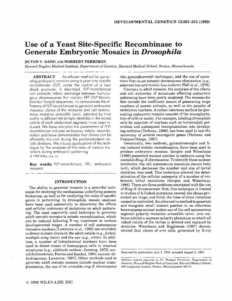
TABLE 2. Parameters of arm Clonal Analysis*
N % A LEC Ane at HS (min) -(min) Total EXP Mosaics Mosaics Defects Clones Denticles 60 180 -240 183 61 58 95 4.2 4.2 t 3 4.5 i 2.9 60 240-300 218 73 56 77 2.7 4.8 t 2.7 3.7 t 2.7 60 300-360 245 82 41 50 3 3.1 * 1.9 3.9 ? 1.8
180 360-540 203 54 12 17 1.2 1.3 2 .5 2.6 ? .8 180 540-720 173 58 0 0 0 0 0
*Eggs of various ages, derived from the cross ofFM7clarmXKzz FRT'O'; +I+ females with FRT'O'IY; FLF~IFLF~ males, were heat shocked at 37°C for 120 min. LEC is the length of egg collection in minutes. Age at HS (heat shock) represents the age of the embryos at the time of heat treatment. The average number of arm clones per animal (A clones) is calculated as the total number of arm clones divided by the total number of larvae with clones (N mosaics). The average number of denticles per clone (A denticles) is the total number of denticles divided by the total number of arm clones. The standard deviation is shown for both averages. Details on the total number of arm clones as well as the total number of denticles per age groups is shown in Figure 3. See Table 1 for additional nomenclature.
540 min are heat shocked for 120 min (Table 2), the size of the clones ranged from 1.8 to 3.4 denticles with an average of 2.6. The average of 2.6 denticles secreted by a single arm cell is in accordance with the results of Wieschaus and Riggleman [1987] who estimated that an epidermal cell gives rise to 3 denticles on average. It is of interest to note that when embryos at ages 540- 720 min are heat shocked, no clones are recovered. This is expected since most mitotic division in the ventral epidermis are completed by 390 min.
Three earlier time points were analyzed in order to assess whether FLP-recombinase can promote mitotic recombination during the first two postblastoderm di- visions (Table 2, Fig. 2). When the heat shock is deliv- ered to embryos at ages 300-360 min, the sizes of the clones ranged from 2.1 to 5.7 denticles with a n average of 3.9 denticles; when the heat shock was performed on embryos a t ages 240-300 min, the sizes of the clones ranged from 1 to 6.4 denticles with an average of 3.7 denticles; and when the embryos are heat treated at ages 180-240 min, the sizes of the clones ranged from 1.6 to 7.4 with an average of 4.5 denticles. The distri- bution of the clone sizes is shown in Figure 3.
Clone Frequency To determine whether the age of the embryos at the
time of the heat shock treatment affects the number of clones per animal, the frequency of arm clones induced in heat shocked arm FRTIOII+ FRT'Ol; FLF81+ em- bryos collected at various embryonic stages was deter- mined (Fig. 2). The efficiency of mosaic induction in- creases when the embryos are heat shocked at younger ages. Few clones are recovered when embryos are heat shocked for 120 min at ages 360 to 540 min (17% of the embryos with the appropriate genotype are mosaics with an average of 1.3 clones per animal), as well as a t ages 300-360 min (50% of the embryos of the appro- priate genotype are mosaics with an average of 3.1 clones per animal). However, more clones are recovered in embryos heat shocked a t ages 240-300 min (77% of the embryos with the appropriate genotype are mosaics
with an average of 4.8 clones per animal) and at ages 180-240 min (95% of the embryos with the appropriate genotype are mosaics with an average of 4.2 clones per animal).
Spatial Distribution of Arm Clones We were concerned that we may be under-estimating
clone sizes due to our inability to detect the full extent of arm clones. The size of large arm clones which over- lap with the segmental denticle band may be under- estimated since wild-type and arm denticles are mor- phologically similar. Additionally, the size of arm clones that span the most posterior third region of the naked cuticle may be under-estimated since arm clones are not found in this domain due to either cell death or cell transformation [Wieschaus and Riggleman, 1987; Klingensmith et al., 19891.
We examined the distribution of small clones (1 to 4 denticles) induced in embryos heat shocked at ages 180-240 min and 240-300 min (Fig. 4). We reasoned that if these clones represented large clones whose sizes have been under-estimated based upon their po- sition within the segmental unit, they should be pref- erentially localized either near the denticle belts or near the most posterior region of the naked cuticle of the segment. Results of this analysis, shown in Figure 4, demonstrate that the small clones are not preferen- tially distributed in the areas that may affect our abil- ity to determine the full size of the arm clones. 90% of the clones are in the central part of the naked region (between 20 and 80% in relative distance from the an- terior and posterior denticle belts). Examples of the position of some of these clones are shown in Figure 5 .
The Mode of Action of FLP-Recombinase Our mosaic analyses demonstrate that the efficiency
of mosaic induction increases when the embryos are heat shocked at younger stages. As expected, since only a minor fraction of epidermal cells undergo a third postblastoderm division [Campos-Ortega and Harten- stein, 19851, we recovered more clones in animals un-

372 DANG AND PERRIMON
A: Heat Shock at1 80-240 rnin 40 I
1 2 3 4 5 6 7 8 9 1 0 1 1 1 2 Cbn Yze
(Number o i Dentrks)
C Heat Shod a t 300-360 rnin 40
Ncbns-128 N dentkies-493
1 2 3 4 5 6 7 8 9 1 0 1 1 1 2 Cbne sue
(Number of dentrles)
6: Heat Shock a t 240-300 rnin
- 1 I N clones-275
1 2 3 4 5 6 7 8 9 1 0 1 1 l Z Clone size
(Number o i Dentdes)
D*: Heat Shock a t 360-540 rnin
50
N clones-1 6 40 N dentrles-31
30
20
10
0 1 2 3 4 5 6 7 8 9 10 11 12
C b n e sue (Number oi dentlcks)
Fig. 3. Distribution of arm clones. The percentage of clones (number of clones with a specific clone sizehotal number of clones) with various clone sizes, estimated by their number of denticles, is depicted at four heat shock time points. N clones is the total number of clones recovered and N denticles is the total number of denticles recovered. These numbers were used to calculate the values of A clones and A denticles shown in Table 2.
*Graph D has a greater Y-axis range than the other graphs.

SITE-SPECIFIC RECOMBINATION AND EMBRYONIC MOSAICS 373
0 20 40 n -
bU
n
N=289 clones Fig. 4. Position of arm clones within the segmental unit [format from Wieschaus and Riggleman,
19871. The position of arm clones ranging in sizes from 1 to 4 denticles induced at ages 180-240 min and 240-300 min is shown. Since no differences were detected between segments (data not shown) all the clones have been pooled together. The position and size of each circle represents the distribution of the
anterior denticle belt of the clone within the naked cuticle. Each circle is shaded to indicate the number of clones that are in that position.
c> 1 clone
a 2-3 clones arm clones found in that region. The bar and values on the right indicate the relative distance from the 0 4-6 clones
7-1 0 clones
10-1 5 clones
0 21 clones
dergoing M1 and M2 than during M3. In addition, we found that more arm clones are recovered when the heat shock is performed during M1 than during M2. This observation is unexpected because more cells di- vide during M2 than M1 [Campos-Ortega and Harten- stein, 19851. This result suggests that FLP-recombi- nase, induced under our heat shock conditions, can exert its effect through more than one postblastoderm mitosis. In this case the size of a clone will represent the additive effect of clones induced during multiple mitoses. Heat shocking embryos at ages 180-240 min and 240-300 min may lead to the recovery of clones induced not only in M1 but also M2 and M3. Similarly, heat shocks performed in embryos at ages 300-360 min may represent the additive effect of clones induced during M2 and M3.
Estimation of the Total Number of Mitotic Clones
The arm marker we utilized to identify the somatic clones only allows us to detect clones in a small fraction of the ventral larval epidermis. The abdominal seg- ments are composed of approximately 90 epidermal precursor cells, half of which secrete the denticle belts [Campos-Ortega and Hartenstein, 1985; Bejsovec and Martinez-Arias, 19911. Since arm clones cannot be de-
main [approximately 6 cells in length; Bejsovec and Martinez-Arias, 19911 which is covered with denticles
- - tected in the most anterior half of the segmental do-
and the most posterior one third of the naked region [approximately 2 cells in length; Wieschaus and Rig- gleman, 1987; Klingensmith et al., 19891, we estimate that there are only 210 epidermal cells (1/3 x 90 x 7) per embryo where arm clones can be found. In this calculation, 7 represents the abdominal regions of the ventral epidermis where arm clones were scored in our analysis (see Materials and Methods). The average number of clones per animal out of these 210 scorable cells is approximately 4.5 or 2.1% (Table 2). Extrapo- lation to the 6,000 cells present at the cellular blasto- derm stage indicates that in the order of 126 cells (2.1% of 6,000) per blastoderm may undergo a site-specific mitotic exchange.
Extension of the Technique As described in the Introduction, the use of mosaics
to determine the effects and cell autonomy of muta- tions affecting embryonic patterning has been poorly investigated. This is due to limitations in the efficiency of generating mosaics, and the paucity of embryonic markers available. In this paper we have shown that FLP-recombinase can be used to solve the first prob- lem. The second problem, however, still needs work. The detection of clones using arm is restricted to a small region of the cuticle and is not appropriate to detect clones of homozygous cells in internal tissues. One method to identify clones in all embryonic cells is to use markers that ubiquitously stain every cell in the

Fig
. 5.
Exa
mpl
e of
arm
clo
nes.
The
cut
icle
of t
he e
mbr
yo s
how
n in
A p
osse
sses
at l
east
5 in
divi
dual
ar
m c
lone
s in
a t
hree
seg
men
t in
terv
al a
nd t
he e
mbr
yo i
n B
exh
ibit
s at
lea
st 4
clo
nes.
Clo
nes
are
indi
cate
d by
arr
ows.

SITE-SPECIFIC RECOMBINATION AND EMBRYONIC MOSAICS 375
embryo. For example, to detect homozygous somatic clones, one could take advantage of the Sex-lethal gene (Sxl). Sxl protein is ubiquitously expressed in female embryos but is not expressed in either male or diplo-X embryos carrying a null Sxl mutation (Sxl’) [Bopp et al., 19911. In this scheme, a lethal mutation (I), located distally to Sxl’ and the FRT element, is crossed with a strain that carries the same FRT element and an au- tosomal FLP-recombinase gene. Following an appro- priate heat shock treatment, a mitotic exchange occur- ring at the level of the FRT element in these 1 S x p FRTIFRT; + IFLP animals, will yield cells homozygous for both 1 and Sxl’ mutations. 111 somatic clones can thus be identified by loss of Sxl protein expression. An alternative method is to use enhancer trap strains, P]lacZ], that ubiquitously express p-galactosidase. In this scheme, flies that carry a 1 mutation located dis- tally to the FRT, are crossed with flies that carry a P[lacZ] inserted distally to the FRT. 111 somatic clones, induced following a heat shock treatment in 1 FRTI P[lacZ] F R T +IFLP animals, can then be identified by loss of p-galactosidase staining.
ACKNOWLEDGMENTS We thank Tze Bin Chou and Elizabeth Wilder for
providing us with essential stocks. We also thank E. Siegfried and L. Perkins for comments on the manu- script. This work was supported by The Stanley J. Sar- noff Endowment for Cardiovascular Science (D.T.D.) and an MOD and NIH grant (N.P.).
REFERENCES Bejsovec A, Martinez-Arias A (1991): Roles of wingless in patterning
the larval epidermis of Drosophila. Development 113:471-486. Bopp D, Bell LR, Cline TW, Schedl P (1991): Developmental distribu-
tion of female-specific sex-lethal proteins in Drosophilu melanogas- ter. Genes Dev 5403-415.
Campos-Ortega JA, Hartenstein V (1985): “The embryonic develop- ment of Drosophila melanogaster.” New YorWBerlin: Springer-Ver- lag.
Chou TB, Perrimon N (1992): Use of a yeast site-specific recombinase to produce female germline chimeras in Drosophila. Genetics. 131: 643-653.
melanogaster: The relationships of cuticular to internal tissues. Dev Biol 85485-504.
Foe V (1989): Mitotic domains reveal early commitment of cells in Drosophilu embryos. Development 107:l-22.
Gergen P, Wieschaus EH (1986): Localized requirements for gene ac- tivity in segmentation of Drosophila embryos: Analysis of a r m - dillo, fused, giant and unpaired mutations in mosaic embryos. Rouxs Arch Dev Biol 195:49-62.
Golic K, Lindquist S (1989): The FLP recombinase of yeast catalyzes site-specific recombination in the Drosophila genome. Cell 59:499- 509.
Golic KG (1991): Site-specific recombination between homologous chromosomes in Drosophila. Science 252:958-961.
Hall J C, Gelbart WM, Kankel DR (1976): Mosaic Systems. In Ash- burner M, Novitsky E (eds): “The Genetics and Biology of Drosoph- ilu.” LondonlNew YorWSan Francisco: Academic Press, Vol la, pp. 265-315.
Haynie JL, Bryant PJ (1977): The effects of X-rays on the prolifera- tion dynamics of cells in the imaginal wing disc of Drosophila mel- anogaster. Wilhelm Rouxs Arch 183235-100.
Hoppe PE, Greenspan RJ (1986): Local function of the Notch gene for embryonic ectodermal pathway choice in Drosophilu. Cell 46:773- 783.
Janning W (1972) Aldehyde oxidase as a cell marker for internal organs in Drosophila melanogaster. Naturwissenschaften, SchaRen
Klingensmith J , No11 E, Perrimon N (1989): The segment polarity phenotype of Drosophila involves differential tendencies toward transformation and cell death. Dev Biol 134130-145.
Lawrence PA, Johnston P, Morata G (1986): Methods of marking cells. In Roberts DB (ed): “Drosophila: A Practical Approach.” Ox- ford/Washington DC: IRL Press, pp. 229-242.
Lawrence PA (1981): A general cpll marker for clonal analysis of Drosophilu development. J Embryo1 Exp Morphol 64:321-332.
Lindsley DL, Zimm GG (1992): “The Genome of Drosophilu meluno- gaster.” California: Academic Press.
Peifer M, Wieschaus E (1990): The segment polarity gene armudillo encodes a functionally modular protein that is the Drosophila ho- molog of human plakoglobin. Cell 63:1167-1178.
Smouse DT, Perrimon N (1990): Genetic dissection of a complex neu- rological mutant, polyhomeotic, in Drosophilu. Dev Biol 139:169- 185.
Technau GM (1986): Lineage analysis of transplanted individual cells in embryos of Drosophila melanogaster. I. The method. Wilhelm Rouxs Arch Dev Biol 195389-398.
Technau GM, Campos-Ortega JA (1987): Cell autonomy of expression of neurogenic genes of Drosophilu melanogaster. Proc Natl Acad Sci (USA) 84:4500-4504.
van der Meer J (1977): Optical clean and permanent whole mount preparations for phase contrast microscopy of cuticular structures of insect larvae. Drosophila Inform Serv 52:160.
Wieschaus E. Rieeleman R (1987): Autonomous reauirements for the
11~516-517.
I -I
segment polarity gene armadillo during Drosophila embryogenesis. Cell 49177-184. Ferrus A, Kankel DR (1981): Cell lineage relationships in Drosophila