use of a double-wire woven uncovered nitinol stent for the …vri.cz/docs/vetmed/62-2-98.pdf · 99...
TRANSCRIPT

98
Case Report Veterinarni Medicina, 62, 2017 (02): 98–104
doi: 10.17221/15/2016-VETMED
Use of a double-wire woven uncovered nitinol stent for the treatment of refractory tracheal collapse in a dog: a case report
H.Y. Yoon1, J.W. Choi1, Ji H. Kim1, Jung H. Kim2*1College of Veterinary Medicine, Konkuk University, Seoul, Republic of Korea2Veterinary Medical Teaching Hospital, Konkuk University, Seoul, Republic of Korea*Corresponding author: [email protected]
ABSTRACT: A 2.7 kg, 11-year-old, castrated male Maltese dog was presented for evaluation of a 2-year history of intractable coughing, dyspnoea and cyanosis. A diagnosis of tracheal collapse with myxomatous mitral valve disease was made on the basis of inspiratory and expiratory thoracic radiographs, fluoroscopy and echocardiogra-phy. Measurement for stent size selection was performed on thoracic radiographs. A 10 mm (diameter) × 70 mm (length) self-expanding double-wire woven uncovered nitinol stent was used for intratracheal implantation and was deployed under fluoroscopic guidance. On thoracic radiography seven days after surgery, the position of the stent remained unchanged. On presentation six months after surgery, the owner reported that the dog was doing well without medical management. Although studies of various intraluminal stents have been reported in dogs, to the authors’ knowledge, use of a double-wire woven uncovered nitinol stent has not been reported previously for the management of a dog with tracheal collapse. Since this particular type of stent with unfixed individual cells provided proper airway patency without stent fracture in the dog in this report, this stent might be used as an alternative to other commercially available nitinol stents in cases of thoracic inlet collapse.
Keywords: canine; collapsing trachea; intraluminal tracheal stenting
Tracheal collapse is a progressive disorder of the airway characterised by laxity of the dorsal tracheal membrane, chondromalacia and collapse of carti-lage rings in middle-aged small and toy dogs, re-sulting in airway obstruction (Gellasch et al. 2002; Moritz et al. 2004; Sun et al. 2008). Collapsed tra-chea initiates ‘honking’ cough, which can lead to life-threatening conditions including severe res-piratory distress, cyanosis, and syncope (Fossum 2007; Durant et al. 2012). Treatments usually in-clude medical therapy that can be considered as the first option in patients with mild clinical signs, and surgical therapy that can be employed for patients with unsatisfactory response to medical therapy (Kim et al. 2008; Sura and Krahwinkel 2008; Beal 2013). The primary goal of surgical therapy for tracheal collapse is to restore normal tracheal lu-
men by supporting the dorsal tracheal membrane and cartilage rings without disruption of blood and nerve supply to the trachea (Nelson 2003; Fossum 2007; Tinga et al. 2015). Various surgical techniques have been described, including plication of the dor-sal tracheal membrane, tracheal ring chondrotomy, extraluminal prostheses and intraluminal stents (Buback et al. 1996; Ayres and Holmberg 1999; Moritz et al. 2004). Compared with conventional surgical techniques, intraluminal tracheal stenting has many advantages, including minimal invasive-ness, immediate improvement in clinical signs and shorter anaesthetic, surgery and recovery times; however, fractured stents have been described as a major complication (Mittleman et al. 2004; Woo et al. 2007; Sun et al. 2008). Although studies of various intraluminal stents have been reported in
Supported by the Konkuk University, Republic of Korea in 2015.
99
Veterinarni Medicina, 62, 2017 (02): 98–104 Case Report
doi: 10.17221/15/2016-VETMED
dogs, to the authors’ knowledge, there is a specific lack of information regarding types of stent that can reduce the risk of stent fracture. The purpose of this case report is to describe successful surgical management of refractory tracheal collapse using a double-wire woven uncovered nitinol stent in a dog.
Case description
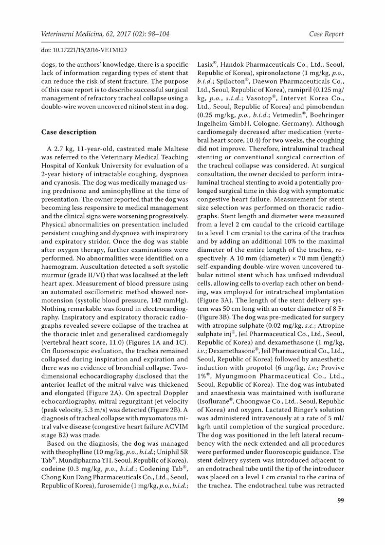
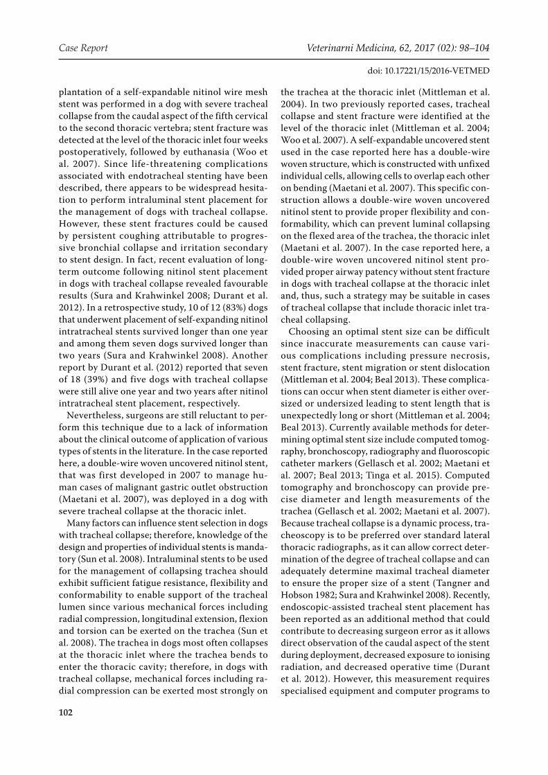
A 2.7 kg, 11-year-old, castrated male Maltese was referred to the Veterinary Medical Teaching Hospital of Konkuk University for evaluation of a 2-year history of intractable coughing, dyspnoea and cyanosis. The dog was medically managed us-ing prednisone and aminophylline at the time of presentation. The owner reported that the dog was becoming less responsive to medical management and the clinical signs were worsening progressively. Physical abnormalities on presentation included persistent coughing and dyspnoea with inspiratory and expiratory stridor. Once the dog was stable after oxygen therapy, further examinations were performed. No abnormalities were identified on a haemogram. Auscultation detected a soft systolic murmur (grade II/VI) that was localised at the left heart apex. Measurement of blood pressure using an automated oscillometric method showed nor-motension (systolic blood pressure, 142 mmHg). Nothing remarkable was found in electrocardiog-raphy. Inspiratory and expiratory thoracic radio-graphs revealed severe collapse of the trachea at the thoracic inlet and generalised cardiomegaly (vertebral heart score, 11.0) (Figures 1A and 1C). On fluoroscopic evaluation, the trachea remained collapsed during inspiration and expiration and there was no evidence of bronchial collapse. Two-dimensional echocardiography disclosed that the anterior leaflet of the mitral valve was thickened and elongated (Figure 2A). On spectral Doppler echocardiography, mitral regurgitant jet velocity (peak velocity, 5.3 m/s) was detected (Figure 2B). A diagnosis of tracheal collapse with myxomatous mi-tral valve disease (congestive heart failure ACVIM stage B2) was made.
Based on the diagnosis, the dog was managed with theophylline (10 mg/kg, p.o., b.i.d.; Uniphil SR Tab®, Mundipharma YH, Seoul, Republic of Korea), codeine (0.3 mg/kg, p.o., b.i.d.; Codening Tab®, Chong Kun Dang Pharmaceuticals Co., Ltd., Seoul, Republic of Korea), furosemide (1 mg/kg, p.o., b.i.d.;
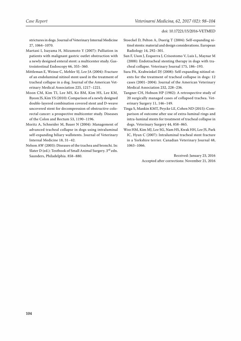
Lasix®, Handok Pharmaceuticals Co., Ltd., Seoul, Republic of Korea), spironolactone (1 mg/kg, p.o., b.i.d.; Spilacton®, Daewon Pharmaceuticals Co., Ltd., Seoul, Republic of Korea), ramipril (0.125 mg/kg, p.o., s.i.d.; Vasotop®, Intervet Korea Co., Ltd., Seoul, Republic of Korea) and pimobendan (0.25 mg/kg, p.o., b.i.d.; Vetmedin®, Boehringer Ingelheim GmbH, Cologne, Germany). Although cardiomegaly decreased after medication (verte-bral heart score, 10.4) for two weeks, the coughing did not improve. Therefore, intraluminal tracheal stenting or conventional surgical correction of the tracheal collapse was considered. At surgical consultation, the owner decided to perform intra-luminal tracheal stenting to avoid a potentially pro-longed surgical time in this dog with symptomatic congestive heart failure. Measurement for stent size selection was performed on thoracic radio-graphs. Stent length and diameter were measured from a level 2 cm caudal to the cricoid cartilage to a level 1 cm cranial to the carina of the trachea and by adding an additional 10% to the maximal diameter of the entire length of the trachea, re-spectively. A 10 mm (diameter) × 70 mm (length) self-expanding double-wire woven uncovered tu-bular nitinol stent which has unfixed individual cells, allowing cells to overlap each other on bend-ing, was employed for intratracheal implantation (Figure 3A). The length of the stent delivery sys-tem was 50 cm long with an outer diameter of 8 Fr (Figure 3B). The dog was pre-medicated for surgery with atropine sulphate (0.02 mg/kg, s.c.; Atropine sulphate inj®, Jeil Pharmaceutical Co., Ltd., Seoul, Republic of Korea) and dexamethasone (1 mg/kg, i.v.; Dexamethasone®, Jeil Pharmaceutical Co., Ltd., Seoul, Republic of Korea) followed by anaesthetic induction with propofol (6 mg/kg, i.v.; Provive 1%®, Myungmoon Pharmaceutical Co., Ltd., Seoul, Republic of Korea). The dog was intubated and anaesthesia was maintained with isoflurane (Isoflurane®, Choongwae Co., Ltd., Seoul, Republic of Korea) and oxygen. Lactated Ringer’s solution was administered intravenously at a rate of 5 ml/kg/h until completion of the surgical procedure. The dog was positioned in the left lateral recum-bency with the neck extended and all procedures were performed under fluoroscopic guidance. The stent delivery system was introduced adjacent to an endotracheal tube until the tip of the introducer was placed on a level 1 cm cranial to the carina of the trachea. The endotracheal tube was retracted

100
Case Report Veterinarni Medicina, 62, 2017 (02): 98–104
doi: 10.17221/15/2016-VETMED
phy seven days after surgery, the stent position remained unchanged (Figures 1B and 1D). The medication was continued for eight weeks, and was thereafter stopped according to the owner’s wishes. On presentation six months after surgery, the owner reported that the dog was doing well without medical management.
DISCUSSION AND CONCLUSIONS
Various stents have been used for the manage-ment of malignant and benign obstruction and these have been reported to alleviate symptoms and improve quality of life in human medicine (Stoeckel et al. 2004; Maetani et al. 2007; Moon et al. 2010). The application of stents designed for use within the biliary, vascular, gastrointestinal
to the level of the cricoid cartilage as the distal 1 cm of the stent was deployed, ensuring that the distal aspect of the stent was positioned at a level 1 cm cranial to the carina of the trachea. The stent was then completely deployed with close attention to the proximal end of the stent. Once the stent was deployed, the endotracheal tube was gently advanced back into the stent to maintain the pa-tient’s airway. Supplemental oxygen was provided as needed.
Postoperative medications included prednisone (0.5 mg/kg, p.o., b.i.d.; Solondo®, Yuhanmedica Co., Ltd., Cheongwon, Republic of Korea) for two weeks and theophylline (10 mg/kg, p.o., b.i.d.), co-deine (0.3 mg/kg, p.o., b.i.d.), furosemide (1 mg/kg, p.o., b.i.d.), spironolactone (1 mg/kg, p.o., b.i.d.), ramipril (0.125 mg/kg, p.o., s.i.d.) and pimobendan (0.25 mg/kg, p.o., b.i.d.). On thoracic radiogra-
Figure 1. Preoperative (A, C) and postoperative (B, D) inspiratory thoracic radiographs: (A, C) severe collapse of the trachea is visible at the thoracic inlet and there is evidence of generalised cardiomegaly (vertebral heart score, 11.0); (B, D) the stent position remains unchanged seven days after surgery

101
Veterinarni Medicina, 62, 2017 (02): 98–104 Case Report
doi: 10.17221/15/2016-VETMED
and respiratory systems in humans has recently described for the management of tracheal collapse, nasopharygeal stenosis, oesophageal stricture, colorectal stricture, ureteral stenosis and urethral stenosis in veterinary medicine (Cook et al. 2013; Lam et al. 2013; Hill et al. 2014; Tinga et al. 2015). The implantation of self-expandable metallic stents appears to be especially promising for intraluminal stabilisation of collapsed trachea in that it can fa-cilitate an immediate improvement of clinical signs, requires only short anaesthetic time, is minimally invasive and allows access to the entire trachea (Beal 2013). Uncovered nitinol wire mesh stents with single-wire woven structures are increasingly
being used for the management of dogs with end stage tracheal collapse (Gellasch et al. 2002; Durant et al. 2012; Tinga et al. 2015). However, despite the major advantages of endotracheal stenting, severe complications including stent fracture have been described (Mittleman et al. 2004; Woo et al. 2007). In one report from 2004, a self-expandable nitinol wire mesh stent was deployed in a dog with se-vere tracheal collapse from the caudal aspect of the fourth cervical to the second thoracic vertebra; stent fracture was observed at the level of the tho-racic inlet six weeks postoperatively, followed by euthanasia 10 weeks postoperatively (Mittleman et al. 2004). In another report from 2007, the im-
Figure 2. Two-dimensional (A) and continuous waved Doppler (B) echocardiography: (A) the anterior leaflet of the mitral valve is thickened and elongated (arrow); (B) on spectral Doppler echocardiography, mitral regurgitant jet velocity (peak velocity, 5.3 m/s) is detected
Figure 3. A double-wire woven uncovered tubular nitinol stent (A, top), commercially available single-wire woven stent (A, bottom) and stent delivery system (B): a single-wire woven stent has fixed cells with braided construction; (A, bottom) a double-wire woven uncovered tubular nitinol stent has unfixed individual cells, which allows cells to overlap each other on bending; (B) the length of the stent delivery system is 50 cm long with an outer diameter of 8 Fr
102
Case Report Veterinarni Medicina, 62, 2017 (02): 98–104
doi: 10.17221/15/2016-VETMED
plantation of a self-expandable nitinol wire mesh stent was performed in a dog with severe tracheal collapse from the caudal aspect of the fifth cervical to the second thoracic vertebra; stent fracture was detected at the level of the thoracic inlet four weeks postoperatively, followed by euthanasia (Woo et al. 2007). Since life-threatening complications associated with endotracheal stenting have been described, there appears to be widespread hesita-tion to perform intraluminal stent placement for the management of dogs with tracheal collapse. However, these stent fractures could be caused by persistent coughing attributable to progres-sive bronchial collapse and irritation secondary to stent design. In fact, recent evaluation of long-term outcome following nitinol stent placement in dogs with tracheal collapse revealed favourable results (Sura and Krahwinkel 2008; Durant et al. 2012). In a retrospective study, 10 of 12 (83%) dogs that underwent placement of self-expanding nitinol intratracheal stents survived longer than one year and among them seven dogs survived longer than two years (Sura and Krahwinkel 2008). Another report by Durant et al. (2012) reported that seven of 18 (39%) and five dogs with tracheal collapse were still alive one year and two years after nitinol intratracheal stent placement, respectively.
Nevertheless, surgeons are still reluctant to per-form this technique due to a lack of information about the clinical outcome of application of various types of stents in the literature. In the case reported here, a double-wire woven uncovered nitinol stent, that was first developed in 2007 to manage hu-man cases of malignant gastric outlet obstruction (Maetani et al. 2007), was deployed in a dog with severe tracheal collapse at the thoracic inlet.
Many factors can influence stent selection in dogs with tracheal collapse; therefore, knowledge of the design and properties of individual stents is manda-tory (Sun et al. 2008). Intraluminal stents to be used for the management of collapsing trachea should exhibit sufficient fatigue resistance, flexibility and conformability to enable support of the tracheal lumen since various mechanical forces including radial compression, longitudinal extension, flexion and torsion can be exerted on the trachea (Sun et al. 2008). The trachea in dogs most often collapses at the thoracic inlet where the trachea bends to enter the thoracic cavity; therefore, in dogs with tracheal collapse, mechanical forces including ra-dial compression can be exerted most strongly on
the trachea at the thoracic inlet (Mittleman et al. 2004). In two previously reported cases, tracheal collapse and stent fracture were identified at the level of the thoracic inlet (Mittleman et al. 2004; Woo et al. 2007). A self-expandable uncovered stent used in the case reported here has a double-wire woven structure, which is constructed with unfixed individual cells, allowing cells to overlap each other on bending (Maetani et al. 2007). This specific con-struction allows a double-wire woven uncovered nitinol stent to provide proper flexibility and con-formability, which can prevent luminal collapsing on the flexed area of the trachea, the thoracic inlet (Maetani et al. 2007). In the case reported here, a double-wire woven uncovered nitinol stent pro-vided proper airway patency without stent fracture in dogs with tracheal collapse at the thoracic inlet and, thus, such a strategy may be suitable in cases of tracheal collapse that include thoracic inlet tra-cheal collapsing.
Choosing an optimal stent size can be difficult since inaccurate measurements can cause vari-ous complications including pressure necrosis, stent fracture, stent migration or stent dislocation (Mittleman et al. 2004; Beal 2013). These complica-tions can occur when stent diameter is either over-sized or undersized leading to stent length that is unexpectedly long or short (Mittleman et al. 2004; Beal 2013). Currently available methods for deter-mining optimal stent size include computed tomog-raphy, bronchoscopy, radiography and fluoroscopic catheter markers (Gellasch et al. 2002; Maetani et al. 2007; Beal 2013; Tinga et al. 2015). Computed tomography and bronchoscopy can provide pre-cise diameter and length measurements of the trachea (Gellasch et al. 2002; Maetani et al. 2007). Because tracheal collapse is a dynamic process, tra-cheoscopy is to be preferred over standard lateral thoracic radiographs, as it can allow correct deter-mination of the degree of tracheal collapse and can adequately determine maximal tracheal diameter to ensure the proper size of a stent (Tangner and Hobson 1982; Sura and Krahwinkel 2008). Recently, endoscopic-assisted tracheal stent placement has been reported as an additional method that could contribute to decreasing surgeon error as it allows direct observation of the caudal aspect of the stent during deployment, decreased exposure to ionising radiation, and decreased operative time (Durant et al. 2012). However, this measurement requires specialised equipment and computer programs to

103
Veterinarni Medicina, 62, 2017 (02): 98–104 Case Report
doi: 10.17221/15/2016-VETMED
calculate relevant tracheal dimensions (Gellasch et al. 2002; Maetani et al. 2007). In addition, the ability to recover from anaesthesia should be taken into account since dogs with tracheal collapse of-ten require emergency management with a focus on the use of a self-expanding stent placement as an emergency procedure (Beal 2013). Inspiratory and expiratory thoracic radiographs can be con-sidered as a safe procedure to determine diameter and length of the tracheal lesion in dogs with end-stage tracheal collapse that fail to respond to rou-tine emergency management strategies (Sura and Krahwinkel 2008; Tinga et al. 2015). As described earlier, a stent diameter that is 10% to 20% larger than the maximum measured tracheal diameter can be chosen (Sura and Krahwinkel 2008). Stent length is more challenging to determine since lodg-ing in the carina or larynx or inadequate coverage of the affected portion of the trachea can result in severe coughing or recurrence of tracheal collapse (Beal 2013). To minimise the risk of complications associated with stent dislocation, two methods have been described: intraluminal stents can be extended about 1–2 cm beyond either end of the affected area; and intraluminal stents can cover the entire trachea (Woo et al. 2007; Durant et al. 2012). To prevent inadequate coverage of the af-fected portion of the trachea caused by shortening of a self-expandable stent or by an inappropriately long stent, intraluminal stents are ideally deployed from 1 cm distal to the cricoid cartilage to 1 cm proximal to the carina.
In the case reported here, stent size was deter-mined based on the measurements from radiographs and the manufacturer’s foreshortening chart with-out general anaesthesia and the stent was inserted with the dog under general anaesthesia. However, shortening of the stent occurred and it expanded by almost as much as the diameter of the trachea. Determining the size of a stent under general anaes-thesia would provide more precise measurements of the trachea; however, in the case reported here, the owner decided not to perform preoperative measurements under general anaesthesia to avoid potential risk to the dog of symptomatic congestive heart failure. Even though the length of the stent was shorter than expected, the collapsed region of the trachea was covered properly, which resulted in improvements of the clinical signs.
To the authors’ knowledge, the use of a double-wire woven uncovered nitinol stent has not been
reported previously for the management of a dog with tracheal collapse. In the dog of this report, this particular type of a stent with unfixed indi-vidual cells provided proper airway patency with-out stent fracture for one year. Finally, this stent might be used as a treatment alternative to other commercially available nitinol stents. Further study on a larger group of patients is needed in order to unambiguously determine whether the double wire nitinol stent is more resistant to fracture than single wire endotracheal nitinol stents.
RefeReNCeS
Ayres SA, Holmberg DL (1999): Surgical treatment of tra-cheal collapse using pliable total ring prostheses: Results in one experimental and 4 clinical cases. Canadian Vet-erinary Journal 40, 787–791.
Beal MW (2013): Tracheal stent placement for the emer-gency management of tracheal collapse in dogs. Topics in Companion Animal Medicine 28, 106–111.
Buback JL, Boothe HW, Hobson HP (1996): Surgical treat-ment of tracheal collapse in dogs: 90 cases (1983–1993). Journal of the American Veterinary Medical Association 208, 380–384.
Cook AK, Mankin KT, Saunders AB, Waugh CE, Cuddy LC, Ellison GW (2013): Palatal erosion and oronasal fistula-tion following covered nasopharyngeal stent placement in two dogs. Irish Veterinary Journal 66, 8.
Durant AM, Sura P, Rohrbach B, Bohling MW (2012): Use of nitinol stents for end-stage tracheal collapse in dogs. Veterinary Surgery 41, 807–817.
Fossum TW (2007): Surgery of the upper respiratory sys-tem. In: Fossum TW (ed.): Small Animal Surgery. 3rd edn. Mosby, St Louis. 906–957.
Gellasch KL, Gomez TDC, McAnulty JF, Bjorling DE (2002): Use of intraluminal nitinol stents in the treatment of tra-cheal collapse in a dog. Journal of the American Veteri-nary Medical Association 221, 1719–1723.
Hill TL, Berent AC, Weisse CW (2014): Evaluation of ure-thral stent placement for benign urethral obstructions in dogs. Journal of Veterinary Internal Medicine 28, 1384–1390.
Kim JY, Han HJ, Yun HY, Lee B, Jang HY, Eom KD, Park HM, Jeong SW (2008): The safety and efficacy of a new self-expandable intratracheal nitinol stent for the tracheal collapse in dogs. Journal of Veterinary Science 9, 91–93.
Lam N, Weisse C, Berent A, Kaae J, Murphy S, Radlinsky M, Richter K, Dunn M, Gingerich K (2013): Esophageal stenting for treatment of refractory benign esophageal
104
Case Report Veterinarni Medicina, 62, 2017 (02): 98–104
doi: 10.17221/15/2016-VETMED
strictures in dogs. Journal of Veterinary Internal Medicine 27, 1064–1070.
Maetani I, Isayama H, Mizumoto Y (2007): Palliation in patients with malignant gastric outlet obstruction with a newly designed enteral stent: a multicenter study. Gas-trointestinal Endoscopy 66, 355–360.
Mittleman E, Weisse C, Mehler SJ, Lee JA (2004): Fracture of an endoluminal nitinol stent used in the treatment of tracheal collapse in a dog. Journal of the American Vet-erinary Medical Association 225, 1217–1221.
Moon CM, Kim TI, Lee MS, Ko BM, Kim HS, Lee KM, Byeon JS, Kim YS (2010): Comparison of a newly designed double-layered combination covered stent and D-weave uncovered stent for decompression of obstructive colo-rectal cancer: a prospective multicenter study. Diseases of the Colon and Rectum 53, 1190–1196.
Moritz A, Schneider M, Bauer N (2004): Management of advanced tracheal collapse in dogs using intraluminal self-expanding biliary wallstents. Journal of Veterinary Internal Medicine 18, 31–42.
Nelson AW (2003): Diseases of the trachea and bronchi. In: Slater D (ed.): Textbook of Small Animal Surgery. 3rd edn. Saunders, Philadelphia. 858–880.
Stoeckel D, Pelton A, Duerig T (2004): Self-expanding ni-tinol stents: material and design considerations. European Radiology 14, 292–301.
Sun F, Uson J, Ezquerra J, Crisostomo V, Luis L, Maynar M (2008): Endotracheal stenting therapy in dogs with tra-cheal collapse. Veterinary Journal 175, 186–193.
Sura PA, Krahwinkel DJ (2008): Self-expanding nitinol st-ents for the treatment of tracheal collapse in dogs: 12 cases (2001–2004). Journal of the American Veterinary Medical Association 232, 228–236.
Tangner CH, Hobson HP (1982): A retrospective study of 20 surgically managed cases of collapsed trachea. Vet-erinary Surgery 11, 146–149.
Tinga S, Mankin KMT, Peycke LE, Cohen ND (2015): Com-parison of outcome after use of extra-luminal rings and intra-luminal stents for treatment of tracheal collapse in dogs. Veterinary Surgery 44, 858–865.
Woo HM, Kim MJ, Lee SG, Nam HS, Kwak HH, Lee JS, Park IC, Hyun C (2007): Intraluminal tracheal stent fracture in a Yorkshire terrier. Canadian Veterinary Journal 48, 1063–1066.
Received: January 23, 2016Accepted after corrections: November 21, 2016