use in plant breeding of acute, chronic or fractionated
TRANSCRIPT

Use in plant breedingof acute, chronic or fractionated dosesof X-rays or fast neutronsas illustrated with leaves of Saintpaulia
C. Broertjes


Stellingen - Assertions
1.
Het verdient aanbeveling om bepaalde gefractioneerde bestralingen, die bij
Saintpaulia een duidelijke verhoging van het aantal mutanten per 100 bestraal-
de bladeren tot gevolg hebben, nader op hun waarde te toetsen bij minder
heterozygote vegetatief vermeerderde gewassen.
It is recommendable to investigate whether given fractionated irradiations,
which, in Saintpaulia, result in an evidently increased number of mutants per
100 irradiated leaves, also do so with less heterozygous vegetatively propag-
ated crops.
Dit proefschrift, p. 64.
2.
De adventief-spruit-methode, waarmede, in vergelijking niet bestraling van
plantedelen met multicellulaire vegetatiepunten, meer en volledige (niet-
chimaere) mutanten verkregen kunnen worden na een mutagene behandeling van af-
gesneden bladeren, is een belangrijk hulpmiddel bij de veredeling van vegeta-
tief vermeerderde gewassen.
The adventitious bud technique, resulting in more and solid, non-chimeral
mutants after a mutagenic treatment of detached leaves, is a better method of
breeding vegetatively propagated crops than the treatment of plant parts with
multicellular buds.
Dit proefschrift, p. 18.
3.
De resultaten van vele in de literatuur beschreven experimenten met gefractio-
neerde bestraling zijn moeilijk te interpreteren en hebben een beperkte weten-
schappelijke waarde omdat met niet of onvolledig gesynchroniseerde celpopula-
ties is gewerkt terwijl bovendien onvolledige reeksen (initiële) doses en

tijd-intervallen zijn toegepast.
The results of many published experiments, describing dose fractionation, are
difficult to interprete and have a restricted scientific value since incom-
pletely synchronized or asynchronous cell populations have been used and more-
over incomplete series of (initial) doses and time intervals have been
applied.
Bijvoorbeeld:D.R. Davies, 1962. Strahlenwirkung und Milieu: 160-170.R.J. Horsley and A. Laszlo, 1971. Int. J. Radiat. Biol. 20: 593-596.W. Sheridan, 1971. Mut. Res. 13: 163-169.
4.
Uit hun" conclusie, dat bij gelijk niveau van lV^-fertiliteit EMS 2 a 3x zoveel
bloeitijd- en chlorophyl mutaties induceert als röntgenstralen, mag niet
zonder meer worden afgeleid, dat dit ook zou gelden voor al die mutaties, die
van belang zijn in de praktische mutatieveredeling.
From their conclusion, that at equal levels of NL-fertility EMS produces 2-3
times more flowering time and chlorophyl mutations than X-rays, it does not
follow that this also applies to all mutations that are important in practical
mutation breeding.
H.A.S. Hussein, 1968. Genetic analysis of mutagen-induced flowering timevariation in Arabidopsis thaliana (L) Heynh. Proefschrift (H. Veen-man en Zn, N.V., Wageningen).
M. Mesken and J.H. van der Veen, 1968. The problem of induced sterility:a comparison between EMS and X--rays in Arabidopsis thaliana.Euphytica 17: 363-370.
5.
Er wordt onvoldoende rekening gehouden met het feit, dat insektenbestrijding
met behulp van de "sterile insect release method" onherroepelijk tot teleur-
stellende resultaten leidt zonder grondige en vooral kwantitatieve veldbiolo-
gische kennis van de betreffende plaag.
Without deep knowledge, in particular quantitative, of the ecology of an
insect species, the "sterile insect release method" will fail.
Insect ecology and the sterile male technique. Proc. FAO/IAEA, Vienna,1969.

6.
Het toevoegen van bepaalde chemische stoffen aan voedsel ter verbetering van
houdbaarheid en kwaliteit dient uit het oogpunt van volksgezondheid onverwijld
te worden gestaakt en vervangen te worden door onschadelijke fysische methoden
zoals bestraling.
The addition of certain chemicals to food to improve the keepability and
quality should be stopped immediately in the interest of public health and re-
placed by physical methods, such as radiation.
7.
De hypothese dat er specifieke orgaan-vormende stoffen in de plant bestaan is
onjuist.
The hypothesis that specific organ-forming substances exist in plants is not
true.
E.W. Sinnott, 1960. Plant morphogenesis (p.413; p.376). McGraw-HillBook Co, N.York-Toronto-London.
8.
Het anoniem beoordelen van met een auteursnaam voorziene wetenschappelijke
manuscripten, voordat deze (al of niet) worden gepubliceerd in een tijdschrift,
is ontoelaatbaar.
Reviewing or refereeing of signed scientific articles as a condition for
acceptance should not be anonymous.
9.
De door Westhoff geuite veronderstelling, dat het aantal potentieel waarde-
volle kamerplanten veel groter is dan dat wat tot nu toe zijn weg naar het
publiek gevonden heeft, moet worden betwijfeld.
Westhoff's supposition that the number of potentially valuable pot plants
greatly exceeds the number so far introduced to the public, seems doubtful.
V.W. Westhoff, 1971. De botanische tuin in de samenleving, p !8 (Redeter gelegenheid van de officiële opening van de Botanische TuinNijmegen).

10.
Het gebruiken van hoogtepunten uit de klassieke muziek bij TV-reclame moestuit cultureel-hygiënische overwegingen worden verboden. Het roept ongewildeassociaties op met wasmiddelen en deodorants bij de concertbezoeker.
The use of the best of classical music in TV commercials should be purged
since at concerts it conjures up unwanted associations with detergents and
deodorants.
Proefschrift van C. Broertjes
Wageningen, 23 juni 1972.

I Nig- H"
Use in plant breeding of acute, chronic or fractionated dosesof X-rays or fast neutrons
as illustrated with leaves of Saintpaulia

Faota canardi sed erunt qui me finxisse loquantur.
OvidiuSy Fasti VI3 3.
(I'll state the facts, but some will say I have imagined things).
(Ik ga ware gebeurtenissen vertellen; maar somnigen zullen zeggen,
dat ik fantaseer).
Dit proefschrift met stellingen van Cornelis Broertjes, landbouwkundigingenieur, geboren te Haarlem op 31 maart 1923, is goedgekeurd door depromotor, dr. ir. J. Sneep, hoogleraar in de leer van de plantenveredeling. |
De Rector Magnificus van de LandbouwhogeschoolJ.M. Polak
Wageningen, 3 mei 1972

C. Broertjes
Use in plant breedingof acute, chronic or fractionated dosesof X-rays or fa t neutronsas illustrated with leaves of Saintpaulia
Proefschrift
ter verkrijging van de graad van
doctor in de landbouwwetenschappen,
op gezag van de Rector Magnificus, Mr. J.M. Polak,
hoogleraar in de rechts- en staatswetenschappen
van de westerse gebieden,
in het openbaar te verdedigen
in de aula van de Landbouwhogeschool te Wageningen
op vrijdag 23 juni 1977 te 16 uur
7972 Centre for Agricultural Publishing and Documentation
Wageningen

ISBN 90 220 0389 2
This thesis will also be published as Agricultural Research Reports 776.
@ Centre for Agricultural Publishing and Documentation, Wageningen, 1972.
No part of this book may be reproduced or published in any form, by print,photoprint, microfilm or any other means without written permission fromthe publishers.
Abstract
Broertjes, C. (1972) Use in plant breeding of acute, chronic or fraction-ated doses of X-rays or fast neutrons as illustrated with leaves ofSaintpaulia. Doctoral thesis, Wageningen. ISBN 90 220 0389 2, (viii) +74 p., 9 tbs, 31 figs, 8 photographs, 134 refs, Eng. and Dutch summaries.Also Agric. Res. Rep. (Versl. landbouwk. Onderz.) 776.
The parameters used were survival of irradiated leaves ofS. ionantlia, production of adventitious plantlets at the base of the pe-tiole and mutation frequency.
The differences between unfiltered X-rays from a 250/25 deep therapyapparatus and fast neutrons from a degraded fissions spectrum was slightand sometimes negligible. A striking dose rate effect was observed,especially with X-rays. In fractionation experiments a protective mechan-ism was induced in the leaves by pre-irradiation with either type ofradiation. This mechanism reached a maximum after 8-12 h. Then the extentof the protection was equivalent to 3 krad X-rays or 1.5 krad fastneutrons after a single pre-treatment with the optimum initial dose of500 rad, X-rays or fast neutrons. A 10-15 fold irradiation with thisdose, separated by 8-h intervals, induced a protection of 6-7 krad X-raysor 3-4 krad fast neutrons.
Several of the treatments result in a higher number of mutants per100 irradiated leaves compared with the optimum acute dose of 3 krad X-rays or 1.5 krad fast neutrons.


Samenvatting
Afgesneden bladeren van het Kaaps Viooltje, Saintpaulia ionantha H. Wendl.
cv. Utrecht, werden acuut, chronisch of gefractioneerd behandeld met X-stralen
of snelle neutronen. Het doel was om de optimale behandelingswijze vast te
stellen voor het verkrijgen van mutanten ten behoeve van de plantenveredeling.
Het effect van de verschillende behandelingen werd uitgedrukt in
1. aantal overlevende bladeren, in % van de controle,
2. aantal adventiefplantjes aan de voet van de bladsteel, per blad en in % van
de controle (produktie), en
3. mutatiefrequentie en aantal mutanten per 100 bestraalde bladeren.
De adventiefplantjes vertonen twee eigenschappen, die ze bijzonder bruik-
baar maken voor dit onderzoek. In de eerste plaats omdat de cellen, die later
betrokken zijn bij de vorming van adventiefplantjes, zich gedurende de gehele
behandeling niet delen. In de tweede plaats omdat het vegetatiepunt van een
adventief plantje uiteindelijk afkomstig is van slechts één epidermiscel. Dit
blijkt uit het feit, dat de gevonden mutanten, behoudens enkele uitzonderingen,
een niet-chimaere structuur vertoonden en dat sectoriaalchimaeren niet werden
aangetroffen.
Uit de resultaten bleek een sterk effect van doseringssnelheid op over-
leving, produktie en mutatiefrequentie. Na 2 rad/min was de LD-Q voor over-
leving ongeveer 100 krad X-stralen, terwijl de LDSO bij 200 rad/min ongeveer
5 krad bedroeg. De "kritische doseringssnelheid", waarbij een maximaal effect
wordt verkregen bij een verandering in doseringssnelheid, bleek ongeveer
7 rad/min te zijn (voor X-stralen).
Experimenten, waarbij de totale dosis in fracties gescheiden door één of
meer tijdintervallen werd gegeven, toonden aan, dat een betrekkelijk lage
initiële dosis een mechanisme induceert, dat de bladeren gedeeltelijk beschermt
tegen een of meer volgende stralingsdoses. De optimale initiële dosis die
maximale bescherming veroorzaakt bleek 500 rad te zijn, zowel bij X-stralen als
snelle neutronen. Het optimale tijdinterval waarna de bescherming een maximum
heeft bereikt bleek 8-12 uur te zijn, echter afhankelijk van de grootte van de
initiële dosis en die van de volgende dosis (doses). De grootte van de bescher-

ming bleek equivalent te zijn met 3-4 krad X-stralen of 1.5-2 krad snelleneutronen, na een eenmalige voorbehandeling met de optimale initiële dosis. Degrootte van de bescherming werd echter ongeveer verdubbeld na een 10-15-voudige voorbehandeling met de optimale initiële dosis, gescheiden door deoptimale tijdinterval van 8 uur. Een acute dosis van bijvoorbeeld 6.5 krad X-stralen was dodelijk. Na een voorbehandeling met 500 rad gaf diezelfde dosis100$ overleving (zie de 6 krad X-lijn in Fig. 12), terwijl een tweede dosisvan 10 krad nog 25? overleving gaf. Na 5 voorbehandelingen met 500 rad X-stralen, gescheiden door 8-urige intervallen, werd echter weer 100% overlevingverkregen na een laatste dosis van 10 krad.
Overleving, produktie zowel als aantal mutanten reageerden over het alge-meen op gelijke wijze op diverse behandelingswijzen. Daar de eerste twee para-meters veel sterker reageerden op een variatie van diverse factoren dan dederde parameter (mutatiefrequentie), bleek het mogelijk te zijn om optimalebehandelingsmethoden te berekenen.
Uitgaande van het aantal mutanten per 100 bestraalde blaadjes, uitgedruktin de overeenkomstige optimale acute dosis (3 krad X-stralen of 1.5 kradsnelle neutronen) werd aangetoond, dat grotere aantallen mutanten verkregenkunnen worden na bepaalde doseringssnelheden of gefractioneerde bestralingendan na een optimale acute bestraling. Voorbeelden zijn een voorbehandeling met500 rad, een tweede dosis van 6 krad, gescheiden door een interval van 8 uurmet X-stralen of een soortgelijke behandeling met snelle neutronen (zieTabel 9). Andere behandelingen zijn niet praktisch of te kostbaar.
Het verschil in effect tussen X-stralen en snelle neutronen (met eengemiddelde energie van 1.7 MeV) bleek verrassend klein te zijn en het is danook moeilijk aan te geven welke straling de voorkeur verdient.
De resultaten beschreven in dit rapport hebben uitsluitend betrekking opSaintpaulia. Bij andere gewassen, die mogelijk minder heterozygoot zijn en dusveel minder mutaties produceren, zou een verhoging van de imitatiefrequentie ofeen verschuiving in het mutatiespectrum wèl duidelijker en van meer betekeniskunnen zijn.
Het belang van de adventiefspruit-methode voor de mutatieveredeling inhet algemeen is speciaal besproken met het oog op de rol van de diplontischeselectie. Er zijn redenen aangevoerd ter ondersteuning van de veronderstelling,dat de diplontische selectie in normale multicellulaire vegetatiepunten groteris dan bij het gebruik van de adventiefspruit-methode.

Curriculum vitae
De auteur behaalde in 1941 het einddiploma van de toenmalige 2e HBS-B
te Haarlem. Vervolgens studeerde hij tropische plantenteelt aan de Landbouw-
hogeschool te Wageningen, met als bijvakken plantenveredeling en erfelijk-
heidsleer.
Na het behalen van het ingenieursdiploma in 1949 was hij een tiental
jaren werkzaam als plantenveredelaar op het Proefstation voor de Boomkwekerij
te Boskoop, aldaar gedetacheerd door het Instituut voor Veredeling van Tuin-
bouwgewarsen te Wageningen.
In 1959 trad hij in dienst van het Instituut voor Atoomenergie in de
Landbouw te Wageningen, dat hem van 1959 tot medio 1960 naar het Brookhaven
National Laboratory op Long Island, U.S.A., zond om zich te specialiseren in
de mutatieveredeling.

Contents
7 Introduction 7
1.1 Factors of importance for mutation breeding 1
1.2 Radiation type and method of irradiation 6
1.3 Diplontic selection 9
1.4 The adventitious bud technique 12
2 Materials and methods 79
2.1 Irradiation facilities 19
2.2 Plant material 20
2.3 Parameters 23
3 Irradiation experiments 26
3.1 Acute irradiation 26
3.1.1 Physiological and genetic effects 27
3.1.2 Practical implications 30
3.2 Chronic irradiations; dose rate phenomena 32
3.2.1 Physiological and genetic effects 32
3.2.2 Practical implications 36
3.3 Fractionated irradiations 36
3.3.1 Physiological effects 36
3.3.1.1 Initial dose 42
3.3.1.2 Time interval 44
3.3.1.3 Extent of protection 45
3.3.1.4 Repeated irradiation 47
3.3.1.5 Conclus ive remarks 51
3.3.2 Genetic effects 52
3.3.3 Practical implications 58
4 Comparison of acutea chronic and fractionated irradiations in
relation to (commercial) plant breeding 60

5 Comparison of X-rays and fast neutrons 63
Summary 65
Aaknowledgements 67
References 68

1 Introduction
The objective of mutation breeding is to obtain the highest possible
frequency of mutants and the best possible mutation spectrum in a given crop.
It enables the plant breeder, on an economically justified basis, to select
for useful or improved genotypes without exceeding certain limits, such as
number of plants to be handled, space and time needed and total costs.
To achieve this objective many factors have to be considered. Three of
them are discussed in section 1.1:
1. the kind of mutagen,
2. the type of mutational events desired, and
3. the problems, related to the choice of the material to be treated.
It is postulated, in Section 1.1, that the nature of the mutational event
depends on the radiation type (X-rays, fast neutrons) as well as on the method
of irradiation (acute, chronic, fractionated). Both are discussed in Section
1.2.
Moreover it is made clear that the kind of plant material treated is a
decisive factor in the mutation frequency, the mutation spectrum as well as in
the ease with which mutational events can be recovered. This recovery refers
to the competition within a multicellular apex (diplontic selection: Section
1.3) and the adventitious bud technique used (the apex of the buds originates
from a single epidermal cell: Section 1.4).
Finally it is thought that, radiation type and method of irradiation may
possibly be manipulated in such a way that the repair of the radiation damage
is optimum (minimum physiological effect) and includes a reasonable quality
and quantity of mutational events. This topic is covered in Chapter 3 (Experi-
ments) after Chapter 2 (Material and methods).
1.1 FACTORS OF IMPORTANCE FOR MUTATION BREEDING
To realize the objective of mutation breeding a number of factors have tobe considered, such as the kind of mutagen, the type of mutational eventdesired and the problems, related to the choice of plant material to be
1

irradiated.
The final decision about the kind of mutagen depends upon the cropinvolved and the plant part to be irradiated. In plants propagated by seedsthat are usually treated, both chemicals and ionizing radiation are effectivemutagens (Gaul, 1963). In vegetatively propagated species when growing plantsor plant parts have to be treated, the more penetrating types of ionizingradiation (X-rays, y-rays, fast neutrons) are better. Chemicals generallygive poor results with vegetatively propagated material because the plantparts take up much less solution when soaked than seeds. Higher concentrationsdo not compensate for this deficiency because they have harmful side-effects(Bowen, 1965; Nybom & Koch, 1965). When using chemicals, the type (baseanalogues - generally, but not exclusively used in studies with micro-organisms - alkylating agents and other mutagenic compounds), concentration,duration, temperature, modifying factors (pH, metallic ions, carrier agentsand after-effects) as well as post-treatments (washing) have to be considered(Heslot, Kamra, Brunner, Konzak, Narayanan, Wickham and de Koek in: Manual onmutation breeding; IAEA Techn. Report 119 (1970): 53-77). But even when amplesolution is taken up under standardized conditions (leaves of African Violet,treated with ethyl methane sulphonate), the mutagenic effect was poor comparedwith ionizing radiation.
The type of mutational event, in other words the mutation spectrum, verymuch depends on the mutagen used. Different mutation spectra can be distin-guished by different ratios between gene mutations, small deficiences andchromosome aberrations. Densely ionizing radiations, like neutrons, producemore aberrations whereas X or gamma rays induce more gene mutations. Likewise,certain chemicals, such as 8-ethoxycaffeine, produce more chromosome breaksand resulting aberrations than ethyl methane sulphonate. But the dose andmodifying factors, which influence the radiosensitivity (such as oxygen), alsochange the mutation spectrum (Micke, 1970; Nilan, 1966; Smith, 1961).
Radiation has a veiy complex effect upon living matter. The energy,dissipated in the cells through ionization, is the starting point of a numberof chain reactions that affect all kinds of cell constituents, such as cellmembranes (rupture of plasma membranes, disorganization of internal mito-chondria membranes or more subtile effects), which are important to maintaincell integrity and viability. Radiation also has an effect on energy metab-olism, reduces the phosphorylation, interferes with the enzyme activity andthe rate of DNA synthesis. The nucleus is also a major site of radiationdamage.

The precise relationship between such cytoplasmic and nuclear factors isnot clear, since the final end-point is also modified by exposure conditions,cell activity during exposure, type of cell and many other circumstances(Bacq & Alexander, 1961; Casarett, 1968; Evans, 1967b).
Part of the damage is restored by repair mechanisms. These repair mech-
anisms - in bacteria there are at least three, but probably more - seem to be
linked to particular enzyme activities with distinct repair time classes,
namely < 2 min, < 15 min, and > 30 min (Kaplan, 1971). Several kinds of
restoration can be distinguished, such as the neutralization of the primary
effects and that of the secondary effects (resynthesis of molecules destroyed
or inactivated by the irradiation). In addition to such intracellular recovery,
intercellular recovery takes place, by which actively dividing cells replace,
after a latent period, the dead ones (Bacq & Alexander, 1961).
The final effect, after the repair of sublethal and reversible damage, is
called the permanent physiological and genetic effect and can be measured in
different ways: the survival of plants or plant parts, the height of seedlings,
the length of plant parts or the dry weight of the plant after a given period.
Biochemical parameters have also been used, such as changes in enzyme activ-
ities (Bacq & Alexander, 1961) or in different types of DNA damage (Ebert &
Howard, 1963), including different types of mutational events (gene mutations,
chromosome rearrangements, reduced fertility).
In this study detached leaves of African Violet, which develop adventi-
tious plantlets at the base of the petiole after rooting, were used to investi-
gate the effect of radiation (see Section 1.4). Two physiological parameters
were used to measure the effects (number of surviving leaves, hereafter called
"survival" and number of adventitious plantlets per leaf, hereafter called
"production") and one genetic parameter (number of visible mutations) (see
Section 2.3).
These parameters were not only selected to express radiation effects, but
mainly because they are of essential importance to the practical breeder, whose
first question is how to obtain (the highest number of) useful mutants.
The physiological parameters will be referred to as the "physiological
effect", whereas the number of mutations will be called "genetic effect" (see
Section 3.1.1). It is realized, however, that this distinction is somewhat
artificial because part of the physiological effect is determined by genetic
elements (e.g. gross chromosome rearrangements as a result of chromosome break-
age reduce fertility as well as vitality).
The physiological effect depends upon a great number of factors, such as

the radiosensitivity of the plant species» total dose, dose rate, type of
radiation and a number of climate and other environmental conditions before,
during and after the treatment (Briggs, 1968; Gaul et al., 1966; Nayaranan &
Konzak, 1969).
The genetic effect upon a given genotype depends on type of radiation,
dose and dose rate, but the role of environmental conditions is much less
obvious and for the greater part unknown. Moreover, it depends greatly on the
genotype involved; in heterozygous diploid plants the mutations, which are
usually recessive, may manifest themselves immediately in the double recessive
form in the irradiated material itself. When homozygous characters are involv-
ed, a second generation is needed after self-fertilizing M« before mutations
can be scored- In polyploid crops (potato, 4 n), Chrysanthemum (6 n), Dahlia
(8 n)) very complex situations may occur when different genes are present in
different ploidy-levels (from multiplex to quadruplex in a tetraploid, for
instance). Moreover, polyploids are less radiosensitive (Sparrow, 1962). These
situations result in an obscure relationship between the treatment and the
result.
Both physiological and genetic effects can thus be modified at will,
relatively speaking. Since repair of physiological and genetic effects a priori
may be the result of (partly) different processes, it might be possible to
manipulate the two effects by a proper treatment so that the physiological
damage (reduced survival, stunted growth, reduced fertility) as well as the
undesirable part of the genetic effect (chromosome aberrations; also effecting
the physiological parameters mentioned before) is minimum whereas the desired
genetic effect (beneficial mutations, favourable chromosome rearrangements) is
maximum. It would indeed be of great importance to the plant breeder if clear-
cut methods were available to produce the optimum number of useful mutants.
In relation to chimera formation and diplontic selection (see Sections
1.4 and 1.3, respectively), the choice of the plant part to be treated is
another important aspect of applied mutation breeding. Generally plant material
with multicellular apices are irradiated, such as seeds, tubers, bulbs,
rhizomes, stolons, scions, cuttings, etc. unless gametes (pollen or eggcells)
or zygotes in the earliest developmental stages are being used.
Since a mutation is a unicellular event, the mutated cell will be exposed
to a competition process in such a multicellular apex. This competition between
the mutated cell and the surrounding non-mutated cells is often lost by the
mutated cell as a result of the general competition between cells in different

regions with different degrees of activity. Possible accompanying damage and
the subsequent reduced growth rate may also go against the mutated cell in the
competitive struggle. When this takes place during haplophase it is called
"haplontic selection", and when it takes place during diplophase it is called
"diplontic selection" (Gaul, 1957). The final result is a lower mutation
frequency and a restricted mutation spectrum so that drastic chromosome aberra-
tions and cells, which carry mutations for compact growth, dwarfness and the
like, disappear automatically. When the breeding programme is aimed at gene
mutations for certain characters without a reduction in growth, fertility and
size, the restricted mutation spectrum is advantageous. When, however, the
programme is aimed at the induction of chromosome breaks and the resulting
aberrations or when chromosome engineering (e.g. the transfer of a piece of
chromosome or gene from a wild species to the genome of a cultivar) is wanted,
the "self-improvement" by this intrasomatic competition is undesirable (see
also Section 1.3).
Moreover, since most apices consist of a number of fairly independent
groups of cell layers, surviving and dividing mutated cells lead to the forma-
tion of mericlinal and periclinal chimeras. The chimeral situation is dis-
continued in seed propagated species after selfing or crossing; in vegetatively
propagated plants, however, it is preserved as so-called budsports which are
directly recognizable when mutations of visible characters are involved. Some-
times mutations, concerning characters of the outer layer(s), such as flower
colour, cannot express themselves because they were induced in the inner
layers. They may be detected years after the induction as the result of
spontaneous or radio-induced uncovering or rearrangement, by which mutated
cells appear in the outer layer(s). This phenomenon even has possibilities for
practical application since the various combinations of mutated and non-mutated
cell layers may result in different phenotypes (Bergann, 1966, 1967; Dommergues
& Gillot, 1964; Klöpfer, 1966; Pötsch, 1966).
Both, diplontic selection and chimera formation very much restrict the
potential of practical mutation breeding as well as more basic studies using
induced mutations in plant material. Such an undesirable situation can to a
great extent be avoided by growing complete plants from single cells, auto-
matically resulting in a high(er) frequency of solid, non-chimeral mutants and
a wide(r) mutation spectrum (see Section 1.4).

1.2 RADIATION TYPE AND METHOD OF IRRADIATION
Various types of ionizing radiations, such as electromagnetic radiation
(X-rays, gamma-rays) and charged or uncharged particles (a-rays, e-rays,
protons, neutrons) are used in radiobiological experiments to study the inter-
action between radiation and biological material (Bacq & Alexander, 1961).
In this study, X-rays were used as a representative of the sparsely ioniz-
ing electromagnetic radiations with a very short wavelength. The energy is
almost entirely dissipated by electrons ejected from the atoms of the material
through which they pass. This process is almost independent of the manner in
which these atoms are combined into molecules and of its elementary composi-
tion.
The penetration of the X-rays depends on the energy used and should be
high enough to prevent a "one-sided effect", as the result of an uneven dose
distribution within the material. The X-rays, used in this study, had an
effective energy of 50-60 keV, when the machine operated at 250 kVp and no
additional filter was applied (the filtering of the apparatus was equivalent to
2-3 mm Al). The decrease in dose rate, caused by one, two or three layers of
African Violet leaves, was measured, and turned out to be 0.9%, 2.5% and 3.51,
respectively. Since in all experiments a one-layer formation was used, where
the leaves overlapped partly at most once, the low percentages indicate that
the energy distribution within the leaves was sufficiently even.
Fast neutrons, having an energy of approx. 10 keV and up, were applied as
a representative of more densely ionizing corpuscular radiations. Fast neutrons
do not produce ionizations directly but mainly react with nuclei of the atoms
of the absorbing material. The reaction with the H-nuclei is the most import-
ant, producing recoil protons which cause ionizations, concentrated along short
tracks, inside the material. Therefore, unlike sparsely ionizing radiations
(such as X-rays), the number of ionizations produced depends largely on the
nature of the elementary composition of the material, hydrogen being the most
important element when using fast neutrons (Bacq & Alexander, 1961; Dertinger
& Jung, 1969). As hydrogen is the major constituent of biological materials,
85-95% of the energy is transferred by the recoil protons. In Saintpaulia
leaves, with a water content of 97-98%, the hydrogen concentration, as well as
its distribution throughout the leaves guarantees an even energy dissipation in
the material (Dertinger & Jung, 1969).
Another reason to compare the effect of X-rays and fast neutrons is that
sparsely ionizing radiation has a mainly indirect effect, whereas densely ion-

izing radiation has a mainly direct effect. The (intracellular) direct andindirect effects are of comparable magnitude in most cases, and depend on thecell function studied. The definition of direct and indirect varies and israther confusing and to a certain extent meaningless. Some authors refer tothe direct effect as an energy dissipation in the molecules involved, whereasin the indirect action these molecules are affected by radiation induced freeradicals (see also Section 1.1). Hence X-rays have a stronger physiologicaleffect and cause fewer chromosome aberrations than fast neutrons (Ehrenberg &Nybom, 1954; van Kampen, 1959; Stone, 1955). Other authors consider theunmodifiable damage as resulting from the direct action and the modifiabledamage as resulting from the indirect action (Dertinger & Jung, 1969). Some-times the direct effect is referred to as the effect upon two separate sitesat the same time by the same electromagnetic ray or particle. This is morelikely to happen with densely ionizing radiation, such as neutrons, than withsparsely ionizing radiation, such as deep therapy X-rays (Chadwick & Leenhouts,1972). In the following part of this study it will be observed that the differ-ence between the fast neutrons and the X-rays used, is much less than generallyreported in the literature. Whether this is because most experiments werecarried out with 14 MeV neutrons is not known, but seems improbable, since14 MeV neutrons have a lower LET (linear energy transfer) and RBE (relativebiological effectiveness) (Bacq & Alexander, 1961) compared with fast neutronsfrom the degraded fission spectrum, with an average energy of approx. 1.7 MeV,used in this study (see Section 2.1).
Various methods of irradiation have been used, namely acute, chronic or
fractionated exposures with X-rays or fast neutrons.
Acute irradiations with X-rays were almost exclusively carried out with a
dose rate of 200 rad/min so that radiation doses of 2-6 krad required only
10-30 minutes exposure times. The fast neutron dose rate (flux density) applied
was equivalent to 1000 rad/h, so that exposure times were 2 to 6 hours. Since
neutrons were not expected to have a dose rate effect, little attention was
paid to this difference at first. But the experimental results as well as a few
irradiations with higher dose rates showed that the dose rate of 1000 rad/h,
used in most experiments, was at the borderline between acute and semi-chronic
irradiations (see also Section 3.2).
Clwonic irradiations with X-rays or fast neutrons Have revealed a striking
dose rate effect in Saintpaulia (Broertjes, 1968b). Dose rate phenomena, on
which voluminous literature is available, have been investigated extensively,

either in mammalian systems (Anonymous, 1968) or in botanical material(Constantin et al., 1970; Sparrow, 1965). From the data presented it appearsthat low rates are less effective than high rates for a given total exposure.This holds true only within certain limits however: above a given dose rate afurther increase does not result in an additional increase in effect.Similarly below a certain low dose rate a further decrease in rate does notdecrease the effect (Bottino & Sparrow, 1970; Constantin et al., 1970; Sparrowet al., 1970).
Somewhere in the range between a low and a high dose rate, there is acritical area which represents a transition from a chronic irradiation (lowdose rate) to an acute one (high dose rate).
Constantin et al. (1970) determined this so-called rate saturation forsoybean seedlings and for mature plants as 50 R/min and 25 R/min, respectively.These results indicate that the phenomenon is dependent on stage of developmentduring irradiation. The end-point measured and the total exposure also provedto be important factors.
Furthermore the rate saturation seems to be dependent on the radio-sensitivity of the plant species involved: sensitive species would undergo asaturation at lower exposure rates than tolerant species. On the basis of theirinterphase chromosome volume, rate saturation constants for a number of specieswere calculated and compared with actual dose rate effect data, which seem tosupport the idea (see Section 3.2 for more details).
Dose fvaotionation was applied to collect information which could be usedin explaining the striking dose rate effect mentioned before.
Dose fractionations, reported in the literature by many authors, arecarried out in various ways. Sometimes a total dose is compared with two frac-tions half its size, separated by various time intervals. In other cases acertain dose is preceeded by various initial doses, separated by one or more(different) time intervals. And still in other cases the effect of a series offractions is studied (Bacchetti, 1965; Bacchetti et al., 1965; Bacq & Alexander,1961; Bewley et al., 1963; Bryant, 1968; Davies & Wall, 1960; Evans, 1966;Evans, 1967a; Fujii, 1968; Generalova, 1968; Generalova, 1969; Howard, 1968;Kabakova et al., 1970; Kada et al., 1966; Kiefer, 1966; Lawrence, 1968; Leonard& Deknudt, 1971; Morris & O'Grady, 1970; Paribok & Krupnova, 1970; Tates, 1968;Tazima & Onimaru, 1966; Withers & Elkind, 1969; Wurgler & Matter, 1968).
Generally the biological effect of fractionation is less than when thetotal dose is applied. This could be explained by part of the damage of thefirst fraction being repaired before the following dose(s) is applied.

In Saintpaulia, using the adventitious bud technique (see Section 1.4),both X-rays and fast neutrons were used to study various elements in fraction-ation experiments: initial dose, time interval, second dose and repeatedirradiations (see Section 3.3) (Broertjes, 1970).
1.3 DIPLONTIC SELECTION
The mutation frequency, expressed as detectable mutated sectors after
irradiation of multicellular apices of African Violet plants, was shown to be
extremely small in the SaintpauHa ionantha cultivar "Utrecht". During one of
the first experiments 100 whole plants were irradiated with 3 krad X-rays
(dose rate 300 rad/min). Only one mutated (flower colour) sector was observed
a few months after regrowth and further development. This observation agrees
with the relatively low number of spontaneous mutants in the greenhouses of
SaintpauHa growers. Similarly Achimenes and Streptooarpus, two other
representatives of the Gesneriaeeae, have few spontaneous mutants. Although it
is claimed that the himdreds and hundreds of cultivars of Saintpaulia ionantha
are the result of both hybridizing and selection among mutants (Moore, 1957),
recently no mutants have been commercialized in Western Europe. In Stvepto-
aarpus a white-flowering mutant, e.g. "Maassens White", arose spontaneously in
the blue-flowering cv. Constant Nymph, whereas in Achimenes the only mutant
recently commercialized was the cv, Willem Maarse, with bright green leaves
and stems (the original form had darker green leaves).
In strong contrast to these facts are the high mutation rates among the
adventitious buds developing at the base of the petioles of irradiated leaves
(> 20% after an X-ray dose of 3 krad). This difference can be explained on the
basis of diplontic selection, in combination with factors related to the
structure of a multicellular apex (Dommergues, 1962; Gaul, 1963) and consists
of the following elements:
1. the general competition between cells in a multicellular apex also as a
consequence of its structure. The apex of angiosperms consists of three fairly
independent cell layers, L^, L- and L, (Satina et al., 1940), each containing
a restricted number of initial cells and a number of less active cells
(méristème d'attente) (Buvat, 1952). Extensive information and details are
given by Balkema (1971), Dommergues (1962) and Yamazaki (1963). It is obvious
that the destination of a mutated cell is defined by its role within the apex
and that a mutation in a cell within a group of cells, not taking part in the
formation of the shoot, plant or axillary bud, will never be observed.
9

2. This also applies to mutations induced in the wrong cell layer. A flower
colour mutation induced in one of the initial cells of the L3 will, under
normal conditions, never manifest itself.
3. The diplontic selection, in a more restricted sense, is the competition
among mutated cells or between a mutated cell and its surrounding non-mutated
ones. It will result in the survival of the fittest since even small differ-
ences in division rate cause the elimination of the more slowly dividing cells
(Gaul, 1957).
The idea of diplontic selection has been opposed by a number of authors
and was recently reviewed by Balkema (1971). She claims that the destiny of
most cells in a multicellular meristem is already fixed and that diplontic
selection plays a role in such cases, where only a few of the many potential
organs can develop, such as with side shoots. In most cases, in her opinion,
diplontic selection can be explained as aspects of normal development or as
the result of differential (mutagen) sensitivity. She demonstrated in
Arabidopsis and sunflower, that by changing the environmental conditions,
chimerism was influenced and so could be manipulated.
Diplontic selection in adventitious buds, however, would appear to be
restricted exclusively to point 3, since neither point 1 nor 2 apply to buds
originating from single cells. It therefore remains to be explained which
factors are responsible for the striking difference between the extremely low
mutation frequency after the irradiation of plants and the high percentage of
mutants, observed after the irradiation of detached leaves and the subsequent
development of adventitious plantlets at the base of the petioles.
An examination of the striking difference in mutation recovery reveals a
few interesting facts. The high mutant frequency, among adventitious plantlets
202 or more after a medium dose, indicates that the majority, if not all, of
the cells must carry one or more mutations.
The 201 mutants are exclusively based on visible changes. There is no
reason to assume that the genes controlling not-directly detectable characters
do not mutate. The fact that we do find "unmutated" plantlets only might mean
that in these plants no detectable mutations are present. It consequently
appears that competition, if any, does not only take place between non-mutated
and mutated cells among the few thousand epidermal cells which are candidates
for the formation of adventitious buds, but that it mainly takes place among
mutated cells. Here, too, a number of cells will be eliminated or are damaged
so badly that they will not be able to form buds.
10

This, however, does not explain the difference between adventitious buds
and multicellular apices, since the same consideration holds true for the
latter: there is no reason to assume that the number of mutations will be
lower in such apices. Is it due to the fact that solid mutants, obtained when
using the adventitious bud technique, are recognized more easily than sectors,
observed after the irradiation of multicellular buds? This could indeed apply
to a number of characters (small changes in size, form or habit) but certainly
not for others (colour of leaf and flower and extreme changes in size, form
and habit). Or is the difference due to a much higher selection pressure in
the multicellular apex, together with the more strict organization within and
between the various layers and the reduced number of initial cells?
Cytological-histological observations of leaf petioles have revealed that
meristems are formed at random around the circumference near the base of the
petiole (on the dorsal side of the petiole slightly less meristems develop).
It therefore seems logical to assume that all epidermal cells in that region
have, in principle, an equal chance to become the origin of an adventitious
bud, except those which are too heavily damaged. Photograph 2, presenting part
of a cross section through the base of the petiole approx. 10 days after cutt-
ing off, shows that most epidermal cells have entered the division stage and
have divided into two or more meristematic cells. Some cells, however, become
omnipotent earlier than others and are the first to enter the division stage.
From that moment almost nothing can stop the cells from becoming the origin of
the apex of an adventitious bud. The surrounding cells, which also are
stimulated to divide, seem to be controlled or suppressed by the mitotic wave,
induced by those first cells. They form the base of the meristem as well as
the connecting tissue between the adventitious bud and the inner part of the
petiole. Photographs 3, 4, 5, 6 and 7 show various developmental stages of the
adventitious bud formation.
One can ask why certain cells become the origin of a bud and others do
not. It is a fact that only a very restricted number of possible candidate
epidermal cells are "chosen": there are approx. 300 epidermal cells around the
circumference of an average petiole which makes 3000 candidates when the
lowest 10 cells are stimulated. Since ultimately about 15 buds are formed,
only 0.5? of the cells have a chance to express themselves.
The question arises whether a certain selection pressure is present
during the very first days after cutting off or if epidermal cells really are
"chosen" at random. It is hard to tell whether the places of origin are pre-
destinated or just at random, since no evidence has been found that shows a
11

relationship between place of origin and position of other elements in thepetiole, such as the vascular bundles for instance.
The results after the irradiation of leaves seem to indicate that thediplontic selection is very restricted. As appears from the figures in Table 7(page 32) a large percentage of the mutants observed belong to the category ofdwarf mutants. This demonstrates that even slow growing mutant cells haveample chances in the competition with faster growing (unmutated) cells, unlessone assumes that almost all cells carry a (great) number of mutations orchromosome aberrations and that the number of "normal" cells is extremelysmall.
This train of thought however is counteracted by the fact that smalldosages, perhaps small dosages in particular, result in a large percentage ofdwarf mutants (Table 7). The low mutation frequency at such a dose, approx. 5Safter 0.5 krad (see Chapter 3) indicates a sufficient supply of normal, un-mutated and undamaged cells, which could have suppressed completely the forma-tion of slow growing dwarf types, even in the case of a slight diplonticselection in favour of the normal cells.
In a multicellular apex, however, the situation is completely different.The organization is much stronger and the number of initial cells much smaller.But the main difference is that the diplontic selection continues during theentire further development and growth of the shoot. Then even extremely smalldifferences in division rate or general fitness ruthlessly eliminate and re-place the weaker or slower growing cells. Such a high pressure during hundredsof cell divisions, in combination with points 1 and 2, leads to plantletswhich resemble the original type in the majority of cases.
1.4 THE ADVENTITIOUS BUD TECHNIQUE
Mitation breeding offers large possibilities in vegetatively propagated
plants, such as ornamentals, numerous fruit crops, potatoes, sweet potatoes,
sugar cane, cassava and grapes. The main advantage is the possibility of
improving one or only a few characters, of an otherwise excellent cultivar,
without altering the remaining genotype. Thus the outstanding cultivars, often
the result of a time-consuming and painstaking cross-breeding programme, can
be further perfected within a reasonable time (Broertjes. 1968a; Nybom, 1961;
Nybom & Koch, 1965).
Such cultivars are often very complicated genotypes which seggregate
after selfing or hybridizing in a very complex way. This makes it, practically
12

Photograph 1. Leaves of Saintpaulia showing developmental stages ofadventitious buds at the base of the petiole.
Photograph 2. Part of a cross section through the base of the petioleapprox. 10 days after detaching. Host epidermal cells have divided.

Photograph 3. First periclinaldivisions (arrows) in epidermalcells at the base of the petioleof the leaf (approx. 5 daysafter the removal of the leaf).
Photographs 4-7. Various later developmental stages leading to theformation of a meristem (after approx. 10 days; Photograph 5) whichforms an apex which develops into a plantlet (Photographs 6 and 7).

-Photograph 5.
Photograph 6.

Photograph 7.
Photograph 8. Saintpaülia •ionantha cv. Utrecht, clone B.

speaking, impossible to recover genotypes in the offspring with the original
gene combination, which, in addition, must moreover carry one or two favour-
able changes. Recovery becomes even more complicated and consequently improb-
able when polyploidy is involved. When only one or two (dominant) simplex
characters have to be changed into a nulliplex, mutation breeding is an almost
ideal tool. This in contrast to the situation where duplex, triplex or quadru-
plex (dominant) characters must be mutated. The irradiation of colchicine-
induced autotetraploid plants, which are more radioresistant, yielded however
surprisingly high percentages of mutants in Aahimenes (Broertjes, 1972b),
Saintpaulia and Streptoaarpus. The higher radioresistance results from the
fact that all kind of gross chromosome aberrations, lethal on diploid level,
are not lethal in (auto)tetraploids. The high mutation frequency is attributed
to the increased number of gross chromosome aberrations, which moreover have a
dominant expression. In addition the chance is increased that identical genes
in the two homologous chromosomes are involved in the mutation. Duplex genes
(most are duplex, because of the heterozygosity of the original material) thus
become nulliplex and manifest themselves in the case of visible characters.
The selection of visible characters generally offers no problems and a
favourable change may soon lead to the commercialization of the mutant. This
holds true for visible changes in ornamentals but also in other crops, such as
fruit colour and spur type in apples and pears, skin colour of potato tubers
as well as growth pattern, size, form and many other directly perceptible
characters in general (Broertjes, 1971; Heslot, 1964).
It is therefore not surprising that the majority of the commercial muta-
tion breeding projects, started since 1960 as co-operative programmes between
the Association Euratom-ITAL at Wageningen and plant breeders or institutions,
deal with vegetatively propagated plants, mostly ornamentals, but also fruit
crops and potatoes (Broertjes, 1965; Broertjes, 1968a).
All kinds of plant parts are irradiated, for example bulbs, tubers,
rhizomes, cuttings, grafts or (small) plants, all of which have in common that
they carry buds with multicellular apices which generally are composed of a
number of fairly autonomous cell layers.
As has already been discussed before, the irradiation of such a complex
apex leads automatically to the formation of chimeras as well as to a low
mutation frequency as the result of diplontic selection. This, the main
stumbling-block, can only be overcome by growing plants from single cells, in
vitro or in vivo, resulting in a higher frequency of solid, non-chimeral,
mutants. It is therefore of utmost importance to look for and to develop such
13

methods.For the commercial plant breeder the most important method at present is
the adventitious bud technique, which makes use of the phenomenon thatadventitious buds on detached leaves ultimately originate from only one(epidermal) cell (Broertjes et al., 1968) as has not only been shown inSaintpaulia (Naylor & Johnson, 1937; Sparrow et al., 1960; Broertjes, 1968b),but also in Aohimenes (Broertjes, 1972b), Kalanokoë (Broertjes & Leffring,1972), Streptooaepus (Broertjes, 1969) and tobacco (de Nettancourt et al.,1971). In all these cases it has been demonstrated, by mutation induction andcytological-histological observations, that neoformation takes place inseveral layers. In the L,, meristems are formed which develop into rootprimordia whereas in the epidermal layer (the L.) cells which have becomededifferentiated and meristematic, develop into shoot primordia after theformation of a meristem (photographs 4-7). Similar phenomena have beenobserved in other species, such as in Lilium hybrids, which almost exclusivelyproduce solid mutants after the irradiation of bulb-scales, preferably removedfrom the bulbs after irradiation (Broertjes & Alkema, 1970). In Endymion,which like Liliwn belongs to the monocotyledons, cytological-histologicalinvestigations have revealed no principle differences in the development ofadventitious bulbils at the base of detached leaves, compared with adventi-tious plantlets in the dicotyledons mentioned before (Broertjes, in prepara-tion) .
Such adventitious buds are exclusively formed by cells at the lowest partof the petiole near the cut end (see Photograph 1). During all our experi-ments, as a routine, the basal 5 mm of the petiole was cut off after thecompletion of the treatment and prior to planting. In this way it was madesure that all epidermal cells which might have entered the division stage as astarting point for the formation of adventitious buds (approx. after 5 days)or meristems in more advanced developmental stages (following prolonged treat-ments) were removed. Consequently, chimera formation and cell competition orcell elimination (as a result of differences in radiosensitivity related tovariation in metabolic activity, cell division stage or lethal chromosome re-arrangement) was avoided during the treatment. In this way, higher situatedepidermal cells on the petiole, which have not yet been stimulated to divide,were stimulated to do so by the removal of the 5 mm long basal part, after theirradiation had been completed. All such epidermal cells consequently haveundergone the whole treatment in a resting, non-dividing stage of development.
The question arises in which stage of the nuclear cycle were these cells.
14

Was it G p the pre-DNA synthesis phase, the S-phase, during which DNA issynthesized, or G-, the post-DNA synthetic period, prior to the actualmitosis? It could also have been a mixed population of cells in differentphases of the cycle. Although labelling with radioactive thymidine and DNAcytophotometry were not applied to investigate the stage of the nuclear cycle,there are some considerations, which indicate that most, if not all epidermalcells, were in G*. One is the fact that an overwhelming majority of theadventitious mutants observed in SaintpauKa, Aahimenes, Streptoearpus,Kalanchoë and other species, were non-chimeral. Irradiation of cells in the(late) S-phase, when the cells are in &j, as far as the DNA structure, but inG2, as far as the amount of DNA is concerned, would have resulted in a muchhigher percentage chimeras. Furthermore it is increasingly frequently report-ed, that in some species all meristematic cells in an embryo of a seedcumulate in G.. during seed maturation {pinus pinea, Laetuoa sativa,Helianthus annuus). In other species, however, cells were almost exclusivelyin G ^ or in G.. and G, in different proportions (d'Amato, 1972).
If the epidermal cells and probably also other cells of the Saintpaulialeaf, indeed appear to be in G, (which is under investigation), it wouldrepresent an ideal synchronization-method, which could profitably be used inradiobiological experiments. Synchronization of animal or plant cells general-ly is achieved by careful handling of temperature fluctuations, to which thecell cultures are exposed, or by using certain chemicals, such as 5-aminouracil, which block DNA-synthesis without interfering with the preceed-ing phases. After thorough washing, all cells start DNA-synthesis simultan-eously. Within a short period, however, desynchronization takes place (Clowes,1965; Prensky & Smith, 1965; Sybenga, 1968).
It is obvious that only by painstaking and laborous procedures can asatisfactory synchronization be obtained and one wonders whether the use ofleaves might be an easier and less expensive method.
It is concluded that the use of leaves of African Violet can be consider-ed a clean method, to study radiation effects and to demonstrate its signific-ance for applied mutagenesis. The most important reasons are the easy re-production of the plantlets, ultimately originating from only one epidermalcell, the subsequent prevention of chimera formation and the decreased oralmost eliminated consequences of differences in radiosensitivity as theresult of different nuclear cycles of the (epidermal) cells.
Many plants can be propagated from adventitious plantlets on detached
15

leaves. Broertjes et al. (1968) list over 350 species, including a variety offamilies, reported in the literature, that belong to that group. Many, however,are not listed, including a number of economically very important plantfamilies, such as Gramineae (cereals) and Solanaoeae (potato, tomato). Manyplants not listed have been tried without success. Many more have never beentried and the breeder should always experiment with both the species and thecultivar(s) he is interested in, since it has been shown that the adventitiousplantlet production varies greatly among different cultivars of one species.In Kalanohoë, for instance, the cultivar Annette produces fewer plantlet.s thanthe cultivar Josine (Table 1) (Broertjes & Leffring, 1972). Similar differenceshave been reported by private breeders using various Chrysanthemum cultivars(pers. commun.). One should therefore not conclude that the technique does notwork in a given species when only one variety or cultivar has been tested.
When testing the ability for adventitious bud formation, variables likerooting-medium (sand, peat, soil, vermiculite or mixtures), environmentalconditions (temperature, humidity, light) and the use of hormones to promoterooting, should be included. One should not overlook various leaf factors, suchas the age of the leaves which proved to be decisive in Kalanahoë (Broertjes &Leffring, 1972). In this study old leaves, just mature leaves and young leaveswere compared. Although all leaves rooted readily, young leaves produced manymore adventitious plantlets and produced them sooner, at the base of thepetiole, than older leaves. The leaf petiole also seems to play a role: leavesof the Kalanohoë cultivar Annette rarely produced plantlets when the petiolewas cut off, whereas the cv. Josine produced some plantlets on leaves of allages, although many more on those with a petiole (see Table 1). (Leaves withouta petiole root and develop plantlets almost exclusively near the midrib.)
Table 1. Number of adventitious plantlets on leaves of twoKalanohoë cultivars1.
cv. Annette cv. Josine
with without with withoutpetiole petiole petiole petiole
Young leaves 27 - 73 24Almost mature leaves 20 3 40 5Old leaves - 3 47 13
i. In all cases 20 leaves were used. All leaves were irradiat-ed with 2 krad X-rays. The number of plantlets was determined7 months after the start of the experiment (March 1970 -November 1970).
16

Table 2. Number of adventitious bulbils on leaves and leaf-parts ofOvnithogalvm thyysotd.es Jacq.
Number of Length Number of bulbils perleaves (cm) leaf or leaf-part
Whole length 10 20 16.2 * 1.4Base 20 10 6.7 * 0.4 larger bulbilsMid-piece 20 10 8.4 * 0.6Top 20 10 8.8 * 0.5
The size of the leaf as well as the leaf part is an important factor for
the number of adventitious buds formed in monocotyledons. In Ornithogaluin,
leaves of approximately 20 cm length were compared with leaf-tops, leaf-bases
and mid-leaf pieces of approximately 10 cm length. In Table 2 it can be seen
that whole leaves produced twice as many adventitious bulbils as any leaf-
piece. Bases produced the lowest number, but the bulbils were larger
(Broertjes, 1972a).
The number of adventitious plantlets is also influenced by the way the
leaves are used: in Streptooarpus the greatest number is obtained when the two
leaf-halves are planted, after taking out the midrib, as compared to planting
the whole leaf without petiole (Broertjes, 1969).
Furthermore the use of modified leaves should not be overlooked. In Lilium
hybrids, such as the Mid Century hybrid cultivars Enchantment and Tabasco, bulb
scales are widely used as propagation material. After irradiation and mutation
induction thus far exclusively solid, non-chimeral mutants have been obtained.
The use of these scales or, in other species, of artificial scales (by cutting
bulbs into pieces) may offer possibilities, especially since the detrimental
effect of fungi can now be very effectively prevented by new fungicides, during
storage, rooting and bulbil formation. Moreover the use of wounded bulbs, a
normal procedure to propagate Hyacinth for instance, also looks promising
(Broertjes & Alkema, 1970).
The use of chemicals is another approach. Rooting is often promoted by the
application of plant hormones but the factors which control adventitious bud
formation are still unknown. Cyto-kinins and other plant regulators certainly
play a role, as has been demonstrated in Begonia (Heide, 1965), but for plants,
not naturally forming adventitious plantlets, so far no practical application
is in sight (Bhojwani & Johri, 1970; Menhenett, 1970; Nag & Johri, 1970;
Plummer & Leopold, 1970).
A convincing example of the value of the adventitious bud technique for
the possibilities of mutation breeding in vegetatively propagated ornamentals
17

has been demonstrated in Streptooar-pus, using the cultivar Constant Nymph.Irradiated or colchicine treated leaf-halves produced a strikingly highpercentage of gene and chromosome mutants or genome mutants respectively: over301 of the adventitious plantlets produced proved to be (solid) mutants whenthe optimum X-ray dose was 3 krad or (non-cytochimeral) tetraploids when thetreatment was for 6 hours with 0.H colchicine. In total 857 mutants wereobtained from which 5 were commercialized, within 3 years after the start ofthe experiment (Broertjes, 1969).
In Aahimenes similar results were obtained. Though less mutable, leafirradiation of the cultivar Paul Arnold produced a number of solid, non-chimeral mutants. Within slightly more than 3 years it resulted in thecommercialization of three mutants, namely two earlier flowering ones("Springtime", and "Early Arnold", both approx. 3 weeks earlier than "PaulArnold") and one compact mutant ("Compact Arnold") (Broertjes, 1972b).
These results demonstrate that mutation breeding of vegetativelypropagated (ornamental) plants can really be a "short cut", provided theadventitious bud technique is available.
The considerations in the foregoing sections have been supported byexperimental results with Saintpaulia ionantha H. Wendl., applying acute,chronic or fractionated treatments with X-rays or fast neutrons. The objectivewas to investigate which treatment yields the highest number of usefulmutants.
18

2 Materials and methods
2.1 IRRADIATION FACILITIES
X-rays were applied with a Philips 250/25 deep therapy apparatus, usually
operating at 250 kVp and 15 mA, without an additional filter. The dose rate
applied was always 200 rad/min. Except for the temperature, which was kept at
* 20°C during the irradiations, no other (climatic) conditions were controlled.
X-ray doses were determined with a Philips Universal Dosimeter connected
to a hose-shaped intracavity ionization chamber (Anonymous, 1963). They were
cross calibrated by using LiF thermoluminescent dosimeters (Puite et al.,
1972). The dose determined is in rads.
The quality of the X-radiation depends a.o. on the voltage applied, on the
use of additional filters and on the own filtering of the tube. The X-ray
machine used has its own filtering value equivalent to 2-3 mm aluminium. This
means that the continuous X-ray spectrum is chopped off partially, in a sense
that the very low energetic rays are missing, whereas with increasing energy a
decreasing part of it is absorbed. The effective energy of an unfiltered beam
consequently has shifted to a higher value and for the machine used was approx.
50-60 keV, when operated at 250 kVp without additional filter. An X-ray spec-
trum of such quality guarantees an even dose distribution within biological
material (see Section 1.2).
Fast neutron irradiations were carried out in the sub core irradiation
room of the BARN (Biological Agricultural Reactor Netherlands).
Fast neutron doses were determined using acetylene equivalent and muscle
tissue equivalent ionization chambers. The Y~contamination of the fast neutron
beam was measured with a magnox-argon ionization chamber. The fast neutron
spectrum is similar to a degraded fission spectrum and has an average energy
of 1.7 MeV. The Y-contamination amounts to 80 rad/h (Chadwick & Oosterheert,
1969).
The fast neutron doses are given as rads in water using the fact that
D (H70) = 1.35 D (CH) (Anonymous, 1967).T i b IX
When operated at full power, a dose rate of 1000 rad/h in FLO was
19

obtained at the irradiation position in the irradiation room of the BARN.
As Saintpaulia leaves contain 97-98% H,0 and because the contribution tothe dose from H is about 100x the one from C, N and O (Anonymous, 1967 p. 75),the dose rate in water may be considered as the dose rate in the material.Moreover the N-content was very low (in % of the dry matter as well as in % ofthe fresh weight, 2.85% and 0.071 respectively).
All fast neutron doses stated in this paper are without y contamination;they therefore must L.e increased with 8% of the dose, divided by the RBEfactor involved.
2.2 PLANT MATERIAL
SaintpauKa, belonging to the Gesneriaaeae, has been selected as anexperimental plant for various reasons. It forms medium-sized plants which canbe grown without difficulty under normal greenhouse conditions throughout theyear. The species reproduces easily from leaf cuttings which, after rooting,produce 10-20 plantlets per leaf from adventitious buds formed at the base ofthe petiole. These adventitious plantlets can be separated from the motherleaf and transplanted into boxes or pots and grown to maturity.
This reproduction system, as has been discussed before, has been chosento avoid the consequences arising from differences in radiosensitivity causedby different cell division stages as well as to prevent chimera formationresulting from mutation induction in multicellular meristems. In general 20leaves per treatment were used.
Saintpaulia originates from tropical East Africa, where it was discoveredin 1892 by Baron Walter von Saint Paul. Most present day cultivars are diploid(2n=28) and are descendents of crosses between species and cultivars of5. ionantha and S. confusa.
The cultivar, which was used for the experiments described, wasSaintpaulia ionantha H. Wendl. cv. Utrecht: a compact growing species withcrenate leaves and blue-violet single flowers. Since the variability of thecommercially grown plants was rather large, as a result of accumulated spontan-eous mutations, a number of clones, each one originating from one single leaf,were produced. After testing their homogeneity as well as their reactions to aseries of acute X-ray doses, one of them was selected and propagated vegetat-ively. Any plant, deviating from the type, was removed (clone B: see Photo-graph 8).
The plants, used for the production of leaves, were grown in a greenhouse
20

with a day temperature of 22-25°C and a night temperature of approx. 19-20°C.During wintertime extra light was applied (Philips TL MF 140 W/33 RS, DoubleFlux; 80 watts per m 2 from 6 h 00 to 22 h 00). The plants were grown in claypots of 10 cm diameter in a mixture of leaf-mold and fertilizer enriched peat.Extra fertilizer was applied every two weeks during active growth of theplants (Pokon: 16% N, 211 P2O5, 27% K2O; 3 g/1).
The leaves were irradiated in sealed polyethylene bags (21 x 23 cm;0.1 mm thick; 20 leaves per bag) and remained in these bags during the wholetreatment in the dark (except during handling and irradiation) at room tem-perature (approx. 20-22°C). Several bags with untreated leaves remained withthe treated ones under the same conditions.
The main purpose of storing the detached leaves in plastic bags is to
prevent water loss by evaporation. Polyethylene of 0.1 mm thickness has a3 -2 -1permeability for water of 140 cm dm per day atm under standard conditions
(20°C and 760 mm mercury pressure). The permeability for oxygen and carbon
dioxide is 20 and 90 cm , respectively (personal information of the manufac-
turer). These values, however, depend on many factors, such as the type of
machine on which the polyethylene is made, the time of the year (the values
vary, for reasons unknown, from sample to sample). But no effect has been
established, which could be attributed to these differences in value. Moreover,
the circumstances during the use play a role: the permeability, when hanging
free in air is much larger than that of packed bags. During the experiments
the bags with leaves were stored in perspex containers between the treatments
and packed upon each other.
Consequently, the loss of water was reduced considerably: at a relative
humidity inside the bags of 50$, for instance, a loss of 10 mg/h per bag was
calculated, whereas 20 leaves, free in air, lose 200 mg/h as has been measured.
The result was that the relative humidity remained high during the whole treat-
ment and that often condensation was observed.
Under these conditions leaves stayed alive for at least 3-4 weeks without
any unfavourable effect on rooting, survival and on production of adventitious
plantlets. On the contrary, the production seemed to be slightly stimulated by
the storage in plastic bags for reasons unknown (Table 3). However, the differ-
ences are far from significant. All experimental results have been expressed
in their respective controls, which were stored and handled in exactly the
same manner as the leaves undergoing the irradiations.
The effect of the storage on the mutation frequency turned out to be nil:
hardly any aberrant types were detected in control plants.
21

Table 3. Effect of duration of storage in polyethylene bags on survivalof leaves and production of plantlets.
Storage time(weeks)
Control1/21234
Number ofleaves
505050505050
Survival(%)
9410096969492
Number ofplantlets
804890883881818819
Number of plantletsper leaf
17.1 *17.8 ±18.4 ±18.35 ±17.4 ±17.8 ±
0.70.60.70.60.90.7
The irradiated leaves were planted in a mixture of peat and leaf-mold
(3:1) in wooden boxes of 37 x 37 cm, 5 rows of 8 leaves each. Later, foam
plastic containers, 30 x 40 cm, were used. After rooting, as soon as the first
plantlets emerged through the peat (3-4 months after planting), the surviving
leaves were transplanted to similar sized boxes or containers. This time they
were planted in 4 rows of 5 leaves each, in a mixture of leaf-mold and peat
(3:1).
As soon as the plantlets were large enough, 3-5 cm tall, they were
separated from the mother leaf, counted (per leaf) and planted in wooden or
foam plastic boxes of 37 x 47 cm; each box containing 30 plantlets. After
approx. 3 months the mutation frequency was determined by counting the number
of visible mutants. The total time, needed per experiment, amounted to approx-
imately 9 months, depending on the season.
Moreover the mutation spectrum was determined by classifying the mutants
into a number of groups, such as:
Plant habit
large (generally the result of larger leaves)
rosette (shorter leaf-petioles combined with smaller leaves)
compact (short petioles and small leaves)
semi-dwarf (size of the plant approx. 50% of the control)
dwarf (size approx. 25%)
super dwarf (extremely small plantlets)
Leaf oharaotevs
long petiole and entire leaf (these two characters almost always appeared
together)
colour (dark green, pale green, yellow, white, brown, speckled, spotted,
variegated)
form (pointed, round, serrate, toothed or entire, uneven blade surface
(corrugated))
22

'1 size (ranging from very small to somewhat larger)- j Flower characters;| colour (different shades of blue and purple)
"1 size (generally smaller to small; seldom larger)v',j form (from open to half-open to closed)
; j number (from free flowering to not flowering).
.!' It was not always easy to classify a certain mutant, since there are no'j sharp and well defined differences between one group or another. Many of the] mutants, carrying more than one mutated character, belonged in different
j groups.!
-r Ï°4 2.3 PARAMETERS
'3
j Three parameters were used to measure the effect of the irradiation,2J namely:1 1. The survival of the irradiated leaves (in % of the control). The majority
I of the leaves which were able to form roots, also produced one or moreii
J adventitious plantlets. Sometimes, however, after a heavy treatment, a leaf•*i
"| only formed one little root and therefore stayed alive. However, such a leaf
'j will never produce a plantlet and moreover will die sooner or later. It there-
I fore was considered to be dead and only those leaves which produced at least
j one plantlet were considered to be alive. Since a number of experiments were
| not repeated, or repeated only once, the statistical variance of survival was
1 not determined.
| However, from all experiments, a series of observations were obtained
I (e.g.. survival and a series of increasing doses or, survival and a series of
increasing time intervals, etc.)* Although the individual significance of a
j given observation cannot be indicated, it can be estimated from its position
! with respect to the neighbouring observations. The distance to the general
| line therefore is a measure of its significance and could be expressed as an
amplitude or as a mean deviation. Both are rather meaningless and this method
of expressing significance has been abandoned.
A general disadvantage of this parameter is that it does not reflect
differences in reaction among treatments which resulted in 1001 survival, in
contrast to the next parameter (production), which sometimes showed a stimula-
tion effect.
2. The number of plantlets produced by each surviving leaf was counted. These
figures were then used to calculate the mean and the variance. This is termed
23

Table 4. The relation between the average number of plantletsper leaf (production) and time of year when leaves are detached.
Time of detaching Number of Av. number of plantletsthe leaves leaves per leaf
February 60 16.9 ± 0.5April 60 13.4 ± 0.4July 60 11-1 " 0.5October 60 9.7 ± 0.4
production and is expressed as the average number per surviving leaf as a I
of the control. This is the most reliable parameter and generally reacts very
sharply to the intensity of the treatment, untreated leaves produce approx-
imately 15 plantlets per leaf, depending on the time of the year.
In a given set of experiments the average number of plantlets per leaf
were as in Table 4. In some cases, however, and for reasons unknown, approx.
20 plantlets per leaf were produced.
In order to eliminate the effect of varying factors as much as possible,
leaves of approximately the same age and size were used, whereas growing and
treatment of the plants were standardized as much as possible.
Since survival and production were expressed in % of the control, a great
part of the influence of time of year and other varying factors was eliminated.
Survival and production were determined 5-6 months after planting at the
time of the separation of the plantlets from the mother leaf.
3. The mutants, directly visible aberrant types, were selected and counted
approximately 3 months after the separation when the plantlets were large
enough to distinguish variations in size, form, and colour of leaf, flower and
plant habit. The number of mutants can be expressed in several ways, namely
as the number of mutants per 100 plantlets, hereafter referred to as the
mutation frequency, and as the number of mutants per W0 irradiated leaves or
in other ways (see Section 3.1.1, and tables 5 and 6, page 28 and 29 resp.).
These parameters are the least reliable, since habit, size and other
visible characters vary with differences in climatic and other conditions and,
in addition, are influenced by the previous treatments. The decision of
whether or not a plant was a mutant was occasionally an arbitrary one.
To investigate whether an aberrant type was or was not a mutant, a number
of normal looking plants as lirell as mutants were propagated vegetatively,
using the same technique as described before. All aberrant types proved to be
stabilized phenotypes after clonal propagation and consequently were the
result of genetic changes rather than teiroorary physiological disturbances.
Some of the normal looking plants, however, could be distinguished from the
24

control when they were compared on a clone basis. Thus small changes in habit,
size or otherwise must have been overlooked at first when comparisons were
made on a one plant basis. Here, too, statistical analysis was not carried out
for the same reasons as have been mentioned for survival.
Most of the data are presented using two linear scales in contrast to the
log-survival curves generally used by other authors (Casarett, 1968; Neary,
1969). They worked with micro-organisms (bacteria, yeast), tissue cultures, or
cell-suspensions of which even extremely low survival rates could be measured.
Consequently the extent of the shoulder and the ultimate slope of the approx-
imately linear later portion of the curves can be determined, which then are
used for measuring extrapolation numbers as a basis for further calculations.
Leaves of a plant, Saintpaulia in this case, are not just a number of
cells, more or less independent of each other, but are organs. When the
percentage of cells, carrying too heavy or lethal damage, is higher than a
certain (unknown) threshold, the whole leaf dies. For complete plants the
threshold, and consequently the extent of the damage, is apparently higher
than for detached leaves. Plants can easily stand doses up to 15 krad X-rays,
as has been observed. They stay alive for a long period, although growth (neo-
formation) is suppressed completely. They become abnormally dark green,
getting large and thick leaves and die, ultimately.
When detached leaves are irradiated the situation is different. Cells
involved in root formation, which consequently have to divide, are much more
radiosensitive: a much less severe radiation damage completely suppresses cell
division and the subsequent neoformation. Then the whole leaf dies. (Survival
is nothing else but shorthand for pertaining the ability to divide). Most
experiments had to be restricted to 20 or at most 40 leaves per treatment,
because of lack of greenhouse space. This is in strong contrast to the astro-
nomic numbers which bacteriologists for instance can handle. Consequently, in
my experiments where survival and production decreased rapidly between 4 and 6
krad X-rays, very insignificant or no data were obtained between 0 and 10%.
Therefore log-survival-curves were useless since the slope of the linear por-
tion of the curve could not be determined accurately enough.
25

3 Irradiation experiments
3.1 ACUTE IRRADIATIONS
As has been discussed in Section 1.2, X-rays and fast neutrons were used
to study the interaction between biological material and the method of
irradiation.
Acute irradiations with X-rays have almost exclusively been carried out
with a dose rate of 200 rad/min. This dose rate is, as will be explained in
Section 3.2, far beyond the rate saturation (see Section 1.2), or the critical
dose rate (see Section 3.2) which both lie in a transition area (called semi-
acute) between chronic irradiation (with low dose rates) and acute irradiation
(with high dose rates). This n-itical area is, as we will see, dependent on
the radiosensitivity of the material and consequently different for different
species (Section 3.2).
Acute irradiations with fast neutrons were usually carried out with a
dose rate of 1000 rad/h (approx. 17 rad/min)-. This rate, as will be demonstrat-
ed, is just about at the borderline between semi-chronic and acute (see Section
3.2). Since the RBE value (relative biological effectiveness) under these
circumstances, using the parameters discussed before, is approx. 2, this dose
rate can be compared with 34 rad/min X-rays, which is just about in the acute
part of the curve (see Fig. 4). (The RBE value is used in radiobiology to
assess the effectiveness of one kind of radiation relative to some "reference"
radiation, usually 200 kVp X-rays. The value is defined as the ratio of the
doses of the radiations producing the same biological response under identical
conditions. It is not clear why RBE values for the same spectrum of neutrons
vary so widely between species and for different biological end-points. The
differences depend on dose and criteria used, such as survival, growth, cyto-
genetic and genetic effects (IAEA Panel, Vienna, Dec. 1971)).
26

&.1.1 Physiological and genetio effects
Fig. 1 represents the X-ray dose effect curves for the three parameters
used, namely survival, production and mutation frequency, with a dose rate of
approx. 200 rad/min. The curves show the normal pattern: there is a linear
response for mutation frequency, as well as for survival between 4.8 and 6
krad and for production between 3-6 krad. The doses 3 krad (50% of the sub-
lethal dose with 100% survival and approx. 903 production) and 6 krad (sub-
lethal dose with approx. 20% survival and 10$ production) have often been
applied after one or more initial doses during fractionation experiments (see
Section 3.3).
Fig. 2 represents the dose effect curves after fast neutron irradiations
in the BARN; the dose rate applied was 1000 rad/h fast neutrons (and a y-
contamination of 80 rad/h).
Surprisingly enough the curves were almost identical with those when X-
rays were used; this phenomenon was not only restricted to acute irradiation
and will therefore be discussed in detail in Chapter 5. From figs. 1 and 2 it
appears that the RBE value for the fast neutrons is approx. 2, a value which
seems to agree reasonably well with those given in the literature (Bacq &
Alexander, 1961 p. 92; Broerse, 1966 p. 93). Its value also indicates that
the fast neutron dose rate was just about between semi-acute and acute when
it is compared with the RBE(=3) value observed after the use of a higher dose
rate (57 rad/min), being well in the acute part of the dose rate response
1009b
80
60
40
20
0 •
W " • - > •production "*
v_survival
V \ •
, V
100%
7 kradX
Fig. 1. Dose effect curves afteracute X-ray treatments (200 rad/min)of African Violet leaves. Parameters:number of surviving leaves, as apercentage of control (survival),average number of adventitious plant-lets per leaf, as a percentage ofcontrol (production) and number ofmutants per 100 adventitious plant-lets (mut.%).
4 kiacjNf
Fig. 2. Dose effect curves afteracute fast neutron treatments (1000rad/h) of African Violet leaves.Parameters: number of survivingleaves, as a percentage of control(survival), average number ofadventitious plantlets per leaf, asa percentage of control (production)and number of mutants per 100adventitious plantlets (mut.%).
27

curve (see Section 3.2.1 and Fig. 9).
The variance in figs. 1 and 2 for production is rather small, because
the figures are based upon data obtained during many irradiation experiments.
Every experiment, either acute, chronic or fractionated, always included a
few standard acute doses, usually 3 and 6 krad X-rays or 1700 and 3400 rad
fast neutrons. (At that time reactor dosimetry was in progress and the fast
neutron doses mentioned, as well as the somewhat unusual doses used in
fractionation experiments, were based upon preliminary dosimetry data, 1700
and 3400 rad being 100 and 200 min irradiation.)
These fairly reasonable standard deviations are in contrast with the much
greater variance of the rest of the data obtained, which are based upon one or
at most two repetitions of a given experiment.
From the figures it would appear that there was a slight stimulation in
production at a low dose, namely at approx. 1.5 krad X-rays and 0.75 krad fast
neutrons. Although it is convincing that both X-rays and fast neutrons showed
this stimulation at comparable doses, calculations have revealed that the
Table 5. Values of various parameters after acute X-ray treatments.
Dose(krad)
00.511.522.533.544.555.566.57
Survival(%)
A
10010010010010010099989687.56438101
Produc-tion(% ofcontrol)
B
100999897969289766248352082
Number ofplantletsper 100irradiatedleaves *
c =foo x 1 5
15001485147014551440138072221117893630336114120.3
Number ofmutantsper 100plantlets
D
048.5121721252933.537.542465054
Number ofmutantsper 100irradiatedleaves
n
59125175245290330324299236141526.00.16
Number ofleaves toirradiateto produce330 mutants
F=^xl00
559264189135114100102110140234635
5500206250
1. On the basis of 15 plantlets per leaf of the control.The data in column A, B and D, as well as the calculated figures in C, E andF, are the averages of all acute irradiations carried out (see text).Figures have been rounded off to nearest whole number when below 1000 and tonearest 5 when above 1000.
28

difference between this point and neighbouring ones was not significant.
Then the question arises how to express the genetic parameter and how to
manipulate the data on which Fig. 1 and Fig. 2 are based, to arrive at
conclusive figures.
There are various ways to express the number of mutants observed:
1. number of mutants per 100 adventitious plantlets (mutation frequency;
column D in tables 5 and 6);
2. number of mutants per 100 irradiated leaves (column E in tables 5 and 6);
3. number of leaves to produce a given number of mutants. This, the number of
mutants, could have been 1000 but for X-rays 330 was chosen, which is the
number produced by the optimum dose of 3 krad (column F); in the case of fast
neutrons the optimum dose of 1.5 krad produced 269 mutants (both numbers apply
for a given set of experiments).
It is evident that the highest possible mutation frequencies are not to
be preferred. The enormous number of leaves needed for the production of for
instance 1000 mutants (> 600.000!) is just not practical. Doses very near the
;' Table 6
Dose(krad)
00.250.50.7511.257.51.7522.252.52.7533.253.5
. Values of
Survival<*)
A
100100100100100100wo988363442393-
various
Produc-tion(% ofcontrol)
B
1009896939186786755443320105-
parameters after acute fast neutron treatments.
Number ofplantletsper 100irradiatedleaves1
r-AxBC-IQOxl5
150014701440139513651290n?o98568541621869142-
Number ofmutantsper 100plantlets
D
-
481115192327313438424650-
Number ofmutantsper 100irradiatedleavesE = C xiló-59115153205245269266212141832961-
Number ofleaves toirradiateto produce269 mutants
F=^xl00
456234176131
no100101127191324928
448026900
1. On the basis of 15 plantlets per leaf of the control.The data in column A, B and D, as well as the calculated figures in C, E andF, are the averages of all acute irradiations carried out (see text).Figures have been rounded off to nearest whole number when below 1000 and tonearest 5 when above 1000.
29

lethal dose should be avoided although they show very high, but insignificant,
mutation frequencies. (The number of adventitious plantlets, after very high
doses, is extremely small which results in very unreliable figures for muta-
tion frequency. They, therefore, have been omitted in further discussions).
They also show an increased physiological effect, as is manifested by the
slower rooting of the leaves and the delayed production as well as weaker
growth of the plantlets. Unforeseen unfavourable (climate) conditions therefore
could have a disastrous effect on the project after the application of such a
heavy dose.
3.1.2 Praatiaal implications
These considerations have led to the conclusion that the optimum treatment
lies around the dose which results in a maximum number of mutants per given
number of irradiated leaves.
In the case of X-rays this is 3 krad, producing 330 mutants per 100 leaves.
In the case of fast neutrons it is 1.5 krad, producing 269 mutants per 100
leaves (see Fig. 3). It should be born in mind that the data used in the graph
and the calculations in Table 5 and Table 6 are the averages of all experiments
in which acute irradiations were applied; the absolute numbers differ from
experiment to experiment as the result of differences in environmental condi-
tions (time of the year and climate in general). During wintertime the effect
of the treatment is somewhat stronger than in a more favourable season, result-
ing in a lower number of plantlets and a lower number of mutants per 100 leaves.
This difficulty is overcome by expressing the results of any treatment in its
corresponding 3 krad acute X-ray treatment.
A somewhat higher dose than the optimum dose of 3 krad could be chosen
when the number of leaves available for irradiation is not restricted, but
where the greenhouse space is the limiting factor. Then a higher concentrationMutants per 100 leaves
400-
300.
200
100
f
Xr
i 2 3 4
1
L
Ik6 6 7 kradX
05 25 35kradNf
Fig. 3. Number of mutants per 100leaves after acute X-ray (200 rad/min) and fast neutron (17 rad/min)treatments.
30

j of mutants among the plantlets might be preferred, again keeping in mind the
j unpractical and limiting high number of leaves to be irradiated using very
I high doses. The optimum dose in such a case therefore lies around 4.5 krad
j X-rays or 2 krad fast neutrons.
I Another conclusion which can be drawn from tables 5 and 6 is that fast
I neutrons apparently are less effective in mutant recovery, when the number of
mutants per 100 leaves is used as parameter: 3 krad X-rays produce an average
of 330 mutants per 100 leaves, whereas 1.5 krad fast neutrons produce only
269. However, all fast neutron experiments were carried out under normal
conditions in the reactor, e.g. at 100 kW power, normal table height, normal
climate conditions. Under these conditions a dose rate of 1000 rad/h (17 rad/
min) was obtained, which, in comparison with X-rays, was low. Higher dose
rates were applied during dose rate experiments, namely 3400 rad/h (57 rad/
min) and 7750 rad/h (129 rad/min). The results of these experiments showed a
somewhat different picture, namely an approx. identical number of mutants per
100 leaves (335 compared to 330 with X-rays), with a dose rate of 57 rad/min
and a total dose of 1 krad. The RBE consequently was higher in such a case,
namely 3 (see 3.2.1 and Fig. 9).
On the basis of number of mutants per krad fast neutrons are superior by
far, at either dose rate.
During the experiments the impression arose that a heavier treatment
caused a shift of the mutation spectrum towards unfavourable drastic mutants,
as expressed by the % of compact, semi-dwarf, dwarf and super dwarf mutants
(supposedly representatives of more drastic chromosome aberrations).
This idea, however, was disproved by testing the spectra of acute X-ray
and fast neutron doses, with the total percentage of such plants taken from a
random sample as a measure for the unfavourable part of the spectrum. These
dwarf types are probably as phenotypes the summation of a combination of
genetic disturbances and are moreover an indisputably, fairly large part of
the spectrum.
As can be seen in Table 7 the average percentage is approx. 58-59% for
X-rays as well as fast neutrons. There seems to be a tendency towards an even
lower percentage at higher doses, which may be due to the elimination of part
of the dwarf mutants (at the higher doses the totality of damage affecting
growth would be greatest). Only extremely high doses, close to a lethal dose,
resulted in 1001 dwarfs. Since at these doses very few, very abnormal buds
were formed, this figure was not significant and has therefore been ignored.
31

Table 7. Percentage of dwarf mutants after various doses of X-rays or fastneutrons.
X-rays
dose(krad)
Control0.511.522.533.5456
TotalAverage
number of mutants(random sample)
04119551
2813161030576101345762
dwarfs(%)
073666359675555545144
58758.7
Fast neutrons
dose(rad)
Control170340500670840100015001700184020002500
TotalAverage
number of mutants(random sample)
015810445311151086432411810770
dwarl
(%)
06477606159576163363764
63958.1
The deviating low percentages of dwarf mutants, after 1840 and 2000 rad
fast neutrons, cannot be explained.
The overall high percentage of dwarf mutants indicates that the majority
of mutants may be the result of drastic chromosome aberrations, rather than
single gene mutations. In fact only a very limited number of mutants were
found, among tens of thousands observed, in which only one visible character
was changed.
3.2 CHRONIC IRRADIATIONS; DOSE RATE PHENOMENA
3.2.1 Physiological and genetic effects
With X-rays, chronic, semi-acute and acute irradiation treatments were
compared, with dose rates ranging from 2 rad/min to 1000 rad/min. Still higher
dose rates, e.g. 10 krad/min, were applied with electrons from a 1.5 MeV Van
de Graaff electron generator. As can be seen in figs. 4 (survival), S (produc-
tion) and 6 (mutation frequency) dose rates of approx. 20 rad/min and up,
result in comparable effects for all three parameters. A drastic change seems
to occur around 7 rad/min, separating acute and semi-acute effects (right part)
from chronic effects (left part). This dose rate, 7 rad/min, has been described
before as the critical dose rate (Broertjes, 1968b) and is comparable with what
Constantin calls rate saturation (Constantin et al., 1970), although our data
32

Total dose (krad)
to 12 14 16 IS 20 100 200 1000 10000Dossrate (rad/mm)
Fig. 4. Survival of irradiatedleaves, as a percentage of control,after various X-ray dose rates.
0 2 4 e 8 10 12 14 16 18 20 100 200 10OO 10000
Dose rate (rad/min)
Fig. 5. Production of plantlets perleaf, as a percentage of control,after various X-ray dose rates.
Total dose Ikrad)
10 12 14 16 18 20 WO 200 1000 10O0O
Dose rate (rad/min)
Fig. 6. Mutation frequency aftervarious X-ray dose rates.
do not fit with his hypothesis (see Section 1.2). On the basis of interphase
chromosome volume (ICV) of soybean and Saintpaulia, respectively being 2.95 mu
(Constantin et al., 1970) and 3.6 mu (Sparrow, 1964) one would expect Saint-
paulia to be more radiosensitive and to have a somewhat lower rate saturation
than soybean. Both are out of line: Saintpaulia was less radiosensitive and
the critical dose rate was much less than expected.
The disagreement between the ICV" found in Saintpaulia and what would be
predicted from the Constantin and Sparrow data may be explained in a variety of
ways. The correct explanation requires considerable additional work. One
possibility is that their conclusions do not have general applicability because
of other complicating factors. Another is the variability of nuclear volume and
interphase chromosome volume among cultivars of the same species and even
within a cultivar at different times (Sparrow et al., 1961; Swift, 1953;
Taylor, 1965; Yamakawa & Sparrow, 1965). Different ways of determining the
interphase chromosome volume may play a role as well. We found an ICV of
33

Mutants per 100 (eaves
500
400
Fig. 7. Number of mutants per 100leaves after various X-ray dose
zoo // V ^». ^xs= - 9<>%DrodUction rates. (From Fig. 5 the doses can beread or interpolated, for any givendose rate, which had to be appliedto reach the various production
lobo levels.)Dose rate I rad/min t
3.1 mu for Saintpaulia, using Craf I + II as fixative, by embedding in
parafin and by staining with safranin and fast green. Sparrow (1964) used
Craf III, Bioloid and methyl violet and orange G, respectively.
The critical dose rate is a fairly sharp breakpoint, in either of the
three parameters used. Although the genetic effect similarly relates to the
dose rate as does the physiological effect it opens up a possibility for the
calculation of an optimum dose rate.
This is shown in Fig. 7, in which the relation between number of i?«jtants
per 100 leaves and the dose rate is presented, either when using a heavy total
dose (10 and 30% production), a medium dose (50 and 70% production) or a light
dose (90% production). (These doses can be read or interpolated for every dose
rate from Fig. 5.) The graphs of Fig. 7 show two maxima, one at 5.3 rad/min
and a total dose of 10.9 krad and a second, somewhat less pronounced one,
around 200 rad/min. This second maximum is caused by a steady survival and
production with increasing dose rate, but with a continuous rise in mutation
frequency per krad (see Fig. 5 in Broertjes, 1968b) with increasing dose rate,
reaching a maximum at 200 rad/min.
The first maximum, 275 mutants per 100 leaves, exceeds by 25% the second
maximum producing 220 mutants per 100 leaves, the latter being the correspond-
ing optimum acute X-ray treatment of 3 krad of the experiments involved.
With fast neutrons similar experiments have been carried out. In contrast
to the literature (see f.i. Elkind, 1970) a clear dose rate effect was observ-
ed, although much less pronounced than that of X-rays. The breakpoint (crit-
ical dose rate; rate saturation) occurred between 4 and 8 rad/min, using survi-
val as end-point, and around 8 rad/min with production and mutation frequency
as parameters (see figs. 8, 9 and 10 in Broertjes, 1968b), which is in agree-
ment with the X-ray data. Calculations showed a maximum number of mutants per
100 leaves at 8 rad/min, namely 317 at a total dose of 1.2 krad, which is
of the 225 mutants, produced by the corresponding optimum acute dose of 1.5
34

Mutants per 100 leaves
8 0 % production
— . . _ _ » * - - . ^ _ 4 0 * production
_ 20% production
57 129Dose rate (rad/min)
Fig. 8. Number of mutants per 100leaves after various fast neutrondose rates.
400
300
200'
100
o
A/
/1 2
\N f \\
3 4
I
\x
5 6 7 kradX
25 kradNI
Fig. 9. Number of mutants per 100leaves after acute X-ray (200 rad/min) and fast neutron (57 rad/min)treatments.
krad of the experiment(s) involved. Partly in agreement with the X-ray data is
a second peak at a high(er) dose rate: 57 rad/min produced 335 mutants per
100 leaves after a total dose of 1 krad; this is 1491 of the corresponding
1.5 krad. At the highest dose rate applied, 129 rad/min, the figures were
lower. In disagreement is a third peak at the very low dose rate of 0.17 rad/
min, namely 333 mutants after a total dose of 6.4 krad; this is 148% in compa-
rison with \i\->. corresponding optimum 1.5 krad (Fig. 8). As has been said
before, the fast neutron data were less pronounced than the X-ray results,
which is also reflected by the 401 and 20% production lines in Fig. 8 that do
not show any significant effect of the dose rates. The data therefore must be
handled with care. The data also suggested that a dose rate of 57 rad/min
could be better compared with the 200 rad/min X-ray treatment than the 17 rad/
min used in most experiments. Also the number of mutants per 100 leaves of
both dose rates are more comparable, as is shown in Fig. 9. From this figure
it appears that neutrons and X-rays have a similar biological effect, that on
a rad basis neutrons are much more effective and that in this case the RBE is
approx. 3 instead of 2, found when using a dose rate of 17 rad/min (see
Fig. 3).
The significance of the Nf-data in Fig. 9 is somewhat questionable
because the fast neutron irradiation experiments with higher dose rates were
carried out only once. (In the case of acute irradiations the final data are
based upon a greater number of observations, such as in figs. 1 and 2, for
instance.)
35

3.2.2 Practical implications
For the practical application of the data a number of considerations haveto be taken into account. Not only must the treatment be efficient and resultin a high number of mutants per 100 leaves, but it must be practical as well,which means that the type of treatment should be available.
With X-rays it is clear that low dose rates, 5.3 and 7.3 rad/min, resultin a somewhat higher aumber of mutants per 100 leaves, namely 275 and 255,respectively, which is 125% and 116% of their corresponding optimum acute dose(3 krad, giving 220 mutants per 100 leaves; see Table 9, page 57). The spec-trum at that dose rate seems to be unchanged: 58 and 53% dwarfs at 5.3 and 7.3rad/min respectively, as compared to 551 at the optimum acute dose of 3 krad(Table 7, page 32). The duration of the treatment, approx. 20 hours at 5.3rad/min (total dose 10.9 krad) and 18 hours at 7.3 rad/min (total dose 13.2krad) is somewhat inconvenient but not impossible. A low dose rate can easilybe made available with any X-ray machine. The conclusion therefore is that theoptimum treatment is applicable for the plant breeder. Whether one shouldbother about the fairly small difference between this treatment and an acuteirradiation is a different question, which may become relevant if irradiationcosts play a role. If they are on a per hour basis, acute irradiation could bepreferable.
With fast neutrons the situation is different. Provided a reactor,producing fast neutrons without a substantial y-contaraination, is available,it seems profitable to investigate the effect of different dose rates. Ourfigures suggest an almost 50% higher number of mutants per 100 leaves at ahigh dose rate (57 rad/min) as well as at a very low dose rate (0.17 rad/min),namely 335 and 333, which is 149% and 148%, respectively, of their correspond-ing optimal acute dose (1.5 krad) (see also Table 9, page 57). However, themutation spectrum, which was not affected by dose rate when using X-rays, isinfluenced very much by the fast neutron dose rate, as appears from thefigures in Table 8.
3.3 FRACTIONATED IRRADIATIONS
3.3.1 Physiological effects
The first experiment in which two equal semi-lethal doses of 3 krad X-
rays each (50% of the sub-lethal dose of 6 krad X-rays) were applied, showed
36

Table 8. Percentage of dwarf mutants aftervarious fast neutron dose rates1.
Dose rate(rad/min)
0.171.7481757129
Number ofmutants
24339886207136611466
Number ofdwarfs
110215401127859250
Dwarfs(%)
45544754588176
1. This table does not present data on the basisof mutants per 100 leaves but is merely a sampleof the mutations taken from some experiments.
that after an interval between doses of more than 8-12 hours the effect of the
two fractions was identical with the effect of only one fraction on both
survival (Fig. 10) and production (Fig. 11). (The two data at 18 and 24 h in
Fig. 10, both being 951, are the result of the loss of one of the 20 leaves
used for each treatment of this experiment.) Although the variability in this
small scale experiment was rather large, it can be seen, especially from
Fig. 11, that some kind of change must have been induced by the first dose
resulting in a mechanism, which reaches a maximum after approx. 12 hours and
gradually breaks down in the following days, although it is still noticeable
Survival
!0O%4
60
•ü
20
46 72 120 144 hours
T«ne interval
Fig. 10. Effect on survival of twosemi-lethal X-ray doses (3 krad;200 rad/min) given at various timeintervals. An acute dose of 3 kradX-rays gives 100% survival ( )and a dose of 6 krad gives 10%survival ( ).
VM%\
60
48 72 96 120 144 hours
Time mtervil
Fig. 11. Effect on production of twosemi-lethal X-ray doses (3 krad;200 rad/min) given at various timeintervals. An acute dose of 3 kradX-rays gives 87% production ( )and a dose of 6 krad gives 20%production ( ).
37

after 5 times 24 hours. (Genetic effects: see section 3.3.2). This conclusion
is drawn from the fact that two doses of 3 krad, separated by for instance
24 hours, have exactly the same effect, as it is drawn in Fig. 11, compared
with only a single dose of 3 krad. One would have expected that the second
3 krad would have added at least the same amount of damage to that of the
first dose (131 decrease in production, measured approx. 6 months after the
irradiation) or probably even more. Instead, no additional damage was observed,
between an 18-h and a 72-h interval, if the form of the curve in Fig. 11 is
correct.
The form of the curve, as it is drawn in Fig. 11, is based upon a number
of considerations, rather than upon the fairly variable data. One is the
comparable form of the curve for survival (Fig. 10), which shows a striking
increase to 100% and stays that way from approx. 12 to 120 hours. It indicates
that after a 12-h interval the second 3 krad had no effect. Whether the 2nd
dose also effected the 1st dose cannot be concluded from Fig. 10 since values
above 100% are not possible with survival. This, however, could have been
considered in Fig. 11 where a few data are above the 3 krad line. The distance
to that line, however, was not significant and moreover it seems unlikely that
the second dose was of influence upon the permanent damage of the first dose
after an interval of for instance 72 hours. Most stages of radiation action in
an aqueous system are extremely short, such as the physical stage, during-13
which the energy absorption takes place (10 sec), the physico-chemical
stage, during which intermolecular energy transfer in excited and ionized
molecules takes place (10~ sec) and the chemical stage, where intramolecular
energy transfer as well as secondary reactions take place (10 sec). The
final one, the biological stage, may take seconds to hours or much longer,
depending on the alteration studied (metabolic alteration, tumors, cell death,
hereditary alterations), on the system irradiated and on the environmental
conditions (storage in liquid nitrogen for instance) (Dertinger & Jung, 1969;
Platzman, 1958).
Comparable experiments strongly suggested that an interval of 8-10 times
24 hours would have resulted in an equally large effect of the two fractions
compared with an acute dose of 6 krad. In Fig. 14, for instance, the two frac-
tions (500 rad X-rays as initial dose and 6 krad X-rays as second dose) are
equally effective, either as an acute dose of 6,5 krad or when separated by
168 hours and up. A similar result was obtained with fast neutrons (see
Fig. 15).
From these considerations and the form of the curves in figs. 10 and 11
38

it is concluded that the main effect was caused by the first irradiation which
induced a mechanism that prevented a second irradiation of 3 krad X-rays
having any effect when applied after a given time interval. (Later in this
section we will see that the extent of the protective effect after a single
pre-treatment happens to be approx. 3 krad (X-rays), which may be one of the
origins of the confusion about the explanation of figs. 10 and 11).
Whether this effect is the result of an improved radio-induced repair
mechanism or of a radio-induced protection is not known. Protection implies
prevention of part of the damage, either by competitive protection or by
restitutive protection. Competitive protection refers to the ability of
certain chemical compounds to compete with the b. omolecules for the diffusible
radicals. They are called radical scavengers, with the characteristic that the
contribution of the indirect effect is reduced. Restitutive protection does
not alter the number of primary lesions but part of the damage is restituted
by the protective agent.
The term "repair" generally refers to enzymatic processes which are under
genetic control. As has been discussed in Section 1.1, several types of repair
mechanisms, each with a distinct repair time class and linked to particular
enzyme activities are recognized. In this context also the word "reactivation"
is used, which refers to the consequences of repair (the actual molecular pro-
cess of the elimination of damage), such as in increase in survival-rate.
Since the words repair, protection and reactivation are used inconsistent-
ly in the literature and also because the mechanism involved in my material
was not investigated and therefore not known, it has been decided to use the
word "protection" to describe the phenomena after fractionated irradiations.
Many publications can be found describing similar phenomena, which are
usually attributed to protective mechanisms, in various organisms such as
Saaoharomyoes (Maisin et al., 1960), chicken (Christian et al., 1965), dogs
(Dimitrow, 1964; Graevskaja & Kejlina, 1956; Ainsworth & Leong, 1966; Page et
al., 1965), hamster (Holloway et al., 1968; Littlebrand et al., 1967), mice
(Cronkite et al., 1950; Hoffman et al., 1965; Sacher & Grahn, 1957; Yuhas,
1968), monkeys (Paterson et al., 1956; Paterson & Matthews, 1952), pigs
(Chaput & Kovacic, 1970; Nachtwey et al., 1967; Page et al., 1965), rabbit
(Leidl & Zankl, 1970; Ramsell & Berry, 1966), rats (Braun, 1957; Braun, 1962;
Taenzer & Krokowski, 1966) and sheep (Page et al., 1965).
Generally whole animals are used to study the effect of one or more pre-
treatments (called initial dose, primary dose or conditioning dose in differ-
ent publications) prior to a second total body (mass) irradiation after
39

various time intervals. Survival or mortality is mostly used as the parameter,
for instance by Christian et al. (1965) with chicken. Some authors irradiated
only part of an animal, such as the skin of pigs, using skin reactions as the
parameter (Bewley et al., 1967). Others use repopulation as the parameter, for
instance in mice (Denekamp et al., 1966; Emery et al.„ 1970).
A number of authors have tried to review all the data on radio-induced
protection and to classify the many different and often confusing observations
obtained by various scientists (Dacquisto, 1959; Maisin et al., 1960; Krokowski
& Taenzer, 1966; Taenzer & Krokowski, 1968). Krokowski (1968) standardized the
results, comparing the LD50's of the radiation effects with and without pre-
irradiation by putting them in a three-dimensional co-ordinate system.
From the data obtained from the literature a number of general conclusions
can be drawn about the effect of dose fractionation upon animals or animal
tissue, namely:
1. pre-irradiation induces a protective mechanism;
2. this protection depends upon the intensity of the initial dose, the optimum
being approx. 10-20% of the LD™;
3. the protection also depends upon the time interval between first and second
dose; generally a time interval of 10-14 days is required for optimum protec-
tion; the protection can last several weeks or even months;
4. a repeated pre-irradiation is less effective than a single initial irradia-
tion.
A peculiar and interesting fact is reported by one author (Krokowski,
1968), namely that radio-induced protection is transmissable by injecting the
serum from pre-irradiated animals into non-irradiated ones. These animals
showed a striking increase in radioresistance (for similar experiments in
Saintpaulia, see p. 51 and 52).
In plants most dose fractionation investigations have included the phenom-
enon of chromosome aberrations (Davies, 1962; Lane, 1952; Sax & Luippold, 1952).
All these experiments confirm the conclusions drawn from previous experiments
that fractionation of an acute X-ray dose always reduces the yield of chromo-
some reunions, usually to that of the sum of the yield of the (equal) fractions.
Lane (1952), using Tradesoantia microspores, found after an interval between
the two fractions of 4 hours a 50% breakage compared with the control. However,
the same amount of breakage, compared with the control, was found after an
interval of 12 hours and a ain a reduced breakage after an interval of 18 hours. j
Lane concluded that radiation affects the initial sensitivity of the cell to
40

chromosome breakage, but could not explain the decreased yield of aberrations
after an 18-h interval. Davies & Wall (1960) studied the V y locus of TvifoUum
repens and found that either fractionation or chronic irradiation were always
less effective than the acute total dose. In terms of the frequency of muta-
tions they concluded that acute irradiation is more effective. Davies (1962)
again working with white clover, studied the effect of a low initial dose on
the protection induced. He found that at 25°C protection was built up rapidly
and reached a maximum after approx. 8 hours and then decreased. At 8°C
(24 hours before, during and 24 hours after the treatment) the protection did
not decrease, not even after 4 days. Furthermore the protection was dependent
on the presence of oxygen; in nitrogen no protection was observed. Ancel (1928)
studied the effect of fractionation on Vioia faba seedlings measuring the
height of the plantlets 4 days after an acute dose and various fractionation
treatments, applying intervals ranging from 1 to 24 hours. Fractionation
always yielded higher plantlets than an acute dose. Sybenga (1964), using
sectorial discoloration of the first leaf of Crotalaria found, unexpectedly,
that fractionation yielded less than twice the effect of a half dose, which
points towards a protective effect of the first dose. Kaplan (1951) reported,
in contrast to the data mentioned before, an increase in sterility after frac-
tionated irradiation of dormant barley seeds compared with an acute dose. The
sterility of the plants also increased viith increasing time interval. Gene
mutations did not react upon fractionation.
Most authors, mentioned before, have worked with complex heterogeneous
systems, such as root tips, seedlings and plants. The disadvantage of such a
system was demonstrated by Miller & Colaiace (1970), who showed, using Vioia
faba roottips, that there was a heterogeneity of the cells to radiosensitivity
and time. Consequently the yields of rings and dicentrics varied with fixation
time and time of irradiation during G1 so that two-hit chromosome aberrations
do not reliably interpret the results of dose-fractionation.
From the results, obtained in the first experiment mentioned before, and
from figs. 10 and 11, various questions arise:
1. Can an "optimum" initial dose be determined which induces a maximum protec-
tive effect in the leaves?
2. What is the relationship between protection and the time interval separating
the initial dose and the second dose?
3. What is the extent of the protection?
4. What is the effect of a repeated irradiation using the "optimum" initial
41

dose separated by the "optimum" time interval?
5. What is the relationship between the various factors and the mutation
frequency?
3.3.1.1 Initial dose
A variety of terms is used in literature to distinguish the first dose
from the next one(s) in a dose fractionation or split dose experiment. They
range from "initial dose", "pre-irradiation" and "pre-treatment" to "primary
dose", "first fraction", "the first of the split doses" and "the conditioning
dose".
Its use depends not only on personal preference but also on the material
used in the experiments, the latter terms mentioned mainly being used in
medically orientated investigations. I prefer "initial dose", replacing it by
"pre-irradiation" or "pre-treatment", when appropriate.
Various initial doses have been applied, ranging from very small ones
(1, 10, 30, 75 and 150 rad X-rays and comparable rad doses of fast neutrons)
up to 3 krad X-rays or fast neutrons. These were followed by a series of second
doses, namely the semi-lethal dose of 3 krad X-rays, the sub-lethal dose of
6 krad and a number of medium and lethal doses (4, 5, 7, 8, 9, and 10 krad X-
rays) or comparable fast neutron doses. The high second doses were given to
test the extent of the protection.
Since no symptoms have been observed that indicate a relation between the
size of the second dose and the time interval, it has been decided to apply a
comparatively short interval between first and second dose, namely 8 hours.
This gives (almost) maximum protection and has been defined, in Section
3.3.1.2, as the "optimum" time interval. The choice was partially conditioned
by practical reasons (most of the fractionation experiments being practicable
within one working-day) and by scientific reasons (a better comparison with
the repeated irradiations, which had to have 8-h intervals; see Section
3.3.1.2).
As can be seen in figs. 12 and 13 (survival and production, respectively,
after various initial doses X-rays or fast neutrons) a very pronounced protect-
ive effect is induced by an initial dose of 250-1000 rad of X-rays of approx.
500 rad using fast neutrons.
Over the optimum initial dose-range the effect of the second dosage of
6 krad is greatly reduced (Fig. 12). The 6 krad X-ray dose, used alone, would
have given a survival of about 201. When used as a second dose the survival is
42

00
20
X-rays
Fig. 12. Effect of various initialdoses on survival. Second doses were6 krad X-rays (200 rad/min) ( ) or3.3 krad fast neutrons (17 rad/min)( ) ; there was an 8-h intervalbetween first and second dose.
Production
100%
60
40
20\ \ N f
X-ray»
3 krad
Initial dose
Fig. 13. Effect of various initialdoses on production. Second doseswere 5 krad X-rays (200 rad/min)( ) or 2.5 krad fast neutrons(17 rad/min) ( ) ; there was an 8-hinterval between first and seconddose.
almost 100% when the first dose was around 500 rad. Thus the effectiveness of
the second dose on survival his been reduced by a factor of 5. At the highest
initial dose used, 3 krad, the effect of the second dose of 6 krad (for a
total dose of 9 krad) is also substantially reduced, here by a factor of about
2.5. It is further interesting to note that a single dose of 9 krad would
normally be completely lethal and produce no survival.
An exact optimum of the initial dose for X-rays is hard to define since a
fairly large dose range, covering a few hundred rads, induces (close to) max-
imum protection. With fast neutrons the optimum initial dose seems to be
approx. 500 rad. Moreover the impression is obtained that the optimum seems to
depend on the extent of the second dose: a larger second dose often requires a
larger initial dose for maximum protection but is not always conclusive. This
indicates a (small) increase in protection with increasing initial dose in the
dose range mentioned before. This phenomenon is demonstrated when repeated
initial doses are applied: see Fig. 23, comparing 5 krad Nf with lower last
doses. However, the unrepaired damage of the increasing first dose interferes
with the protection that increases less. They mask the effect of the protec-
tion, especially when higher initial doses are applied, and cause a diffuse
optimum, especially in the case of X-rays.
The somewhat arbitrary choice of the "optimum" initial dose has therefore
fallen on an initial dose in the lower region of the dose range, namely 500
rad X-rays or fast neutrons. This dose minimizes the effect of the first dose
and induces (close to) maximum protection.
43
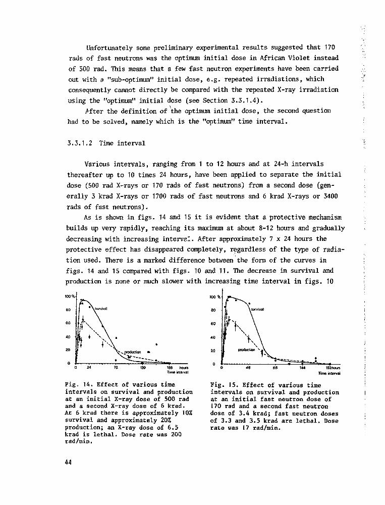
Unfortunately some preliminary experimental results suggested that 170
rads of fast neutrons was the optimum initial dose in African Violet instead
of 500 rad. This means that a few fast neutron experiments have been carried
out with a "sub-optimum" initial dose, e.g. repeated irradiations, which
consequently cannot directly be compared with the repeated X-ray irradiation
using the "optimum" initial dose (see Section 3.3.1.4).
.After the definition of the optimum initial dose, the second question
had to be solved, namely which is the "optimum" time interval.
3.3.1.2 Time interval
Various intervals, ranging from 1 to 12 hours and at 24-h intervals
thereafter up to 10 times 24 hours, have been applied to separate the initial
dose (500 rad X-rays or 170 rads of fast neutrons) from a second dose (gen-
erally 3 krad X-rays or 1700 rads of fast neutrons and 6 krad X-rays or 3400
rads of fast neutrons).
As is shown in figs. 14 and 15 it is evident that a protective mechanism
builds up very rapidly, reaching its maximum at about 8-12 hours and gradually
decreasing with increasing interval. After approximately 7 x 24 hours the
protective effect has disappeared completely, regardless of the type of radia-
tion used. There is a marked difference between the form of the curves in
figs. 14 and 15 compared with figs. 10 and 11. The decrease in survival and
production is none or much slower with increasing time interval in figs. 10
I100%
40
Fig, 14. Effect of various timeintervals on survival and productionat an initial X-ray dose of 500 radand a second X-ray dose of 6 krad.At 6 krad there is approximately 10%survival and approximately 20%production; an X-ray dose of 6.5krad is lethal. Dose rate was 200rad/min.
100%
60
40
20
Pig. 15. Effect of various timeintervals on survival and productionat an initial fast neutron dose of170 rad and a second fast neutrondose of 3.4 krad; fast neutron dosesof 3.3 and 3.5 krad are lethal. Doserate was 17 rad/min.
44

• 1
I and 11 than in figs. 14 and 15: after 72-96 hours both survival and production
j are below 10$ in contrast to the high %'s in figs. 10 and 11 (1001 survival
and approx. 871 production).
| This may be the result of a somewhat better protective capacity by the
I high(er) initial dose in figs. 10 and 11 (3000 rad) (see also Section 3.3.1.1)
| but probably is mainly to be attributed to the higher second doses used in
| figs. 14 and 15, which has tested the protection much more seriously.
j Figures 14 and 15 moreover indicate that after a relatively long interval
| (168 hours and •„ 3), the effect of two fractions (500 and 6000 rad) is equal to
j that of the total acute dose (6500 rad). Consequently, the effects of the two
fractions are completely cumulative. This is evident because the lines for
survival and production otherwise would have gone back to a point between the
lethal point and their respective base lines (for survival 10$ and for produc-
tion 20$). In this case fractionation does not result in a lower effect,
provided the right time interval is chosen. Although the system used may have
something to do with it (no cell division during the whole experiment and thus
no cell elimination) one wonders whether thp statement in the beginning of
Section 1.2, page 8 ("generally the biological effect of fractionation is
less ") would still hold true if experiments reported in the literature
had been carried out with more different and longer time intervals.
In relation to the repeated irradiations planned (Section 3,3.1.4), the
"optimum" time interval was defined as the shortest time needed for maximum
protection. The interval of 8 hours was therefore selected and permitted 60
repeated irradiations of the material with the optimum initial dose within a
20 day period. Intervals of 12 hours would have extended the experiments to
30 days which is too long for the separated leaves to remain in sealed plastic
bags without a considerable loss of vitality.
3.3.1.3 Extent of protection
To test the extent of the radio-induced protection a series of second
doses, the highest of which are lethal when given as an acute single dose,
were applied. As can be seen in Figure 16 even after an initial dose of 600
rad X-rays (or 500 rad fast neutrons) and a second dose of 9 krad X-rays (or
4.5 krad fast neutrons) there is a surviving fraction which corresponds with
an acute single dose approximately 3 krad X-rays or 1.5 krad fast neutrons
lower. In other words: a comparatively low initial dose of 500-600 rad induces
a protection equivalent to approximately 3 krad X-rays or 1.5 krad fast
45

100%
BO
60
40
20
0
\ \
without pre-irradiatton*
acuteNf
- — acute X
(both taken from
fig-slar>d2t
\ \
\ \ \ ^
vs. - ^
lOktad X6 kradNf
Fig. 16. Extent of protection induc-ed by an initial X-ray dose of 600rad (-•_) or an initial fast neutrondose of 500 rad (—A—), tested byapplying various second doses of X-rays or fast neutrons after an 8-hinterval. Dose rate was 200 rad/min(X-rays) and 17 rad/min {fastneutrons).
neutrons.
This protection is also demonstrated by an experiment in which an optimum
initial dose (500 rad X-rays) or the "sub-optimum" fast neutron dose of 170
rad preceeded a series of second doses, either fast neutrons or X-rays, after
an 8-h interval. Thus, an initial X-ray dose was followed either by a second
X-ray dose or by a second fast neutron dose. Similarly, initial doses of fast
neutrons were followed by a second dose of either fast neutrons or X-rays.
The data show (figs. 17 and 18) that the distance between the acute lines
and the other parallel lines is equal to approximately 3 krad X-rays or 1.5
krad fast neutrons. Looking at Fig. 17 another way we can see, as before, that
60 •
40
20
With-irradiatton
X-acute
Nf< acute
(both taken from
figs land2)
14 krad x
1 2 3 4 5 6 7 kradNf
Fig. I7. Effect on survival of the(optimum) initial dose (500 rad ofX-rays or 170 rad of fast neutrons)after various second doses of X-raysor fast neutrons. -A-, both initialand second doses of X-rays, ~k~.,initial dose of X-rays, second doseof fast neutrons, — • — , both initialand second doses of fast neutrons;— O — , initial dose of fast neutrons,second dose of X-rays. Dose rate was200 rad/min (X-rays) or 17 rad/min(fast neutrons).
20
krtdNf
Fig. J8. Effect on production of the(optimum) initial dose (500 rad ofX-rays or 170 rad of fast neutrons)after various second doses of X-raysor fast neutrons. -A-, both initialand second doses of X-rays; -A-,initial dose of X-rays, second doseof fast neutrons; — • — , both initialand second doses of fast neutrons;— 0 — , initial dose of fast neutrons,second dose of X-rays. Dose rate was200 rad/min (X-rays) or 17 rad/min(fast neutrons).
46

at 6 krad of an acute single dose there is only * 10% survival. When 6 krad is
the second dose, survival is raised to approx. 100$. For production (Fig. 18)
the figures are somewhat lower, except in the case of X-ray.* preceeding fast
neutrons where a pronounced stimulation can be noted. The origin of this, not
significant, stimulation is not known and relates to a similar phenomenon
which has been discussed in Section 3.3.1. There the question was raised
whether the second dose influences the repair of part of the effect of the
first dose. However, if the "X-Nf line" in Fig. 18 is drawn correctly, it
means more than the (even complete) repair of the small first dose: in addi-
tion a stimulation of one kind or another has been induced which results in a
production above the 1001 of the control.
In any case one important conclusion can he drawn from figs. 17 and 18,
namely that both X-rays and fast neutrons are equally effective in the induc-
tion of a comparatively equally large protection against a second dose of
radiation, either X-rays or fast neutrons.
3.3.1.4 Repeated i rradiat ion
To investigate whether a repeated irradiation with the optimum initial
dose, separated by optimum time intervals would result in an increased,
decreased or equal protection, 500 rad X-ray or 170 rad fast neutron doses
were repeatedly applied at 8-h intervals. During the treatment, the leaves,
sealed in plastic bags, were kept in the dark except when handled and
irradiated. The temperature was kept at approximately 20°C; the effect of
other temperatures has not been studied.
After 1, 5, 10, 15 etc. up to 60 repetitions a series of last doses (1.5, 3,
4.5, 6, 8, and 10 krad X-rays or 0.8, 1.7, 2.5, 3.4, 5 and 6.8 krads of fast
neutrons) were applied to test the protection. A decrease would mean that a
single pre-treatment yields the maximum possible protection value which dis-
appears in time, independent of subsequent irradiations. A steady level would
indicate that the protection is kept maximum after the single pre-irradiation
and stays that way until 60 repetitions, during which every irradiation com-
pensates for the decrease, which goes on continuously. An increase would in-
dicate that every irradiation adds some (the same amount?) protection to the
system, which is bound to reach a maximum when the decrease in protection,
induced by the first irradiations, sets in.
It can be seen from Fig. ]9 that the protection increases rapidly: a last
dose of 10 krad X-rays, being lethal after a single pre-irradiation f the "X-X
47

Survival
100%
80
60
40
20
020 30 50 60 » 500 rad X
Repetitions
0 0 *
80
60
40
20
n
without pie- irradic tion\
\
(taken \fromligiA
1»50O tadX-. , •;
i '
' ' , :
\ '; 'f -
Fig. 19. Effect on survival ofrepeated X irradiations of 500 radat 8-h intervals; various last dosesof X-rays were applied, namely 6krad ( ), 8 krad ( ) or 10 krad( ). Dose rate was 200 rad/min.
10 krao X
Fig. 20. Effect on survival ofrepeated X irradiations of 500 radat 8-h intervals; various last doseswer.- applied. Dose rate was 200 rad/min.
lines" in figs. 17 and 18) results in 1001 survival after 5 repetitions with
the optimum initial dose of 500 rad X-rays and stays that way until 20 repeti-
tions.
In Fig. 20, which is based on the same data as Fig. 19 but presented
differently, an impression about the extent of the protection can be obtained.
The distance between the "acute line" and the "one pre-treatment line" is
approx. 3-4 krad, whereas the distance between the latter and the following
one (5 repetitions) is probably about the same. The X-ray data, however, are
incomplete because the series of high last doses was not long enough.
With fast neutrons the range of last doses was sufficiently long to
determine the increasing protection with increasing number of repetitions. As
is shown in Fig. 21 maximum protection is obtained after 10 repetitions. (In
fact the "10 x 170 rad line" in Fig. 21 represents the lines for 10, 15 and
20 repetitions, which were almost identical. The exact optimum number of
repetitions will be defined and discussed later.) The distance between the
10x170 fad
Fig. 21. Effect on survival ofrepeated fast neutron irradiationsof 170 rad at 8-h intervals; variouslast doses were applied. The "500rad line" is taken from Fig. 16.Dose rate was J7 rad/min.
48

"10 x 170 rad line" and the acute line is approx. 3 krad, which is substan-tially more than after only one pre-treatment. But we must not forget that theinitial dose of 170 rad fast neutrons used for the repeated irradiations, hasinduced a smaller protection after only one pre-treatment as was the case withan optimum initial dose (see Fig. 16). It might therefore take a greater num-ber of repetitions to reach maximum protection when using a sub-optimum dose.This would explain the relatively small distance between the acute line andthe "1 x line", and the relatively large distance between the "1 x line" andthe "5 x line". The, again small, distance between the "5 x line" and the"10 x line" indicates a saturation point, where the protection has reached itsabsolute maximum value, following the type of repeated irradiation applied.(Other doses separated by shorter (?) time intervals might result in a higher(?) maximum value.)
Figures 22 and 23 (survival and production, respectively, after repeatedirradiations with 170 rad fast neutrons) show that the protection reaches amaximum after approx. 15 repetitions and then decreases fairly rapidly. A doseof 5 krad fast neutrons, for instance (solid line, closed circles), appliedafter 15 repetitions (with 170 rad fast neutrons) results in approx. 90%survival (Fig. 22) and 401 production (Fig. 23). After only a single pre-treatment these figures are just about 0%, both for a pre-irradiation with 170rad fast neutrons (Fig. 18, the Nf-Nf line) and for a pre-irradiation with theoptimum initial dose of 500 rad fast neutrons (Fig. 16).
From these figures it would appear that fast neutrons are less effective
Survival
100%
80
60
40
20
0
I 1j Ik 1
* É
\ ' \\
\
•—1
"-.4
20 30 40 60 «WOraBNf
Repetitions
Fig. 22. Effect on survival ofrepeated fast neutron irradiationsof 170 rad at 8-h intervals. Lastdoses of fast neutrons were 6.7 krad(—o—), 5 krad (--•—), 3.4 krad(—A—), 1.7 krad (—A—), 170 rad(—••—). Dose rate was 17 rad/min.
B0 60 xlTOradNf
Repetitions
Fig. 23. Effect on production ofrepeated fast neutron irradiationsof 170 rad at 8-h intervals. Lastdoses of fast neutrons were 6.7 krad(—o—), 5 krad (--•—), 3.4 krad(—A—), 1.7 krad (—A--), 170 rad(—D—). Dose rate was 17 rad/min.
49

in the induction of protection than X-rays. (5 krad fast neutrons as a last
dose never results in 100% survival (Fig. 22), whereas 10 krad X-rays as a last
dose reaches 1001 survival after 5 repetitions and stays that way until 20
repetitions (Fig. 19)). It seems to be confirmed by Fig. 12, where the maximum
of the 3.3 krad fast neutron dose stays below that of the (somewhat less
heavier) 6 krad X-ray dose.
The impression, from figs. 22 and 23 (fast neutrons) compared with figs.
19 and 20 (X-rays), that fast neutrons are less effective in the induction of
protection does not agree with the results presented in figs. 17 and 18. As has
been discussed in Section 3.3.1.3 the four lines (X-X, X-Nf, Nf-Nf and Nf-X)
are completely comparable with each other, especially when the "Nf-Nf-line"
(the lowest of the four) is replaced by the "Nf-line" of Fig. 16 (a similar
treatment, but with the optimum initial dose of 500 rad fast neutrons). This
line is closer to the average of the three others and therefore better fits the
conclusion in Section 3.3.1.3 that neutrons are equally effective compared with
X-rays in inducing protection.
The impression, after repeated irradiation, that neutrons are less effect-
ive is probably the result of the use of a sub-optimum (initial) dose of 170
rad which becomes more apparent after repeated irradiation than after a single
pre-treatment.
The observation that maximum protection is obtained after 10-15 repeti-
tions (figs. 22 and 23) seems logical. The effect of the first irradiation with
the initial dose starts to break down substantially after 2-3 days, as can be
seen in Fig. 15. This rapid breakdown of the protection, induced by the first
dose(s), sets in after 6-9 repetitions (2-3 days) and apparently is hardly
compensated by the (probably decreasing) additions to the protection by the
subsequent doses. The final result is a maximum that is reached a restricted
number of repetitions later. From thereon the protective value slowly decreases
as the result of the increasing extent of the unrepaired damage or of the
decreasing protective value, or of a combination of the two. The foregoing is
based upon results with fast neutrons and probably also holds true for X-rays.
The last doses with X-rays were, unfortunately, not high enough (Fig. 19) to
prove this assumption.
It would have been an interesting experiment to irradiate repeatedly, up
to 15-20 times for instance, and then test the extent of the protection by the
irradiation uith high last doses at the same periods as used in my experiments.
One wonders whether the lines would decrease earlier (faster) or later (slower).
50

Earlier and/or faster would indicate that the protection decreases earlier ormore rapidly because the lacking repetitions do not add protection anymore.Later and/or slower would indicate that the protection reaches a stable valueby a given number of repetitions and stays that way because it is not anymoredamaged by subsequent irradiations, whereas also no unrepairable damage isadded to the system anymore.
3.3.1.5 Conclus ive remarks
The data presented above agree in part with the data in the literature,
using animals or animal (mammalian) tissues, listed in Section 3.3.1. The
optimum initial dose mentioned there is approx. 15? of the LD™ and the extent
of the protection induced (increase in tolerance by ca. 1.7) seems to fit
fairly well with the data presented here. In contradiction, however, is the
fact that the protection is reported to stay active in animals over an extreme-
ly long period whereas in African Violet most of the protective effect has dis-
appeared after 5 days in the case of a single pre-treatment. Also in contra-
diction is the fact that in animals a single pre-irradiation induces a better
protection than a repeated one. The data presented here clearly demonstrate a
maximum protection after a 10-15 fold repetition of the pre-irradiation dose.
The comparison of African Violet with animals or animal tissues is not
justified. Animal cells react differently from plant cells. The partly agreeing
results, reported in the literature, are, however, striking enough to justify
further investigations with animals or animal (mammalian) tissue, in particular
with relation to radiotherapy. Some scientists, however, are not convinced by
the literature reviews mentioned in Section 3.3.1, in my opinion as the result
of the incompleteness of many split dose experiments, in which generally a very
restricted number of doses are separated by a sometimes restricted and arb-
itrary number of different time intervals. As I have seen with my material, it
is of utmost importance to choose the right first and subsequent doses and the
right series of time intervals to demonstrate the effects of the first dose. A
wrong choice would have given confusing and unexplicable results.
In connection with the results of Krokowski (1968), who reported a trans-
missable radio-induced protection (Section 3.3.1) I tried to investigate
whether this phenomenon also takes place in African Violet. Grafting (irradiat-
ed) leaves on an unirradiated plant turned out to be impossible, so the experi-
ments were discontinued. It would, however, be interesting to carry out a
number of experiments to further investigate this phenomenon such as to irrad-
S1

iate one half of a complete plant with 500 rad for instance and to check theradioresistance of leaves of the unirradiated part, after given time intervals.Another method would be to irradiate the leaf disc of a detached leaf and totest the radioresistance of the leaf petiole by irradiating it with a series ofhigh doses after a given time interval. Both experiments may bring to light theexistence of a transportable protection within a plant (part). If so, one couldtry to transfer the component(s). responsible by pulping irradiated leaves andextracting the juice with a subsequent uptake of the extract by detached un-treated leaves.
In plants, as has been discussed in Section 3.3.1, very few dose fraction-ation studies have been reported. Just before the final version of this studyHorsley and Laszlo published results of dose fractionation experiments withalgae (1971). They reported an unexpected additional recovery after a first X-ray dose, using a synchronous cell culture of Oedogoniwi. A sharp rise insurvival was obtained in a split dose experiment (first dose 1200 rad X-rays,second dose 4200 rad X-rays) after a 2-h interval, reaching a maximum between6-8 hours before falling steeply. The explanation suggested was an efficientrecovery mechanism, set into play by the first dose of radiation.
3.3.2 Genetic effects
As has been said before (Section 2.3) the third parameter, expressed as
mutation frequency (number of mutants per 100 plantlets) or as the number of
mutants per 100 irradiated leaves, is the least reliable. The reason being the
long time between the irradiation and the observation of the mutants (9-10
months), during which many things may happen to part of the experiment. Another
reason is that, especially after heavy treatments, it is difficult to decide
whether a plantlet grows slowly or abnormally because of either genetic or
physiological disturbances. Moreover, the small number of plantlets obtained
after heavy treatments, results in a poor significance of the figures for
mutation frequency, reflected in the far from ideal form of the curves in figs.
24, 27 and 29. Fortunately, most experiments included a range of second doses
(in the case of a single pre-treatment) or a range of last doses (in the case
of repeated irradiations). The procedure to obtain the best possible results,
using this parameter, was as follows: to eliminate the defects in the data of
the individual lines the average of all second or last doses was calculated.
Every single line was then compared with the average line and the ones that
fitted the general tendency best was selected for the calculations to determine
52

the number of mutants per 100 irradiated leaves, in which all three parameters
were discounted. The cpnversion from one figure to the other, such as Fig. 24
into parts of figs. 25 and 26, thus gives an impression of the effect of
fractionation upon the genetic parameters. No absolute value should therefore
be attached to the lpies. On the other hand, the persistent tendency obtained,
makes it plausible that the truth has not been violated too much.
In Fig. 24 the/effect of initial dose of either X-rays or fast neutrons
upon the mutation frequency is shown. As the RBE, for survival and production,
was 1 (figs. 12 ana 13), I have expressed the mutation frequency on the same
basis. The abscissa of the graph therefore gives the initial dose for either
X-rays or fast neutrons.
With increasing initial dose, an increasing protection against genetic
effect builds up, reaching a maximum for X-rays around 1.5 krad and for fast
neutrons between 0.5 and 1 krad, reflected in minimum mutation frequencies.
(The first minima, at an initial dose of approx. 150 rad, have been neglected
because the low survival and production in that case result in a very poor
significance of the mutation frequency among the few plantlets produced.) With
still increasing dose the protection seems to stay relatively unchanged for X-
rays, but decreases rapidly for fast neutrons.
With still increasing initial dose the protection stays relatively un-
changed for X-rays.
When the results in figs. 12 and 13 are compared with those in Fig. 24
maximum protection is found around an initial dose of 500 rad, resulting in
maximum survival and production. For mutation frequency maximum protection,
resulting in minimum mutation frequency, seems to be shifted towards a higher
initial dose, especially for X-rays.
Furthermore the more drastic reaction with fast neutrons rather than X-
Mut.%
00%
80
SO
40
20
'v
•2.5 krad Nf
; A 7 krad Nf
•^i?~-~-~. •——~ï*wi%
05 1 1.5 2 25 3
Httf don
Fig. 24. Effect of various initialX-ray or fast neutron doses onmutation frequency. Second dose was6 krad X-rays or 1.7 and 2.5 kradfast neutrons respectively; 8-hintervals between first and seconddose were applied. The scale ofinitial doses applies to both X-raysand fast neutrons on the basis of anRBE is 1. Dose rate was 200 rad/min(X-rays) or 17 rad/min (fastneutrons). (The highest point of the"2.5 krad Nf-line" is very question-able.)
53

Mutants per 100 leaves
500
Fig. 25. Number of mutants per 100leaves after various initial andsecond doses of X-rays. Dose ratewas 200 rad/min. The intervalbetween first and second dose was8 hours.
Mutants per 100 leaves
500
400
300
200
WO
0.5 1 15 krad Nfmid dose
Fig. 26. Number of mutants per 100leaves after various initial andsecond doses of fast neutrons. Doserate was 17 rad/min. The intervalbetween first and second dose was8 hours.
rays is striking in all three diagrams (representing the three parameters
used) so that identical or almost identical (biochemical) reactions may be the
origin of the correlation between the behaviour of the three parameters.
For both X-rays and fast neutrons the number of mutants per 100 leaves
was calculated as mentioned before. The result is shown in figs. 25 and 26,
which demonstrate, that 500 rad X-rays, in combination with 6 krad X-rays as a
second dose and after an 8-h interval, produces the maximum number of mutants
per 100 leaves, namely 415, being 1631 of the corresponding optimum acute dose
of 3 krad X-rays. An initial dose of 250 rad fast neutrons, in combination
with a second dose of 1.7 krad fast neutrons, produces the highest number cf
mutants per 100 leaves ever obtained, namely 505, which is 146$ of the corres-
ponding optimum acute dose of 1.5 krad fast neutrons. (With the number of
mutants per 100 leaves as parameter, the RBE values for fast neutrons would
again be approx. 2, and not 1, as for survival and production as parameter.)
The effect of time interval upon mutation frequency, as is shown in Fig.
27, behaves in a similar way: a rapidly increasing protection with still
larger intervals, which was already demonstrated for survival and production
in figs. 14 and 15. Consequently a maximum number of mutants is to be expected
just before survival and production reaches a maximum. This is demonstrated in
Fig. 28 where 500 rad X-rays, in combination with 6 krad X-rays, produced the
maximum number of mutants per 100 leaves after a 10-h interval. The absolute
number, 240, however, is low in comparison with the corresponding optimum
acute dose of 3 krad (270) as well as compared with the initial dose experi-
ments, described just before (415). Since the whole 6 krad line is lower than
54

192 240 hoursTime interval
Fig. 27. Effect of various timeintervals on mutation frequency.Initial dose was 500 rad X-rays or170 rad fast neutrons; second dosewas 6 krad X-rays or 3.4 krad fastneutrons respectively. Dose ratewas 200 rad/min (X-rays) or 17 rad/min (fast neutrons).
Mutants per 100 leaves
500
400
240 hours
Time interval
Fig. 28. Effect of various timeintervals on number of mutants per100 leaves. Initial dose was 500 radX-rays or 170 rad fast neutrons;second dose was 6 krad X-rays or 3.4krad fast neutrons, respectively.Dose rate was 200 rad/min (X-rays)or 17 rad/min (fast neutrons).
expected, the only possible explanation is that this second dose had a more
damaging effect than usual, for reasons unknown.
The conclusion, namely that the optimum time interval approximately lies
around 10 hours, however, is valid.
With fast neutrons a similar picture was obtained: a high number of
mutants between 8 and 48 hours with a maximum at a time interval of 24 hours.
An initial dose of 170 rad, in combination with a second dose of 3.4 krad fast
neutrons, produced 355 mutants per 100 leaves, being 142% in comparison with
the corresponding acute dose of 1.5 krad fast neutrons.
The effect of repeated irradiation with the optimum initial dose, separat-
ed by the optimum time interval, is shown in Fig. 29 for X-rays. Only the high
last dose showed that the protection against genetic damage increased with
number of repetitions, reaching a maximum at 5-20 times, reflected as low muta-
Mut.%
100%
80
60
40
20 '
O krad X
. .53 krad X -'• ».—r
X> 20 40 x 600 rad X
Repetitions
Fig. 29. Effect on mutation frequencyof repeated X irradiation with 500rad at 8-h intervals. Second dose was10 krad and 5.3 krad (= average ofall second doses). Dose rate was 200rad/min.
55

tion frequencies. Lower last doses did not respond to number of repetitions,
as is demonstrated by the 5.3 krad X line, being the average of all last doses
applied. Fast neutrons responded exactly in the same way.
Since survival as well as production clearly reacted upon increasing
number of repetitions (figs. 20, 21, 22 and 23), it is a priori to be expected
that the number of mutants per 100 leaves will increase with increasing number
of repetitions. This can be seen in figs. 30 and 31, which demonstrate an
optimum number of mutants per 100 leaves around 25-30 repetitions. With X-rays,
the highest number was obtained after 30 repetitions, separated by 8-h inter-
vals, and applying a last dose of 4.5 krad X-rays, namely 435 mutants per 100
leaves. This is 1301 in comparison with the corresponding optimum acute dose
of 3 krad (Table 9). With fast neutrons similar results were obtained (Fig.
31): the highest number, which happened to be the same as with X-rays, namely
435, was obtained after 25 repetitions with 170 rad and a last dose of 1.7
krad. In comparison with the corresponding optimum acute dose of 1.5 krad the
highest number amounted to 1821 (see Table 9).
The "170 rad Nf line" of Fig. 31 seems to be an indication that the
gradually decreasing production (Fig. 23) with increasing number of repetitions
is compensated by an increasing mutation frequency, resulting in a more or less
steady number of mutants per 100 leaves of that line (the survival stays 1001
throughout the whole treatment: Fig. 22).
By comparing Fig. 30 with Fig. 31 it is obvious that fast neutrons,
despite their similar action to X-rays upon the material, do have a somewhat
more direct effect, demonstrated by the almost horizontal lines of Fig. 30
Mutants per too leaves
500
400-4.5 krad
Mutants per WO leaves
500
400
300
200
100
60
170 raj
\ X"*jSkrad*~^taad V '-,-<tf.fcra|
x5O0radRepetitions
SO 60 x170 rad Nf
Repetitions
Fig. 30. Effect on number ofmutants per 100 leaves of repeatedX-irradiation with 500 rad at 8-hintervals; various last doses wereapplied. Dose rate was 200 rad/min.
Fig. 31. Effect on number of mutantsper 100 leaves of repeated fastneutron irradiation with 170 rad at8-h intervals; various last doseswere applied. Dose rate was 17 rad/min.
56

Table 9. Number of mutants per 100 leaves (n) and percentage of dwarfs aftervarious X-ray or fast neutron treatments1.
Treatment
X-rays
ACUTE (200 rad/min)3 krad (= optimum acute dose)
n of the n of thetreatment corresponding(n ) optimum acute
dose (n )
n.ln
°
330 330
Fast neutrons
ACUTE (17 rad/min)1.5 krad (= optimum acute dose) 270 270
100
100
dwarfs
55
CHRONIC5.3 rad/min;7.3 rad/min;
FRACTIONATED
Initial dose500 rad-8 h-6
Repeated30 x 500 rad;
10,9 krad13.2 krad
krad
4.5 krad
275255
415
435
220220
255
335
125116
163
130
5853
57
59
61
CHRONIC0.17 rad/min; 6.4 krad8 rad/min; 1.2 krad57 rad/min; 1 krad
FRACTIONATED
Initial dose250 rad-8 h-1.7 krad
Interval170 rad-24 h-3.4 krad
Repeated25 x 170 rad; 1.7 krad
1. Compilation of data from
333317335
505
355
435
225225225
345
250
238
various experiments.
148141149
146
142
182
455481
52
58
56
(after approx. 20 repetitions with 500 rad X-rays) and the rapidly decreasing
lines of Fig. 31, after reaching the optimum. But here, again, we have to
remember that the fast neutron repeated irradiations were carried out with the
sub-optimum dose of 170 rad.
The foregoing was a description of the experimental results which, though
interesting, do not elucidate much of the origin of the phenomena. What actual-
ly takes place in plants is not known, since no or very scanty and incomplete
investigations in other angiosperms have been reported. The comparison with the
more extensive and more complete radiobiological investigations in bacteria and
57

animals or animal (mammalian) tissues is risky and probably not justified, ashas been discussed before (Section 3.3.1.5).
3.3.3 Fraatioal implications
Although a clear-cut protective effect of fractionated irradiation isdemonstrated, the practical results, measured as number of mutants per 100leaves in percentage of the corresponding optimum acute dose, are somewhatdisappointing. The number of mutants can be increased, in the case of X-rays,to 130% by applying 30 repetitions of 500 rad X-rays, separated by 8-h inter-vals and a last dose of 4.5 trad and to 1631 by using an initial dose of 500rad, 8 hours later followed by 6 krad. However, this high figure is somewhatquestionable when compared with a similar treatment, which resulted in only90S (see discussion in Section 3.3.2 under time interval). More important isthat the mutation spectrum, expressed as percentage of dwarfs (last column,Table 9), turned out to be unchanged in comparison to acute treatments.
The same holds true for fast neutrons which, compared with the optimumacute dose of 1.5 krad at 17 rad/min, resulted in 1821 (25 repetitions of 170rad, followed by 1.7 krad), 1421 (170 rad - 24 hours - 3.4 krad) and 1461(250 rad - 8 hours - 1.7 krad). However, when these figures are expressed inthe optimum 1 krad acute dose administered with a higher dose rate (57 rad/min), giving 149% as compared to the optimum 1.5 krad acute dose at 17 rad/min, the situation becomes different. Then only the repeated irradiation showsa surplus in number of mutants per 100 leaves, namely 122% (182/149 x 100).The mutation spectrum, if the % of dwarfs is taken as a standard for it (dis-cussed in Section 3.1.2), is unchanged (last column, Table 9) and completelycomparable with that after X-rays (Table 7, page 32).
However, the missing links in these considerations are fractionatedirradiations with the optimum initial dose of 500 rad fast neutrons. A singlepre-treatment or a repeated irradiation with that dose might have rendered anequally larger number of mutants per 100 leaves (even when expressed in theabsolute optimum acute dose of 1 krad at 57 rad/min) as has now been found,using 170 rad as initial dose (and compared to 1.5 krad at 17 rad/min). Inother words, it is not justified to base a decision about the practical valueof a given fast neutron treatment at a dose rate of 17 rad/min on the compar-ison with the absolute optimum acute dose of 1 krad at 57 rad/min.
Therefore it could be worthwhile to consider a pre-treatment, with theoptimum initial X-ray dose, before a heavy second dose. A repeated irradia-
58

tion, in the case of fast neutrons, seems promising, but since it is too
costly and time consuming, it has to be rejected as impractical.
59

4 Comparison of acute, chronic and fractionatedirradiations in relation to (commercial) plant breeding
As has been discussed in the introduction (Section 1.1), one of thestarting-points of my investigations was the expectation that repair of physio-logical and genetical effects might be the result of partly different processes.Consequently it was anticipated that, by controlling the various factors ofinfluence on these processes, it would be possible to improve the efficiency ofthe mutagenic treatment in such a way, that more and possibly also more usefulmutants were obtained from a given number of irradiated leaves.
The general impression from the data presented in the foregoing chaptersis, that this expectation has not been realized. The difference between acuteand chronic irradiations (Table 9) are slight and not interesting in the caseof X-rays, and larger, but sometimes impractical, in the case of fast neutrons(6.4 krad at a dose rate-of 0.17 rad/min requires approx. 26 full days irradia-tion!; 1.2 krad at a dose rate of 8 rad/min, however, is applicable since itrequires only 2.5 hours).
The previous statement at first appears to be in contrast with most of thecurves, which show an optimum value for the parameter studied. However, onfurther consideration, the optima in figs. 25, 26, 28, 30 and 31, whichrepresent the number of mutants per 100 leaves, are the result of the multi-plication of two physiological parameters (survival and production) and onegenetic parameter (mutation frequency). This can be seen in the last but onecolumn of Table 5 (page 28), combined with the 4th column: the number ofmutants per 100 leaves is A x B x D x 15.10" , in which A represents survival,B represents production and D represents mutation frequency.
The reaction of either survival and production, which show a high degreeof correlation with each other, consists of a generally drastic increasingpercentage after the gradual increase of certain factors, such as initial dose,time interval and number of repetitions. Reversely, mutation frequency shows adecreasing and less pronounced percentage, following the gradual increase ofthe factors mentioned before. Consequently, an optimum is the result, when thethree parameters are combined, as is the case when the result of a given treat-ment is expressed as the number of mutants per 100 irradiated leaves.
60

Because of the difference in intensity upon changing factors, such an
optimum would also be obtained when only one physiological parameter (or the
square root of the product of survival and production) is multiplied by the
mutation frequency. Moreover, one gets the impression that the mutation
frequency does not only react less intensively upon varying factors but also
slower, when compared with survival and production. In Fig. 24 for instance
the minimum and maximum mutation frequency, after various initial doses, varies
from approx. 20$ to approx. 60-801 for fast neutrons (the high figures for
mutation frequency are not significant) whereas it varies from approx. 351 to
701 for X-rays. This is much less in comparison with the magnitude observed
when survival and production are used as parameter (figs. 12 and 13: survival
ranges from 20-100% (X-rays) and from 0-approx. 701 (fast neutrons), whereas
production ranges from approx. 10-80% (X-rays) and 0-60% (fast neutrons)).
In the same Fig. 24 the minimum mutation frequency lies around an initial
dose of 0.8 krad for fast neutrons and 1.5 krad for X-rays (the minima after an
initial dose of approx. 150 rad have been neglected: see Section 3.3.2). This
is in contrast to the optimum initial dose of 500 rad for both X-rays and fast
neutrons in the case of survival and production (figs. 12 and 13). This in-
dicates, that maximum protection against genetic damage is obtained after a
higher initial dose as compared with the protection against damage of a more
physiological nature.
This causes the optimum which is obtained by the multiplication of
survival, production and mutation frequency to be somewhat diffuse and to vary
with variations in treatment and circumstances. But the fact remains, that the
adventitious bud technique, in which survival, production and mutation
frequency are logical and inseparable factors, does result in maximum number
of mutants per given number of leaves following a certain treatment.
For commercial mutation breeding purposes a few questions arise, namely:
1. which treatment is to be preferred taking into consideration the number and
quality of the mutants,
2. which treatment should be selected, taking into account the availability of
leaves in connection with the availability of greenhouse space (with other
words: a very heavy treatment, requiring many leaves and less greenhouse space
to produce a given number of mutants or a light treatment, requiring less
leaves but more space to obtain a similar number of mutants, based on the
assumption that the % of dwarfs is a measure for the quality of the mutation
spectrum), and
3. which irradiation facility is to be preferred, taking into account the
61

availability as well as economical aspects.
As is shown in Table 9, page 57, and as has been discussed in variousChapters before, the only practical treatment with X-rays is either an acutedose of 3 krad or a heavy dose of approx. 6 krad preceeded by an initial doseof 500 rad and an 8-h interval between first and second dose. The mutationspectrum seems to be alike in all cases.
Using fast neutrons, a semi-acute irradiation with 8 rad/min and a totaldose of 1.2 krad seems to give the best results; an acute irradiation with57 rad/min unfortunately is accompanied by an unfavourable mutation spectrum(8U dwarfs, last column Table 9). Two other treatments give similar results(the fractionated treatments in Table 9) and might also be considered. Allother treatments, however, are impractical or too costly (repeated irradiation,for instance).
The choice between a light or heavy dose depends on the availability of
leaves to be irradiated, and greenhouse space. Since the latter is usually the
limiting factor a heavy dose seems to be the best choice. When both are not
limited, a light dose is to be preferred, mainly with respect to a possible
disastrous effect of unforeseen unfavourable (climate) conditions as well as
the possibility of an unfavourable effect upon the mutation spectrum, both
discussed before. Moreover, the chance that a favourable mutation is accom-
panied by one or more unfavourable ones, is reduced.
62

5 Comparison of X-rays and fast neutrons
The qualitative difference in the underlying work between X-rays and fast
neutrons has been shown to be extremely small, although the reactions of the
material after neutrons were not as clear-cut as with X-rays, which point
towards a more direct effect of fast neutrons. The difference between both
radiation types is reflected by the fact that neither the dose rate, nor dose
fractionation has an effect upon the mutation spectrum when X-rays are used;
fast neutrons, however, show an unfavourable shift of the spectrum when high
dose rates are applied (Table 8, page 37).
When, however, the effects of X-rays and fast neutrons (upon survival,
production and mutation frequency) are compared on a rad basis, fast neutrons
are sometimes more efficient, which can be expressed in terms of an RBE
(relative biological effectiveness). For acute irradiations, using 17 rad/min,
fast neutrons have an RBE = 2, compared with X-rays (figs. 1, 2 and 3). When a
higher fast neutron dose rate is applied, namely 57 rad/min, the RBE value is
3 (Fig. 9). In fractionation experiments a more complicated picture was observ-
ed. For the induction of a protective effect the RBE = 1, when survival and
production are used as parameters (Fig. 6). When the number of mutants is the
end-point, the RBE = 2 for initial dose, and 1 for number of repetitions.
A striking equality between the two radiation types is shown in figs. 17
and 18 where it is shown that it does not make much difference, upon survival
and production, whether X-rays or fast neutrons are used, either as initial
dose or as second dose. This is an unexpected result, since the completely
different way of energy dissipation in (biological) material of X-rays (sparse-
ly ionizing) and fast neutrons (densely ionizing mainly through recoil protons)
a priori presupposes a more pronounced difference in effect. The only explana-
tion is, that despite the different ways of energy dissipation, the changes in
the end-points measured are the result of a series of indirect reactions, which
largely follow the same pathways.
Consequently it is hard to say which of the two radiation-types is to be
preferred in our material. The heterozygosity of the Saintpaulia cultivar used
is apparently so large, that an increase in mutation frequency (by using
63

various treatments or by using neutrons) is limited, probably because cells,
carrying too many genetic changes disappear. This may explain the almost
constant percentage dwarf mutants (Table 9) in spite of the different treat-
ments and the different radiation methods used. (The only exception is the
high % of dwarfs after a fast neutron irradiation with 57 rad/min (Table 8,
page 37 and Table 9, page 57).
In species, however, which have a much lower degree of heterozygosity, it
might pay to investigate whether or not fast neutrons (with a high dose rate)
are superior to X-rays.
64

Summary
Detached leaves of African Violet, Saintpaulia ian<mtha H. Wendl. cv.
Utrecht, have been exposed to acute, chronic or fractionated irradiations with
X-rays or fast neutrons to define the optimum treatment for commercial mutation
breeding. The effects of the various treatments were expressed as:
1. survival of the irradiated leaves,
2. production of adventitious plantlets at the base of the petiole and
3. mutation frequency and number of mutants per 100 leaves.
The plantlets formed on the petiole of Saintpaulia leaves have two
characteristics which make them especially useful for such a study. First, one
can be certain that throughout the time of irradiation and treatment all cells
likely to be involved in the production of adventitious plantlets are in a non-
dividing state. Second, the apex of each adventitious plantlet can be traced
back ultimately to cells derived from a single epidermal cell. The evidence
for this is that all mutants (with a few exceptions discussed in this report)
are complete: sectors are virtually never found.
The results show a striking dose rate effect on survival, production and
mutation frequency. At a rate of 2 rad/min the LD5Q for survival is obtained
after approx. 100 krad X-rays as compared to approx. 5 krad X-rays at a rate
of 200 rad/min. The critical dose rate, at which there is a maximum effect
from a change in dose rate proved to be approx. 7 rad/min fX-rays).
Dose fractionation experiments (experiments in which more than one
irradiation is made, with the irradiations separated by a time interval) have
demonstrated, that a relatively low initial dose induces a mechanism which
protects the leaves against part of the effect of one or more subsequent radia-
tion doses. The optimum initial dose for maximum protection was found to be
500 rad of either X-rays or fast neutrons. The optimum interval, after which
the protection has reached a maximum, was 8-12 hours, depending on the size of
the initial dose and also on the size of the subsequent dose(s). The extent of
the protection was shown to be equivalent to 3-4 krad X-rays or 1.5-2 krad
fast neutrons, after a single pre-treatment with the optimum initial dose. The
protection was approx. double that amount after 10-15 repeated treatments with
65

the optimum initial dose, separated by 8-h intervals.There is another way of considering the magnitude of the protection
afforded by the initial dose. Normally a dose of 6.5 krad of X-rays is lethal.After one initial treatment with 500 rad (of X-rays) one gets approx. 1001survival (see the 6 krad X line in Fig. 12), whereas 251 sur/ival is obtainedwith a second dose as high as 10 krad. After 5 repeated irradiations with thesame initial dose, separated by 8-h intervals, 1003 survival is obtained aftera last dose of 10 krad.
Generally, survival and production as well as number of mutants reactedsimilarly upon different types of treatments. Since the first two parameters,however, reacted much more intensively upon variation of various factors,compared with the third parameter, mutation frequency, it was possible tocalculate and define optimum treatments.
On the basis of number of mutants per 100 irradiated leaves, as a percent-age of the corresponding optimum acute dose (3 krad X-rays or 1.5 krad fastneutrons) it was shown that a higher number of mutants can be obtained withcertain dose rate treatments or fractionated irradiations compared with anoptimum acute irradiation.
Examples are a pre-treatment with 500 rad and a second dose of 6 krad,separated by an 8-h interval in the case of X-rays or a similar treatment withfast neutrons (Table 9). Other treatments are impractical or too expensivebecause of prolonged irradiations.
The difference between X-rays and fast neutrons (average energy of approx.1.7 MeV) has shown to be surprisingly small and one can hardly find a reasonto prefer one type of radiation to the other.
The results described in this report are valid only for SaintpauHa,however. In other crops, which might be less heterozygous and thus produce jfewer mutants, an increase in number of mutants or a shift in the mutation !
spectrum may be more significant.
The significance of the adventitious bud technique for mutation breedingin general has been discussed especially with regard to the role of diplonticselection. Reasons are presented to support the belief that there is much lessdiplontic selection with the adventitious bud technique than in normal multi-cellular apices.
66

Acknowledgements
Leaves of African Violet were first irradiated late in 1960 and have
continued sine* on an ever increasing scale. Many persons assisted, so that it
is hard to recollect everybody's true contribution. Nevertheless I express my
sincere gratitude to:
Seymour Shapiro, for introducing me to the field of mutagenesis and radio-
biology and for his continued interest.
Dick de Zeeuw, for his confidence in my work and, above all, in giving me
so much freedom of action.
Professor Sneep, for the many inspiring and constructive discussions.
Many of my colleagues, who patiently listened for years to Saintpaulia
stories over coffee, lunch and tea-breaks and on many other occasions and very
many other ITAL staff members: my technicians Anneke, Heleen, Marian and Emmy;
the greenhouse attendants Leo, Adri, Wim and Hans; all the reactor staff, who
at times worked night and day for long periods on some complicated (fast)
neutron or X-ray experiments; the typists and during the final stage Hans in
particular, who typed and retyped texts; Jan for the drawings; Menno for the
many fine pictures; and many others, who in one way or another have contributed
to the final result.
Pudoc, for helping and advising with form, content, style and English.
And last but not least my wife and children, who understandingly accepted
my sometimes poor behaviour as husband and father during the times I was think-
ing and dreaming of her majesty Saintpaulia and her adventitious buds.
67

References
Ainsworth, E.J. & G.F. Leong, 1966. Recovery from radiation injury in dogs asevaluated by the split-dose technique. Radiat. Res. 29: 131-142.
Amato, F. d1, 1972. Morphogenetic aspects of the development of meristems inseed embryos. In: Dynamics of meristem cell populations; Plenum Press,N. York (in press).
Ancel, S., 1928. L'effet du fractionnement des doses de rayons X sur desgraines germinées. C.r. Séanc. Soc. Biol. 98: 223-224.
Anonymus, 1963. Irradiation facilities of the Association Euratom-ITAL 1: X-ray machine.
Anonymus, 1967. Neutron irradiation of seeds, IAEA techn. Rep. 76.Anonymus, 1968. Dose rate in mammalian radiation biology. U.S.A. EC-DTI Conf.Anonymus, 1970. Manual on mutation breeding. FAO/IAEA techn. Rep. Ser. 119.
Vienna.Bacchetti, S., 1965. Recovery from sublethal X-ray damage in surviving yeast
cells. Radiat. Res. 25: 103-114.Bacchetti, S., R. Elli, R. Falchetti & F. Mauro, 1965. Recovery from X-ray
damage in yeast cells. Ann. Meeting Eur. Soc. Radiat. Biol., Utrecht,The Netherlands.
Bacq, Z.M. & P. Alexander, 1961. Fundamentals of radiobiology. Pergamon Press,Oxford, London, New York, Paris.
Balkema, G.H., 1971. Chimerism and diplontic selection. A.A. Balkema, Rotter-dam-Kaapstad.
Bergann, F., 1966. The relative instability of chimerical clones - the basisfor further breeding. Gatersleben Symp. on induced mutations and theirutilization: 287-301.
Bergann, F., 1967. Mutations-ChimSren: Rohmaterial ziichterischer Weiterbehand-lung. Umschau 67: 791-797.
Bewley, D.K., J.F. Fowler, R.L. Morgan, J.A. Silvester, B.A. Turner & R.H.Thomlinson, 1963. Experiments on the skin of pigs with fast neutrons and8 MeV X-rays, including some effects of dose fractionation. Br. J. Radiol.36: 107-115.
Bewley, D.K., S.B. Field, R.L. Morgan, B.C. Page & C.J. Parnell, 1967. Theresponse of pigskin to fractionated treatments with fast neutrons and X-rays. Br. J. Radiol. 40: 765-770.
Bhojwani, S.S. & B.M. Johri, 1970. Cytokinin-induced shoot bud differentiationin mature endosperm of Saupvula pulverulenta. Z. Pfl. Physiol. 63: 269-275.
Bottino, P.J. & A.H. Sparrow, 1970. The effects of exposure time and rate onthe survival and yield of lettuce, barley and wheat. In: (Bensen &Sparrow, ed.); Survival of food crops and livestock in the event ofnuclear war: 306-324. Upton, N.Y.
Bowen, H.J.M., !9S5. Mutations in horticultural chrysanthemums. In: The use ofinduced mutations in plant breeding. Rep. FAO/IAEA techn. Meeting Rome,1964: 695-700. Pergamon Press, Oxford.
Braun, H., 1957. Experimentelle Untersuchungen iiber die Wirkung von Röntgen-ganzbestrahlungen auf den tierischen Organismus bei nachfolgender Bestrah-
68

lung mit Letaldosen. Oncologia 10: 272.Braun, H., 1962. Über die Steigerung der Leistungsfahigkeit der Ratten nach
Ganzkörperbestrahlung, geprüft ira Schwimmversuch. Strahlungstherapie 119:462.
Briggs, R.W., 1968. Effects of post-treatment hydration and dehydration ofethyl methane sulphonate-treated maize seeds. In: Mutations in plantbreeding 2: 47-57. IAEA, Vienna.
Broerse, J.J., 1966. Effects of energy dissipation by mono-energetic neutronsin mammalian cells and tissues. Radiobiologisch Instituut TNO, Rijswijk.
Broertjes, C , 1965. The organization and co-ordination of mutation breedingresearch in the Netherlands. Meded. LaadbHogesch. Gent 30: 907-975.
Broertjes, C , 1968a. Mutatieveredeling bij land- en tuinbouwgewassen. Land-bouwk. Tijdschr. 80: 162-168.
Broertjes, C , 1968b. Dose-rate effects in Saintpaulia. In: Mutations in plantbreeding 2: 63-71. IAEA, Vienna.
Broertjes, C., 1969. Mutation breeding of Streptoaappus. Euphytica 18: 333-339.Broertjes, C , 1970. Dose fractionation studies and radiation-induced protec-
tion phenomena in African Violet. In: (Bensen & Sparrow, ed.); Survival offood crops and livestock in the event of nuclear war: 325-342. Upton, N.Y.
Broertjes, C , 1971. Possibilities of mutation breeding in ornamentals. In:Eucarpia meeting on ornamentals: 7-15, Wageningen.
Broertjes, C , 1972a. Improvement of vegetatively propagated plants by ionizingradiation. In: Induced mutations and plant improvement: 293-299. IAEA,Vienna.
Broertjes, C , 1972b. Mutation breeding of Aohimenes. Euphytica 21: 48-63.Broertjes, C. & H.Y. Alkema, 1970. Mutation breeding of flowerbulbs. In: First
int. Symp. on flowerbulbs 2: 407-411, Noordwijk/Lisse.Broertjes, C , B. Haccius & S. Weidlich, 1968. Adventitious bud formation on
isolated leaves and its significance for mutation breeding. Euphytica 17:321-344.
Broertjes, C. & L. Leffring, 1972. Mutation breeding of Kalanahoë. Euphytica(in press).
Bryant, P.E., 1968. Survival after fractionated doses of radiation: modifica-tion by anoxia of the response of Chlamydomonas. Nature, Lond. 219: 75-77.
Buvat, R., 1952. Structure, evolution et fonctionnement du mëristème apical dequelques dicotylêdones. Annls. Sci. nat. (Bot.), lie sér. 13: 202-203.
Casarett, A.P., 1968. Radiation biology. Prentice-Hall, N.Yersey. US AEC.Chaput, R.L. & R.T. Kovacic, 1970. Miniature pig performance after fractionated
supralethal doses of ionizing radiation. Radiat. Res. 44: 807-820.Chadwick, K.H. & H.P. Leenhouts, 1972. The molecular target theory and the
analysis of survival curves (in preparation).Chadwick, K.H. & W.F. Oosterheert, 1969. Neutron spectrometry and dosimetry in
the subcore facility of a swimming pool reactor. Atompraxis 15: 178-180.Christian, E.J.B., S.Ph. Steamer & M.H. Sanderson, 1965. Mechanisms of y-ray
induced radio-resistance to early mortality in the 3-day chick. Radiat.Res. 25: 179.
Clowes, F.A.L., 1965. Synchronization in a meristem by 5-amino-uracil. J. exp.Bot. 16: 581-586.
Constantin, M.J., D.D. Killion & E.G. Siemer, 1970. Exposure rate effects onsoybean plant responses to gamma radiation. In: (Bensen & Sparrow, ed.);Survival of food crops and livestock in the event of nuclear war: 277-286.Upton, N.Y.
Cronkite, E.P., C.R. Sipe, O.C. Eltzholtz, W.H. Chapman & F.W. Chambers Jr.,1950. Increased tolerance of mice to lethal X-irradiation as a result ofprevious sub-lethal exposures. Proc. Soc. Exp. Biol. Med. 73: 184-186.
Dacquisto, M.P., 1959. Acquired radioresistance. A review of the literatureand report of a confirmatory experiment. Radiat. Res. 20: 118-129.
69

Davies, D.R., 1962. The effect of dose-fractionation on mutation induction.Strahlenwirkung und Milieu: 160-170. ,
Davies, D.R. & E.T. Mall, 1960. Induced mutations at the V y locus of Trifoliimrepens. I. Effects of acute, chronic and fractionated doses of gammaradiation on induction of somatic mutations. Heredity 15: 1-15.
Denekamp, J., J.F. Fowler, K. Kragt, C.J. Parnell & S.B. Field, 1966. Recoveryand repopulation in mouse skin after irradiation with cyclotron neutronsas compared with 250-keV X-rays or I5-MeV electrons. Radiat. Res. 29: 71-84.
Dertinger, H. & H. Jung, 1969. Molekulare Strahlenbiologie. Springer, VerlagBerlin-Heidelberg.
Dimitrov, A., 1964. Untersuchungen an Tieren mit "Immunitat" gegen ionisieren-den Strahlen. Strahlentherapie 124: 114.
Dommergues, P., 1962. La destinée de la cellule mutee: consequences dans le casdes plantes a multiplication vegetative et dans Ie cas des plantes a re-production sexuée. Proc. 3rd Eucarpia Congr., Paris: 115-139.
Dommergues, P. & J. Gillot, 1964. Variation de reaction des boutures d'oeilleta 1'irradiation gamma. In: The use of induced mutations in plant breeding,Rome: 713-718.
Ebert, M. & A. Howard, 1963. Radiation effects in physics, chemistry andbiology. North-Holland Publishing Co., Amsterdam.
Ehrenberg, L. & N. Nybom, 1954. Ion density and biological effectiveness ofradiations. Acta agric. scand. 4: 396-418.
Elkind, M.M., 1970. Damage and repair processes relative to neutron (andcharged particle) irradiation. Curr. Topics Radiat. Res. Q. 7: 1-44.
Emery, E.W., J. Denekamp & M.M. Ball, 1970. Survival of mouse skin epithelialcells following single and divided doses of X-rays. Radiat. Res. 41: 450-466.
Evans, H.J., 1966. Repair and recovery from chromosome damage after fraction-ated X-ray dosage. In: Genetical aspects of radiosensitivity: mechanismsof repair: 31-48. IAEA, Vienna.
Evans, H.J., 1967a. Repair and recovery from chromosome damage induced byfractionated X-ray exposures. Radiat. Res. (Proc. 3rd internat, congr. ofradiat. research; Cortina d'Ampezzo, 1966). Ed. G. Silini; North-HollandPubl. Co. - Amsterdam: 482-501.
Evans, H.J., 1967b. Repair and recovery at chromosome and cellular levels:similarities and differences. In: Recovery and repair mechanisms in radio-biology. Brookhaven Symposia in Biology, Number 20: 111-131.
Fujii, Y«, 1968. Fractionation experiments with yrays and neutrons in mais.Ann. Rep. natn. Inst. Genet. Jap. 19: 78-79.
Gaul, H., 1957. Zur Frage der ontogenetischen Elimination mutierter Zeilen nachRontgenbestrahlung von Samen. Naturwissenschaften 44: 566.
Gaul, H., 1963. Mutationen in der Pflanzenziichtung. Z. PflZücht. 50: 194-307.Gaul, H., K. Bender, E. Ulonska & M. Sato, 1966. EMS-induced genetic variabi-
lity in barley; the problem of EMS-induced sterility, and a method toincrease the efficiency of EMS-treatment» In: Mutations in plant breeding:63-85. IAEA, Vienna.
Generalova, M.V., 1968. The effect of the X-ray fractionated dose on thechromosome aberrations yield in Cvepis oapillarie. I. Irradiation at theGj-stage. Genetika 4: 17-23.
Generalova, M.V., 1969. The effect of fractionated X-ray doses on the yield ofchromosome aberrations in Cvepis eapillaris. II. Irradiation at the stagesGj, S and G2. Genetika 5: 84-92.
Graevskaja, B.M. & R.J. Kejlina, 1956. Die Herabsetzung der Empfindlichkeit vonTieren gegen Einwirkung von Röntgenstrahlen in letaler Dosis bei ihrervorhergehenden Bestrahlung mit nicht letalen Dosen. Biofizika 1: 230.
70

Heide, O.M., 1965. Interaction of temperature, auxins and kinins in the re-generation ability of begonia leaf-cuttings. Physiologia PI. 18: 891-920.
Heslot, H., 1964. L1induction experimentale de mutations chez les plantesflorales. C.r. Acad. Agric. Fr., Séance du 16 Décembre: 1281-1308.
Hoffman, N.A., F.F. Gollin & K.H. Clifton, 1965. Gross tumor response versusquantitative cell survival following irradiation; comparison and inter-pretation. Radiat. Res. 25: 199.
Holloway, R.J., G.F. Leong, E.J. Ainsworth, M.L. Albright & S.J. Baum, 1968.Recovery from radiation injury in the hamster as evaluated by the split-dose technique. Radiat. Res. 33: 37-49.
Horsley, R.J. & A. Laszlo, 1971. Unexpected additional recovery following afirst X-ray dose to a synchronous cell culture. Int. J. Radiat. Biol. 20:593-596.
Howard, A., 1968. The oxygen requirement for recovery in split-dose experimentswith Oedogonivm. Int. J. Radiat. Biol. 14: 341-350.
Kabakova, N.M., Yu.G. Kapul'tsevitch & V.I. Korogodin, 1970. Influence dufractionnement des doses d'irradiation sur la survie des cellules delevures (en Russe). Radiobiologyia 10: 300-303.
Kada, T., C O . Doudney & F.L. Haas, 1966. Mutation frequency response to asecond radiation exposure. Mutation Res. 3: 118-128.
Kampen, E.J. van, 1959. Ioniserende straling. Chem. Weekbl. 55: 153-158, 165-170.
Kaplan, R.W., 1951. Chromosomen- und Faktormutationsraten in Gerstenkörnernbei verschiedenartigen Quellungsbehandlungen oder Kalte wahrend oder nachder Röntgenbestrahlung sowie bei Dosisfraktionierung. Z. indukt. Abstamm.-u. VererbLehre 83: 347-382.
Kaplan, H.S., 1971. Chemical inhibitors of the repair of X-ray induced singlestrand breaks in cellular DNA. Rep. 8th ann. Meet. Eur. Soc. Radiat. Res.,Basko Polje, Yugoslavia.
Kiefer, J., 1966. X-ray effects on barley roots. II. Single and fractionatedexposure. Int. J. Radiat. Biol. 11: 373-382.
Klöpfer, K., 1966. Methods of demonstrating and breeding chimerical potatoclones. Gatersleben Symp. on induced mutations and their utilization: 305-309.
Krokowski, E., 1968. Extraregionale Strahlenwirkungen. Strahlentherapie 135:193-201.
Krokowski, E. & V. Taenzer, 1966. Der radiogene Strahlenschutzeffekt. Strahlen-therapie 130: 139-145.
Lane, G.R., 1952. Interpretation in X-ray chromosome breakage experiments.Genetics 37, suppl.: 23-34.
Lawrence, C.W., 1968. Effect of fractionated doses of radiation on recombina-tion. Heredity 23: 143-146.
Leidl, W. & H. Zankl, 1970. Studies about the effect of interval length infractionated irradiation of rabbit testes. Strahlentherapie 139: 548-552.
Leonard, A. & Gh. Deknudt, 1971. The rate of translocations induced in spermato-gonia of mice by two X-irradiation exposures separated by varying timeintervals. Radiat. Res. 45: 72-79.
Littlebrand, B. & L. Révész, 1967. Effect of a second exposure to X-rays onpost-exposure recovery. Nature, Lond. 214: 841-842.
Maisin, J., E. van Duyse, A. Dunjic, J. van der Merckt, A. Wambersie & D.Werbrouck, 1960. Acquired radio-resistance, radio-selection and radio-adaptation. Int. J. Radiat. Bot., suppl. 183-194.
Menhenett, R., 1970. Effects of adenine on the formation of roots and buds inleaf squares. New Phytol. 69: 537-547.
Micke, A., 1970. Factors influencing the mutant spectrum and the quality ofmutants. Type of mutagen and dose. In: Manual on mutation breeding. IAEA
71

techn. Rep. 119: 125-129.Miller, M.W. & J.S. Colaiace, 1970. Radiation dose fractionation, mitotic index
and multiple fixation studies on chromosome aberration in Vioia faha.Mutation Res. 10: 449-462.
Moore Jr., H.E., 1957. African violets, gloxinias and their relatives. A guideto the cultivated gesneriads. MacMillan, New York.
Morris, T. & S.E. O'Grady, 1970. Dose-response curve for X-ray induced trans-locations in mouse spennatogonia. II. Fractionated doses. Mutation Res. 9:411-415.
Nachtwey, D.S., E.J. Ainsworth & G.F. Leong, 1967. Recovery from radiationinjury in swine as evaluated by the split-dose technique. Radiat. Res. 31:353-367.
Nag, K.K. & B.M. Johri, 1970. Effects of cytokinins and injury on the formationof shoots by leaves of Denchpophthoe falaata. Planta 90: 360-364.
Narayanan, K.L. & C.F. Konzak, 1969. Influence of chemical post-treatments onthe mutagenic efficiency of alkylating agents. In: FAO/IAEA, Induced muta-tions in plants: 281-304. Pullman, Wash.
Naylor, E. & B. Johnson, 1937. A histological study of vegetative reproductionin Saintpaulia ionantha. Am. J. Bot. 24: 673-678.
Neary, G.J., 1969. The interpretation of survival curves in relation to radia-tion quality and cellular repair mechanisms. Proc. 2nd Symp. Microdosi-metry, Stresa, Italy: 51-74.
Nettancourt, D. de, P. Dijkhuis, A.J.G. van Gastel & C. Broertjes, 1971. Thecombined use of leaf irradiation and of the adventitious bud technique forinducing and detecting polyploidy, marker mutations and self-compatibilityin clonal populations of Niootiana alata Link and Otto. Euphytica 20: 508-521.
Nilan, R.A., 1966. Nature of induced mutations in higher plants. GaterslebenSymp. on induced mutations and their utilization: 5-20.
Nybom, N., 1961. The use of induced mutations for the improvement of vegetat-ively propagated plants. In: Mutations and plant breeding: 252-294.National Academy of Sciences, Washington.
Nybom, N. & A. Koch, 1965. Induced mutations and breeding methods in vegetat-ive ly propagated plants. In: The use of induced mutations in plant breed-ing. Rep. FAO/IAEA techn. Meeting, Rome, 1964: 661-678. Pergamon Press,Oxford.
Page, N.P., D.S. Nachtwey, G.F. Leong, E.J. Ainsworth & E.L. Alpen, 1965.^Recovery from radiation injury in sheep, swine and dogs as evaluated bythe split-dose technique. Radiat. Res. 25: 54 (abstract).
Paribok, V.P. & G.F. Krupnova, 1970. The nature of the effect of fractionatedX-irradiation of meristematic cells. Citologya 12: 525-533.
Paterson, E., C.W. Gilbert & M. Haigh, 1956. Effects of paired doses of wholebody irradiation in the rhesus monkey. Br. J. Radiol. 29: 218.
Paterson, E. & J. Matthews, 1952. Time intensity factors and whole body irra-diation. Br. J. Radiol. 25: 427.
Platzman, R.L., 1958. Radiation biology and medicine. Reading, Addison-WesleyPress.
Plummer, T.H. & A.C. Leopold, 1970. Chemical treatment for bud formation inSaintpaulia. Proc. Am. Soc. hort. Sci. 70: 442-444.
Pötsch, J., 1966. On the dissociation of chimerical shoot variants by the useof X-rays. Gatersleben Symp. on induced mutations and their utilization:301-304.
Prensky, W. & H.H. Smith, 1965. The mechanism of 5-amino-uracil induced syn-chrony of cell division in Vioia faba root meristems. J. Cell Biol. 24:401-414.
Puite, K.J., D.L.J.M. Crebolder, J.J. Broerse & B. Hogeweg, 1972. Inter-
72

comparisons of absorbed dose and absorbed dose distribution for X-irradia-tions using mailed LiF thermoluminescent dosimeters. Phys. Med. Biol. (inpress).
Ramsell, T.G. & R.J. Berry, 1966. Recovery from X-ray damage to the lens. Theeffects of fractionated doses observed in rabbit lens epithelium irradiat-ed in vivo. Br. J. Radiol. 39: 853-858.
Sacher, G.A. & D. Grahn, 1957. Response of mice to conditioning doses of wholebody irradiation. Rep. Argonne natn. Lab. biol. med. biophys. Div. 5841:95-97.
Satina, S. & A.G. Avery, 1940. Demonstration of the three germ layers in theshoot apex of Datura by means of induced polyploidy in periclinal chimeras.Am. J. Bot. 27: 895-905.
Sax, K. & H. Luippold, 1952. The effect of fractional X-ray dosage on thefrequency of chromosome aberrations. Heredity 6: 127-133.
Smith, H.H,, 1961. Mutagenic specificity and directed mutation. In: Mutationsand plant breeding. NAS-NRC 891: 413-436.
Sparrow, A.H., 1962. The role of the cell nucleus in determining radiosensit-ivity. Brookhaven Lecture Series 17, BNL 766 (T-287).
Sparrow, A.H., 1964. Relationship between chromosome volume and radiationsensitivity in plant cells. In: Symp. on fundamental cancer research andcellular radiation biology. Williams and Wilkins Co., Baltimore: 199-218.
Sparrow, A.H., 1965. Comparisons of the tolerances of higher plant species toacute and chronic exposures of ionizing radiation. Jap. J. Genet. 40,suppl.: 12-37.
Sparrow, A.H., R.C. Sparrow & L.A. Schairer, 1960. The use of X-rays to inducesomatic mutations in Sainbpautia. Afr. Violet Mag. 13: 32-37.
Sparrow, A.H., R.L. Cuany, J.P. Miksche & L.A. Schairer, 1961. Some factorsaffecting the: responses of plants to acute and chronic radiation exposures.Radiat. Bot. 1: 10-34.
Sparrow, A.H., S.S. Schwemraer & P.J. Bottino, 1970. The effects of externalgamma radiation from radioactive fall out on plants with special referenceto crop production. In: (Bensen & Sparrow, ed.). Survival of food cropsand livestock in the event of nuclear war: 670-711. Upton, N.Y.
Stone, W.S., 1955. Indirect effects of radiation on genetic material. In:Brookhaven Symp. on biology 8: mutation: 171-190.
Swift, H., 1953. Quantitative aspects of nuclear nucleoproteins. Int. Rev.Cytol. 2: 1-76.
Sybenga, J., 1964. Quantitative analysis of radiation-induced sectorial dis-coloration of the first leaf of Crotalaria intermedia. Radiat. Bot. 4:127-139.
Sybenga, J., 1968. Cytogenetica. A. Oosthoek, Utrecht.Taenzer, V. & E. Krokowski, 1966. Anderung der Radiosensibilitat durch Vorbe-
strahlung. Strahlentherapie 129: 1.Taenzer, V. & E. Krokowski, 1968. Acquired radioresistance following whole body
irradiation. Acta radiol. ther. phys. biol. 7: 88-96.Tates, A.D., 1968. The lowering effect of dose-fractionation on recessive
lethal frequencies in Drosophila spermatocytes. Mutation Res. 5: 109-116.Taylor Jr., F.G., 1965. Nuclear volume changes in south-eastern tree species
before spring growth. Radiat. Bot. 5: 61-64.Tazima, Y, & K. Onimaru, 1966. Repair of radiation induced permutational
damages revealed by fractionated irradiation of silkworm spermatids. Ann.Rep. natn. Inst. Genet. Japan 17: 91-92.
Withers, H.R. & M.M. Elkind, 1969. Radiosensitivity and fractionation responseof crypt cells of mouse jejunum. Radiat. Res. 38: 598-614.
Würgler, F.E. & B.E. Matter, 1969. Split-dose experiments with stage-14 oocytesof Drosophila melanogaster. Mutation Res. 6: 484-486.
73
Yamakawa, K. & A.H. Sparrow, 1965. Correlation of interphase chromosome volumeand reduction of viable seed set by chronic irradiation of 21 cultivatedplants during reproductive stages. Radiat. Bot. 5: 557-566.
Yamazaki, K., 1963. Zonal structure of shoot apices. In: Gamma Field Symposia(Ohmiya-machi, Ibaraki-ken) 2: 1-11.
Yuhas, J.M., 1968. The central nervous system syndrome of the mouse: effectsof strain, age and conditioning exposure. Radiat. Res. 31: 714-721.
74