uropatagium venation pattern in bats as diagnostic character (by...
TRANSCRIPT

© RUSSIAN JOURNAL OF THERIOLOGY, 2015Russian J. Theriol. 14(2): 129–132
ever, features of hirsuties on uropatagium in differentspecies are described (Kuzyakin, 1950: 233), but thefeature of the venation is not mentioned even there. Inbeautifully illustrated book on bats of Europe (Dietz &von Helversen, 2004) the authors also did not payattention on the differences in venation in uropatagium.
The venation pattern on a bat’s membrane is formedby muscles (or separate muscle fibers), nerves, bloodvessels, and the system of the connective tissue fibers(Kowtun, 1979). The latter (collagen and elastic fibers)are the most abundant components of venation in pla-giopatagium and present in all of its parts. This vena-tion component in the representatives of different gen-era and species of bats is the most variable of all theabove described. This refers to the number of connec-tive tissue fibers in different parts of the bat’s mem-brane, their direction and thickness (Kowtun, 1979).An example of three species of European pipistrellesseparation on the features in plagiopatagium venation isshown by Dietz and von Helversen (2004).
Among the bats from Russian Far East there is oftena difficulty in M. petax and M. sibirica identification.This happens due to their similar sizes and instability ofcertain diagnostic features used as identification keys.For example, on the dry skin, it may be impossible todetermine the exact point of the wing membrane attach-ment to the hind foot. These species do not differ also insome dental characters (presence of protocones on the
Introduction
When it comes to diagnostics of bats, morphologi-cal peculiarities of tail membrane (uropatagium) areused in very limited cases. Additional skin structures(e.g. postcalcareal lobe near spur) in most species ofnorthern Myotis are undeveloped, except Myotis fraterG. Allen, 1923 and M. ikonnikovi Ognev, 1912 (Strel-kov, 1963). Free edge of uropatagium between the endof a spur and tail in Myotis is thin, smooth, and devoidof any morphological structures. Only a few speciesincluding M. nattereri (Kühl, 1817) and M. bombinusThomas, 1906, where “uropatagium’s free edge be-tween the ends of spurs and tail thickened, serrated, anddensely covered with stiff bristles located on the edgein two parallel rows” are exception here (Bobrinskiy etal., 1965: 91). This character is extremely stable andallows to distinguish accurately these bats from theother Palearctic Myotis (Kuzyakin, 1950). A good di-agnostic character for separation of M. gracilis Ognev,1927 (=M. sibirica Kaschenko, 1905, see Kruskop(2012)) and M. ikonnikovi proved to be the shape of theveins in the uropatagium (Kondo & Sasaki, 2005; Kawaiet al., 2006; Kawai, 2009). Myotis gracilis has a “straighttype” venous vessel, whereas M. ikonnikovi has a “dog-leg type” one. These examples actually confine theusage of these morphological structures in the uropat-agium as diagnostic characters. In species essays, how-
Uropatagium venation pattern in bats as diagnostic character(by the example of genus Myotis)
Nikolai E. Dokuchaev
ABSTRACT. By the example of genus Myotis it was shown that venation pattern on uropatagium of bats insome cases can be used as diagnostic character. Venation features on uropatagium allow easy to separateDaubenton’s bat, eastern water bat, big-footed bat, and pond bat from Brandt’s bat, Siberian bat, whiskeredbat, and Ikonnikov’s bat.
KEY WORDS: bats, Chiroptera, Myotis, uropatagium, uropatagium venation.
Nikolai E. Dokuchaev [[email protected]], Institute of Biological Problems of the North, Far Eastern Branch ofthe Russian Academy of Sciences, Portovaya str. 18, Magadan 685000, Russia.
Рисунок жилкования межбедренной перепонки летучих мышейкак диагностический признак (на примере рода Myotis)
Н.Е. Докучаев
РЕЗЮМЕ. На примере рода Myotis показано, что рисунок жилкования на межбедренной перепонкелетучих мышей в ряде случаев может быть использован как диагностический признак. Особенностижилкования на уропатагиуме позволяют легко разделять такие виды как водяная, восточная, длин-нопалая и прудовая ночницы с одной стороны и ночницы Брандта, сибирская, усатая, Иконникова сдругой.
КЛЮЧЕВЫЕ СЛОВА: летучие мыши, ночницы, Myotis, жилкование межбедренной перепонки.

130 N.E. Dokuchaev
molars) (Tiunov, 1984, 1997). Meanwhile, by studyingthe collection of Myotis from Russian Far East, I dis-covered that the venation pattern on uropatagium makesit easy to distinguish these species. In this paper, typesof the uropatagium venation are considered that allowto easy separation of some pairs of the Myotis species.
Material and methods
Dry Myotis skins have been investigated in Zoolog-ical Institute of the Russian Academy of Sciences (RAS)(Saint Petersburg), Institute of Animals Systematicsand Ecology of the Siberian Branch of RAS (Novosi-birsk), and the Institute of Biological Problems of theNorth of the Far Eastern Branch of RAS (Magadan),and for some species, pictures of uropatagium havebeen made. The following species had been studied: M.brandtii (Eversmann, 1845) — 50 specimens, M. dasy-
cneme (Boie, 1825) — 78, M. daubentonii (Kühl,1817) — 48, M. ikonnikovi — 5, M. mystacinus (Kühl,1817) — 134, M. nattereri — 25, M. petax Hollister,1912 — 53, M. sibirica — 57, M. macrodactylus (Tem-minck, 1840) — 8 specimens.
Results and discussion
Venation pattern of the wing membrane within Chi-roptera is very variable, both qualitatively and quantita-tively. Quantitative differences can be well expressedeven in closely related species (Yablokov et al., 1974;Kowtun, 1979). Without going into fine structure, wefocus only on the external morphology of the venation.Throughout their length, more or less evenly spacedspots are clearly visible on the lumen (Fig. 1). In eachof them there is one hair, or a group of hairs. Since hairfollicles combined with sebaceous and sweat glands,these structures form a spot in the complex. Actually,these spots with their internal structures (muscle andcollagen fibers) create the pattern of the membranevenation.
Venation number on uropatagium varies consider-ably in different bats species. E.g., on uropatagium ofM. nattereri we can count 6 venation rows (Fig. 2A).Only venations running parallel to the outer edge ofuropatagium are numbered in this figure, although wecan also see other venations oriented differently. In M.petax (Fig. 2B) and M. daubentonii, on the contrary,venation is frequent, and rows number is even difficultto calculate. In any case, their number is no less than 20.The same venation pattern was observed in M. dasyc-neme and M. macrodactylus. In M. sibirica and M.brandtii venations arranged more sparsely. It looks likethey have densely populated venation spots alternatedwith those where spots are located very rarely, althoughtheir general direction is visible (Figs 1 and 2D). Also
Fig. 1. Fragment of Myotis sibirica uropatagium (enlarged).
Fig. 2. Venation patterns of uropatagium in Myotis: A — M. nattereri, B — M. petax, C — M. brandtii, D — M. sibirica.
131Uropatagium venation pattern in Myotis
other morphological characteristics. Most certainly itrequires further development and accumulation of dataon the number of veins in different species. At the sametime it is obviously promising, particularly for quickdiagnostics by gathering biological material from liv-ing animals.
ACKNOWLEDGMENTS. I would like to thank allreviewers for their helpful comments and useful recom-mendations regarding early versions of the manuscript.I am grateful also to curators of mammal collections inZoological Institute of the Russian Academy of Scienc-es and Institute of Animals Systematics and Ecology ofthe Siberian Branch, Russian Academy of Sciences forthe opportunity to work there.
References
Bobrinskiy N.A., Kuznetsov B.A. & Kuzyakin A.P. 1965.[Identification Guide to the Mammals of the USSR].Moscow: Prosveshchenie. 384 p. [in Russian].
Dietz C. & von Helversen O. 2004. Illustrated IdentificationKey to the Bats of Europe. Electronic Publication. Ver.1.0.Tuebingen & Erlangen, Germany. Available at http://www.uni-giessen.de/cms/fbz/fb08/Inst/tsz/st/downloads/bats-identification-key.
Kawai K., Kondo N., Sasaki N., Fukui D., Dewa H., Sato M.& Yamaga Y. 2006. Distinguishing between cryptic spe-cies Myotis ikonnikovi and M. brandtii gracilis in Hok-kaido, Japan: evaluation of a novel diagnostic morpho-logical feature using molecular methods // Acta Chi-ropterologica. Vol.8. No.1. P.95–102.
Kawai K. 2009. Myotis gracilis Ognev, 1927, Myotis ikonni-kovi Ognev, 1912 // Ohdachi S.D., Ishibashi Y., IwasaM.A. & Saitoh T. (eds.). The Wild Mammals of Japan.Kyoto: Shoukadoh Book Sellers. P.96–100.
there are differences in the nature of venation location.In some Myotis, a distance between the venations isapproximately the same throughout the uropatagium(Fig. 2C), whereas in others may be observed narrow-ing of the space between them in the direction from theedge of the membrane to the body (Fig. 2D). Judging bythe labels, such differences may occur in the samespecies from the one local population. However, thenumber of veins and their placement on the uropat-agium was stable enough.
It should be noted that recently separated by themolecular genetic methods (Matveev et al., 2005;Kruskop et al., 2012), such species pairs as M. petaxand M. daubentonii on a one hand, and M. sibirica andM. brandtii on the other, do not differ in the uropat-agium venation.
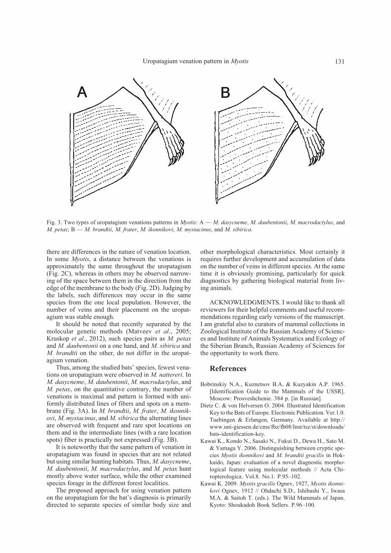
Thus, among the studied bats’ species, fewest vena-tions on uropatagium were observed in M. nattereri. InM. dasycneme, M. daubentonii, M. macrodactylus, andM. petax, on the quantitative contrary, the number ofvenations is maximal and pattern is formed with uni-formly distributed lines of fibers and spots on a mem-brane (Fig. 3A). In M. brandtii, M. frater, M. ikonnik-ovi, M. mystacinus, and M. sibirica the alternating linesare observed with frequent and rare spot locations onthem and in the intermediate lines (with a rare locationspots) fiber is practically not expressed (Fig. 3B).
It is noteworthy that the same pattern of venation inuropatagium was found in species that are not relatedbut using similar hunting habitats. Thus, M. dasycneme,M. daubentonii, M. macrodactylus, and M. petax huntmostly above water surface, while the other examinedspecies forage in the different forest localities.
The proposed approach for using venation patternon the uropatagium for the bat’s diagnosis is primarilydirected to separate species of similar body size and
Fig. 3. Two types of uropatagium venations patterns in Myotis: A — M. dasycneme, M. daubentonii, M. macrodactylus, andM. petax; B — M. brandtii, M. frater, M. ikonnikovi, M. mystacinus, and M. sibirica.

132 N.E. Dokuchaev
Kondo N. & Sasaki N. 2005. An external taxonomic charac-ter suitable for separating live Myotis ikonnikovi and M.mystacinus // Mammal Study. Vol.30. P.29–32.
Kowtun M.F. 1979. [On the nature of venation of the pat-agium in Chiroptera] // Zoologicheskii Zhurnal. Vol.58.No.2. P.207–217 [in Russian].
Kruskop S.V. 2012. Order Chiroptera // Pavlinov I.Ya. &Lissovsky A.A. (eds.). The Mammals of Russia: A Taxo-nomic and Geographic Reference. Moscow: KMK Sci.Press. P.73–126.
Kruskop S.V., Borisenko A.V., Ivanova N.V., Lim B.K. &Eger J.L. 2012. Genetic diversity of northeastern Palae-arctic bats as revealed by DNA barcodes // Acta Chi-ropterologica. Vol.14. No.1. P.1–14.
Kuzyakin A.P. 1950. [Bats]. Moscow: Sovetskaya nauka.444 p. [in Russian].
Matveev V.A., Kruskop S.V. & Kramerov D.A. 2005. Re-validation of Myotis petax Hollister, 1912 and its new
status in connection with M. daubentonii (Kuhl, 1817)(Vespertilionidae, Chiroptera) // Acta Chiropterologica.Vol.7. No.1. P.23–37.
Strelkov P.P. 1963. Order Chiroptera // Gromov I.M., Gu-reev A.A., Novikov G.A., Sokolov I.I., Strelkov P.P.,Chapsky K.K. [Mammals of the USSR Fauna. Vol.1].Moscow & Leningrad: Izdatelstvo Akademii nauk SSSR.P.122–218 [in Russian].
Tiunov M.P. 1984. Order Chiroptera Blumenbach, 1779 //Krivosheev V.G. (ed.). [Terrestrial Mammals of the USSRFar East. Identification Key]. Moscow: Nauka. P.73–101 [in Russian].
Tiunov M.P. 1997. [Bats of the Russian Far East]. Vladivos-tok: Dalnauka. 134 p. [in Russian].
Yablokov A.V., Panyutin K.K. & Panina G.A. 1974. [Vena-tion features of the wing membrane in some bats (Chi-roptera, Mammalia)] // Vorontsov N.N. (ed.). [Theriolo-gy]. Novosibirsk: Nauka. Vol.2. P.48–56 [in Russian].