update on who projects
DESCRIPTION
Update on WHO Projects. Update on WHO Projects. WHO HBV-genotype panels HBsAg HBV-DNA WHO IS antiHBc. Worldwide distribution of HBV genotypes. Worldwide Frequency Distribution of Chronic HBV Infections. HBsAg Prevalence. 8% - High. 2-7% - Medium.TRANSCRIPT

Update on WHO Projects
Update on WHO Projects
•WHO HBV-genotype panels•HBsAg•HBV-DNA
•WHO IS antiHBc
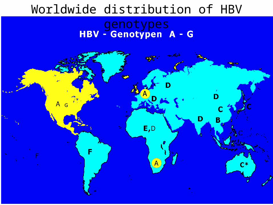
Worldwide distribution of HBV genotypes
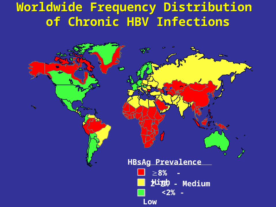
Worldwide Frequency Distribution of Chronic HBV Infections
Worldwide Frequency Distribution of Chronic HBV Infections
HBsAg Prevalence
8% - High 2-7% - Medium
<2% - Low

• WHO IS HBsAggenotype A, subtype A2
• WHO IS HBV-DNAgenotype A
representing only 1% of HBV-infected population
WHO International Standards
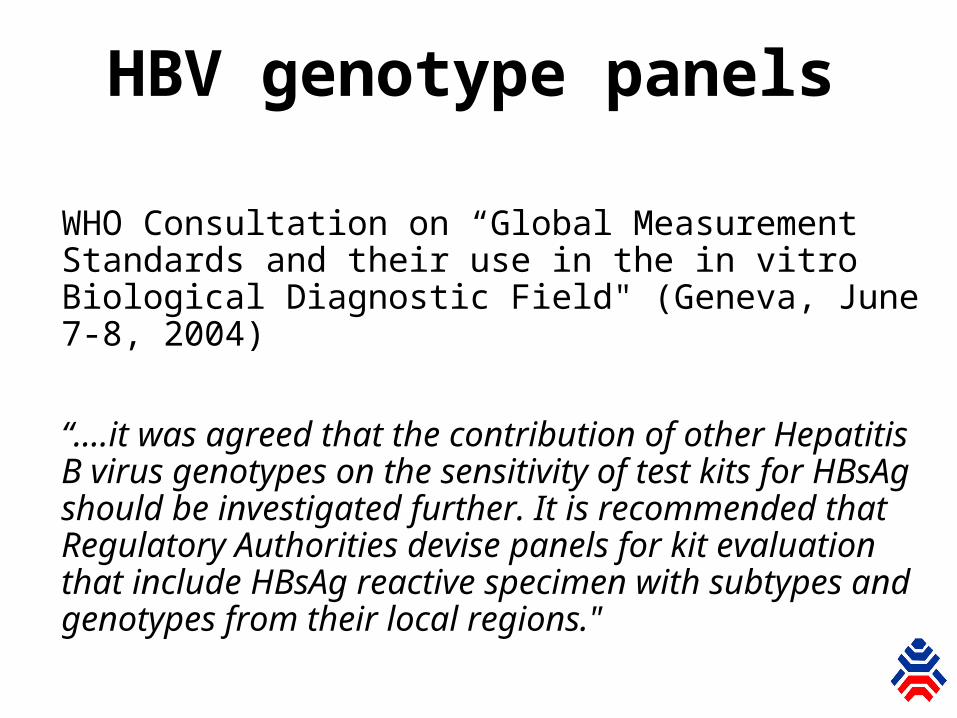
WHO Consultation on “Global Measurement Standards and their use in the in vitro Biological Diagnostic Field" (Geneva, June 7-8, 2004)
“….it was agreed that the contribution of other Hepatitis B virus genotypes on the sensitivity of test kits for HBsAg should be investigated further. It is recommended that Regulatory Authorities devise panels for kit evaluation that include HBsAg reactive specimen with subtypes and genotypes from their local regions."
HBV genotype panels

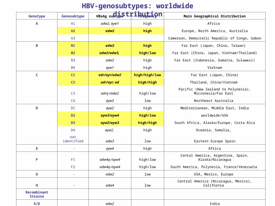
Genotype Genosubtype HBsAg subtype Frequency Main Geographical Distribution
A A1 adw2, ayw1 high Africa
A2 adw2 high Europe, North America, Australia
A3 Cameroon, Democratic Republic of Congo, Gabon
B B1 adw2 high Far East (Japan, China, Taiwan)
B2 adw2/adw3, high/low Far East (China, Japan, Vietnam/Thailand)
B3 adw2 high Far East (Indonesia, Sumatra, Sulawesi)
B4 ayw1 high Vietnam
C C1 adr/ayr/adw2 high/high/low Far East (Japan, China)
C2 adr/ayr; ad high/high Thailand, China/Vietnam
C3 adrq-/adw2 high/low Pacific (New Zealand to Polynesia), Micronesia/Far East
C4 ayw3 low Northeast Australia
D D1 ayw2 high Mediterranean, Middle East, India
D2 ayw3/ayw4 high/low worldwide/USA
D3 ayw2/ayw3 high/high South Africa, Alaska/Europe, Costa Rica
D4 ayw2, high Oceania, Somalia,
not identified adw3 low Eastern Europe Spain
E - ayw4 high Africa
F F1 adw4q-/ayw4 high/low Cental America, Argentina, Spain, Alaska/Nicaragua
F2 adw4q-/ayw4 high/low South America, Polynesia, France/Venezuela
G - adw2 low USA, Mexico, Europe
H - adw4 low Central America (Nicaragua, Mexico), California
Recombinant Strains
A/D adw2 India
C/D ayw2 Tibet
C/? adw2 Vietnam
HBV-genosubtypes: worldwide distribution
Aim
• HBsAg panel • HBV-DNA panel
from the same hi(+) unitsreflecting all major HBV-genotypes / major genosubtypes
if lyophilisation: validationno inactivation step
project introduced to and accepted by ECBS 2005
Cooperation: Prof W. Gerlich (Univ. Giessen)
HBV genotype Panels

• collection of plasma units worldwide– China, Taiwan, Japan; Egypt; Iran; Russia; Brasil,
South Africa…
– plasma from Russia, Germany, Japan, Brasil arrived at PEI
– represent genotypes A, B, C, D, (F) and mixed genotypes
– plasma from South Africa (genotype E?), Brasil (genotype F?) and Iran has been announced
HBV genotype panels-current status-
Genotype Genosubtype HBsAg subtype Frequency Main Geographical Distribution
A A1 adw2, ayw1 high Africa
A2 adw2 high Europe, North America, Australia
A3 Cameroon, Democratic Republic of Congo, Gabon
B B1 adw2 high Far East (Japan, China, Taiwan)
B2 adw2/adw3, high/low Far East (China, Japan, Vietnam/Thailand)
B3 adw2 high Far East (Indonesia, Sumatra, Sulawesi)
B4 ayw1 high Vietnam
C C1 adr/ayr/adw2 high/high/low Far East (Japan, China)
C2 adr/ayr; ad high/high Thailand, China/Vietnam
C3 adrq-/adw2 high/low Pacific (New Zealand to Polynesia), Micronesia/Far East
C4 ayw3 low Northeast Australia
D D1 ayw2 high Mediterranean, Middle East, India
D2 ayw3/ayw4 high/low worldwide/USA
D3 ayw2/ayw3 high/high South Africa, Alaska/Europe, Costa Rica
D4 ayw2, high Oceania, Somalia,
not identified adw3 low Eastern Europe Spain
E - ayw4 high Africa
F F1 adw4q-/ayw4 high/low Cental America, Argentina, Spain, Alaska/Nicaragua
F2 adw4q-/ayw4 high/low South America, Polynesia, France/Venezuela
G - adw2 low USA, Mexico, Europe
H - adw4 low Central America (Nicaragua, Mexico), California
Recombinant Strains
A/D adw2 India
C/D ayw2 Tibet
C/? adw2 Vietnam
HBV-genosubtypes: worldwide distribution
Current work
• Characterization of candidate materials (HBsAg, HBV-DNA, Sequencing)
• Pilot experiments to separate HBsAg from infectivity (HBV-DNA) and to fully characterize candidate materials (Prof W Gerlich, Univ. Giessen)
HBV genotype panels
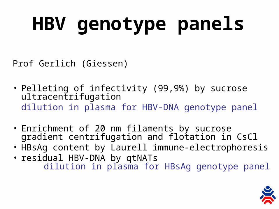
Prof Gerlich (Giessen)
• Pelleting of infectivity (99,9%) by sucrose ultracentrifugation
dilution in plasma for HBV-DNA genotype panel
• Enrichment of 20 nm filaments by sucrose gradient centrifugation and flotation in CsCl
• HBsAg content by Laurell immune-electrophoresis• residual HBV-DNA by qtNATs
dilution in plasma for HBsAg genotype panel
HBV genotype panels

To be done….
• Selection of panel members• Selection of target concentrations (HBsAg,HBV-
DNA)• Preparation• Design of collaborative study
• Characterization of panels
HBV genotype panels
Update on WHO Projects
•WHO HBV-genotype panels•HBsAg•HBV-DNA
•WHO IS antiHBc

• serological marker for past HBV-infection• in rare cases HBV-DNA (low+)• in rare cases HBV-transmissions reported from antiHBc-
pos donors to recipients • blood screening marker in several countries
– previously as surrogate marker for NANBH– considerations to rely in future on antiHBc combined
with ID HBV-NAT
WHO IS antiHBc
antiHBc

• „heterogenous“ antiHBc-assays– competitive / non-competitive assays– reducing agents in specimen diluent for increase of specificity?– often same antigen source– „confirmation“ of unspecific results by „different“ assays
• no confirmation assay for antiHBc• no common algorithm for „antiHBc-positive“
WHO IS antiHBc
What is the ideal candidate material ?
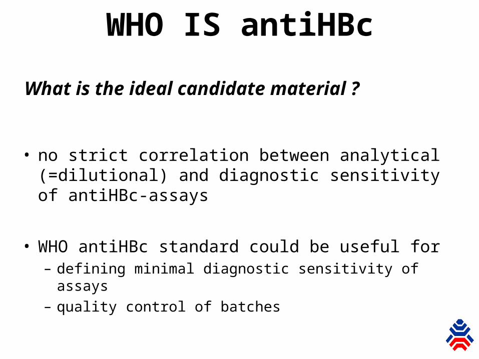
• no strict correlation between analytical (=dilutional) and diagnostic sensitivity of antiHBc-assays
• WHO antiHBc standard could be useful for– defining minimal diagnostic sensitivity of assays– quality control of batches
WHO IS antiHBc
What is the ideal candidate material ?
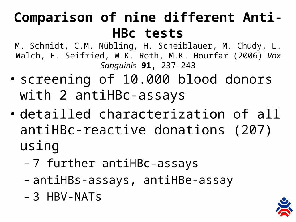
• screening of 10.000 blood donors with 2 antiHBc-assays
• detailled characterization of all antiHBc-reactive donations (207) using– 7 further antiHBc-assays– antiHBs-assays, antiHBe-assay– 3 HBV-NATs
Anti-HBc screening of blood donors – Comparison of nine different Anti-HBc tests
M. Schmidt, C.M. Nübling, H. Scheiblauer, M. Chudy, L. Walch, E. Seifried, W.K. Roth, M.K. Hourfar (2006) Vox Sanguinis 91, 237-243

antiHBc-reactivity in 9 different assays
9/9 assays ≥5/9 assays <5/9 assays
antiHBc only 27 (13%)HBV-DNA pos: 1
10 17
one second marker pos
antiHBc + antiHBs
67 (32%)>100 mIU antiHBs/ml: 49<100 mIU antiHBs/ml: 18
49 11 7
antiHBc + antiHBe
7 (3%) 7
both second markers pos
antiHBc + antiHBs + antiHBe
106 (51%)>100 mIU antiHBs/ml: 98<100 mIU antiHBs/ml: 8
106
207173 (84%) antiHBs-pos
162 (78%) 21 (10%) 24 (12%)
Characterization of 207 antiHBc-reactives
high lows/co-values antiHBc
Anti-HBc screening of blood donors – Comparison of nine different Anti-HBc tests
M. Schmidt, C.M. Nübling, H. Scheiblauer, M. Chudy, L. Walch, E. Seifried, W.K. Roth, M.K. Hourfar (2006) Vox Sanguinis 91, 237-243
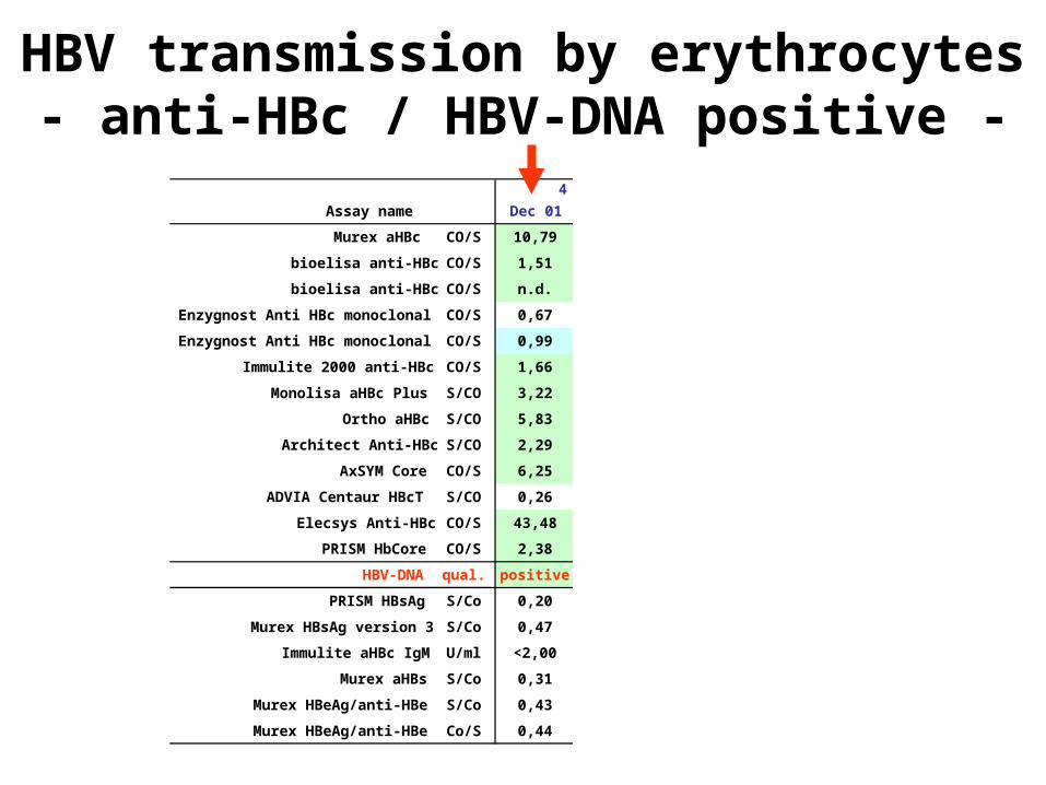
HBV transmission by erythrocytes- anti-HBc / HBV-DNA positive -
4 Plasma units from 1 donor
Assay name Dec 01 Sep 27 Jul 06 Apr 29
Murex aHBc CO/S 10,79 9,78 10,79 10,79
bioelisa anti-HBc CO/S 1,51 1,48 1,40 1,34
bioelisa anti-HBc CO/S n.d. n.d. 0,99 0,96
Enzygnost Anti HBc monoclonal CO/S 0,67 0,95 0,89 0,91
Enzygnost Anti HBc monoclonal CO/S 0,99 1,04 n.d. n.d.
Immulite 2000 anti-HBc CO/S 1,66 1,16 1,54 1,50
Monolisa aHBc Plus S/CO 3,22 2,97 2,96 2,97
Ortho aHBc S/CO 5,83 5,85 5,81 5,26
Architect Anti-HBc S/CO 2,29 2,24 2,32 2,59
AxSYM Core CO/S 6,25 6,02 5,88 4,98
ADVIA Centaur HBcT S/CO 0,26 0,16 0,96 1,00
Elecsys Anti-HBc CO/S 43,48 34,48 47,62 50,00
PRISM HbCore CO/S 2,38 2,27 2,56 2,27
HBV-DNA qual. positive positive positive positive
PRISM HBsAg S/Co 0,20 0,23 n.d. n.d.
Murex HBsAg version 3 S/Co 0,47 0,44 n.d. n.d.
Immulite aHBc IgM U/ml <2,00 <2,00 n.d. n.d.
Murex aHBs S/Co 0,31 0,24 n.d. n.d.
Murex HBeAg/anti-HBe S/Co 0,43 0,45 n.d. n.d.
Murex HBeAg/anti-HBe Co/S 0,44 0,44 n.d. n.d.
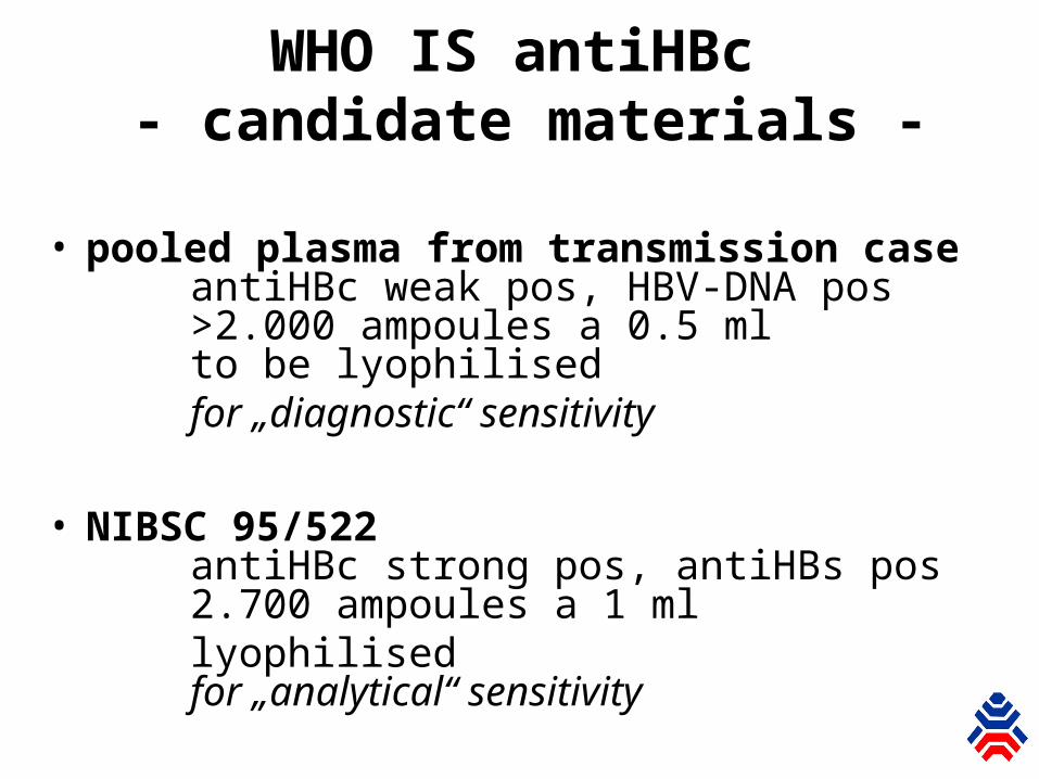
WHO IS antiHBc - candidate materials -
• pooled plasma from transmission caseantiHBc weak pos, HBV-DNA pos>2.000 ampoules a 0.5 mlto be lyophilisedfor „diagnostic“ sensitivity
• NIBSC 95/522 antiHBc strong pos, antiHBs pos2.700 ampoules a 1 mllyophilisedfor „analytical“ sensitivity
Acknowledgment
• G. Unger, M. Chudy (PEI)• W. Gerlich (Giessen)• M. Otani (Sao Paulo)• J. Tanaka , H. Yoshizawa (Hiroshima)• Prof. Zhibourt (Moscow)
• M. Schmidt (Frankfurt
• H. Scheiblauer, S. Nick (PEI)• M. Ferguson (NIBSC)
HBV genotype panels
WHO
IS antiHBc