unusual presentation of hereditary coproporphyria: a case report · 2019-07-10 · case...
TRANSCRIPT

Remedy Publications LLC.
Neurological Disorders and Stroke International
2018 | Volume 1 | Issue 2 | Article 10101
Unusual Presentation of Hereditary Coproporphyria: A Case Report
OPEN ACCESS
*Correspondence:Giovanna Graziadei, Rare Diseases
Center, IRCCS Ca 'Granda Foundation - Maggiore Policlinico Hospital, Via F.
Sforza 35, 20122 Milano, Italy,E-mail: giovanna.graziadei@policlinico.
mi.itReceived Date: 28 Jun 2018Accepted Date: 23 Jul 2018Published Date: 30 Jul 2018
Citation: Graziadei G, Spinelli D, Granata
F, Brancaleoni V, Di Pierro E. Unusual Presentation of Hereditary
Coproporphyria: A Case Report. Neurol Disord Stroke Int. 2018; 1(2): 1010.
Copyright © 2018 Giovanna Graziadei. This is an open access
article distributed under the Creative Commons Attribution License, which permits unrestricted use, distribution,
and reproduction in any medium, provided the original work is properly
cited.
Case ReportPublished: 30 Jul, 2018
AbstractHereditary Coproporphyria (HCP) is a rare genetic disease that is characterized by neurological acute attacks after puberty with fatal systemic complications; it is rarely accompanied by cutaneous symptoms. This report describes a case in which HCP was suspected and confirmed by raised urinary and fecal porphyrins and a novel missense variant in the coproporphyrinogen oxidase gene, the c.1348 A>G (p.Arg450Gly) mutation, which segregated in three other family members. This case suggests clearly that when a patient is admitted at Emergency room with atypical unexplained neurologic/psychiatric and/or gastrointestinal symptoms, porphyria must be always considered in the differential diagnosis. We remark that our patient did no presented urinary Porphobilinogen (PBG) and 5-Aminolevulinic Acid (ALA) alterations; this could be due to starting treatment with glucose solution a week before testing urine porphyrins. The suspicion was based mainly on clinical signs and anamnestic history; including recent treatment with claritromicin, confirming that porphyrias are a lot of times misdiagnosed diseases, mimicking many other acute medical and psychiatric conditions. A correct and early diagnosis is required to develop a management plan that is appropriate to the patient’s needs.
Keywords: Hereditary coproporphyria; Acute attack; Skin lesions; Diplopia; Retro-orbital cellulitis
IntroductionHereditary Coproporphyria (HCP) is a form of acute porphyria, in which the symptoms are
mainly neurovisceral and occur intermittently. Porphyrias are a group of rare inherited metabolic disorders, each resulting from a partial deficiency of a specific enzyme in the heme biosynthesis pathway. Attacks typically start in the abdomen with low-grade pain that slowly increases over a period of days with nausea progressing to vomiting. In some individuals, the pain is predominantly in the back or extremities. When an acute attack is untreated, a motor neuropathy may develop over a period of days or few weeks. The neuropathy first appears as weakness proximally in the arms and legs, then progresses distally to involve the hands and feet. Some individuals experience respiratory insufficiency due to the loss of innervations of the diaphragm and muscles of respiration. Acute attacks are associated commonly with use of certain medications, caloric deprivation, and changes in female reproductive hormones. About 20% of those with an acute attack also experience photosensitivity associated with bullae and fragility of light-exposed skin [1].
Case PresentationA 23 years old young woman was admitted to the Emergency Room (ER) because of diplopia
in the left gaze, pain in the posterior left orbit and pain in the lower limbs with postural instability. During the medical investigations, the patient complained of abdominal pain, fever, vomiting and closed alvus. In the week before admission, the patient referred tracheo-bronchitis treated with claritromicine.
In 1992, the patient underwent general anesthesia, because of a major stomatological surgery, without any problem. In the past history recurrent pharyngo-tonsillitis and uterine fibroma were referred; a thalassemia trait was described as well. Since 2003 the patient assumed estroprogestinic therapy and later on she suffered of desfoliant and urticarial skin lesions treated with topic therapy and photoprotection. Two siblings, brother and sister respectively of 25 and 17 years of age were affected by Thalassemia Major and regularly underwent hemotrasfusions. Because of the recent
Giovanna Graziadei1*, Diana Spinelli2, Francesca Granata1, Valentina Brancaleoni1 and Elena Di Pierro1
1Rare Diseases Center, IRCCS Ca 'Granda Foundation - Maggiore Policlinico Hospital, Italy
2Radiology Residency School, University Milano-Bicocca, Italy
Giovanna Graziadei, et al., Neurological Disorders and Stroke International
Remedy Publications LLC. 2018 | Volume 1 | Issue 2 | Article 10102
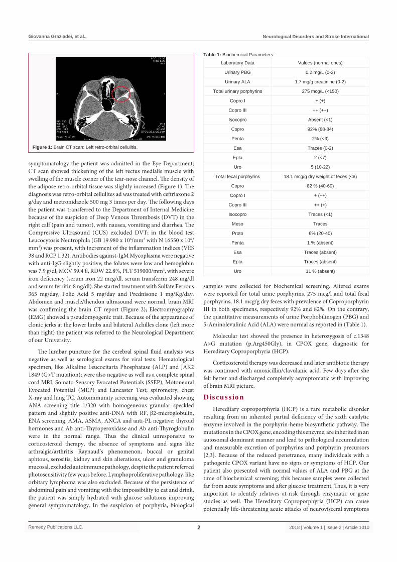
symptomatology the patient was admitted in the Eye Department; CT scan showed thickening of the left rectus medialis muscle with swelling of the muscle corner of the tear-nose channel. The density of the adipose retro-orbital tissue was slightly increased (Figure 1). The diagnosis was retro-orbital cellulites ad was treated with ceftriaxone 2 g/day and metronidazole 500 mg 3 times per day. The following days the patient was transferred to the Department of Internal Medicine because of the suspicion of Deep Venous Thrombosis (DVT) in the right calf (pain and tumor), with nausea, vomiting and diarrhea. The Compressive Ultrasound (CUS) excluded DVT; in the blood test Leucocytosis Neutrophila (GB 19.980 x 10⁶/mm3 with N 16550 x 106/mm3) was present, with increment of the inflammation indices (VES 38 and RCP 1.32). Antibodies against-IgM Mycoplasma were negative with anti-IgG slightly positive; the folates were low and hemoglobin was 7.9 g/dl, MCV 59.4 fl, RDW 22.8%, PLT 519000/mm3, with severe iron deficiency (serum iron 22 mcg/dl, serum transferrin 248 mg/dl and serum ferritin 8 ng/dl). She started treatment with Sulfate Ferrous 365 mg/day, Folic Acid 5 mg/day and Prednisone 1 mg/Kg/day. Abdomen and muscle/thendon ultrasound were normal, brain MRI was confirming the brain CT report (Figure 2); Electromyography (EMG) showed a pseudomyogenic trait. Because of the appearance of clonic jerks at the lower limbs and bilateral Achilles clone (left more than right) the patient was referred to the Neurological Department of our University.
The lumbar puncture for the cerebral spinal fluid analysis was negative as well as serological exams for viral tests. Hematological specimen, like Alkaline Leucocitaria Phosphatase (ALP) and JAK2 1849 (G>T mutation); were also negative as well as a complete spinal cord MRI, Somato-Sensory Evocated Potentials (SSEP), Motoneural Evocated Potential (MEP) and Lancaster Test; spirometry, chest X-ray and lung TC. Autoimmunity screening was evaluated showing ANA screening title 1/320 with homogeneous granular speckled pattern and slightly positive anti-DNA with RF, β2-microglobulin, ENA screening, AMA, ASMA, ANCA and anti-PL negative; thyroid hormones and Ab anti-Thyroperoxidase and Ab anti-Thyroglobulin were in the normal range. Thus the clinical unresponsive to corticosteroid therapy, the absence of symptoms and signs like arthralgia/arthritis Raynaud’s phenomenon, buccal or genital aphtous, serositis, kidney and skin alterations, ulcer and granuloma mucosal, excluded autoimmune pathology, despite the patient referred photosensitivity few years before. Lymphoproliferative pathology, like orbitary lymphoma was also excluded. Because of the persistence of abdominal pain and vomiting with the impossibility to eat and drink, the patient was simply hydrated with glucose solutions improving general symptomatology. In the suspicion of porphyria, biological
samples were collected for biochemical screening. Altered exams were reported for total urine porphyrins, 275 mcg/l and total fecal porphyrins, 18.1 mcg/g dry feces with prevalence of Coproporphyrin III in both specimens, respectively 92% and 82%. On the contrary, the quantitative measurements of urine Porphobilinogen (PBG) and 5-Aminolevulinic Acid (ALA) were normal as reported in (Table 1).
Molecular test showed the presence in heterozygosis of c.1348 A>G mutation (p.Arg450Gly), in CPOX gene, diagnostic for Hereditary Coproporphyria (HCP).
Corticosteroid therapy was decreased and later antibiotic therapy was continued with amoxicillin/clavulanic acid. Few days after she felt better and discharged completely asymptomatic with improving of brain MRI picture.
DiscussionHereditary coproporphyria (HCP) is a rare metabolic disorder
resulting from an inherited partial deficiency of the sixth catalytic enzyme involved in the porphyrin-heme biosynthetic pathway. The mutations in the CPOX gene, encoding this enzyme, are inherited in an autosomal dominant manner and lead to pathological accumulation and measurable excretion of porphyrins and porphyrin precursors [2,3]. Because of the reduced penetrance, many individuals with a pathogenic CPOX variant have no signs or symptoms of HCP. Our patient also presented with normal values of ALA and PBG at the time of biochemical screening; this because samples were collected far from acute symptoms and after glucose treatment. Thus, it is very important to identify relatives at-risk through enzymatic or gene studies as well. The Hereditary Coproporphyria (HCP) can cause potentially life-threatening acute attacks of neurovisceral symptoms
Figure 1: Brain CT scan: Left retro-orbital cellulitis.
Laboratory Data Values (normal ones)
Urinary PBG 0.2 mg/L (0-2)
Urinary ALA 1.7 mg/g creatinine (0-2)
Total urinary porphyrins 275 mcg/L (<150)
Copro I + (+)
Copro III ++ (++)
Isocopro Absent (<1)
Copro 92% (68-84)
Penta 2% (<3)
Esa Traces (0-2)
Epta 2 (<7)
Uro 5 (10-22)
Total fecal porphyrins 18.1 mcg/g dry weight of feces (<8)
Copro 82 % (40-60)
Copro I + (++)
Copro III ++ (+)
Isocopro Traces (<1)
Meso Traces
Proto 6% (20-40)
Penta 1 % (absent)
Esa Traces (absent)
Epta Traces (absent)
Uro 11 % (absent)
Table 1: Biochemical Parameters.

Giovanna Graziadei, et al., Neurological Disorders and Stroke International
Remedy Publications LLC. 2018 | Volume 1 | Issue 2 | Article 10103
that mimic many other acute medical and psychiatric conditions [4]. Lack of clinical recognition often delays effective treatment, and inappropriate diagnostic tests may lead to misdiagnosis and inappropriate treatment.
In literature three ways to classify the different types of porphyrias are reported: Erythopoietic and hepatic forms, according to the major site of expression of the specific enzymatic deficiency; cutaneous and non-cutaneous forms, acute and non-acute or chronic forms. The three most important and characterizing clinical entities are the acute porphyric attack, the acute and the chronic skin symptoms. The acute porphyrias are comprised of Acute Intermittent Porphyria (AIP) [5], Variegate Porphyria (VP) [6], Hereditary Coproporphyria (HCP) [7] and Delta-Aminolevulinic Acid Dehydrates (ALAD) Deficiency Porphyria, which is also known as plumboporphyria or Doss porphyria [8].
All patients suffering from an acute porphyria can manifest a broad spectrum of often-unspecific clinical symptoms, independently from the specific type of acute porphyria. These symptoms include long-lasting colicky abdominal pain, nausea and vomiting, diarrhea, tachycardia, hypertension, seizures, muscle weakness, a variety of other neurological and psychiatric signs. An acute porphyria should be considered in many patients with unexplained abdominal pain or other characteristic symptoms. However, porphyrias are rare diseases and are rarely considered in differential diagnosis [9]; prompt and accurate diagnosis and treatment greatly improve prognosis and may prevent development of severe or chronic neuropathic symptoms. The diagnosis can be rapidly confirmed by the demonstration of a markedly increased urinary PBG level by using a single-void urine specimen and then by the quantitative measurement of PBG, ALA, and total porphyrin levels too. Precipitating factors, including porphyrinogenic drugs [10], alcohol, hormonal changes, recurrent or chronic infection and reduced caloric intake due to fasting or diets [11], should be eliminated, and appropriate supportive and symptomatic therapy should be initiated. Intravenous hemin therapy is the most effective treatment. Intravenous glucose alone [12] is appropriate only for mild attacks (mild pain, no paresis or hyponatremia) or until hemin is available [13,14].
Thus, particularly, VP and HCP are also referred to as Neurocutaneous porphyrias, characterize by the presence of increased photosensitivity, with abnormal skin vulnerability on the sun-exposed areas of the skin, blistering, erosions, scars and post inflammatory hyper pigmentation. The skin findings in VP and HCP are identical to those encountered in non-acute porphyria, like Porphyria Cutanea Tarda (PCT). By contrast, however, AIP and ALAD deficiency porphyria do not present with cutaneous symptoms [2,3]. The non-acute or chronic porphyrias consist of PCT, the most frequent type of porphyria worldwide, Erythropoietic Protoporphyria (EPP), Congenital Erythropoietic Porphyria (CEP) and Hepatoerythropoietic Porphyria (HEP), the recessively inherited variant of PCT.
In our case considering the coexistence of the acute neurological finding and the presence of other apparently chronic mild different visceral and cutaneous symptoms and signs, we suspected the diagnosis of acute neuro-visceral and cutaneous porphyria, even if the presentation of this form of acute porphyria (Hereditary Coproporphyria, HCP) was really not typical and till now not completely understood yet.
ConclusionAcute porphyrias are rare and sometimes misdiagnosed, because
various symptoms and signs mimic other diseases. Once porphyria is suspected, biochemical analyses easily detect porphyrins and their precursors from blood, urine or feces; the alterations of these specimens are more evident if collected during the acute phase. Otherwise, molecular analysis can be done at the quiescent phase of the disease. Pathogenetic mechanisms and clinical manifestations differ in each individual and in the different forms of porphyrias, most of which require a specific treatment. Early diagnosis and information about precipitating factors can decrease mortality and prevent subsequent attacks among patients with acute porphyrias, so mutation screening is recommended for family members.
AcknowledgementThis research was supported in part by grants from the Italian
Ministry of Health (GR–2011–02347129 to EDP) and from
Figure 2: MRI: Panel A shows coronal section of STIR imagesPanel B coronal section of T2-weighted images showing orbital apex crowdingPanel C assial section of STIR imagesPanel D and E report coronal and assial sections showing complete resolution.
Giovanna Graziadei, et al., Neurological Disorders and Stroke International
Remedy Publications LLC. 2018 | Volume 1 | Issue 2 | Article 10104
Fondazione IRCCS Ca’ Granda Ospedale Maggiore Policlinico (RC2018).
References1. Bissell DM, Wang B, Cimino T, Lai J. Hereditary Coproporphyria.
In: Pagon RA, Bird TD, Dolan CR, Stephens K, Adam MP, editors. GeneReviews. Seattle (WA): University of Washington, Seattle; 1993-2018, USA; 2012.
2. Bickers DR, Frank J. The porphyrias. In: Dermatology in General Medicine. New York: McGraw Hill, USA; 2003:1435-1466.
3. Anderson KE, Sassa S, Bishop DF, Desnick RJ. Disorders of heme biosynthesis: X-linked sideroblastic anemia and porphyrias. In: The Metabolic and Molecular Bases of Inherited Disease. New York: McGraw Hill, USA; 2001:2991-3062.
4. Elder GH, Hift RJ, Meissner PN. The acute porphyrias. Lancet. 1997;349(9065):1613-7.
5. Kauppinen R, Von und zu Fraunberg M. Molecular and biochemical studies of acute intermittent porphyria in 196 patients and their families. Clin Chem. 2002;48(11):1891-900.
6. Frank J, Christiano AM. Variegate porphyria: past, present and future. Skin Pharmacol Appl Skin Physiol. 1998;11(6):310-20.
7. Martasek P. Hereditary coproporphyria. Semin Liver Dis. 1998;18(1):25-32.
8. Doss M, Von Tiepermann R, Schneider J, Schmid H. New type of hepatic porphyria with porphobilinogen synthase defect and intermittent acute manifestation. Klin Wochenschr. 1979;57(20):1123-7.
9. Kauppinen R. Porphyrias. Lancet. 2005;365(9455):241-52.
10. Anderson KE, Bloomer JR, Bonkovsky HL, Kushner JP, Pierach CA, Pimstone NR, et al. Recommendations for the diagnosis and treatment of the acute porphyrias. Ann Intern Med. 2005;142(6):439-50.
11. Poblete-Gutierrez P, Kunitz O, Wolff C. Diagnosis and treatment of the acute porphyrias: an interdisciplinary challenge. Skin Pharmacol Appl Skin Physiol. 2001;14:393-400.
12. Han Handischin C, Lin J, Rhee J, Peyer AK, Chin S, Wu PH, et al. Nutritional regulation of hepatic heme biosynthesis and porphyria through PGC- 1alpha. Cell. 2005;122(4):505-15.
13. Bonkovsky HL, Healey JF, Lourie AN, Gerron GG. Intravenous heme-albumin in acute intermittent porphyria: evidence for repletion of hepatic hemoproteins and regulatory heme pools. Am J Gastroenterol. 1991;86(8):1050-6.
14. Tenhunen R, Mustajoki P. Acute porphyria: treatment with heme. Semin Liver Dis. 1998;18(1):53-5.