unstable os odontoideum in young children
TRANSCRIPT
0008-3194/93/141-144/$2.00/©JCCA 1993
Unstable os odontoideumin young childrenSt6phane Lefebvre, DC*Dwight Vallee, DC**J David Cassidy, DC, MSc (Orth), FCCS(C)***Anne K Dzus, MD, FRCS(C)****
Os odontoideum is a rare anomaly ofthe odontoidprocess.When unstable, it can be a source ofneckpain that can result inserious neurological injury. In young children, the presence ofa transverse lucency at the base ofthe dens represents thenormal developing growth plate. However, os odontoideumshouldbe considered in the differential diagnosis in a child withrecurrent or persistent neckpain. A case ofan unstable osodontoideum in a two-year-old child is presented to illustratethis point.(JCCA 1993; 37(3):141-144)
KEYWORDS: os odontoideum, instability, neck pain.
L'os odontoide est une anomalie rare de l'apophyse odontoide.Lorsqu'elle est instable, elle cause parfois une douleurcervicale qui peut entramner des blessures neurologiquesgraves. Chez les enfants en bas age, la presence d'une lucencytransverse a la base de la dent de l'axis represente une zonecartilagineuse normale d'accroissement des os longs.Toutefois, ilfaut tenir compte de l'os odontoide lors de laformulation du diagnostic differentiel d'un enfantqui souffre dedouleurs cervicales recurrentes ou persistantes. Pour illustrercette question, voici maintenent l'exemple d'un enfant age dedeux ans dont l'os odontoide est instable.(JCCA 1993; 37(3):141-144)
MOTS CLS: Os odontoide, instabilite, douleur cervicale.
IntroductionOs odontoideum is a rare anomaly of the odontoid process. Itsprevalence is unknown, as many cases remain asymptomaticand hidden for life. It may be described as a wide transverse gapof fibrous tissue separating the main body of the dens from thecentrum of the axis. Once believed to represent a defect ofunionof the ossification center of the odontoid to the body of axis,there is evidence suggesting that it results from an early fractureof the odontoid secondary to trauma. I
Os odontoideum is not clinically significant unless it presentswith atlanto-axial instability. This instability may progress and
cause severe neurologic deficit.2 Pain is the most commoncomplaint and restricted neck mobility the most common phys-ical finding.3 In adults, if routine radiographic views disclose atransverse lucency at the base of the dens further investigationfor atlanto-axial instability is necessary. In children, the sametransverse lucency represents the odontoid growth plate. There-fore, children complaining of recurrent or persistent neck painshould have flexion and extension views to rule out an unstableos odontoideum. The purpose of this case report is to illustratethe value of flexion and extension radiographs in children com-plaining of recurrent or persistent neck pain.
Case reportA two-and-a-half-year old boy was referred to the paediatricorthopaedic outpatient clinic at the Royal University Hospitalfor evaluation of recurrent neck pain. The first episode of neckpain had begun following a fall at a playground when he was oneand a half years old. At that time, radiographs of his cervicalspine had not disclosed any abnormality. (Figure 1) Musclestrain was diagnosed and the child was treated with medicationby his family doctor. Six months later, the boy was still com-plaining of recurrent neck pain, but additional radiographs didnot reveal any obvious change. Since he continued to have neckpain 11 months after the initial injury, he was referred for
* Clinical Resident, Canadian Memorial Chiropractic College,Toronto, Ontario.
** Private practice of Chiropractic, Saskatoon, Saskatchewan*** Research Associate, Department of Orthopaedics, Royal University
Hospital, Saskatoon, Saskatchewan.*** Assistant Professor, Department of Orthopaedics, Royal University
Hospital, Saskatoon, Saskatchewan.Reprint request to: Dr. J.D. Cassidy, Department of Orthopaedics,Royal University Hospital, University of Saskatchewan, Saskatoon,S7N OXO.
© JCCA 1993.
The Journal of the CCA / Volume 37 No.3 / September1993 141

Os odontoideum
.ill*_ll_s:Y , , : _r ,j,.s.X_.,-...........~~~~~~~~~~~~~~~~~~~~~~~~~~~~~~~~~~~~~~~~~~~~~~~~~~~~~~~~~~~~~~~~~~~~~~~~~~~~~~~~~~~~~~~~~~~~~~~~~~~~~~~~~~~~~~~~~~~~~.......
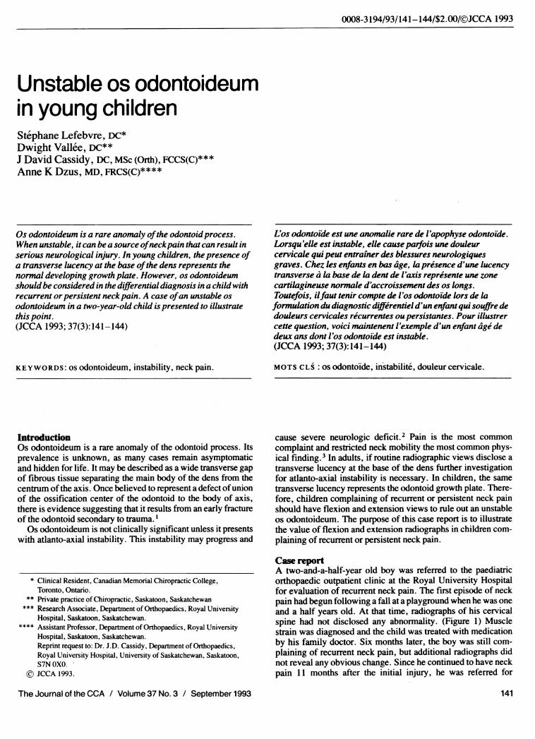
Figure 1 Anteroposterior and lateral radiographs of the cervical spine taken at the initial presentation show no evidence of abnormality.
Figure 2 The flexion and extension radiographs taken two years later show 5 to 6 mm of anterior displacement of C1 on C2. Note the anteriordisplacement of the os odontoideum with the anterior arch of atlas during flexion. (arrow)
The Journal of the CCA / Volume 37 No. 3 / September 1993142

S Lefebvre, D Vallee, JD Cassidy, AKDzus
Fiure 3 The tomogram shows that theodontoid is not united to the body of C2.(arrow)
orthopaedic evaluation.The boy was otherwise healthy except for occasional asthma
attacks. He was the product of a full-term pregnancy and un-complicated delivery. He had achieved the normal develop-mental milestones.
Examination revealed a normally developed boy for his age.Motion of the cervical spine was normal, except for extension,which he was reluctant to perform. The upper extremities wereneurologically intact. Palpation of the cervical spine revealed notenderness.
Radiographs of the cervical spine, including flexion andextension views, revealed 5 to 6 mm of displacement of C1 onC2. (Figure 2) Tomograms added evidence of a fibrous defect atthe base of the odontoid. (Figure 3) A magnetic resona,nceimage obtained to assess the state of the spinal cord was unre-markable.
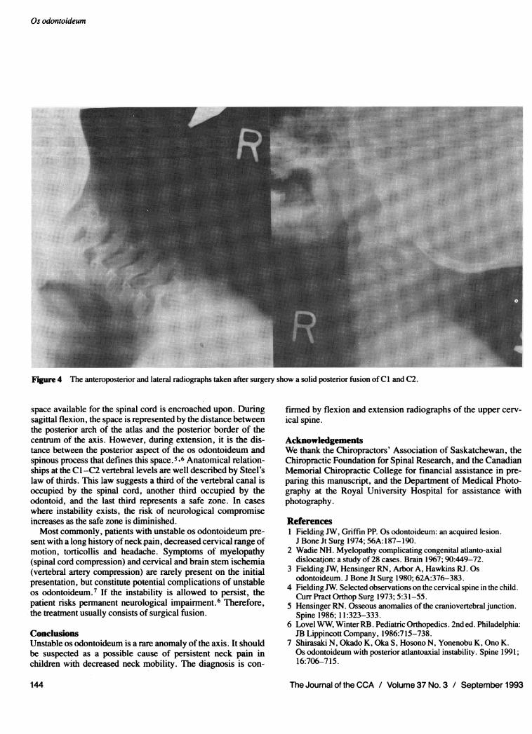
Surgical fusion of C1 to C2 was scheduled to relieve thepersisting pain and to prevent neurological compromise. Oneyear after surgery, the patient was completely pain-free, exceptafter extreme physical exertion. On examination, range ofmotion of the cervical spine was full and painless. He remainedneurologically intact. Radiographs showed a solid fusion of Cland C2. (Figure 4)
DiscussionIn young children, the radiographic appearance of an os odon-toideum and a normally developing odontoid process are indis-tinguishable. In fact, the growth plate of a normally developingaxis and the bone defect characteristic of an os odontoideumboth appear lucent on plain film radiographs. This radiographiclucency persists until the growth plate starts to close, some timeduring the third year of life. By age four the growth plate hascompletely closed in about 50% of children, and the remainingclose by age six.4 Therefore, prior to age six, it is not possible todiagnose a clinically significant os odontoideum without flexionand extension radiographs.
Usually atlanto-axial instability is assessed by measuring theatlanto-dental interspace. This method is invalid in cases of osodontoideum. This remains true even when flexion and exten-sion radiographs are used. The reason is that the transverseligament fixes the free ossicle of the os odontoideum to theanterior arch of the atlas, thus maintaining the normal atlanto-dental interspace throughout flexion and extension. In order todetermine instability in cases of os odontoideum, movementmust be observed between the centrum of the axis and the osodontoideum in the sagittal plane on plane radiographs.
There is the potential for neurological compromise when the
The Journal of the CCA / Volume 37 No.3 / September 1993 143
Os odontoideum
-~~~~~~~~~~~~~~~...,~~~~~~~~~~~~~~~~~...
~~~~~~~~~~~~~~~~~~~~~~~~~~~~~~~~~~~~~~~~~~~~~~~... ....
~~~~~~~~~~~~~~~~~~~~~~~~~~~~~~~~~~~~~~~~~~~~~~~~~'.~~~~~~~~~~~~~~~~~~~~~~~~~~~~~~~~~~~~~~~~~~~~~~~~~~~~~~~~~~~~~~~~~~~~~~~~~~~~~~~~~~~.......I~~~~~~~~~~~~~~~~~~~. .. .. ...
Figure 4 The anteropostenor and lateral radiographs taken after surgery show a solid posterior fusion of Cl and C2.~~~~~~~~~~~~~~~~~~~~~~~~~~~~~~~~~~~~~~~~~~~~~~~~~~~~~~~~~~~~~~~~~~~~~~~~~~~~~~~~~~~~~~~~~~~~~~~~~~~~~~~~~~~~~~~~~~~~~~~~...........
space available for the spinal cord is encroached upon. Duringsagittal flexion, the space is represented by the distance betweenthe posterior arch of the atlas and the posterior border of thecentrum of the axis. However, during extension, it is the dis-tance between the posterior aspect of the os odontoideum andspinous process that defines this space.56 Anatomical relation-ships at the Cl-C2 vertebral levels are well described by Steel'slaw of thirds. This law suggests a third of the vertebral canal isoccupied by the spinal cord, another third occupied by theodontoid, and the last third represents a safe zone. In caseswhere instability exists, the risk of neurological compromiseincreases as the safe zone is diminished.Most commonly, patients with unstable os odontoideum pre-
sent with a long history ofneck pain, decreased cervical range ofmotion, torticollis and headache. Symptoms of myelopathy(spinal cord compression) and cervical and brain stem ischemia(vertebral artery compression) are rarely present on the initialpresentation, but constitute potential complications of unstableos odontoideum.7 If the instability is allowed to persist, thepatient risks permanent neurological impairment.6 Therefore,the treatment usually consists of surgical fusion.
ConclusionsUnstable os odontoideum is a rare anomaly of the axis. It shouldbe suspected as a possible cause of persistent neck pain inchildren with decreased neck mobility. The diagnosis is con-
firmed by flexion and extension radiographs of the upper cerv-ical spine.
AcknowledgementsWe thank the Chiropractors' Association of Saskatchewan, theChiropractic Foundation for Spinal Research, and the CanadianMemorial Chiropractic College for financial assistance in pre-paring this manuscript, and the Department of Medical Photo-graphy at the Royal University Hospital for assistance withphotography.
References1 Fielding JW, Griffin PP. Os odontoideum: an acquired lesion.
J Bone Jt Surg 1974; 56A:187-190.2 Wadie NH. Myelopathy complicating congenital atlanto-axial
dislocation: a study of 28 cases. Brain 1967; 90:449-72.3 Fielding JW, Hensinger RN, Arbor A, Hawkins RJ. Os
odontoideum. J Bone Jt Surg 1980; 62A:376-383.4 Fielding JW. Selected observations on the cervical spine in the child.
Curr Pract Orthop Surg 1973; 5:31-55.5 Hensinger RN. Osseous anomalies of the craniovertebral junction.
Spine 1986; 11:323-333.6 Lovel WW, Winter RB. Pediatric Orthopedics. 2nd ed. Philadelphia:JB Lippincott Company, 1986:715-738.
7 Shirasaki N, Okado K, Oka S, Hosono N, Yonenobu K, Ono K.Os odontoideum with posterior atlantoaxial instability. Spine 1991;16:706-715.
144 The Journal of the CCA / Volume 37 No.3 / September 1993