unknown location or unknown viabilitymedia01.commpartners.com/2018/aium/february_2018... · early...
TRANSCRIPT

Page 1
Early Pregnancies ofUnknown Location or
Unknown Viability
Carol B. Benson, MD No Disclosures
Early First Trimester Ultrasound
Is the pregnancy intrauterine?OR
Is the pregnancy ectopic?
Is the pregnancy viable*?OR
Is the pregnancy a miscarriage
*Has potential to result in live born infant
Terminology & Definitions
Intrauterine Pregnancy of UnknownViability (IPUV)
Ultrasound findings: Intrauterine gestational sac with no
embryonic heartbeat
Pregnancy of Unknown Location (PUL)hCG & Ultrasound findings:
No intrauterine gestational sac oradnexal mass
Early pregnancy failureNot: Miscarriage, spontaneous abortion…
Usually seen Transvaginallyby 5.0 weeks
Usually seen Transabdominallyby 5.5 weeks
Usually when hCG > 1000 mIU/ml(1st or 3rd IRP)
Intrauterine Sac-Like Structure1st Sign of Pregnancy
Beyene Double sac TV
Early Pregnancy – Transvaginal Scan
Published Ultrasound Signs ofEarly Pregnancy
Double sac sign* (reported 1982)
Intradecidual sign* (reported 1984)
*If present, diagnosis = IUP*If absent, does not mean no IUP
IUP = intrauterine pregnancy
Page 2
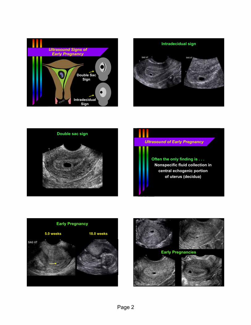
Ultrasound Signs ofEarly Pregnancy
Double SacSign
IntradecidualSign
Areizaga Double sac TV
Intradecidual sign
Perez doublesac sign
Double sac sign
Ultrasound of Early Pregnancy
Often the only finding is . . .
Nonspecific fluid collection in
central echogenic portion
of uterus (decidua)
Brown 5w
Early Pregnancy
5.0 weeks 18.0 weeks
Early IUPs from PD10,11,13,14
Early Pregnancies

Page 3
Problem
If nonspecific fluid collection incentral echogenic portion of uterus
reported as no intrauterine pregnancyor the possibility of
a “pseudogestational sac”↓
Clinician concludes ectopic pregnancy↓
Patient treated with Methotrexateor dilatation & curettage (D&C)
Problem
When early intrauterine pregnancyis exposed to Methotrexate (MTX)
↓Follow up: intrauterine pregnancy
with heartbeat
↓Miscarriage or fetal malformations
Two readers assessed
199 proven intrauterine gestations
fluid in uterus, no YS or embryo
embryonic heartbeat on follow up
First trimester outcome
148 (74.4%) live
51 (25.6%) miscarriage
Study of Ultrasound Signs ofEarly Pregnancy
Signs of early
pregnancy
Double Sac Sign Intradecidual Sign
No Sign“Nonspecific”
Signs (or Absence of Signs) of Early Pregnancy
Reader 1 Reader 2DSS present* 32% 30%IDS present** 23% 39%Neither sign 57% 48%
Kappa = *0.24 & **0.23 (poor inter-observer agreement)
No relationship between outcome and presence/absence of DSS & IDS(p > 0.10, Fisher exact test)
Study of Ultrasound Signs ofEarly Pregnancy Early Intrauterine Gestation
Early intrauterine gestations oftenhave a nonspecific appearance(>50% in our study)
Even in the absence of an intradecidual sign or a double sac sign,it’s most likelyan early intrauterine gestation

Page 4
Early Intrauterine Gestation
How should one report anIntrauterine fluid collection with
no yolk sac or embryo andnormal adnexa?
“Intrauterine sac-like structure that isalmost certainly an intrauterinegestational sac”
OR“Probable early intrauterine gestation.
Follow up ultrasound suggestedfor definitive confirmation”
“Pseudogestational Sac”
Definition:
Fluid in the uterine cavity
mimicking a gestational sac
with ectopic pregnancy
Misuse of Term Pseudogestational Sac
Definition: fluid in uterine cavity
with ectopic pregnancy
Frequency with ectopic pregnancy
1979 report: 20%
1990 reports: 10%
Intrauterine Fluid in Ectopic Pregnancy: A Reappraisal
All proven ectopic pregnancies July 2008 to August 2011 = 229 cases
Fluid, when present, characterized by Shape:
pointy-edged or smooth*Location:
clearly in cavity* or uncertainFluid contents:
echoes & debris or anechoic*
*Features of early intrauterine pregnancy
Intrauterine Fluid in Patients With Ectopic Pregnancy
No fluid83.4%
Fluid16.6%
Intrauterine Fluid in Ectopic Pregnancy: Fluid Characterization
Fluid inconsistent with gestational sacpointy-edgedclearly within uterine cavity
(not the decidua)containing echoes or debris
Fluid similar to a gestational sacsmooth marginsnot clearly within uterine cavityanechoic

Page 5
Intrauterine Fluid in Ectopic Pregnancy: Inconsistent with Gestational sac
Pointy-edged Internal echoes Within cavity or debris not decidua
30/38 28/38 7/38
38 ectopics (16.6%) had fluid in cavity
No fluid83.4%
Fluid: Inconsistent with gestational sac
13.5%
Fluid: Similar to gestational sac
3.1%
Intrauterine Fluid in Patients With Ectopic Pregnancy
Jimenezdecid cyst & EP
7 with fluid similar to a gestational sac5 had adnexal mass of ectopic
(2 no mass)
RO
Intrauterine Fluid in Patients With Ectopic Pregnancy
Fluid: Similar to gestational sac(“Nonspecific”)
&No adnexal mass
(extraovarian)0.9%
Calculations
Intrauterine gestations 98%Nonspecific fluid 50%
Ectopic pregnancies (per CDC) 2%Nonspecific fluid & no mass 1%
For nonspecific fluid collection in cavity& no adnexal mass
Do the math…99.9% likelihood of intrauterine
pregnancy (>1000 to 1)
hCG & Nonspecific Intrauterine Fluid CollectionGestational Sac or Pseudogestational Sac?
11 dayslater
7 dayslater
16 dayslater
Page 6
Pseudogestational Sac
Little or no value, avoid the term If definite ectopic pregnancy
term has no value Otherwise, odds are strongly
in favor of intrauterine gestation(99.9%)
Report nonspecific fluid collectionin uterus as “almost certainly anintrauterine gestational sac”
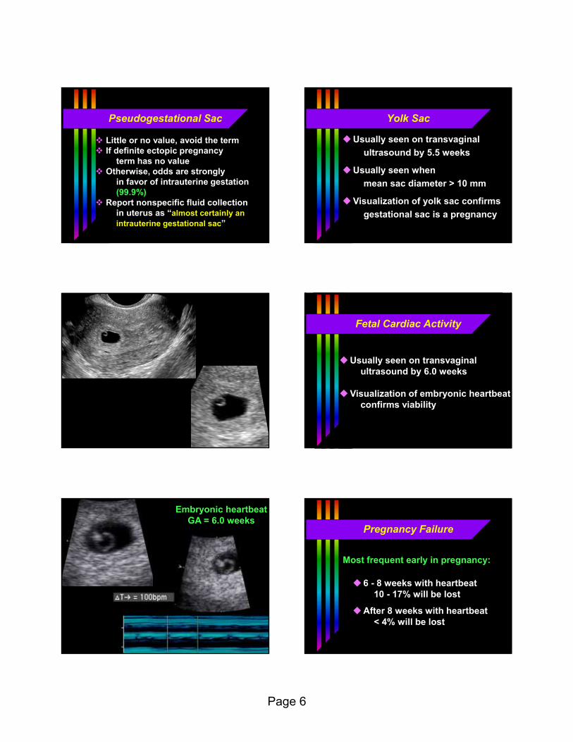
Yolk Sac
Usually seen on transvaginal
ultrasound by 5.5 weeks
Usually seen when
mean sac diameter > 10 mm
Visualization of yolk sac confirms
gestational sac is a pregnancy
Thompson YS TV
Fetal Cardiac Activity
Usually seen on transvaginalultrasound by 6.0 weeks
Visualization of embryonic heartbeatconfirms viability
Kelly early FH
Embryonic heartbeatGA = 6.0 weeks
Pregnancy Failure
Most frequent early in pregnancy:
6 - 8 weeks with heartbeat10 - 17% will be lost
After 8 weeks with heartbeat< 4% will be lost
Page 7
Increased loss ratesExisting conditions
Prior miscarriages
Uterine duplication anomalies
Fibroids
Advanced maternal age
Pregnancy Failure
Increased loss ratesOnce pregnant
Bleeding
Slow fetal heart rate
Subchorionic hematoma
Pregnancy Failure
Definitive diagnosis
Embryo ≥ 7 mm* with no heartbeatMean sac diameter ≥ 25 mm*
with no heartbeatNo heartbeat & gestational age
2 wks after gestational sac seen*(GA >7 wks by prior ultrasound)
Sliding sac
Pregnancy Failure
*SRU Consensus Panel on Dx of Early Pregnancy Failure 2012
Pregnancy Failure
Findings on follow up ultrasound
*SRU Consensus Panel on Dx of Early Pregnancy Failure 2012GS = gestational sac; YS = yolk sac
Rationale for ≥ 7 mm cutoffSet value to virtually eliminate
any false positive diagnoses(100% specificity)
Prior criteria not stringent enoughBased on small numbers of cases
Need to account for interobservervariability (± 15%)
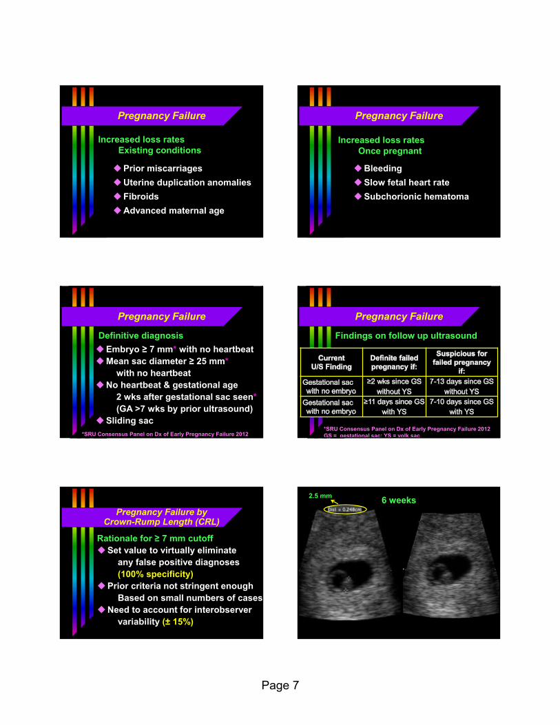
Pregnancy Failure by Crown-Rump Length (CRL)
Catlin CRL 2mmNo FH; +FH at F/U
6 weeks2.5 mm

Page 8
Catlin CRL 2mmNo FH; +FH at F/U
One week later (7 weeks)
Schultz 7.5 mmNo FH
7.5 mm embryo
Rationale for ≥ 25 mm cutoffSet value to virtually eliminate
any false positive diagnoses(100% specificity)
Prior criteria not stringent enoughBased on small numbers of cases
Need to account for interobservervariability (± 19%)
Pregnancy Failure by Mean Sac Diameter (MSD)
Schwartzbergfailed preg large MSD
Failed pregnancy
Mean sac diameter (MSD)(35 + 20 + 28) 3 = 28 mm
Sliding Intrauterine Gestational Sac
Gestational sac within uterinecavity, not embedded in decidua
Shifts position within uterinecavity on realtime scanning
Zhang SAB sliding sac
Failed pregnancy – Sliding sac

Page 9
Suspicious but not definitive
Embryo < 7 mm with no heartbeat
larger the embryo, higher the risk
Mean sac diameter 16 - 24 mm
with no heartbeat
> 6 weeks gestation by LMP with
gestational sac, but no embryo
Pregnancy Failure
Gradineau 6mmNo FH
6.2 mm embryo
Costi TVlarge GS
Suspicious for Failed pregnancyEnlarged empty gestational sac (MSD = 19.3 mm)
High likelihood of subsequentpregnancy failure
Small sac size (MSD – CRL < 5 mm)even with heartbeat
Embryonic bradycardia(the slower the rate,
the greater the risk)Large subchorionic hematoma
size > 50% gestational sac size
Pregnancy Failure
Sullivan small GS then demise
Suspicious for Failing PregnancySmall sac size
MSD = 10.1CRL = 11.2
∆ = ─ 1.1 mm
Sullivan small GS then demise
Suspicious for Failing PregnancySmall sac size
Demise 10 days later
Page 10
Associated with first
trimester pregnancy loss
Especially for FHR < 90 bpm
Slow Fetal Heart Rate Slow FH
6 weeks
Subchorionic Hematoma& Live Embryo
Prognosis (risk of failed pregnancy)*Hematoma sizeGestational age at diagnosisMaternal age
Gestational Age (Weeks)6.0-7.0 7.1-8.0 8.1-11.0
Demise % 19.6% 14.6% 3.6%Maternal Age (Years)<35 ≥35
Demise % 9.6% 19.6%*n=434; demise by end of 1st trimester
Which method is best for assessinghematoma size? Subjective: small, moderate, large? Size compared to gestational sac?
(%): ≤10; 10-25; 25-50; >50 Fraction of gestational sac
surrounded by hematoma? (%): ≤10; 10-25; 25-50; >50
3 orthogonal measurements tocalculate “volume”?
Subchorionic Hematoma& Live Embryo
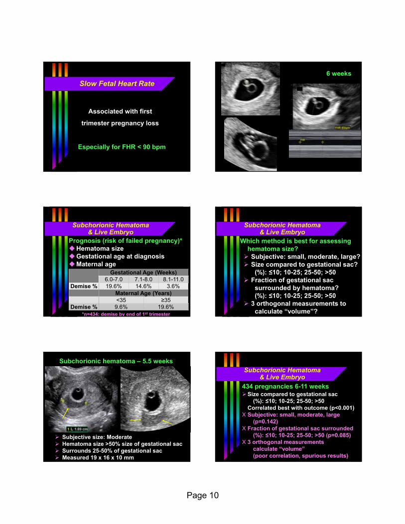
Diaz 5.5 w SCH mod
Subchorionic hematoma – 5.5 weeks
Subjective size: Moderate Hematoma size >50% size of gestational sac Surrounds 25-50% of gestational sac Measured 19 x 16 x 10 mm
Size compared to gestational sac(%): ≤10; 10-25; 25-50; >50
Correlated best with outcome (p<0.001)X Subjective: small, moderate, large
(p=0.142)X Fraction of gestational sac surrounded
(%): ≤10; 10-25; 25-50; >50 (p=0.085)X 3 orthogonal measurements
calculate “volume” (poor correlation, spurious results)
Subchorionic Hematoma& Live Embryo
434 pregnancies 6-11 weeks

Page 11
434 pregnancies 6-11 weeks*
Hematoma Size as Fraction of Gestational Sac Size (p<0.001)≤10% 10-25% 25-50% >50%
Live* 114 112 66 89Demise 7 11 8 27
Demise % 5.8% 8.9% 10.8% 23.3%
*Live at end of 1st trimester
Subchorionic Hematoma& Live Embryo
Hussar SCH large
Subchorionichematoma
6 weeks 12 weeksResolved
Recommended follow up of suspicious but not definitive findings (IPUV*)
Ultrasound, not hCG
7-10 days (in most cases)
Pregnancy Failure
*Intrauterine pregnancy of unknown viability
hCG & No Intrauterine Gestational Sac
Ultrasound diagnosis
Pregnancy of Unknown Location (PUL)hCG & Ultrasound findings:
No intrauterine gestational sac oradnexal mass
Ectopic PregnancyNo intrauterine gestational sac &
findings of ectopic pregnancy
Ectopic Pregnancy
Transvaginal Ultrasound Findings
No intrauterine gestation
Adnexal mass separate from ovary
Tubal ring
Internal yolk sac
Embryo with heartbeat
Free fluid
hCG Discriminatory Level
Rationale for setting this:
If hCG > discriminatory level
and
No intrauterine gestation (IUP) seen
↓Safe to treat suspected ectopic
Will not damage a normal pregnancy
Page 12
hCG Discriminatory Level
Levels proposed (mIU/ml):
6,500 (1981)
3,600 (1985)
1,000-2,000 (1990 and beyond)
Improved equipment and
transvaginal probes
Our Study
Methods:
Transvaginal sonograms 2000-2010
Positive pregnancy test and
No intrauterine fluid collection
Subsequent live intrauterine gestation
hCG at time of no IUP recorded
Pregnancy outcomes collected
hCG levels
hCG N Live at 14w*
< 1000 162 89.9%
1000-1499 19 88.6%
1500-1999 12 86.6%
≥ 2000 9 80.6%
*No difference in miscarriage ratesamong hCG groups (p>0.05)
9 Cases with hCG ≥2000
hCG Pregnancy outcome2215 Term singleton2217 Term twins2374 Term singleton2530 Live at 21w, loss from chorio2539 Term singleton2993 Term singleton4336 Term singleton4476 Live at 7.6w, loss before 14w6567 Live at 7.1w, slow FH, 8w demise
hCG levels
6567 = Highest hCG
with subsequent live IUP
4336 = Highest hCG
with subsequent live born
hCG & Ectopic Pregnancy*
*SRU Consensus Panel on Dx of Early Pregnancy Failure 2012
US Finding Key Points
Nointrauterine
fluid collection
Normaladnexa
• A single hCG in a woman with an “empty” uterus on U/S:
• DOES NOT reliably distinguish between ectopic and intrauterine pregnancy
• DOES NOT definitively exclude a potentially normal IUP (though normal IUP is unlikely if >3000 mIU/ml)
• DOES NOT exclude an ectopic pregnancy(even if <1000 mIU/ml)
• DOES NOT justify presumptive treatment for ectopic pregnancy, using methotrexate or other medical/surgical means
• The concept of a "discriminatory" hCG level – an hCG value for ruling in or ruling out ectopic pregnancy at a single point in time – should not be used to guide management decisions

Page 13
Safe Rule
No matter what the hCG,
definitive interventions, like D&C
or MTX, should be avoided
in suspected but unproven
ectopic pregnancy
Unless patient is unstable,
get f/u ultrasound and/or
hCG before intervention
Ectopic Pregnancy
Transvaginal Ultrasound Findings
No intrauterine gestation
Adnexal mass separate from ovary
Tubal ring
Internal yolk sac
Embryo with heartbeat
Free fluid
Broadbent Ectopicempty uterus
Ectopic Pregnancy
Delcarmen Ectopicempty uterus
Ectopic Pregnancy
231 Ectopic Pregnancies
Ultrasound findings present in 94.8%
Adnexal mass 94.4%
Nonspecific mass 54.1%
Tubal ring (no YS or heartbeat) 24.7%
Yolk sac (no heartbeat) 8.3%
Live embryo 7.4%
Free fluid: Moderate – large 0.4%(no adnexal mass)
Frates et al. J Ultrasound Med 2014
Ectopic Pregnancy
Ultrasound findings Likelihoodof ectopic
Intrauterine pregnancy (IUP) ~0%
No IUP, normal pelvis/simple cyst 1%
No IUP, complex mass 92%
No IUP, tubal ring 95%
Live embryo outside uterus 100%
Page 14
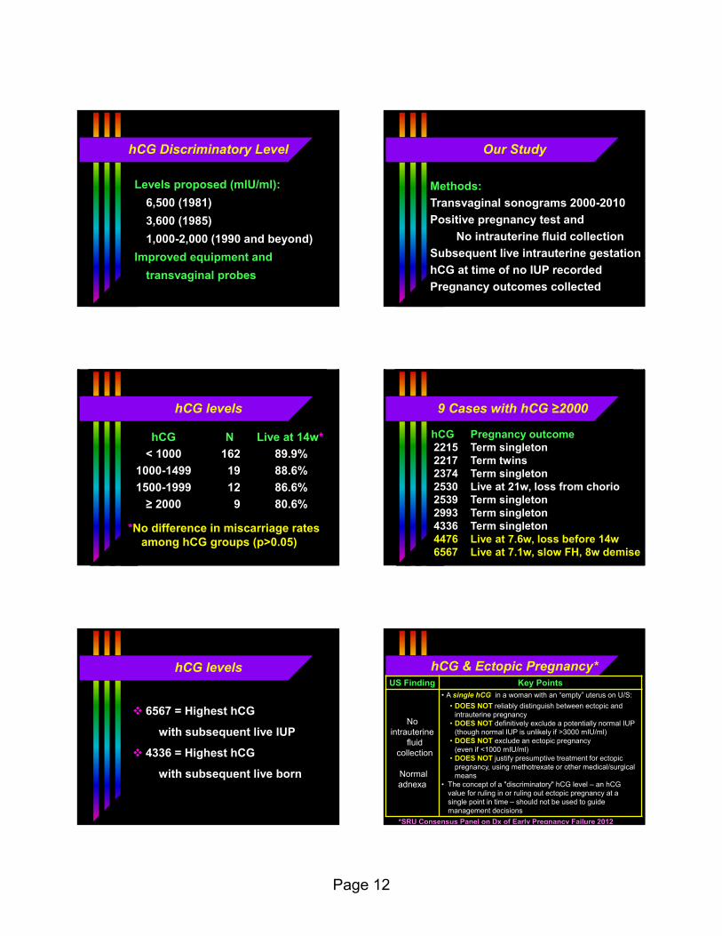
Doppler for Ectopic Pregnancy
Ultrasound findingsAdnexal mass
Color Doppler findingsRim of color around mass
Spectral Doppler findingsLow resistance waveform
CautionIntraovarian lesion with rim ofcolor is likely corpus luteum
Doppler has limited use over and above ultrasound.
If there is an adnexal mass, Doppler does not assist the diagnosis, because the interpretation should already be probable ectopic.
Doppler for Ectopic Pregnancy
Jabba ectopic
Ectopic Pregnancy
Unusual Ectopic Pregnancies
Abdominal
Interstitial (Cornual)
Cervical
Ovarian
Heterotopic (IUP & ectopic)
Cesarean section scar/defect
implantation
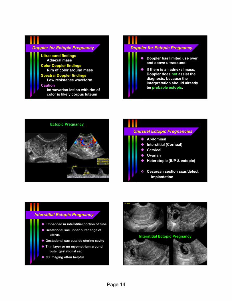
Interstitial Ectopic Pregnancy
Embedded in interstitial portion of tube
Gestational sac upper outer edge of
uterus
Gestational sac outside uterine cavity
Thin layer or no myometrium around
outer gestational sac
3D imaging often helpful
Truong cornual ectopic
Interstitial Ectopic Pregnancy

Page 15
Truong cornual ectopic
Interstitial Ectopic Pregnancy
Rosengarten bicorn IUPmimics interstitial
Pregnancy inBicornuate uterus
Heterotopic Pregnancy
Intrauterine & ectopic pregnanciescoexistant
Incidence~ 1 / 4,000 - 8,000 (natural)10x – 20x higher (Rx infertility)
Ultrasound findingsIntrauterine gestationEctopic pregnancy
Murray heterotopicinjected
Heterotopic pregnancy
Intrauterinegestation
Interstitial ectopicpregnancy
Cervical Ectopic Pregnancy
Gestational sac implanted in the cervix
Differential diagnosis includes
miscarriage in progress if no embryo
with heartbeat
May slide within cervical canal
May pass on short-term follow up
Hancock cx ectopic
CervicalEctopic
Pregnancy