university of zurich - semantic scholar of zurich ... marcus j. schultz, md, phd, fccs, marcel levi,...
TRANSCRIPT

University of ZurichZurich Open Repository and Archive
Winterthurerstr. 190
CH-8057 Zurich
http://www.zora.uzh.ch
Year: 2008
Acute coagulopathy of trauma: hypoperfusion induces systemicanticoagulation and hyperfibrinolysis
Brohi, K; Cohen, M J; Ganter, M T; Schultz, M J; Levi, M; Mackersie, R C; Pittet, J F
Brohi, K; Cohen, M J; Ganter, M T; Schultz, M J; Levi, M; Mackersie, R C; Pittet, J F (2008). Acute coagulopathyof trauma: hypoperfusion induces systemic anticoagulation and hyperfibrinolysis. Journal of Trauma,64(5):1211-1217.Postprint available at:http://www.zora.uzh.ch
Posted at the Zurich Open Repository and Archive, University of Zurich.http://www.zora.uzh.ch
Originally published at:Journal of Trauma 2008, 64(5):1211-1217.

Acute coagulopathy of trauma: hypoperfusion induces systemicanticoagulation and hyperfibrinolysis
Abstract
BACKGROUND: Coagulopathy is present at admission in 25% of trauma patients, is associated withshock and a 5-fold increase in mortality. The coagulopathy has recently been associated with systemicactivation of the protein C pathway. This study was designed to characterize the thrombotic, coagulantand fibrinolytic derangements of trauma-induced shock. METHODS: This was a prospective cohortstudy of major trauma patients admitted to a single trauma center. Blood was drawn within 10 minutesof arrival for analysis of partial thromboplastin and prothrombin times, prothrombin fragments 1 + 2(PF1 + 2), fibrinogen, factor VII, thrombomodulin, protein C, plasminogen activator inhibitor-1 (PAI-1),thrombin activatable fibrinolysis inhibitor (TAFI), tissue plasminogen activator (tPA), and D-dimers.Base deficit was used as a measure of tissue hypoperfusion. RESULTS: Two hundred eight patientswere studied. Systemic hypoperfusion was associated with anticoagulation and hyperfibrinolysis.Coagulation was activated and thrombin generation was related to injury severity, but acidosis did notaffect Factor VII or PF1 + 2 levels. Hypoperfusion-induced increase in soluble thrombomodulin levelswas associated with reduced fibrinogen utilization, reduction in protein C and an increase in TAFI.Hypoperfusion also resulted in hyperfibrinolysis, with raised tPA and D-Dimers, associated with theobserved reduction in PAI-1 and not alterations in TAFI. CONCLUSIONS: Acute coagulopathy oftrauma is associated with systemic hypoperfusion and is characterized by anticoagulation andhyperfibrinolysis. There was no evidence of coagulation factor loss or dysfunction at this time point.Soluble thrombomodulin levels correlate with thrombomodulin activity. Thrombin binding tothrombomodulin contributes to hyperfibrinolysis via activated protein C consumption of PAI-1.

Title:
Acute coagulopathy of trauma: hypoperfusion induces systemic
anticoagulation and hyperfibrinolysis
Short Title: Acute Traumatic Coagulopathy
Authors:
Karim Brohi, FRCS, FRCA, Mitchell J. Cohen, MD, Michael T. Ganter, MD,
Marcus J. Schultz, MD, PhD, FCCS, Marcel Levi, MD, PhD, Robert C.
Mackersie, MD, and Jean-Franc¸ois Pittet, MD
Author affiliations:
From the Department of Surgery, The Royal London Hospital (K.B.), London, United
Kingdom; Departments of Surgery (M.J.C., R.C.M., J.F.P.) and Anesthesia (M.T.G.,
J.F.P.), San Francisco General Hospital, University of California San Francisco, San
Francisco, California; and the Departments of Intensive Care, Laboratory of
Experimental Intensive Care and Anesthesiology (M.J.S.) and Internal Medicine (M.L.),
Academic Medical Center, Amsterdam, The Netherlands.
Corresponding Author:
Karim Brohi
Department of Surgery, The Royal London Hospital
Whitechapel Road, London, E1 1BB, United Kingdom
Phone: 020 7377 7695 Fax: 020 7377 7044
E-mail: [email protected]

Sources of Support:
This work was supported in part by grant R01 GM62188 from the National Institute of
Health. Assays of prothrombin fragments 1+2, thrombomodulin, protein C, tissue
plasminogen activator and plasminogen activator inhibitor-1 were performed by Ely Lilly
and Company. The supporting bodies had no role in the conduct of the study or the
drafting of the manuscript.
Meetings Presented:
Part of this work was presented at ‘Trauma, Shock, Inflammation & Sepsis 2007’,
Munich, Germany, March 2007.

Abstract
Background: Acute traumatic coagulopathy, present on admission in 25% of
trauma patients, is associated with shock and a 5-fold increase in mortality. The
coagulopathy has recently been associated with systemic activation of the
protein C pathway. This study was designed to fully characterize the
coagulopathy of shock and the resultant thrombotic, coagulant and fibrinolytic
derangements.
Methods: This was a prospective cohort study of major trauma patients admitted
to a single trauma center. Blood was drawn within 10 minutes of arrival for
analysis of partial thromboplastin and prothrombin times, prothrombin fragments
1+2 (PF1+2), fibrinogen, factor VII, thrombomodulin, protein C, plasminogen
activator inhibitor-1 (PAI-1), thrombin activatable fibrinolysis inhibitor (TAFI),
tissue plasminogen activator (tPA) and D-dimers. Base deficit was used as a
measure of tissue hypoperfusion.
Results: 208 patients were studied. Systemic hypoperfusion was associated
with anticoagulation and hyperfibrinolysis. Thrombin generation was related to
injury severity, but acidosis did not affect Factor VII or PF1+2 levels.
Hypoperfusion-induced increase in soluble thrombomodulin levels was
associated with reduced fibrinogen utilization, reduction in protein C and an
increase in TAFI. Hypoperfusion also resulted in hyperfibrinolysis, with raised
tPA and D-Dimers. This was associated with the observed reduction in PAI-1
and not alterations in TAFI.

Conclusions: Acute traumatic coagulopathy is due to systemic hypoperfusion
and is characterized by systemic anticoagulation and hyperfibrinolysis. There
was no evidence of coagulation factor loss or dysfunction at this time point.
Soluble thrombomodulin levels appear to correlate with thrombomodulin activity.
Thrombin-thrombomodulin leads to hyperfibrinolysis via activated protein C
consumption of PAI-1.

Introduction
Acute traumatic coagulopathy is present immediately on admission in 25% of
trauma patients and is associated with a 5-fold increase in mortality1. Accepted
causes of traumatic coagulopathy are consumption of clotting factors, acidosis
and hypothermia leading to reduced activity, and dilution from intravenous fluids
and packed cell administration. However acute coagulopathy is present early in
the post-injury phase, prior to fluid administration and in normothermic patients1.
Further, while acidosis per se affects coagulation protease function, clot time and
maximum clot firmness are only impaired at very low pH (<6.8)2.
We have recently demonstrated that only patients who are in shock are
coagulopathic on admission3. Increased severity of hypoperfusion was
associated with an increase in plasma thrombomodulin and a reduction in protein
C levels. This suggests that acute coagulopathy is due to systemic
anticoagulation due to activation of the protein C pathway.
The overall goal of this study was to fully characterize the coagulopathy of shock,
and in particular to examine the interplay of shock, anticoagulation and the
fibrinolytic system. Secondly, we wished to determine whether coagulation factor
consumption or dysfunction due to acidosis were responsible for coagulopathy
prior to massive fluid and blood transfusion. Third, we hypothesized that
thrombomodulin has a central role in traumatic coagulopathy, complexing
thrombin and resulting in anticoagulation and hyperfibrinolysis. Finally, there has

been some debate from basic science studies as to whether the hyperfibrinolysis
resulting from thrombin-thrombomodulin formation is due to activation of
Thrombin Activatable Fibrinolysis Inhibitor (TAFI)4 or activated protein C
consumption of PAI-15,6. We sought to understand which pathway was more
important in clinical practice.
Methods
This was a prospective cohort study of consecutive major trauma patients
admitted to a single level 1 trauma center. The methodology has been described
previously3. Briefly, a 10 ml sample of blood was drawn by a designated member
of the trauma team immediately on admission to the emergency department. The
sample was spun down, plasma extracted and frozen at -80oC. For this study
we assayed plasma levels of Factor VII (+++++), tissue Plasminogen Activator
(tPA) - Asserachrom tPA, Diagnostica Stago (normal range 3-13 ng/ml) and TAFI
ELISA, Enzyme Research Laboratories (normal range: 2.8 - 9.2 mcg/ml) in
addition to previously described measurements of Prothrombin Fragments 1+2
(PF1+2), Fibrinogen, soluble Thrombomodulin (sTM), protein C, Plasminogen
Activator Inhibitor-1 (PAI-1) and D-Dimer levels. For the D-Dimer assay, levels
above 0.22 μg/ml were reported as 0.22 μg/ml.
Data were collected prospectively on patient demographics, injury time,
mechanism (blunt or penetrating) and severity, prehospital fluid administration,
the time of arrival in the trauma room and admission vital signs. In the absence

of a biochemical marker, the injury severity score was used as a surrogate
measure of the degree of tissue injury. A full blood count, coagulation profile and
arterial blood gas were drawn at the same time as the research sample as part of
the standard management of major trauma patients. The degree of shock and
systemic tissue hypoperfusion was assessed with the base deficit. Admission
base deficit is a clinically useful early marker of tissue hypoperfusion in trauma
patients and an admission base deficit greater than 6 mEq/l has previously been
identified as predictive of worse outcome in trauma patients.7,8
Statistical analyses were performed with Microsoft Excel 2003 with the WinSTAT
plug-in. Normal-quantile plots were used to test for normal distribution.
Parametric data are expressed as mean ± 95% confidence intervals. Non-
parametric data are given as median (inter-quartile range). Two-group analysis
was performed with a two-tailed unequal variance Student’s t-test. The χ2 test
was used for dichotomous data analysis. A p-value of 0.05 was chosen to
represent statistical significance.
Results
Blood was drawn on 208 consecutive patients immediately on arrival to the
trauma room. Median prehospital time was 28 minutes (interquartile range - IQR:
23-29) and 150 mls of fluids (0-200) were administered in the field. Median injury
severity score was 17 (9-26) and 25% were penetrating.

Patients without shock (base deficit - BD≤6) were not coagulopathic, regardless
of injury severity (Fig 1, A&B). 2.6% of patients with a BD≤6 had a prolonged PT
(>18s) compared to 19.6% of patients with BD>6 (p=0.001, χ2) and only 1.9% of
patients with a BD≤6 had a prolonged PTT (>60s) compared to 12.5% of patients
with BD>6 (p=0.007, χ2). For patients in shock, both PT and PTT were
prolonged as injury severity increased (Fig 1, A&B). In contrast, there was
activation of fibrinolysis without shock (Fig 1C). However shock increased the
degree of fibrinolysis in all patients (Fig 1D). Acute coagulopathy is therefore a
consequence of shock and is characterized by systemic anticoagulation and
hyperfibrinolysis.
Prothrombin fragments 1+2 (PF1+2) were assayed to assess the degree of
thrombin generation. PF1+2 increased with injury severity (Fig 2A). There was
no reduction in PF1+2 levels as acidosis increased (fig 2B), suggesting that
reduction in coagulation factor activity due to acidosis does not make a
significant contribution to the coagulopathy of shock. Factor VII levels were also
unchanged by acidosis (Fig 2C), and sufficient for clot generation. Combined,
these data also suggest that consumption of factors is not clinically significant at
this time point and does not contribute to acute coagulopathy.
We previously demonstrated that increasing shock with hypoperfusion was
associated with a rise in plasma levels of soluble thrombomodulin (sTM) and a
decrease in protein C levels3. Thrombomodulin complexes with thrombin and

switches it to an anticoagulant function. Thrombin is therefore not free to cleave
fibrinogen to form fibrin. As thrombomodulin levels rise, fibrinogen levels also
rise (figure 3A). Figure 3B shows how with low thrombomodulin levels, there is a
dose-dependent reduction in fibrinogen levels. This effect is abolished when
thrombomodulin is high. Thus in the presence of shock and high
thrombomodulin levels, fibrin production is minimal, regardless of clotting factor
activity.
Further consequences of formation of the thrombin-thrombomodulin complex
include activation of protein C. We did not measure activated protein C in this
study but can demonstrate falling protein C levels with increasing sTM (Fig 3C)
and have previously shown that this is likely to represent protein C activation due
to observed effects on anticoagulation3. Thrombin complexed to thrombomodulin
also activates TAFI, and we can demonstrate an increase in TAFI levels with
increasing sTM (Fig 3D). Together these results support a central role for
thrombomodulin in acute traumatic coagulopathy.
We have demonstrated that systemic fibrinolysis is also a component of this
coagulopathy (Fig 1C). Tissue Plasminogen Activator (tPA) is released from the
endothelium, and was significantly elevated in patients with shock, irrespective of
the amount of thrombin generated (Fig 4, A&B). tPA levels were significantly
higher when PAI-1 was low (Fig 4C) and increasing tPA levels were correlated
with increasing D-Dimers, as expected (Fig 4D).

When present in excess, activated protein C is a potent inhibitor of PAI-14 and
we have previously shown that patients in shock have low levels of PAI-1 and a
direct correlation between protein C and PAI-1 levels, suggesting that protein C
activation leads to PAI-1 consumption3. It has previously been suggested that
the de-inhibition of fibrinolysis seen with protein C is not due to this mechanism
but to a competitive reduction in TAFI activation by the thrombin-thrombomodulin
complex5,6. We can demonstrate this competitive binding of T-TM to either
protein C or TAFI by an inverse correlation between protein C and TAFI levels
(Fig 5A). However while we can demonstrate an inverse relationship between
PAI-1 and the D-Dimer level (Fig 5B) there is no such correlation between TAFI
and D-Dimers (Fig 5C) suggesting that in the clinical setting the protein C - PAI-1
interaction is more important for the observed hyperfibrinolytic state.
Discussion
Acute traumatic coagulopathy occurs in patients who are shocked and is not due
to coagulation factor consumption or dysfunction due to acidosis or dilution.
These factors may be important later in the clinical course, after massive
transfusion or the development of severe acidosis. However shock itself is
associated with a coagulopathy that is due to the systemic activation of
anticoagulant and fibrinolytic pathways.

We have demonstrated previously that the protein C pathway is implicated in this
process3, and show here the central role of thrombomodulin in the conversion of
thrombin from its coagulant role to a regulator of clot formation. Thrombomodulin
is an endothelial protein that is present in normal endothelial cells. Theoretically,
activation leads to increased thrombomodulin expression on the surface of the
endothelium, where it complexes thrombin which cleaves protein C at the
endothelial protein C receptor. This ‘anticoagulant thrombin’ is no longer
available to cleave fibrinogen to form fibrin, as we have demonstrated. This has
significant implications for current practice. All efforts to correct traumatic
coagulopathy are currently directed at augmenting the clotting factor pathway,
through the administration clotting factors and platelets, e.g. fresh frozen plasma9
or recombinant Factor VIIa10. In theory, while patients are shocked and
thrombomodulin is present in excess, thrombin that is generated will be
anticoagulant, and stable clot will not be formed. Although it may be possible to
overwhelm thrombomodulin with massive thrombin generation, this would also be
associated with widespread activation of protein C. This would lead to
consumption of PAI-1 and increased fibrinolysis, breaking down the clot that had
formed. Further, activated protein C has a relatively long half-life11, and the
anticoagulant environment might persist and result in re-bleeding. Further
studies will be needed to ascertain whether this is mechanism is important
following augmentation of the extrinsic pathway during shock.
There has been some debate in the literature about the relationship between
soluble thrombomodulin (sTM) and endothelial-bound thrombomodulin (eTM).
Studies variously suggest that sTM does not reflect endothelial TM activity but is
simply a marker of endothelial injury12, is itself active13,14 or that it is in fact
inhibitory to the accepted role of thrombomodulin15,16. Our data would suggest
that plasma sTM levels do reflect overall thrombomodulin activity, as increased
sTM levels appear to be associated with decreased fibrinogen utilization and
activation of protein C & TAFI.
Finally, we have demonstrated that the increased fibrinolysis associated with
injury is also due to shock and is mediated through de-inhibition of tPA through
the consumption of PAI-1. As mentioned above, it has been suggested that TAFI
is the main driver of fibrinolysis inhibition, and that reduction in TAFI activation by
the competitive binding of protein C to thrombin-thrombomodulin is the
mechanism for de-repression of fibrinolysis with activation of protein C. Although
we can demonstrate an increase in TAFI levels with thrombomodulin, and a
competition between TAFI and protein C, there was no observable correlation
between TAFI and D-Dimer levels. Thus the consumption of PAI-1 by activated
protein C appears to be the more important cause of hyperfibrinolysis.
This study has several limitations that have been alluded to previously3. The PT
and PTT are very crude methods of identifying coagulopathic patients, and do
not describe the global fibrinolytic state at all. Comparing these biochemical

markers to more functional methods such as viscoelastic coagulation analyses
(e.g., thrombelastography™) might reveal more clinically relevant changes in
coagulation. Further, this is an investigation of the state of the coagulation
system at a single time point. Alterations in response to continued hemorrhage
or successful resuscitation are worthy of further study. Previous investigations at
later time points have identified that patients become hypercoagulable17,18 and
are at risk of thromboembolic complications19. It is possible that this is a result of
depletion of protein C following systemic activation, indeed a previous study has
identified admission coagulopathy as an independent risk factor for later venous
thromboembolism20.
In summary we have identified that acute traumatic coagulopathy occurs only in
the presence of shock and is characterized by systemic anticoagulation and
hyperfibrinolysis mediated through the activation of thrombomodulin. This has
significant implications for the management of traumatic hemorrhage, and
suggests that hypoperfusion must be corrected before the coagulation system’s
hemostatic balance can be restored.

References
1. Brohi K, Singh J, Heron M et al. Acute traumatic coagulopathy. J Trauma
2003; 54:1127--1130.
2. Engstrom M, Schott U, Romner B, Reinstrup P. Acidosis impairs the
coagulation: A thromboelastographic study. J Trauma. 2006;61:624-8
3. Brohi K, Cohen MJ, Ganter MT, Matthay MA, Mackersie RC, Pittet JF. Acute
Traumatic Coagulopathy: Initiated by Hypoperfusion, Modulated Through the
Protein C Pathway? Ann Surg 2007;245:818-8
4. Rezaie AR. Vitronectin functions as a cofactor for rapid inhibition of activated
protein C by plasminogen activator inhibitor-1. Implications for the mechanism of
profibrinolytic action of activated protein C. J Biol Chem. 2001; 276:15567--70.
5. Bajzar L, Jain N, Wang P, Walker JB. Thrombin activatable fibrinolysis
inhibitor: Not just an inhibitor of fibrinolysis. Crit Care Med 2004;32:S320-4
6. Bajzar L, Nesheim ME, Tracy PB. The Profibrinolytic Effect.of Activated
Protein C in Clots Formed From Plasma Is TAFI-Dependent. Blood
1996;271:22949-52
7. Rutherford EJ, Morris JA Jr, Reed GW et al. Base deficit stratifies mortality
and determines therapy. J Trauma 1992; 33:417--423.
8. Davis JW, Parks SN, Kaups KL et al. Admission base deficit predicts
transfusion requirements and risk of complications. J Trauma 1996; 41:769--
774.
9. Gonzalez EA, Moore FA, Holcomb JB et al. Fresh frozen plasma should be
given earlier to patients requiring massive transfusion. J Trauma 2007;62:112-9

10. Boffard KD, Riou B, Warren B et al. Recombinant factor VIIa as adjunctive
therapy for bleeding control in severely injured trauma patients: two parallel
randomized, placebo-controlled, double-blind clinical trials. J Trauma 2005; 59:8-
-18.
11. Okajima K, Koga S, Kaji M et al. Effect of protein C and activated protein C
on coagulation and fibrinolysis in normal human subjects. Thromb Haemost
1990;63:48-53.
12. Ishii H, Uchiyama H, Kazama M. Soluble thrombomodulin antigen in
conditioned medium is increased by damage of endothelial cells. Thromb
Haemost. 1991;65:618-23.
13. Ohlin AK, Larsson K, Hansson M. Soluble thrombomodulin activity and
soluble thrombomodulin antigen in plasma. J Thromb Haemost. 2005;3:976-82.
14. Conway EM, Van de Wouwer M, Pollefeyt S et al. The lectin-like domain of
thrombomodulin confers protection from neutrophil-mediated tissue damage by
suppressing adhesion molecule expression via nuclear factor κB and mitogen-
activated protein kinase pathways. J Exp Med 2002; 196:561--564
15. Faust SN, Levin M, Harrison OB et al. Dysfunction of endothelial protein C
activation in severe meningococcal sepsis. N Engl J Med 2001; 345:408--416.
16. Tanaka KA, Fernandez JA, Marzec UM et al. Soluble thrombomodulin is
antithrombotic in the presence of neutralising antibodies to protein C and reduces
circulating activated protein C levels in primates. Br J Haematol. 2006;132:197-
203.
17. Boldt J, Papsdorf M, Rothe A et al. Changes of the hemostatic network in
critically ill patients--is there a difference between sepsis, trauma, and
neurosurgery patients? Crit Care Med 2000; 28:445--450.
18. Gando S. Serial studies of protein C in trauma patients. Jpn J Thromb
Hemost 1996; 7:312--318.
19. Geerts WH, Code KI, Jay RM et al. A prospective study of venous
thromboembolism after major trauma. N Engl J Med 1994; 331:1601--1606.
20. Knudson MM, Collins JA, Goodman SB et al. Thromboembolism following
multiple trauma. J Trauma 1992; 32:2--11.

Fig 1: Shock induces anticoagulation and hyperfibrinolysis
20
30
40
50
60
70
<16 16-30 >30
ISS
PTT BD≤6
BD>6
* +
10
12
14
16
18
20
22
<16 16-30 >30
Injury Severity Score
Prot
hrom
bin
Tim
e (s
)
BD≤6BD>6
+
+**
A
B
0
5
10
15
20
<16 16-30 >30
Injury Severity Score
D-D
imer
BD≤6BD>6
*
*+*
C
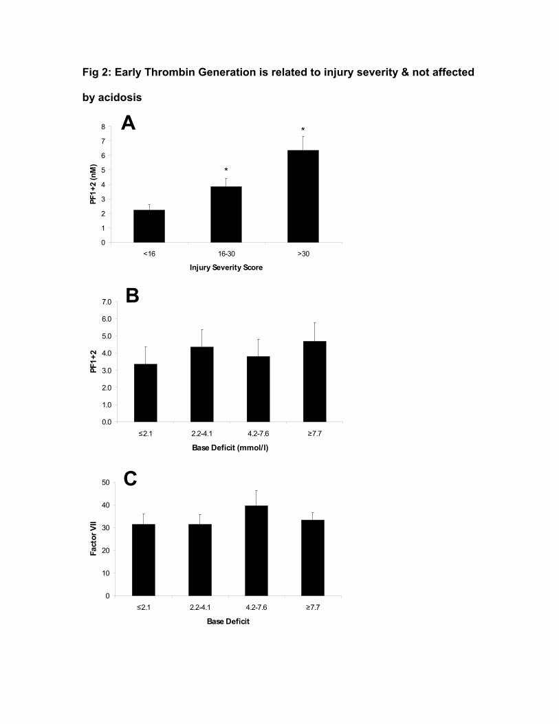
Fig 2: Early Thrombin Generation is related to injury severity & not affected
by acidosis
0
1
2
3
4
5
6
7
8
<16 16-30 >30
Injury Severity Score
PF1+
2 (n
M)
*
*
A
0.0
1.0
2.0
3.0
4.0
5.0
6.0
7.0
≤2.1 2.2-4.1 4.2-7.6 ≥7.7
Base Deficit (mmol/l)
PF1+
2
B
0
10
20
30
40
50
≤2.1 2.2-4.1 4.2-7.6 ≥7.7
Base Deficit
Fact
or V
II
C

Fig 3: Increased soluble thrombomodulin is associated with reduced fibrinogen utilization, reduction in protein C
(activation) and an increase in TAFI.
1.5
2.0
2.5
3.0
3.5
<28 28-34 34-43 >43
Thrombomodulin
Fibr
inog
en
*
1.8
2.2
2.6
3.0
3.4
3.8
<2 2-4 >4
Prothrombin fragments 1+2 (nM)
Fibr
inog
en
TM≤43TM>43
*
50
60
70
80
90
100
<28 28-34 34-43 >43
Thrombomodulin (ng/ml)
Prot
ein
C (%
)
*
4.0
4.5
5.0
5.5
6.0
6.5
7.0
7.5
<28 28-33 34-42 >42
Thrombomodulin
TAFI
**
A B
C B D

0
10
20
30
40
50
60
≤2.1 2.2-4.1 4.2-7.6 ≥7.7
BD
tPA
*
0
10
20
30
40
50
60
<2 2-4 >4
PF1+2
tPA BD≤6
BD>6
**
+
*
0
10
20
30
40
50
<1.1 1.1-5.1 5.2-13.1 >13.1
PAI-1
tPA * *
*
0
2
4
6
8
10
12
<12.3 12.3-18.6 18.7-28.7 >28.7
tPA
D-D
imer
s
**
D
BFig 4: Activation of fibrinolysis
A
C

Fig 5: Hyperfibrinolysis is due to consumption of PAI-1, not a reduction in
TAFI
4.0
4.5
5.0
5.5
6.0
6.5
7.0
<58 58-78 79-101 >102
Protein C
TAFI
*
A
0.0
5.0
10.0
15.0
<1.1 1.1-5.1 5.2-13.1 >13.1
PAI-1
D-D
imer
s **
*
0
2
4
6
8
10
12
<4.7 4.7-5.8 5.9-7.1 >7.1
TAFI
D-D
imer
s
B
C