university of nairobi college of health sciences …
TRANSCRIPT

UNIVERSITY OF NAIROBI
COLLEGE OF HEALTH SCIENCES
SCHOOL OF MEDICINE
DEPARTMENT OF OBSTETRICS AND GYNAECOLOGY
THE PATTERN OF LAPARASCOPIC FINDINGS AND MANAGEMENT OFFERED
TO WOMEN WITH INFERTILITY AT THE KENYATTA NATIONAL HOSPITAL
2009-2013
DR. GIDEON MUTISO MUTUA

ii
THE PATTERN OF LAPARASCOPIC FINDINGS AND MANAGEMENT OFFERED
TO WOMEN WITH INFERTILITY AT THE KENYATTA NATIONAL HOSPITAL
2009-2013
A RETROSPECTIVE CROSS SECTIONAL STUDY
A DISSERTATION IN PARTIAL FULFILMENT OF A DEGREE IN MASTERS OF
MEDICINE IN OBSTETRICS AND GYNECOLOGY
Principal Investigator:
Dr. Gideon Mutiso Mutua. M.B.Ch.B
Senior House Officer and Student, Department of Obstetrics and Gynecology, University of
Nairobi.
Reg No. H58/70608/2011
Supervisors:
1. Prof. S.B.O. Ojwang’, M.B.Ch.B, M. Med OBSGYN, Dip Gynaeoncology
Professor, Department of Obstetrics and Gynecology, College of Health Sciences,
University of Nairobi.
Consultant Obstetrician and Gynecologist, Kenyatta National Hospital.
2. Dr. Frank Kagema, M.B.Ch.B, M. Med OBSGYN
Lecturer, Department of Obstetrics and Gynecology, College of Health Sciences,
University of Nairobi.
Consultant Obstetrician and Gynecologist, Kenyatta National Hospital.

iii
DECLARATION
I declare that this is my original work and has not been presented for a degree in any other
university.
DR. GIDEON MUTISO MUTUA
STUDENT MASTERS OF MEDICINE IN OBSTETRICS AND GYNAECOLOGY
DEPARTMENT OF OBSTETRICS AND GYNAECOLOGY
SCHOOL OF MEDICINE, COLLEGE OF HEALTH SCIENCES
UNIVERSITY OF NAIROBI
Signed ………………………………………………Date………………………………

iv
CERTIFICATE OF SUPERVISION
This is to certify that the thesis presented in this book was researched upon by Dr. Gideon
Mutiso Mutua under my guidance and supervision, and the thesis is submitted with my
approval.
PROF. S.B.O. OJWANG’, M.B.CH.B, M. MED OBSGYN, DIP GYNAEONCOLOGY
PROFESSOR, DEPARTMENT OF OBSTETRICS AND GYNECOLOGY,
COLLEGE OF HEALTH SCIENCES, UNIVERSITY OF NAIROBI.
CONSULTANT OBSTETRICIAN AND GYNECOLOGIST, KENYATTA NATIONAL
HOSPITAL.
Signed…………………………………………………..Date……………………………………
DR. FRANK KAGEMA,M.B.CH.B, M. MED OBSGYN
LECTURER, DEPARTMENT OF OBSTETRICS AND GYNECOLOGY,
COLLEGE OF HEALTH SCIENCES, UNIVERSITY OF NAIROBI.
CONSULTANT OBSTETRICIAN AND GYNECOLOGIST, KENYATTA NATIONAL
HOSPITAL.
Signed ……………………………………. …………….Date…………………………………

v
CERTIFICATE OF AUTHENTICITY
This is to certify that Dr. Gideon Mutiso Mutua, a masters of medicine in obstetrics and
gynecology student registration number H58/70608/2011, researched upon this thesis in the
Department of Obstetrics and Gynecology, School of Medicine ,College of Health Sciences,
University of Nairobi ,under the guidance and supervision of Prof. S.B.Ojwang’ and Dr Frank
Kagema.
PROF. ZAHIDA QURESHI
ASSOCIATE PROFESSOR OF OBSTETRICS AND GYNAECOLOGY,
CHAIRPERSON DEPARTMENT OF OBSTETRICS AND GYNAECOLOGY,
SCHOOL OF MEDICINE, COLLEGE OF HEALTH SCIENCES,
UNIVERSITY OF NAIROBI
Signed……………………………………………….Date ……………………………………..

vi
ACKNOWLEDGEMENTS
First of all I wish to thank the Lord Jesus for being my Ebenezer. I would also like to express my
gratitude to the following:
To my supervisors Prof. S.B.O.Ojwang’ and Dr. Frank Kagema for their invaluable advice and
guidance.
To my wife, Beatrice Nyawira Muthui and our two sons Joshua Mutua and John Muthui for
their ceaseless support, encouragement and understanding.

vii
List of Abbreviations
ART Assisted reproductive technology
HSG Hysterosalpingography
ICSI Intra-cytoplasmic sperm injection
IHH Idiopathic hypothalamic hypogonadism
IUD Intrauterine device
IUI Intra uterine insemination
IVF In-Vitro Fertilization
KNH Kenyatta National Hospital
LEEP Loop electrosurgical excision procedure
MTRH Moi Teaching and Referral Hospital
PCOS Polycystic ovary syndrome
PCR Polymerase Chain Reaction
PID Pelvic inflammatory disease
POF Premature ovarian failure
SLE Systemic lupus erythematosus
STI Sexually Transmitted Infection
UON University of Nairobi
WHO World Health Organization

viii
LIST OF FIGURES AND TABLES
Figure 1. File retrieval and analysis of records of women with infertility …………....………..19
Table 1. Socio-demographic characteristics of women with infertility………………………....20
Figure 2. Types of infertility of women………..………………………………………………..21
Table 2. Duration of infertility………………………………………………………………….21
Figure 3: Seminalysis abnormalities of husbands of women with infertility…………………...22
Table 3. History of presenting illness of women with infertility………………………………..23
Table 4. Pre-operative diagnosis of women with infertility…………………………………….24
Table 5.Laparoscopic findings of women with infertility……………………………………....24
Table 6. Association of patient characteristics and tubal blockage……………………………..25
Table 7. Association of patient characteristics and endometriosis……………………………...26
Table 8.Comparison between pre-operative and intra-operative diagnosis………………...….27
Table 9.Management offered to women with infertility……………………………………..…28
Table 10: Complications in women with infertility who underwent laparoscopy……………...29

ix
DEFINITIONS
1. Fecundity is the probability of achieving a live birth in one menstrual cycle.
2. Fecundability is the probability of being pregnant in a single menstrual cycle.
3. Infertility is the inability of a couple to conceive within one year of unprotected
intercourse of reasonable frequency. Reasonable frequency in this context refers to sexual
frequency of 2-3 times a week.
4. Male infertility is inability of a man to achieve a conception with his partner after 12
months of regular unprotected sexual intercourse.
5. Primary Infertility applies to those who have never conceived, whereas secondary
Infertility designates those who have conceived at some time in the past.
6. Sterility is an intrinsic inability to achieve pregnancy.
7. Sub-fertility occurs when the couple has difficulty in conceiving because both partners
have reduced fecundity.
8. Unexplained Infertility is the diagnosis given to couples after a thorough evaluation has
not revealed a cause.

x
TABLE OF CONTENTS
List of Abbreviations……………………………………………………..………………….vii
List of figures and tables………………………………………………………..…….……..viii
Definitions……………………………………………………………………………………ix
Abstract…………………………………………………………………..…………………..xi
Introduction and Literature review……………………………………………..………….1
Conceptual framework………………………………………………………..…………….10
Problem Statement and Justification………….. …………………………..………………12
Research Question…………………………………………………………...………………13
Objectives……………………………………………………………………...……………..13
Main objective…………………………………………………………….…………………..13
Specific objectives…………………………………………………………...………………..13
Materials and Methods……………………………………………………………………...14
Study design……………………………………………………………………………14
Study site and setting…………………………………………….…………………….14
Study population……………………………………………………………………….14
Inclusion criteria……………………………………………….…………………14
Exclusion criteria…………………………………………..……………………..14
Study sample size and sampling procedure……………………………..……………..15
Data variables…………………………………………………………………………..16
Data collection and management…………………………………………..…………..16
Data analysis…………………………………………………………...……………….17
Study Limitations………………………………………………………….…………………18
Ethical Considerations………………………………………………………………………18
Results……………………………………………………………….……………………….19
Discussion……………………………………………………………….……………………29
Conclusion……………………………………………………………………...…………….34
Recommendation…………………………………………………………...………………..35
References…………………………………………………………..………………………..36
Appendices…………………………………………………………………..……………….41
Appendix (I) Data Retrieval Form……………………………………………………………41
Appendix (III) Ethics Letter…………………………………………………………………..45

xi
ABSTRACT
Background
Infertility is a common condition with important medical and socioeconomic implications.
Infertility affects 10 to 15 percent of reproductive-aged couples worldwide. In Kenya the exact
magnitude is not known although according to the Infertility in Kenya Survey Report 2008,
gynecologists spent 31% of their consultations on infertility in both Kenyatta National Hospital
and Moi Teaching and Referral Hospital, the two largest hospitals in Kenya. Laparoscopy is the
gold standard approach for direct inspection and assessment of pelvic pathology. It allows both
diagnosis and immediate surgery for conditions such as tubal blockage, pelvic adhesions and
endometriosis.
Methodology
The main objective of this retrospective cross sectional study was to describe the pattern of
laparoscopic findings and management offered to women with infertility at the Kenyatta
National Hospital. A total of 402 patients’ records of women with infertility who underwent
laparoscopy between January 2009 and December 2013 were analyzed. The information in the
records was retrieved using a pre-tested and precoded data retrieval form. Data entry was made
and analyzed after verification using SPSS version 21.0
Results
The mean age of the women with infertility was 32.0 years which shows an increasing age shift.
The main laparoscopic finding were tubal blockage (74.40%), endometriosis (11.20%) and
genital tuberculosis (5.22%). Genital Tuberculosis, pelvic adhesions and endometriosis were the
intra-operative diagnoses most likely to be missed pre-operatively with kappa values of 0.0,
0.002 and 0.1 respectively. Nearly a third of all patients (29.67%) were eventually recommended
to pursue Assisted Reproductive Technologies (ART) services.
Conclusion and Recommendations
There is a shift in the mean age of women with infertility therefore prompt evaluation and
appropriate care is critical. Tubal blockage is an important cause of infertility as are
endometriosis and genital tuberculosis. There should be a standardized way of evaluating and
reporting laparascopic findings such as endometriosis since this has implications on
management.The general evaluation of women with infertility should include endometriosis and
genital tuberculosis.There is need for affordable low cost ART services in Kenya as envisioned
in the National Reproductive Health Strategy 2009-2015 in KNH since it is not only the premier
referral and teaching hospital in the country and region but also the largest hospital in eastern
Africa.

1
INTRODUCTION AND LITERATURE REVIEW
Infertility is a common condition with important medical and socio-economic implications. In
addition, infertility treatment is not covered by insurance. Infertility is defined as failure of a
couple to conceive after twelve months of regular intercourse without use of contraception (1,2).
Fecundability is the probability of achieving a pregnancy in one menstrual cycle (2).
Infertility can either be primary or secondary. Primary infertility refers to no prior pregnancies
while secondary infertility refers to infertility following at least one prior conception (2).
Infertility affects 10 to 15 percent of reproductive-aged couples worldwide (2). In Kenya the
exact magnitude is not known although in a national survey, gynecologists spent 31% of their
consultations on infertility in both Kenyatta National Hospital (KNH) and Moi Teaching and
Referral Hospital (MTRH) (3). An earlier study by Mati et al had found that 60% of patients in
the Gynecology Outpatient Clinic at KNH presented with infertility (4).
Approximately 50 % of women will be pregnant at 3 months, 75 % will be pregnant at 6 months
and more than 85 % will be pregnant by 1 year (2). Globally, Secondary infertility is more
common than primary infertility (2). This is also true in Kenya (5). Most couples are more
correctly considered to be subfertile, rather than infertile, as they will ultimately conceive if
given enough time (2). This concept of subfertility can be reassuring to couples. However, there
are exceptions, such as in bilaterally obstructed tubes or azoospermia (2).
Infertility is a unique medical condition since it involves a couple and as such both need to be
involved since the treatment modality would be determined by the couple’s individual fertility
prospects.The evaluation of a couple with infertility involves the assessment of four key aspects
for fertility: the sperm (male factor), the oocyte (ovulatory factor and ovarian reserve), transport
and implantation of ova (pelvic factor including fallopian tubes) and the uterus (2).

2
Etiology of Infertility in the Female
Successful pregnancy requires a complex sequence of events including ovulation, ovum pick-up
by a fallopian tube, fertilization, transport of a fertilized ovum into the uterus and implantation
into a receptive uterine cavity. In addition, sperm of adequate number and quality must be
deposited at the cervix near the time of ovulation.
In general, infertility can be attributed to the female partner one third of the time, the male
partner one third of the time and both partners in the remaining one third (2).
In a WHO study of 8502 infertile couples the most common factors of female infertility
included; ovulatory disorders (25%), tubal blockage and other tubal abnormalities (22%),
endometriosis (15%), pelvic adhesions (12%) and hyperprolactinemia (7%) (6).
In Kenya, Okumu found the causes of infertility in the female to be pelvic adhesions and
bilateral tubal obstruction (61%), anovulation (16%), hyperprolactinemia (10%) and ovulatory
oligomenorrhea at 6% (7).
When ovulation is infrequent (oligoovulation) or absent (anovulation) the number of oocytes
available for fertilization is reduced. Women with regular monthly menses and menstrual
molimina such as breast tenderness, dysmenorrhea and bloating generally have ovulatory cycles.
If menses and molimina are irregular or absent, pregnancy or other conditions associated with
oligoovulation/anovulation are likely to be the cause.
Ovulatory dysfunction may be caused by abnormalities in the hypothalamus, anterior pituitary,
or ovaries (2). Hypothalamic dysfunction or improper migration of the hypothalamic
gonadotropin-releasing hormone neurons may be inherited; such as in Idiopathic Hypothalamic
Hypogonadism (IHH) or Kallman syndrome. Other hypothalamic disorders may be due to
lifestyle, for example, excessive exercise, eating disorders, or stress. Thyroid disease and

3
hyperprolactinemia may also contribute to menstrual disturbances (2,6). Other important causes
of ovulatory disorders are polycystic ovary syndrome (PCOS) and medication such as
contraceptive pills.
Infertility is also linked to age due to the loss of viable oocytes with increasing age. At
midgestation, a normal human female fetus has approximately seven million oocytes, which will
decrease to between two and three million by birth. Ongoing atresia of nondominant follicles
proceeds throughout a woman's reproductive life span, with approximately 300,000 follicles at
puberty and less than 1,000 follicles at the onset of menopause (2). The rate of follicle loss
accelerates after the mid-thirties (8).
Tubal and pelvic factors are important causes of infertility (2, 6). Tubal disease may be caused
by pelvic infection, endometriosis or prior pelvic surgery. Adhesions can prevent normal tubal
movement, ovum pick-up and transport of the fertilized egg into the uterus (2).
A history of pelvic inflammatory disease (PID), abortion and Sexually Transmitted infection
(STI) is suspicious for damage to the fallopian tubes or pelvic adhesions. In their study Gachuno
et al showed that a history of STI and abortion was associated with tubal infertility (9).
The most common causes of tubal disease are infection with Chlamydia trachomatis or Neisseria
gonorrhea (2). Tuberculosis is a common cause of both tubal and intrauterine disease in countries
with endemic infection. Tubal infertility has been estimated to follow in 12 %, 23 %, and 54 %
of women following one, two, or three cases of PID, respectively (2). Conversely, a considerable
number of patients have been shown to have tubal damage with no history of prior PID (2).
Endometriosis is thought to be responsible for 10-25 % of infertility (2, 6, 10,11). Endometriosis
is defined as the presence of endometrial glands and stroma at extrauterine sites. These ectopic

4
endometrial implants are usually located in the pelvis but can occur nearly anywhere in the body
(10). It is a common estrogen dependent disorder which may present with chronic pelvic pain
and infertility (10). It may also be symptomless.
Endometriosis results in inflammation and chronic bleeding within implants leading to
obstruction of fallopian tubes or development of severe pelvic adhesions (2, 11). The mechanism
for impaired fertility may involve anatomic distortion from pelvic adhesions and endometriomas
or production of substances such as cytokines which are "hostile" to normal ovarian function
(10). Thus ovulation, fertilization and implantation may be impeded (10,11).
Congenital and acquired uterine abnormalities can lead to infertility. Acquired anomalies include
fibroids, intrauterine polyps and Asherman syndrome (2). Congenital uterine anomalies include
uterine septum, bicornuate uterus, unicornuate uterus and uterine didelphys. Septate uterus is
associated with the poorest reproductive outcome (12).
Fibroids may prevent normal implantation depending on their size and location (2, 13). Fibroids
may obstruct a fallopian tube, distort the uterine cavity or fill the uterine cavity. The less vascular
endometrium and myometrial dysfunctional contractility contribute to lower rates of successful
pregnancy (2).
Asherman Syndrome, the presence of intrauterine adhesions or synechiae can lead to infertility
(2). It occurs most frequently in women with a history of uterine dilatation and curettage.
Endometrial polyps are estimated to be present in 3 to 5 percent of infertile women (2). Their
removal has been shown to improve pregnancy rates following intrauterine insemination (2, 14).

5
Abnormalities in cervical mucus production are most frequently observed in women who have
undergone cryosurgery, cervical conization, or Loop Electrosurgical Excision Procedure (LEEP)
for treatment of an abnormal Pap smear. Cervical infection is thought to negatively impact
mucus quality (2,14,15). Causative microorganisms include Chlamydia trachomatis, Neisseria
gonorrhea, Ureaplasma urealyticum and Mycoplasma hominis (2).
Other causes of female infertility include genetic disorders and autoimmune diseases.The most
common aneuploidies associated with infertility are 45, X (Turner syndrome) in women and 47,
XXY (Klinefelter syndrome) in men (16). The former is associated with gonadal dysgenesis
while the latter is associated with primary hypogonadism in the male. Gonadal dysgenesis is the
most common cause of premature ovarian failure (POF) (2).
Some women with premature ovarian failure have been found to have autoimmune diseases such
as systemic lupus erythematosus (SLE), celiac disease and myasthenia gravis. Such women may
have autoimmune oophoritis as part of polyglandular autoimmune failure (16).
Unexplained infertility is the diagnosis given to couples after a thorough evaluation has not
revealed a cause (2). It may be due to small contributions from multiple factors such as subtle
oocyte and sperm dysfunction (17).
Laparoscopic Findings and Treatment Options for Women with Infertility
Laparoscopy is the gold standard approach for direct inspection and assessment of pelvic
pathology (2). It allows both diagnosis and immediate surgery for conditions such as tubal
blockage, pelvic adhesions and endometriosis. Chromotubation is performed for assessment of
tubal patency. A dilute dye is injected through an acorn cannula on the cervix or a balloon
catheter positioned in the uterus and then tubal spill evaluated through the laparoscope (2).

6
Since laparoscopy is an invasive procedure, hysterosalpingography (HSG) and ultrasonography
are performed as part of the initial infertility evaluation.
Laparoscopy is more superior in the diagnostic work up of infertility in addition to providing an
opportunity to institute immediate treatment. Mbura and Mgaya in their study at Muhimbili
Medical Centre showed that HSG has a 50% failure rate in detecting pelvic adhesions (18).
Gichuhi in his study at KNH also showed that HSG demonstrated tubal patency in 44.7% of
patients while laparoscopy demonstrated patency on 47% of patients. In addition, the same study
showed corneal occlusion on 26.2% of patients against 31.9% by laparoscopy (19).
A stepwise approach where HSG is done first and then laparoscopy is more cost-effective
(2,20). Furthermore HSG and laparoscopy tend also to be complimentary for diagnostic work up.
Laparoscopic surgery is better than laparotomy for a number of reasons: the surgery is minimal
and thus shorter hospital stay, there are less adhesions which is important in infertility since
adhesions contribute to infertility (2). In addition, there tends to be a fair agreement in the
assessment of tubal patency and occlusion between laparoscopy and laparotomy as shown by
Ouko and Ndavi in a study in KNH (5).
Lastly, direct visualization by laparoscopy is the gold standard diagnosis of endometriosis (1).
Granted that laparoscopic diagnosis of endometriosis does not always correlate with the
histopathological diagnosis as shown by Sikolia et al in their study at Aga Khan Hospital,
negative histopathology does not rule endometriosis (21). In cases where laparoscopy does not
yield sufficient access or bleeding is not adequately controlled conversion to laparotomy is done
to optimize treatment. Laparoscopic surgical treatment for women with infertility includes
several procedures.

7
Laparoscopy has also been shown to pick pathology which has been missed on HSG and
therefore institute appropriate surgical treatment at once or refer patients early for other
treatment options such as ART. Early referral is critical for older women since the ability to
conceive in a woman diminishes with advancing age (22). Corson and his colleagues found that
in a 100 patients with a negative reproductive work-up up to the point of laparoscopy, 68 had
significant pathology: intrinsic tubal disease 24, peritubal adhesive disease 34, and endometriosis
43, some in combination.
In cases where uterine synechiae or submucosal fibroid is suspected, hysteroscopy can also be
performed to evaluate the uterine cavity and resection done. Hysteroscopy is to be performed in
cases where submucosal fibroid or polyp occupies less than 50% of the intrauterine cavity from a
prior evaluation such as with HSG (2,20).
In a study of 281 women by Heise et al, 46 women (16.4%) were referred for laparoscopy after
initial HSG. Eight (17.3%) of them were noted to have unilateral tubal blockage while 28
(60.8%) had bilateral tubal blockage. Unilateral and bilateral salpingectomy was done
respectively during laparoscopy. Salpingolysis was performed on 7 (15.2%) women while 3
(6.7%) women had untreatable adhesions (23).
Distal tubal blockage is treated by fimbrioplasty or salpingectomy. In an evaluation of 35 women
who had undergone laparoscopic fimbrioplasty, Audebert et al found the intrauterine pregnancy
rate was 51%, live birth rate was 37% and ectopic pregnancy rate was 23% after two years (24).
Terminal salpingostomy is performed to relieve tubal obstruction associated with hydrosalpinx.
Efficacy for improving fertility depends on ampullary dilation, presence of mucosal folds,
percentage of ciliated cells in the fimbrial end, and peritubal adhesions. Salpingectomy is

8
recommended for significant adnexal adhesions or where hydrosalpinges are dilated more than 3
cm especially in preparation for IVF (2,20).
A Cochrane review including three randomized trials showed the odds of pregnancy were
increased with laparoscopic salpingectomy for hydrosalpinges prior to IVF (OR = 1.75, 95% CI
1.07-2.86), as were the odds of ongoing pregnancy/live birth (OR = 2.13, 95% CI 1.24-3.65)
(25).
Tubal anastomosis is done for reversal of sterilization, midtubal block secondary to pathology,
tubal occlusion from ectopic pregnancy and salpingitis isthmica nodosa. Rock et al found that the
pregnancy rate after tubal anastomosis is 75 % in women with tubal length of 4 cm or more, but
only 19 % in those with shorter tubes (26).
Pelvic adhesions especially peri-adnexal adhesions are treated through adhesiolysis which has
been shown to improve pregnancy rates (27). Endometriosis can be ablated or excised
laparoscopically. The most effective treatment of endometriomas is excision. Medical therapy
for endometriomas larger than 1 cm has been shown not to be effective (28). In addition to
improving pregnancy outcomes in endometriosis, laparoscopic excision also relieves pain (28).
Laparoscopic ovarian drilling is done for patients with polycystic ovary syndrome (PCOS)-
related anovulatory infertility where medical treatment has failed (2).
Laparoscopy has some complications such as visceral perforation and bowel injury. Ndovi in his
study in KNH found the rate of complications to be 1.2% (29). Carbon dioxide subcutaneous
emphysema and carbon dioxide gas embolism may also occur as highlighted by Obura (30).

9
Etiology of Infertility in the Male
The evaluation of infertility needs to involve the couple as either of them or both may be the
cause of infertility. In addition, involvement of the couple together from the beginning is critical
for them to support each other emotionally and financially. Causes of male infertility include
abnormalities of sperm production, abnormalities of sperm function and obstruction of the
ductal outflow tract (2). In addition to history and physical examination, seminalysis is the core
investigation in the evaluation of male infertility. Additional investigations are guided by the
history and physical examination (2,6).

10
CONCEPTUAL FRAMEWORK
Infertility in a couple may be due to female, male, combined causes or unexpelained causes. In
the evaluation of infertility the couple is evaluated together. The evaluation begins with a history
and then physical examination followed by investigations and finally the appropriate treatment
and care offered to the couple.
In the evalauation of the female a careful history and physical examination which then guides the
investigations to be carried out is done. The investigations done should in most cases be used to
confirm or build up on the diagnosis made in the history and physical examination.
The causes of female infertility may be tubal factors and ovulatory causes which may manifest
as polycystic ovarian syndrome (PCOS). The other causes are endometriosis, genital
tuberculosis. These causes lead to the situation of infertility as shown below in the conceptual
framework.
The causes of infertility or the diagnoses may be confirmed or found at laparoscopy as tubal
blockage, pelvic adhesions, polycystic ovaries, endometriosis. In addition other findings at
laparoscopy may be uterine malformations or anomalies such as hypoplastic uterus and asherman
syndrome seen at hysteroscopy. Tubal blockage may also manifest with hydrosalpinges and
peritubal adhesions.
Once the diagnosis or impression is made at laparoscopy various procedures (interventions) are
then made. These include tuboplasty for tubal blockage, adhesiolysis for pelvic adhesions,
excision and ablation for endometriosis and hysteroscopy for asherman syndrome (see
conceptual framework below).

11
CONCEPTUAL FRAMEWORK
Intervention
Laparoscopic
Findings
Situation
Causes
Adhesiolysis Tuboplasty
Salpingectomy Excision
Tubal blockage Hydrosalpinges Endometriosis Adhesions
INFERTILITY
Ovulatory Factors Tubal Factors Pelvic adhesions Endometriosis
12
PROBLEM STATEMENT AND JUSTIFICATION
Infertility is one of the most common reasons for gynecology visits not only globally but also in
Kenya (2, 4). Infertility is unique in that it involves a couple. It cuts across all racial, religious
socio-economic strata of life and has significant psychological, social and economic impact on
the individual. Infertility is also one of the nine components of the National Reproductive Health
Policy of Kenya (31). The implementation of this policy with regard to infertility is further
elaborated in the National Reproductive Health Strategy 2009-2015 (32).
There are various studies done globally on the subject of infertility on the etiology and treatment
modality at or after laparoscopy. However, there are few local studies on laparoscopic findings
and management offered to women with infertility.
Lastly, advances in medical technology have brought to the fore various diagnostic and treatment
options for infertility such as ART. In addition, laparoscopy techniques for both diagnosis of
infertility and treatment have improved over time. The purpose of this study therefore was to fill
this knowledge gap in Kenya and provide a basis for more studies in the field in future. This was
a retrospective cross sectional study because of time factor. This study helped audit laparscopic
services offered to gynecology patients with the aim of providing optimal services.
In addition, it is hoped that this study will help other centers in the country that have or intend to
have this facility improve on the experience gained in Kenyatta National Hospital (KNH).
Currently, KNH and Moi Teaching and Referral Hospital (MTRH) are the only public facilities
in Kenya that offer laparoscopic services for infertility management.

13
RESEARCH QUESTION
What is the pattern of laparoscopic findings and management offered to women with
infertility at the Kenyatta National Hospital?
OBJECTIVES
Main objective
To describe the pattern of laparoscopic findings and management offered to women with
infertility at the Kenyatta National Hospital
Specific objectives
1. To describe the sociodemographic characteristics of women with infertility undergoing
laparoscopy at the Kenyatta National Hospital.
2. To describe the pattern of laparoscopic findings in women with infertility at the Kenyatta
National Hospital.
3. To compare the pre-operative and post-operative diagnosis in women undergoing
laparascopy for infertility at the Kenyatta National Hospital.
4. To describe the management offered to women with infertility after undergoing
laparoscopy at the Kenyatta National Hospital.
5. To describe complications in women who were done laparoscopy for infertility at the
Kenyatta National Hospital.
14
MATERIALS AND METHODS
Study design
This was a hospital based retrospective descriptive cross sectional study where a total of 402
patient records of women who underwent laparoscopy between January 2009 and December
2013 were analyzed.
Study site and Setting
The study was carried out at Kenyatta National Hospital; located 3.5 km from Nairobi Central
Business District. Kenyatta National Hospital (KNH) has a bed capacity of 1900 and is the
largest hospital in East and Central Africa. It is a teaching and referral facility that serves Kenya
and other neighboring countries.
Study population
The study population was women who presented with infertility and underwent laparoscopy.
Patients with infertility are seen in the Gynecology Outpateint Clinic (GOPC) and the Infertility
clinic at the Kenyatta National Hospital (KNH) outpatient department. The patients with an
indication for laparoscopy are then admitted to ward IB for preparation for surgery.
Inclusion criteria
The following inclusion criteria was used:
Women with infertility who underwent laparoscopy.
Patients with complete information with regard to demographic characteristics, complete
documentation on laparoscopic findings and treatment options offered.
Women with infertility who underwent hysteroscopy as part of the fertility management.
Exclusion criteria
The following exclusion criteria was used:

15
Women with infertility who were scheduled for laparoscopy but instead laparotomy was
done without prior laparoscopy.
Women with infertility who were scheduled for laparoscopy but did not undergo the
procedure.
Study sample size
The sample size for the study was calculated using Fisher’s formula (33) as follows:
N = Z2(P)(1-P)
D2
Where:
N= The desired sample size
Z=The standard normal deviation usually set at 1.96 which corresponds to 95% confidence
interval.
P= Estimated proportion taken as 0.5 (50%). This is because there are several
expectedlaparascopy findings such as tubal blockage, adhesions, endometriosis and
hydrosalpinges.
D=Degree of accuracy with which P is determined set at 0.05
Thus N =1.96×1.96(0.5×0.5)
0.05×0.05
The sample size was therefore 384.

16
Sampling Procedure
A total of 402 patient files were analyzed. There were 487 patients who underwent laparoscopy
between January 2009 and December 2013 according to theatre records. Out of these, 51 files
did not meet the inclusion criteria while 34 files were missing. Therefore the retrieval rate was
93.02%. All the files of patients who underwent laparoscopy for infertility and fulfilled the
inclusion criteria were retrieved and then convenience sampling done where every consecutive
file was reviewed for anaylsis.
Retrieval rate = total number of patients done laparoscopy minus missing files×100
total number of patients done laparoscopy
= 487-34 × 100
487
= 93.02%
Data variables
The data variables included age, marital status, type of infertility, duration of infertility, pre-
oprative diagnosis, intra-operative diagnosis and treatment procuderes.
Study instrument
A pre-tested and pre-coded data retrieval form was used to collect data from the patients’
records.
Data collection and Management
Two research assistants were trained on the data retrieval form and then recruited to help in the
collection of data from the records department of KNH. Data was collected by the principal
investigator and the two research assistants. Data entry and statistical analysis were done using

17
the Statistical Package for Social science (SPSS) version 21.0. Data validation was carried out
before analysis.
Data Analysis
Descriptive statistics were used in result presentation. Continuous data such as age was described
using means, standard deviations, medians, proportions and frequency distribution. Categorical
data was analyzed using percentages.Kappa statistics were used to compare the pre operative and
post operative diagnoses. Analyzed data was presented in the form of tables and pie charts.In the
analysis, confidence interval was set at 95% and P value below 0.05 was taken as statistically
significant.

18
STUDY LIMITATIONS
There were four study limitations. Firstly there was incomplete data on the various data variables
of the study. Secondly, the file retrieval rate was 93.02% thus information on the missing files
was not reviewed and analyzed.
Thirdly, hysterosalpingography and laparoscopy are done by different radiologists and
gynecologists respectively therefore introducing information bias.
Lastly, this study was carried out in one facility and even though this is the main teaching and
referral hospital in Kenya, the study findings may not be generalized in the Kenyan population.
Nevertheless this was a retrospective cross sectional study where there was no Hawthorne's
effect and thus the findings are likely to be a true reflection of the services offered.
ETHICAL CONSIDERATIONS
The study was carried out after approval from the University of Nairobi/Kenyatta National
Hospital Scientific and Ethical Review Committee and the Department of Reproductive Health,
Kenyatta National Hospital.
This was a low risk study unlikely to be injurious to patients since it was a retrospective cross
sectional study. In addition, the data was reviewed without any contact with the patients who
had been treated.
All information obtained from the study was handled in confidence and used only for the
intended purpose. The research findings will be communicated to the primary care providers of
the institution through the annual Infertility Management symposium at the Kenyatta National
Hospital.

19
RESULTS
A total of 402 files of female patients who presented with infertility and had laparoscopy done
between January 2009 and December 2013 were analyzed. This was a retrieval rate of 93.02% as
shown in the flow chart below.
Figure 1. Flow chart depicting retrieval of records of women with infertility who had
laparoscopy done at the KNH between January 2009 and December 2013
Women who were done
laparoscopy for infertility
between January 2009 and
December 2013
n=487
Did not meet
inclusion
criteria n=51
Missing files
n=34
Available for analysis
n=402
(93.02%)

20
Table 1.Socio-demographic characteristics of women with infertility done laparoscopy at
the KNH between Jan 2009 and Dec 2013 (n=402)
Variable Frequency %
Age (years)
Mean (Standard deviation)
Median (Inter-quartile range)
Min-Max
32 (5.0)
32 (29-35)
21-51
------
------
------
Age group
21-24
25-29
30-34
35-39
>=40
26
89
172
91
24
6.5
22.1
42.8
22.6
6.0
Marital status
Married
Single
Divorced
Separated
372
18
4
8
92.5
4.5
1.0
0.2
If divorced or separated was infertility a contributing factor
Yes
No
Unknown
10
4
16
33.3
13.3
53.3
Residence
Urban
Peri-urban
Rural
Other
297
54
50
1
73.9
13.4
12.4
0.2
Educational level
None
Primary
Secondary
Tertiary
2
101
227
102
0.5
25.7
56.5
25.3
Religion
Protestant
Catholic
Muslim
African traditional
290
101
10
1
72.1
25.1
2.5
0.2
Occupation
Unemployed/housewife
Casual worker
Self-employed
Formal employment
73
6
203
118
18.2
1.5
51.0
29.4

21
Table 1.above shows the sociodemographic characteristics of the patients. The mean age was
32.0 years. Most of the patients were married (90.5%). In the category of those who were
divorced or separated, a third of them (33.3%) attributed the cause of their separation or divorce
to infertility.
Type of infertility
Figure 2. Type of infertility of women who underwent laparoscopy at the KNH between
January 2009 and December 2013 (n=402)
Most of the patients had secondary infertility (54.7%) as shown in the figure 1. above.
Duration of Infertility
Table 2. Duration of infertility of women who underwent laparoscopy at the KNH between
January 2009 and December 2013 (n=402)
Variable Frequency %
Duration of infertility (years)
Mean (SD)
Min-Max
5 (3-8)
1-20
------
------
Number of years of infertility
1-3
4-6
7-9
>=10
119
143
65
75
29.6
35.6
16.2
18.7
SD-Standard deviation
The average duration of infertility was 5 years as shown in table 2 above.

22
Seminalysis Abnormalities
Figure 3: Seminalysis abnormalities of husbands of women with infertility done
laparoscopy at the KNH between January 2009 and December 2013 (n=41)
Azoospermia represented 12.2% of abnormalities on seminalysis as shown in figure 2.above.

23
History of Presenting Illness
Table 3.History of presenting illness of women with infertility done laparoscopy at the
KNH between January 2009 and December 2013 (n=402)
Variable Frequency %
History of Pelvic Inflammatory Disease (PID)
Yes
No
Unkown
156
228
18
38.8
56.7
4.5
History of Sexually Transmitted Infection (STI)
Yes
No
Unknown
23
326
53
5.6
81.1
13.2
Systemic Illness
None
Diabetes
Hypertension
TB
Asthma
HIV/AIDs
Peptic Ulcer Disease (PUD)
378
2
2
11
2
5
2
94.03
0.49
0.49
2.73
0.49
1.24
0.49
History of Infertility treatment
None
Laparoscopic tuboplasty
Open tuboplasty
In-Vitro Fertilization (IVF)
Intra-Uterine Insemination (IUI)
385
8
6
2
1
95.78
1.99
1.49
0.49
0.24
Slightly more than a third (38.8%) of the patients had a history of Pelvic Inflammatory Disease
(PID) and 5.6% of them had history of a sexually transmitted infection (STI) as shown in table
3. above.
24
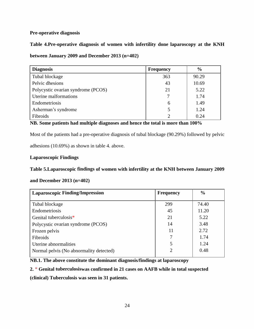
Pre-operative diagnosis
Table 4.Pre-operative diagnosis of women with infertility done laparoscopy at the KNH
between January 2009 and December 2013 (n=402)
NB. Some patients had multiple diagnoses and hence the total is more than 100%
Most of the patients had a pre-operative diagnosis of tubal blockage (90.29%) followed by pelvic
adhesions (10.69%) as shown in table 4. above.
Laparoscopic Findings
Table 5.Laparoscopic findings of women with infertility at the KNH between January 2009
and December 2013 (n=402)
Laparoscopic Finding/Impression Frequency %
Tubal blockage
Endometriosis
Genital tuberculosis*
Polycystic ovarian syndrome (PCOS)
Frozen pelvis
Fibroids
Uterine abnormalities
Normal pelvis (No abnormality detected)
299
45
21
14
11
7
5
2
74.40
11.20
5.22
3.48
2.72
1.74
1.24
0.48
NB.1. The above constitute the dominant diagnosis/findings at laparoscopy
2. * Genital tuberculosiswas confirmed in 21 cases on AAFB while in total suspected
(clinical) Tuberculosis was seen in 31 patients.
Diagnosis Frequency %
Tubal blockage
Pelvic dhesions
Polycystic ovarian syndrome (PCOS)
Uterine malformations
Endometriosis
Asherman’s syndrome
Fibroids
363
43
21
7
6
5
2
90.29
10.69
5.22
1.74
1.49
1.24
0.24

25
Tubal blockage was the most common laparoscopic finding (74.4%) followed by endometriosis
(11.2%) and genital tuberculosis (5.22%) as shown in table 5. above.
Association of patient charecterists with tubal blockage
Table 6. Association of patient characteristics and tubal blockage in women with infertility
who had laparoscopy done at the KNH between Jan 2009 and Dec 2013 (n=402)
Characteristics
With tubal blockage
N=299
Without tubal blockage
N=103
OR (95% CI) P value
n (%) Mean (SD)/
Median (IQR)
n (%) Mean (SD)/
Median (IQR)
Age 32.1(4.6) 31.4 (5.2) 0.161
Type of infertility
Primary
Secondary
132 (44.1)
167 (55.9)
50 (48.5)
53 (51.5)
0.8 (0.5-1.3)
1.0
0.439
Duration of infertility 5(3-8) 4 (3-7) 0.011
PID
Yes
No
149 (49.8)
150 (50.2)
59 (57.3)
44 (42.7)
0.7 (0.5-1.2)
1.0
0.192
Marital status
Married
Unmarried
278 (93.0)
21(7.0)
94 (91.3)
9 (8.7)
1.3 (0.6-2.9)
1.0
0.568
STI
Yes
No
80 (26.8)
219 (73.2)
39 (37.9)
64 (62.1)
0.6 (0.4-1.0)
1.0
0.033
P value=0.05
SD-Standard deviation
IQR-Inter-quartile range
PID-Pelvic inflammatory disease
STI-Sexually transmitted infection
Patients who were married were 1.3 times more likely to have tubal blockage than those who
were not married as shown in table 6.above. This was not statistically significant. (OR 1.3, p
value= 0.568).

26
Association of patient characterists with endometriosis
Table 7. Association of patient characteristics and endometriosis in women with infertility
who had laparoscopy done at the KNH between Jan 2009 and Dec 2013 (n=402)
Characteristics
With endometriosis
N=45
Without endometriosis
N=357
OR (95%
CI)
P value
n (%) Mean (SD)/
Median (IQR)
n (%) Mean (SD)/
Median (IQR)
Age 32.8 (5.2) 31.9 (4.7) 0.531
Type of infertility
Primary
Secondary
17 (37.8)
28 (62.2)
165 (46.2)
192 (53.8)
0.7 (0.3-1.3)
1.0
0.284
Duration of infertility 4 (3-6) 5 (3-8) 0.149
PID
Yes
No
18 (40.0)
27 (60.0)
190 (53.2)
167 (46.8)
0.6 (0.3-1.1)
1.0
0.094
Marital status
Married
Unmarried
37 (82.2)
8 (17.8)
335 (93.8)
22 (6.2)
0.3 (0.1-0.7)
1.0
0.012
Unemployed
Employed
6 (13.3)
39 (86.7)
67 (18.8)
290 (81.2)
0.7 (0.3-1.6)
1.0
0.373
STI
Yes
No
15 (33.3)
30 (66.7)
104 (29.1)
253 (70.9)
1.2 (0.6-2.4)
1.0
0.561
P value=0.05
SD-Standard deviation
IQR-Inter-quartile range
PID-Pelvic inflammatory disease
STI-Sexually transmitted infection
Patients with STI were 1.2 more times likey to have endometriosis compared to those without
STI although this was not statiscally significant as shown in table 7. above (OR 1.2, p=0.561)

27
Comparison between pre-operative and intra-operative diagnosis
Table 8.Comparison between pre-operative and intra-operative diagnosis in women with
infertility who had laparoscopy done at the KNH between January 2009
and December 2013 (n=402)
Variable Pre-operative
diagnosis
n (%)
Intra-operative
diagnosis
n (%)
Kappa statistic,
P value
Tubal blockage Yes
No
363 (90.3)
39 (9.7)
299 (74.4)
103 (25.6)
0.3, <0.001
Pelvic adhesions Yes
No
43 (10.7)
359 (89.3)
222 (55.2)
180 (44.8)
0.002, 0.934
PCOS Yes
No
21 (5.2)
381 (94.8)
14 (3.5)
388 (96.5)
0.7, <0.001
Endometriosis Yes
No
6 (1.5)
396 (98.5)
45 (11.2)
357 (88.8)
0.1, 0.002
Uterine abnormalities Yes
No
7 (1.5)
395 (98.5)
5 (1.2)
397 (98.8)
0.9,1.000
Genital Tuberculosis Yes
No
0 (0.0)
402 (100.0)
21 (5.2)
381 (94.8)
0.0, <0.001
Fibroids Yes
No
2 (0.5)
400 (99.5)
7 (1.7)
395 (98.3)
0.4, 0.067
PCOS=Polycystic ovarian syndrome
P value=0.05
Genital Tuberculosis, pelvic adhesions and endometriosis were the intra-operative diagnoses
most likely to be missed pre-operatively with kappa values of 0.0, 0.002 and 0.1 respectively as
shown in table 8.above.

28
Management Option Offered
Table 9.Management offered to women with infertility who had laparoscopy done at the
KNH between January 2009 and December 2013 (n=402)
Procedureat Laparoscopy Frequency %
Adhesiolysis
Adhesiolysis and salpingostomy
Adhesiolysis, fimbrioplasty
Excision and ablation for endometriosis
Salpingectomy
Laparoscopic ovarian drilling
Conversion to laparatomy (myomectomy, tuboplasty)
Hysteroscopy
157
113
98
45
44
33
31
4
29.90
21.52
18.67
8.57
8.38
6.29
5.90
0.76
Eventual management option offered
Conceive normally with no further intervention
In-Vitro Fertilization (IVF)
Open Tuboplasty
Adoption
Intra-Uterine Insemination (IUI)
Interventional Radiology
229
118
28
20
4
3
56.97
29.35
6.97
4.98
0.99
0.75
NB: A total of 82 patients had failed tuboplasty with completely blocked tubes, 11 had
frozen pelvis so no laparoscopy done. Multiple procedures were done in some patients.
Adhesiolysis alone then adhesiolysis and salpingostomy were the most common laparoscopic
procedures (29.90% and 21.52% respectively). The eventual management offered to most of the
patients was to conceive normally without further intervention (56.97%) while 29.35% of the
patients were advised to pursue IVF as shown in table 9. above.

29
Complications of laparoscopic surgery
Table 10: Complications in women with infertility who had laparoscopy done at the KNH
between January 2009 and December 2013 (n=402)
Complications of laparoscopic surgery Frequency %
None
Gut injury
Urinary tract injury
Wound sepsis
396
3
2
1
98.5
0.7
0.5
0.2
Most of the patients (98.5%) did not experience any complications from laparoscopic surgery as
shown in table 10. above.

30
DISCUSSION
This was a retrospective cross sectional descriptive study where a total of 402 files of patients
who underwent laparoscopy between January 2009 and December 2013 were analyzed. The
mean age of the study population was 32.0 years (SD 5.0) (table 1). Earlier studies in KNH on
infertility had shown younger ages. The mean age in a study by Nderitu in 2007 (34) was 30.0
years whereas the mean age was 27.1 years ina study by Gichuhi in 1995 (19). This shift inage
could be attributed to postponement of getting children latter in life by women due to education
and employment (35).
This increasing age of women with infertility has an important implication because of decreased
fecundability of a woman with increasing age especially from 30 years (36). Therefore prompt
and appropriate care is critical (36,37).
A third (10 out of 30) of the women in the category of those who were divorced or separated in
this study attributedthe cause of divorce or separation to infertility (table 1). Dhont et al in
Rwanda also found that a third of dissolution of unions was due to infertility (38). Social stigma
is attached to infertility and divorce is likely especially if the woman is unemployed (3).
In this study, most of the patients (54.7%) had secondary infertility (figure 1). Okwelogu et al in
Nigeria also found secondary infertility to be more common than primary infertility (39).This is
also true globally (2). Women with secondary infertility are more likely to succeed in conception
especially in In-Vitro Fertilization (IVF) (8). This is important since 29.35% of the patients in
this study were recommended to pursue IVF.
Tubal blockage was the most common laparoscopic finding (74.40%) in this study (table 5). In
Africa, tubal factor is the most common cause of female infertility whereas globally tubal

31
blockage and other tubal abnormalities are the second most common cause of female infertility
after Ovulatory disorders (6,39).
In this study, women with PID were not more likely to have tubal blockage compared to those
women without PID although it was statistically significant (OR 0.1, p=<0.001).This might be
attributed to the fact that this study may not have had sufficient power to detect this association.
Endometriosis was found in 11.20% of the patients at laparoscopy but was not suspected in most
of the patients before surgery (table 5). There were few cases where the severity of
endemetriosis was graded. Chirchir in his study at Aga Khan Hospital here in Kenya found the
prevalence of endometriosis to be 35% among Caucasians and 14% among Black Africans (40).
In this study 100 % of the study population were black Africans. In an earlier study by Gichuhi
in 1995 in KNH no endometriosis was found in women with infertility at laparoscopy (19). This
could be due to evolving technique in diagnosis of endometriosis or there may be a difference in
the two study populations.
Mboudou et al in Cameroon found the prevalence of endometriosis to be 13.53% (41). In
contrast, Osefo and Okeke found the prevalence of endometriosis among the Igbo women of
Nigeria to be much lower at 4.3% (42). Globally, the prevalence of endometriosis is 15% (6)
There was no association between patients’ characteristics such as age, marital status, parity,
type of infertility and endometriosis in this study (table 7). Endometriosis has been associated
with lower parity and to be more common in causcasians and Asians than in black women (43-
46). This study may not have had sufficient power to detect differences in parity. All the patients
were also black women.

32
Genital tuberculosis was seen in 5.22% of the patients in this study (table 5) and was the
diagnosis most likely to be missed before laparoscopy. These patients with TB were referred to
the TB clinic at the hospital for treatment and also followed up in the infertility clinic. In india
some studies have shown prevalence of 19% among women with infertility (47). The global
prevalence of genital tuberculosis is 2-6% with a rising incidence, partly as a result of HIV
pandemic and emergence of resistant strains (48).
Genital TB has profound implications because the multidrug therapy may adversely affect the
pregnancy outcome (48). It is therefore important to probe for TB in a patient with infertility in a
setting of HIV pandemic such as Kenya. In addition, a combination of methods including
polymerase chain reaction (PCR) is more accurate (47-51).
Laparoscopic treatment for tubal and pelvic disease as was done in this study has been shown to
avoid progression to In-Vitro Fertilization (IVF) (2,20). In their study, Levy et al found that
when laparoscopy was recommended in cases with suspected bilateral tubal occlusion on HSG, it
altered the original treatment plan in 30% of the patients from IVF to induction of ovulation with
intra uterine insemination (IUI) (22).
Ablation and excision for endometriosis was done in 8.57% of the patients in this study(table 9).
This procedure has been shown to improve fertility and pregnancy rates even in patients who
may require IVF (52-54). In contrast, several randomized control trials have shown that hormone
therapy for endometriosis does not improve fertility or pregnacy rates (55). Laparoscopic ovarian
drilling was done in 6.29% of the patients in this study. This is associated with an ovulation rate
of 80% and pregnancy rates of 82% at 24 months (56, 57).

33
In this study, most of the patients (56.97%) were advised to conceive without further intervention
while 29.35% of the patients were advised to pursue IVF (table 9). There is certainly a great
need for low cost Assisted Reproducitve Technologies (ART) in developing countries including
Kenya (3,58). The provision of such services however need to be done with public education and
awareness (39).
Adoption as a management option was recommended to 4.98% (20 out of 402) of the patients
(table 9). Assuming the patients chose adoption, this adoption rate after infertility treatment is
comparable with other studies done elsewhere. In a study of 1338 couples with infertility in
USA, adoption of a child occurred in 5.9% of the couples (59). There is however a low
acceptance rate of adoption in Kenya. Ondieki in her study in KNH found that 80% of couples
with infertility did not favour adoption (60). The Infertility Survey in Kenya also showed that
adoption was not favoured by infertile couples (3). In contrast, Wole in a rural survey in Uganda
found that adoption was acceptable (61).
A small number of the patients (0.75%) were recommended for interventional radiology i.e tubal
catheterization under fluoroscopic guidance (table 9). In a study of 110 infertile women on
therapeutic effectiveness of tubal catheterization and fertility outcome, Papaioannou et al showed
that 36.2% of the women conceived without IVF or Intra-cytoplasmic sperm injection (ICSI)
after tubal catheterization (62).
The rate of complications from laparoscopy was found to be quite low in this study at only 1.5%.
In contrast, Nderitu in his study at KNH in 2007 found that 8.7% of the patients had
complications (34). However, in the study by Nderitu the sample size was 82 as compared to 402

34
in this study. Globally, complications of laparoscopy in gynecologic patients range from 0.1% to
10% (63).

35
CONCLUSION
There are significant findings in this study. Firstly, there is a shift in the mean age of women
with infertility which calls for prompt evaluation and appropriate care. Secondly, tubal blockage
was the most common laparoscopic finding and therefore appropriate management of PID and
STI is important since these significantly contribute to tubal blockage and peritubal adhesions.
Thirdly genital tuberculosis and endometriosis are important causes of infertility in women.
HIV/AIDS which is endemic in Kenya has been shown to increase the prevalence of tuberculosis
globally (37, 54). According to the Kenya Aids Indicator Survey (KAIS) 2012, the prevalence of
HIV among adults aged 15 to 64 years is 5.6% (64). Granted that in this study the link between
HIV/AIDS and tuberculosis was not established. Nevertheless this calls for a high index of
suspicion in the evalauation of women with infertility.
Lastly, nearly a third of the patients were recommended to pursue ART services. Therefore there
is a great demand for affordable ART services in Kenya.These services need to be developed
together with an effective referral system for infertility care in general. Despite the limitations of
this study due to its retrospective design, it provides important knowledge and also forms a
basis for other studies in the field of infertility.

36
RECOMMENDATIONS
1. There should be a standardized way of evaluating and reporting laparascopic findings
such as endometriosis since this has implications on management.
2. The general evaluation of women with infertility should include probing for
endometriosis and genital tuberculosis.
3. In cases of suspected (clinical) TB, a combination of diagnostic methods including use
of Lowenstein-Jensen medium, pelvic and endometrial PCR in a TB endemic region
such as Kenya to improve the diagnosis.
4. Histological evaluation of endometriosis should be done in a teaching and referral centre
such as KNH.
5. There is need for affordable low cost ART services in Kenya as enviosined in the
National Reproductive Health Strategy 2009-2015 especially in KNH since it is not only
the premier referral and teaching hospital in the country and region but also the largest
hospital in the eastern African region.
37
REFERENCES
1. Practice Committee of the American Society for Reproductive Medicine. Definitions of
infertility and recurrent pregnancy loss. FertilSteril 2008; 90:560.
2. Halvorson, L.M. Evaluation of the infertile couple in: William’s Gynecology 22nd
ed. The
McGraw-Hill Companies Inc, 2012. pp 506-528.
3. Infertility in Kenya Survey Report, 2008. Ministry of Health, Kenya.
4. Matthews, T.,Mati, J.K. and Fomulu, J.N. A study of infertility in Kenya. Result of
Investigation of the infertile couple in Nairobi. EAMJ 1981; 58:288-97.
5. Ouko, D.O and Ndavi, P.M. Laparoscopic and Laparotomy Findings in Patients
Undergoing Tuboplasty for Infertility at Kenyatta National Hospital. J. Obst. Gyn. East.
Cent. Afr. 2006; 19: 1-7.
6. WHO Technical Report Series. Recent Advances in Medically Assisted Conception
Number 820, 1992, pp 1-111.
7. Okumu, C.V. Social background of tubal infertility at KNH, Nairobi, MMed. Thesis
1989, University of Nairobi.
8. Richardson, S.J., Senikas, V. and Nelson, J.F. Follicular depletion during the
menopausal transition: evidence for accelerated loss and ultimate exhaustion. J
ClinEndocrinolMetab 1987; 65:1231.
9. Gachuno, O.W., Rukaria, R.R and Wanyoike, G. Risk factors associated with tubal
infertility at Nazareth Hospital. J. Obst. Gyn. East. Cent. Afr. 2009; 21: 1-6.
10. Bulun, S.E. Endometriosis. N Engl J Med 2009; 360:268.
11. Gupta, S., Goldberg, J.M, Aziz N., et al. Pathogenic mechanisms in endometriosis-
associated infertility. FertilSteril 2008; 90:247.
12. Homer, H.A., Li T.C. and Cooke, I.D. The septate uterus: a review of management and
reproductive outcome. FertilSteril 2000; 73:1.
13. Pritts, E.A. Fibroids and infertility: a systematic review of the evidence. ObstetGynecol
Surv 2001; 56:483.
14. Perez-Medina, T., Bajo-Arenas, J., Salazar, F.et al: Endometrial polyps and their
implication in the pregnancy rates of patients undergoing intrauterine insemination: a
prospective, randomized study. Hum Reprod 20:1632, 2005 [PMID: 15760959].

38
15. Katz, D., Slade, D. and Nakajima, S.: Analysis of pre-ovulatory changes in cervical
mucus hydration and sperm penetrability. AdvContracept 13:143, 1997 [PMID:
9288332].
16. Jackson, J.E, Rosen, M., McLean, T.et al. Prevalence of celiac disease in a cohort of
women with unexplained infertility. FertilSteril 2008; 89:1002.
17. Clementini, E., Palka, C., Iezzi, I.et al. Prevalence of chromosomal abnormalities in 2078
infertile couples referred for assisted reproductive techniques. Hum Reprod 2005; 20:437.
18. Mbura, J.S.I. and Mgaya, H.N. Comparison of hysterosalpingography and laparoscopy in
evaluation of tubal factor in infertility. J. Obst. Gyn. East. Cent. Afr. 1988;7: 78.
19. Gichuhi, L.N. A study to compare hysterosalpingography and laparoscopic findings in
the investigation of female infertility at KNH. M.Med. Thesis. University of Nairobi,
1995.
20. Kumar, A. et al. Infertility in:Current Diagnosis & Treatment Obstetrics &
Gynecology10th
ed, The McGraw-Hill Companies Inc.2007. pp 374.
21. Sikolia, Z. W., Evan, S.andSamuel, G. M. Correlation between laparoscopic and
histopathologic diagnosis of endometriosis. International Journal of Gynecology and
Obstetrics 2011; 115:272–275.
22. Levy, Y., Lev-Sagie, A., Holtzer, H. Should laparoscopy be a mandatory component of
the infertility evaluation in infertile women with normal hysterosalpingogram or
suspected unilateral distal tubal pathology? Eur J ObstetGynecolReprod Biol. 2004;
114(1):64-8.
23. Heis, M., Amarin, Z., Ibrahim, A. Y. et al: Uterine and tubal anatomical abnormalities in
infertile women: diagnosis with routine hysterosalpingography prior to selective
laparoscopy. South African Journal of Radiology.2005; 14(2):4-8.
24. Audebert, A.J., Pouly, J.L. and Von Theobald, P. Laparoscopic fimbrioplasty: an
evaluation of 35 cases. Hum Reprod. 1998; 13:1496.
25. Bildirici, I., Bukulmez, O., Ensari, A. et al: A prospective evaluation of the effect of
salpingectomy on endometrial receptivity in cases of women with communicating
hydrosalpinges. Hum Reprod. 2001; 16:2422.
26. Rock, J.A., Guzick, D.S., Katz, E. et al: Tubal anastomosis: pregnancy success following
reversal of fallopian monopolar cautery sterilization. FertilSteril. 1987; 48:13.

39
27. Tulandi, T., Collins, J.A., Burrows, E. et al: Treatment-dependent and treatment-
independent pregnancy among women with periadnexal adhesions. Am J Obstet
Gynecol.1990; 162:354.
28. Donnez, J., Wyns, C. and Nisolle, M. Does ovarian surgery for endometriomas impair the
ovarian response to gonadotropin? FertilSteril. 2001; 76:662.
29. Ndovi, E.D. Dye laparascopy at KNH. M.Med. Thesis. University of Nairobi, 1980.
30. Obura, T. Laparoscopic surgery: risks and informed consent. J. Obst. Gyn. East. Cent.
Afr. 2009; 21: 29-36.
31. National Reproductive Health Policy of Kenya October 2007, Ministry of Health, Kenya.
32. National Reproductive Health Strategy 2009-2015, August 2009,Ministry of Health,
Kenya.
33. Fisher, A.A., Laing, E.J., Stoeckel, E. J. Handbook for Family Planning Operations
Research Design. 1991, New York: USA: Population Council.
34. Nderitu,C.M. Outcome of Open versus laparoscopic tuboplasty at Kenyatta National
Hospital. M.Med. Thesis. University of Nairobi, 2007.
35. Melinda, M., Ronald, R.R., McDonald, P., et al,Why do people postpone parenthood?
Reasons and social policy. Human Reproduction Update. 2011. 11(6): 848-860.
36. Committee on Gynecologic Practice of American College of Obstetricians and
Gynecologists, Practice Committee of American Society for Reproductive Medicine.
Age-related fertility decline: a committee opinion. FertilSteril. 2008; 90:486.
37. Faddy, M.J., Gosden, R.G., Gougeon, A., et al. Accelerated disappearance of ovarian
follicles in mid-life: implications for forecasting menopause. Hum Reprod. 1992; 7:1342.
38. Dhont, N., Wijgert, J., Coene, G., Gasarabwe, A., Temmerman, M. ‘Mama and papa
nothing’: living with infertility among an urban population in Kigali, Rwanda. Human
Reproduction. 2011;26(3): 623-629.
39. Sciarra, J.J. Sexually transmitted diseases: global importance. International Journal of
Gynecology & Obstetrics. 1997;58 (1):107–119.
40. Chirchir, A. The prevalence of endomtriosis among women undergoing laparscopy at
Aga Hospital Kenya. MMed Thesis,2005. University of Nairobi.

40
41. Mboudou, E. T., BelleyPriso, E., Mayer, F. E.The Prevalence of Endometriosis in
women with infertility Presenting for Laparoscopy, Clinics in Mother and Child Health.
2007; 4 (2): 233-236.
42. Osefo J., Okeke B. Endometriosis: Incidence among the Igbos of Nigeria. International
Journal of Obstestrics and Gynecology.1989; 30 (4):349-53.
43. Treloar, S.A., Bell, T.A., Nagle, C.M. Early menstrual characteristics associated with
subsequent diagnosis of endometriosis. Am J ObstetGynecol 2010; 202:534.
44. Missmer, S.A., Hankinson, S.E., Spiegelman, D. Incidence of laparoscopically confirmed
endometriosis by demographic, anthropometric, and lifestyle factors. Am JEpidemiol
2004; 160:784.
45. Hediger, M.L., Hartnett, H.J., Louis, G.M. Association of endometriosis with body size
and figure. FertilSteril 2005; 84:1366.
46. Kennedy, S., Bergqvist, A., Chapron, C. ESHRE guideline for the diagnosis and
treatment of endometriosis. Hum Reprod 2005; 20:2698.
47. Gupta,N., Sharma,J.B., Mittal, S.,Singh, N., Misra, R., Kukreja, M. Genital tuberculosis
in Indian infertility patients. International Journal of Gynecology & Obstetrics.2007; 97
(2): 135–138.
48. Shaheen, R., Subhan, F., Tahir, F. Epidemiology of genital tuberculosis in infertile
population. J Pak Med Assoc. 2006;56:306-309.
49. Doody K.J. Treatment of the infertile couple in: William’s Gynecology 22nd
ed. The
McGraw-Hill Companies Inc. 2008; 256-268.
50. John, R.C., Kristina, C.H., David, P.H., Susannah, D. C.Diagnosis of pelvic tuberculosis
in a patient with tubal infertility. Fertility and Sterility. January 2011;95(1):28917–
28920.
51. Baxi, A., Neema, H., Kaushal.,Sahu, P., Baxi, D. Genital Tuberculosis in Infertile
Women:Assessment of Endometrial TB PCR Results with Laparoscopic and
Hysteroscopic Features.The Journal of Obstetrics and Gynecology of India. 2011;301-
306.
52. Hull, M.G., Williams, J.A., Ray, B. et al: The contribution of subtle oocyte or sperm
dysfunction affecting fertilization in endometriosis-associated or unexplained infertility: a

41
controlled comparison with tubal infertility and use of donor spermatozoa. Hum Reprod.
1998; 13:1825.
53. Camus, E., Poncelet, C., Aucouturier, J.S., et al. [Hydrosalpinx and fertilization in vitro-
embryo transfer: abstention or salpingectomy? Abstention, salpingectomy or
salpingostomy?]. GynecolObstetFertil. 2001; 29:466.
54. Bianchi, P.H., Pereira, R.M., Zanatta, A. Extensive excision of deep infiltrative
endometriosis before in vitro fertilization significantly improves pregnancy rates. J
Minim Invasive Gynecol 2009; 16:174.
55. Parazzini, F., Fedele, L., Busacca, M. Postsurgical medical treatment of advanced
endometriosis: results of a randomized clinical trial. Am J ObstetGynecol1994; 171:1205.
56. Felemban, A., Tan, S,L. and Tulandi, T. Laparoscopic treatment of polycystic ovaries
with insulated needle cautery: a reappraisal. FertilSteril. 2000; 73:266.
57. Corson, L. S., Cheng, A. and Jacqueline, N. G. Laparoscopy in the “Normal” Infertile
Patient: A Question Revisited. The Journal of the American Association of Gynecologic
Laparoscopists. August 2000;7(3):317–324.
58. Boogard,L.S. Infertility and the provision of infertility medical services in developing
countries. Human Reproduction Update. 2009;14 (6):605-621.
59. Pinborg, A., Hougaard, C., Andersen, A. et al. Prospective longitudinal cohort study on
cumulative 5-year delivery and adoption rates among 1338 couples initiating infertility
treatment,Human Reproduction. 2009;24 (4): 991-999.
60. Ondieki, D.K. Male Involvment in the management of infertile couples at Kenyatta
national Hospital,M.Med. Thesis. University of Nairobi, 2012.
61. Wole, T.M. Infertile patients’ knowledge, attitude and practice regarding infertility as
seen in a gynecology clinic in northern Uganda M.Med. Thesis. University of Nairobi,
1997.
62. Papaioannou, S., Afnan, M., Girling, A. et al. Diagnostic and therapeutic value of
selective salpingography and tubal catheterization in an unselected infertile population.
Fertility and Sterility. 2003;79 (3): 613–617.
63. Magrina, J.F. Complications of laparoscopic surgery. ClinObstetGynecol 2002; 45:469.
64. Kenya Aids Indicator Survey (KAIS) 2012. Ministry of Health, Kenya.

42
APPENDICES
APPENDIX I:DATA RETRIEVAL FORM
A. Basic Information
1. Date ………………………
2. Study No ………………….
3. Hospital No ………………
B. Socio-demographic Data
4. Age ……………………
5. Educational level
1. None
2. Primary
3. Secondary
4. Tertiary
6. Religion……………………
1. Protestant
2. Catholic
3. Muslim
4. Other…………….
7. Occupation …………………………
1. Unemployed
2. Casual worker
3. Self employed
4. Formal employment
8. Residence…………………………….
1. Urban
2. Peri-urban

43
3. Rural
4. Other---------------------------------------------------
C. History of Presenting Illness
9. Duration of Infertility (Years)…………………………
10. Type of Infertility
1. Primary Infertility
2. Secondary Infertility
11. Seminalysis Done for Husband
1. Yes
2. No
12. Results of spouse
1. Normal
2. Abnormal
13. If secondary infertility, history of abortion
1. Yes
2. No
14. History of Prior Pelvic Inflammatory Disease
3. Yes
4. No
15. History of Prior Sexually Transmitted Infection (STI)
1. Yes
2. No
If yes, indicate type……………………………………

44
16. Any systemic illness
1. Diabetes
2. Hypertension
3. Other (Indicate)………………………………………….
17. History of previous surgery
1. Yes
2. No
If yes indicate type of surgery ………………………………………..
D. Pre-operative diagnosis
18. What was the pre-operative diagnosis (HSG)
1. Tube blockage (indicate whether right or left)
Proximal Distal
Unilateral Bilateral
2. Uterine anomalies (Indicate type) -----------
E. Laparoscopic Findings
19. Date surgery planned………………………………………….
Date surgery done……………………………………………….
20. Pelvic/Tubal Pathology (indicate whether right or left)
1. Tubal blockage
Unilateral Bilateral
2. Hydrosalpinx
Unilateral Bilateral
3. Pelvic adhesions
Yes No

45
4. Endometriosis
Yes No
5. Other Pelvic Pathology………………………………………………..
6. No Pelvic Pathology noted ……………………………………………
F. Treatment Option
21. Treatment Option given
1. Salpingectomy
2. Salpingostomy
3. Adhesiolysis
4. Fimbriolysis
5. Hysteroscopy
6. Conversion to laparotomy……………………………………….. (Mention procedure
done)
7. Assisted Reproductive Technology (ART)-------------------------
(Mention whether IVF, IUI etc.)
8. Other treatment option………………………....... (Mention option given)
G. Complications of laparoscopy
22. Complications of laparoscopy
1. None
2. Gut injury
3. Urinary tract injury
4. Other injury (mention type……)

46
APPENDIX II: ETHICS APPROVAL LETTER

47

48