university of groningen sec protein‐conducting channel and
TRANSCRIPT

University of Groningen
Sec Protein‐Conducting Channel and SecASluis, E.O. van der; Nouwen, N.; Driessen, A.J.M.
Published in:Molecular Machines Involved in Protein Transport across Cellular Membranes
IMPORTANT NOTE: You are advised to consult the publisher's version (publisher's PDF) if you wish to cite fromit. Please check the document version below.
Document VersionPublisher's PDF, also known as Version of record
Publication date:2007
Link to publication in University of Groningen/UMCG research database
Citation for published version (APA):Sluis, E. O. V. D., Nouwen, N., & Driessen, A. J. M. (2007). Sec Protein‐Conducting Channel and SecA. InR. E. Dalbey, C. M. Koehler, & F. Tamanoi (Eds.), Molecular Machines Involved in Protein Transport acrossCellular Membranes (25 ed., pp. 35-68). (The Enzymes; No. 25).
CopyrightOther than for strictly personal use, it is not permitted to download or to forward/distribute the text or part of it without the consent of theauthor(s) and/or copyright holder(s), unless the work is under an open content license (like Creative Commons).
Take-down policyIf you believe that this document breaches copyright please contact us providing details, and we will remove access to the work immediatelyand investigate your claim.
Downloaded from the University of Groningen/UMCG research database (Pure): http://www.rug.nl/research/portal. For technical reasons thenumber of authors shown on this cover page is limited to 10 maximum.
Download date: 12-11-2019
brought to you by COREView metadata, citation and similar papers at core.ac.uk
provided by University of Groningen

2
Sec Protein-Conducting Channel
and SecAELI O. VAV DER SLUIS � NICO NOUWEN �ARNOLD J.M. DRIESSEN
Department of Molecular Microbiology
Groningen Biomolecular Sciences and Biotechnology Institute
University of Groningen
9751 NN Haren, The Netherlands
I. Abstract
The Sec machinery facilitates protein translocation membrane insertionand into biological membranes of organisms from all three domains of life.The mechanism of the cotranslational mode of translocation is conservedacross the domains, whereas the components involved in posttranslationaltranslocation differ. In addition, significant differences are observed in thecomposition of the Secmachinery within the bacterial domain. Here, we willreview these differences in an evolutionary context, and discuss the latestinsights into the structure and dynamics of the translocon and the bacterialmotor protein SecA, with emphasis on their oligomeric state(s) duringprotein translocation.
II. Introduction
Every cell contains at least one membrane that separates the cytoplasmfrom the extracellular environment and its intracellular organelles. Embed-ded within these membranes is a variety of different transport systems thatselectively allow passage of molecules, thereby enabling the cell to carefully
33THE ENZYMES, Vol. XXV ISSN NO: 1874-6047# 2007 Elsevier Inc. All rights reserved. DOI: 10.1016/S1874-6047(07)25002-4

control the (bio)chemical composition on both sides of the membrane.Proteins are the largest and most complex molecules that are transportedacross membranes, and several different transport systems exist that canhandle this class of substrates. The Sec machinery is the only proteintransport system that is conserved across all three domains of life. It enablesprotein translocation across the cytoplasmic membrane of bacteria andarchaea, the endoplasmic reticulum (ER) membrane of eukarya, and thethylakoid membrane of photosynthetic eukarya [1].
A property that distinguishes the Sec machinery from other transportsystems is its ability to transport substrates toward two different cellularcompartments: the aqueous environment on the trans side of the membraneor the hydrophobic environment of the membrane itself. In line withthat property, the spectrum of substrates that is transported by the Secmachinery ranges from highly hydrophobic to highly hydrophilic proteins.The only feature that all substrates have in common is a hydrophobicN-terminal signal sequence or a transmembrane segment (membrane anchorsignal) that ensures substrate recognition and initiation of the translocationprocess.Most signal sequences are cleaved off by a signal peptidase to convertthe preprotein into the mature form, whereas N-terminal transmembranesegments remain attached to the substrate.
The most conserved part of the Sec machinery is the ‘‘translocon,’’ amembrane integrated channel that allows the passage of the (pre)proteinsacross the hydrophobic lipid bilayer [2]. All translocons consist of threeevolutionarily related subunits, but nevertheless archaeal and eukaryotictranslocons can be distinguished from bacterial and thylakoid transloconson the basis of their amino acid sequences [3]. The translocon can associatewith different partners to mediate two conceptually different modes ofprotein translocation: cotranslational and posttranslational translocation.The first is mainly employed for the insertion of integral membrane proteins(IMPs), and the latter mainly for translocation of secretory proteins [4].Cotranslational translocation requires the translocon to associate with theribosome, allowing a direct coupling between synthesis and translocation ofthe (pre)protein [5]. This process is conserved in all domains of life [6]and driven by ongoing protein synthesis at the ribosome. To prevent synthe-sis of membrane proteins in the cytoplasm, ribosome nascent chain com-plexes (RNCs) are targeted to the translocon via the signal recognitionparticle (SRP) in conjunction with its membrane-bound receptor (SR) [7].In eukaryotes, protein synthesis is slowed down or arrested until the nascentchain has been transferred from SRP to the translocon [8]. For more detailson the mechanism of SRP-dependent targeting, the reader is referred to oneof the reviews that have appeared [9, 10].
Posttranslational protein translocation occurs by definition after proteinsynthesis has been completed and requires the translocon to associate with
34 ELI O. VAV DER SLUIS, ET AL.

a motor protein to provide the driving force for the translocation reaction.In this mode of translocation, the Sec machineries in the various domains oflife differ substantially from each other. Posttranslational translocation inbacteria and chloroplasts is driven by the cis-acting ATPase SecA [11],whereas in ER membranes it is driven by a trans-acting Hsp70-like ATPasetermed BiP or Kar2 [12]. Given this topological difference, the molecularmechanism underlying posttranslational translocation is expected to differlargely between the ER and the bacterial cytoplasmic membrane. Post-translational protein translocation has also been suggested to occur inarchaea, but these organisms lack a SecA homologue and no apparentenergy source is available for a trans-acting motor protein.
III. Outline
The overall mechanisms of the two modes of protein translocation havebeen unraveled by groundbreaking studies in the early nineties, employingreconstituted systems from Escherichia coli and Saccharomyces cerevisiae.The last 5 years have led to a tremendous increase in our insights into thestructural basis of protein translocation through the elucidation of high-resolution crystal structures from individual components [13–17] and low-to medium-resolution electron microscopy (EM) structures of a variety offunctional complexes [18–21]. These structural and biochemical data haveyielded detailed insights into the molecular mechanism underlying proteintranslocation. The purpose of this review is to present an overview of ourcurrent understanding of the structural dynamics of the bacterial Secmachinery during protein translocation. We will focus on conformationalchanges that occur within the translocon, how they might be induced by(pre)proteins, the ribosome or SecA, and wewill highlight major unresolvedquestions. Some of these issues have received considerable attention inreviews [2, 22–24], and therefore additional emphasis will be on two issuesthat have not been addressed extensively, that is variations that are observedbetween Sec machineries of different bacteria and the controversyconcerning the oligomeric state(s) of the translocon and SecA duringprotein translocation.
IV. Variationand Evolutionof theSecMachinery
A. THE CANONICAL BACTERIAL SEC MACHINERY
In addition to the motor protein SecA and the three translocon proteins(SecY, SecE, and SecG), the Sec machinery of the vast majority of bacteriaconsists of YidC, SecD, SecF, and YajC. YidC is involved in the insertion of
2. SEC PROTEIN-CONDUCTING CHANNEL AND SECA 35

IMPs into the lipid bilayer by contacting the transmembrane segmentsof nascent IMPs shortly after they leave the SecYEG translocon [25].In addition, YidC functions independently of SecYEG in the integration ofsmall IMPs such as the FoC subunit of ATP synthase and the bacteriophagecoat protein M13 [26]. The mitochondrial YidC homologue Oxa1p fromS. cerevisiae has been shown to directly interact with the ribosome [27, 28]but thus far ribosome binding has not been demonstrated for YidC, while thecytoplasmic domain of Oxa1p implied in ribosome binding is absent in YidC.
With the exception of some lactic acid bacteria, all completely sequencedbacterial genomes encode for the proteins SecD, SecF, and YajC. SecD-FYajC forms a trimeric complex that is involved in protein translocationand associates with SecYEG [29, 30]. Two studies have indicated thatSecDF might be both functionally and physically coupled to SecG[31, 32], but the exact function of SecDFYajC has remained elusive [33].It has been proposed that SecDFYajC is involved in release of preproteinsfrom the translocon, regulation of SecA cycling, and maintenance of theproton motive force. The latter proposal has been shown to be based on apolar effect of the growth conditions used with a SecDF depletion strain,rather than on the functional defects of the depletion of SecDF itself [34].Further experiments are required to (dis)prove the other proposed func-tions of SecDF. In contrast to SecD and SecF, YajC is not required for cellviability. YajC alone has been shown to exist as a homooligomeric complexin the inner membrane of E. coli [35], but the functional importance ofthis complex is unknown.
B. EVOLUTIONARY HISTORY OF THE E. COLI SEC MACHINERY
Although the most intensively studied bacterial Sec machinery is thatfrom E. coli, some characteristics of this system are not representative forthe vast majority of bacteria. There are at least three components thatdistinguish the E. coli Sec machinery from that of other bacteria: SecB,SecM, and SecE. The tetrameric cytoplasmic protein SecB is a secretionspecific chaperone that prevents intracellular aggregation of (pre)proteins[36]. SecB slows down the folding of preproteins by binding to their matureregion [37], and it targets them to the extreme C-terminus of SecYEG-bound SecA [38]. Once translocation of the preprotein has been initiated,SecB is released from the translocon and able to start a new targeting cycle[11]. SecB is not essential for cell viability [39], but it is thought to berequired for translocation of a subset of (pre)proteins [40]. Thus far, noclear amino acid motifs have been identified that render (pre)proteins SecBdependent [41], but it has been shown that SecB-binding sites are enrichedin aromatic and basic residues [42].
36 ELI O. VAV DER SLUIS, ET AL.

The second component that distinguishesE. coli frommost other bacteriais SecM, a small regulatory protein (formerly known as gene X) that isencoded directly upstream of SecA [43]. Under secretion-deficient condi-tions, SecM induces a pause in translation of the secM–secA messengerRNA by means of an arrest sequence in its C-terminus [44] that is sensedby the interior of the ribosome [45]. This results in prolonged exposure ofthe SecA ribosome-binding site and consequently an upregulation of theamount of cellular SecA. In addition, SecM is involved in localizingthe expression of SecA to the vicinity of SecYEG [46]. SecM contains asignal sequence at its N-terminus, and thus the ribosome carrying a secM–secAmessenger and the arrested nascent chain is targeted to the translocon.The SecA molecules that are subsequently synthesized in the vicinity ofSecYEG are more active in protein translocation than SecA molecules thatare synthesized without a functional secM gene in cis [46]. This SecApopulation possibly corresponds to the ‘‘membrane integral’’ form ofSecA [47, 48]. SecM is not required for cell viability provided that sufficientSecA is supplied in trans [43].
SecE is the third component that distinguishes E. coli from many otherbacteria; E. coli SecE consists of three transmembrane segments (TMSs),whereas most of its homologues are single spanning membrane proteins[49]. The additional two TMSs might specifically facilitate protein translo-cation at low temperatures, since E. coli cells containing a variant of SecElacking these two TMSs are cold sensitive for growth [50].
An extensive genome analysis has revealed that SecB, SecM, and SecEwith three TMSs are not unique toE. coli as they are present in several otherproteobacteria, but not in any other bacterial divisions [51]. It is tempting tospeculate that an optimized Sec machinery could be particularly beneficialto the frequently pathogenic proteobacteria, but it should be noted that themicrobial genome-sequencing projects are strongly biased toward patho-genic organisms in general. Interestingly, the genomic distribution of SecB,SecM, and SecE with three TMSs reveals a part of the evolutionary historyof theE. coli Secmachinery. By combining the genomic distributionwith thephylogenetic relationships between the proteobacterial subdivisions inwhich each component is present (Figure 2.1), it was revealed that the Secmachinery has most likely evolved in the following successive steps: withinthe proteobacteria, the canonical Sec machinery (containing only SecYEG,SecA, SecDFYajC, and YidC) was first supplemented with SecB, then SecEwas extended with two TMSs, and finally SecM was introduced. Hence, theE. coli Sec machinery represents the end product of a stepwise evolutionaryprocess. Intermediate compositions with only SecB or SecB in combinationwith a three TMS-containing SecE are also observed, but neither theextended SecE nor SecM is ever observed without SecB, and SecM is
2. SEC PROTEIN-CONDUCTING CHANNEL AND SECA 37

never observed without extended SecE. It has been proposed that both SecEwith three TMSs and SecM could specifically improve SecB-dependentprotein translocation by maximizing the amount of SecYEG-bound SecAthat forms the receptor for preprotein–SecB complexes. This can be accom-plished in two ways: (1) by increasing the affinity of SecA for SecYEG (viaSecE) or (2) by carefully regulating and localizing the expression of SecA(via SecM) [51]. Further biochemical studies are required to investigate thepossible synergistic contribution of SecB, SecM, and extended SecE toprotein translocation.What should be kept inmind is that the Secmachineryof the model organism E. coli is of much greater complexity than that ofmost other bacteria.
C. SEC PARALOGUES
Noncanonical compositions of the Sec machinery-containing paraloguesof one or more components are also observed in many bacteria. Severalgenomes of organisms belonging to the divisions Actinobacteria (e.g.,Mycobacterium tuberculosis) [52] and Firmicutes (e.g., Listeria monocyto-genes and Streptococcus gordonii) [53, 54] encode for paralogues of SecA,and few of those bacteria encode for paralogues of SecY, SecE, and/orSecG as well. The genomes of the proteobacteria Gluconobacter oxydansand Francisella tularensis encode for SecB paralogues [51]. The genomicdistribution of these paralogues has not yet been investigated in an
FIG. 2.1. Genomic distribution of accessory features of the Sec machinery in proteobacteria
in combination with bacterial phylogeny. The distribution suggests that the Sec machinery
has evolved in a stepwise fashion by sequentially acquiring SecB, the SecE extension, and
SecM [51].
38 ELI O. VAV DER SLUIS, ET AL.

evolutionary context, and SecA2 is the only paralogue that has beenstudied genetically. It has been shown in both M. tuberculosis [55] and inL. monocytogenes [56] that SecA2 is important for pathogenicity but not forviability. These observations have led to the speculation that the accessorySec machinery components of these Gram-positive bacteria might befunctional equivalents of the pathogenicity related Type II–IV secretionsystems found in many Gram-negative bacteria [56]. The thus far identifiedSecA2-dependent substrates do not have any functional characteristics incommon. However, several substrates contain an atypical signal sequenceor become glycosylated before translocation [56–62]. Interestingly, someSecA2-dependent substrates do not contain a signal sequence at all [55, 56].It will be of great interest to investigate these and other features thatdistinguish SecA2 and the other paralogues from the canonical Secmachinery.
V. SecAStructure,Function,andDynamics
A. THE INVOLVEMENT OF SECA IN COTRANSLATIONAL
PROTEIN TRANSLOCATION
The motor protein SecA is one of the largest and most complex bacterialproteins. It consists of multiple domains and it interacts with nearly allthe other components involved in protein translocation: (pre)proteins,SecYEG, SecB, nucleotides, the cytoplasmic membrane, and possibly theribosome. Although co- and posttranslational translocation reactions aremostly studied as individual pathways in S. cerevisiae and E. coli, respec-tively, there are several indications that the two pathways overlap. MostIMPs are translocated cotranslationally, but several IMPs contain largeextracytoplasmic domains that are translocated in a SecA-dependent man-ner [25, 63–66]. This implies that SecA and the ribosome can either bind tothe translocon simultaneously or that they can bind alternating to thetranslocon. Although simultaneous binding of SecA and the ribosome toSecYEG is structurally difficult to envisage (see Sections 15.4 and 17), it hasbeen shown that ribosomes and SecA do not compete for binding toSecYEG [67]. In addition, it has been demonstrated that SecA has a lowbut intrinsic ribosome-binding capacity, either alone [68, 69] or in conjunc-tion with SecYEG [67]. Interestingly, ATP hydrolysis by SecA appears toinduce the release of the ribosome from the translocon [67]. In this context,it should be stressed that during translocation of a large extracytoplasmicdomain of an IMP by SecA, the ribosome would remain tethered to thetranslocon via the nascent chain rather than being truly released. The latter
2. SEC PROTEIN-CONDUCTING CHANNEL AND SECA 39

would favor rebinding of the ribosome to the translocon for cotransla-tional continuation of the translocation process. Taken together, theco- and posttranslational protein translocation pathways are likely to beintertwined. Therefore, in vitro membrane protein insertion studies withSecA-dependent membrane proteins of varying topologies are eagerlyawaited to further unravel this intricate process. In particular, special atten-tion should be paid to the role of YidC and SecDFyajC during membraneinsertion of SecA-dependent IMPs.
B. THE OVERALL MECHANISM OF POSTTRANSLATIONAL
PROTEIN TRANSLOCATION
In contrast to its possible role in cotranslational protein translocation,the role of SecA in posttranslational translocation is understood inmuch more detail due to extensive biochemical studies with purifiedcomponents. This has resulted in the following widely accepted workingmodel (Figure 2.2): In SecB-containing organisms, the cycle of posttransla-tional translocation starts with binding of a (pre)protein–SecB complexto SecYEG-bound SecA [11], on which the preprotein is transferred toSecA [70]. In organisms lacking SecB, the preproteins either bind directlyto SecYEG-bound SecA, or are targeted to the translocon via bindingto cytoplasmic or lipid-bound SecA. The subsequent binding of ATP toSecYEG-bound SecA induces a conformational change that results ininsertion of the signal sequence into the translocon, and release of SecB(if present). At the same time, SecA is thought to insert partially into thetranslocon [71], and around 2.5 kDa of the mature domain of the preproteinis translocated [72, 73]. ATP hydrolysis results in release of the (pre)protein
SecB
SecA
SecYEG
ADP
N
ADP
2 3 4 5 6 71
ADPADP
ADP
ADPc
ATPATP ATP
ATP
ATP
PMF PMF
Azide Azide
Pi
Pi
FIG. 2.2. Schematic representation of posttranslational protein translocation in E. coli.
See text for details.
40 ELI O. VAV DER SLUIS, ET AL.

from SecA and deinsertion of SecA from the translocon, in a step that canbe inhibited by the commonly used antibacterial compound azide [74].Next, rebinding of SecA to the partially translocated polypeptide chaincan drive the translocation of another 2.5 kDa of the mature preproteindomain [72, 73]. Depending on the length of the (pre)protein, multiplecycles of ATP binding and hydrolysis and SecA binding and release arerequired to completely translocate the substrate across the membrane.
C. STRUCTURE OF THE SECA PROTOMER
The working model described above is still rather abstract, but ourinsight into the molecular details of the mechanism has become increasinglyclear due to the availability of crystal structures from SecA [13, 17, 75],SecB [14, 15, 76], and an archaeal SecYEG homologue [16]. Three differentcrystal structures of SecA are available, two from B. subtilis and one fromM. tuberculosis. The actual motor function of SecA, that is conversion ofchemical energy into movement, is initiated by a ‘‘DEADmotor’’ core thatis also present in DNA/RNA helicases [77]. The DEAD motor consists oftwo similarly folded domains that are referred to as nucleotide-bindingfolds (NBF1 and NBF2), each resembling the recombination proteinRecA. At the interface of these two domains a single ATP molecule canbe bound and hydrolyzed, which induces the conformational changes inSecA that ultimately results in the translocation of preproteins. SecAinteracts with preproteins via the preprotein-binding domain (PBD, alsoreferred to as ‘‘preprotein cross-linking domain’’ (PPXD) [78]) that isinserted into the amino acid sequence of NBF1 (Figure 2.3A), but forms aseparate domain in the SecA structure [79, 13] (Figure 2.3B). The remain-der of the SecA structure can be subdivided into four regions: the helicalscaffold domain (HSD), the helical wing domain (HWD), the C-terminallinker (CTL), and the SecB-binding domain ‘‘SecAc.’’ The HSD forms along scaffold to which NBF1, NBF2, the PBD, and the HWD areconnected, the HWD is a loosely attached domain with unknown function,and the CTL forms the connection with SecAc at the extreme C-terminus[13] (Figure 2.3A and B).
1. Oligomeric State of SecA
In order to understand the working mechanism of any protein on amolecular level, it is not only essential to know its structure and the exactlocation of the interaction sites for all its ligands but also to elucidate thefunctional oligomeric state of the protein itself. The oligomeric state of bothSecA and SecYEG during protein translocation has become a controversial
2. SEC PROTEIN-CONDUCTING CHANNEL AND SECA 41

N
NBF1a
PBD
PBD
NBF1b
NBF1
NBF2
NBF2
HSDa
HSD
HSDb SecAc
HWD
HWD
CTL
CTL
C
A
B C
D E
FIG. 2.3. Structure of SecA. (A) Schematic overview of the domain structure of SecA. NBF:
nucleotide-binding fold; PBD: preprotein-binding domain; HSD: helical scaffold domain;
HWD: helical wing domain; CTL: C-terminal linker; SecAc: SecB-binding motif. (B) Crystal
structure of SecA protomer from B. subtilis with individual domains colored as in (A) [13].
(C) Crystal structure of SecA from B. subtilis in an open conformation, possibly representing
the (pre)protein-bound state [75]. The conformational changes with respect to the structure
42 ELI O. VAV DER SLUIS, ET AL.

topic, and the complexity of the matter is schematically depicted inFigure 2.4. In an attempt to enlighten both discussions, we will address thetopics individually, starting with SecA. For clarity, we have grouped theexperimental data according to the following three subquestions:
1. What is the oligomeric state of soluble SecA?2. What is the oligomeric state of SecYEG-bound SecA?3. What is the oligomeric state of translocation-engaged SecA?
2. The Oligomeric State of Soluble SecA
It has been shown with various techniques that purified SecA exists in adynamic equilibrium between a monomeric and a dimeric form, and thedissociation constant (KD) has been estimated to be around 0.1 mM underphysiological conditions [80]. The cellular concentration of SecA is �8 mM[81], and thus SecA is expected to be largely dimeric in vivo. Higher orderSecA oligomers have also been reported, but only under nonphysiologicalconditions or with truncated SecA mutants [17, 82]. Three reports haveshown that translocation ligands can induce monomerization of SecAdimers, which raises the question whether the cellular predominant SecAdimer is also the functional state. Fluorescence- and cross-linking studieswith purified SecA have shown that themonomer–dimer equilibrium can beshifted toward the monomer by the addition of certain lipids or detergents[83, 84], or signal peptides [83, 85], although a different view has been
depicted in (B) are indicated by arrows. (D) Crystal structure of dimeric SecA from B. subtilis
that most likely represents the physiologically active dimer [13]. The two intradimeric HSD–
HSD contacts that are maintained during protein translocation are depicted in red [95].
(E) Crystal structure of M. tuberculosis SecA. (See color plate.)
FIG. 2.4. Schematic overview depicting the complexity of the debate concerning the oligo-
meric states of SecA and SecYEG. The experimentally demonstrated equilibria between the
different oligomeric states are indicated by arrows, and all the possible interactions are
indicated by dashed lines. See text for details.
2. SEC PROTEIN-CONDUCTING CHANNEL AND SECA 43

published, suggesting that signal peptides induce oligomerization of SecA[84]. Lipid-bound SecA has been shown to exist mainly in a dimeric formthat can be dissociated on binding of nucleotides [86]. Although all thesestudies underscore the dynamic and sensitive nature of the SecAmonomer–dimer equilibrium, it remains questionable whether any of these observedchanges in oligomeric state are functionally relevant since no SecYEG nor(pre)proteins were present in these studies.
3. The Oligomeric State of SecYEG-Bound SecA
The oligomeric state of SecA while bound to SecYEG detergent solutionhas been addressed by native gel electrophoresis and gel filtration [87–89].It was shown that both monomeric and dimeric SecA can bind to SecYEG,provided that SecYEG is stabilized in a dimeric form either by covalentlinkage [88] or by an antibody [89]. Unstabilized SecYEG in detergent onlyretains monomeric SecA after a preprotein has been trapped inside thechannel before solubilization of the membrane [88]. These results should beinterpreted carefully, however, since the monomer–dimer equilibrium ofSecA has been shown to be highly sensitive to detergents [83].
The oligomeric state of SecA bound to membrane-embedded SecYEGhas been addressed by chemical cross-linking [90] and surface plasmonresonance (SPR) [91]. Dimeric SecA can be detected after binding tourea-treated inverted membrane vesicles (IMVs) [90], but could not bedetected with SecYEG-containing proteoliposomes [83]. The concentrationof SecA added in the latter experiment however was far below physiological(5 nM vs 8 mM), and thus the results obtained with the IMVs appear to bemore reliable. Chemical cross-linking of the population of SecA that copuri-fies with IMVs revealed mainly SecA monomers [90], while the fraction ofSDS-resistant dimers dramatically increases on overexpression of SecYEG[91]. SPR measurements also suggest that SecA is dimeric while it is boundto membrane-embedded SecYEG, since wild-type SecA binds to SecYEG-overexpressing IMVs similarly to a covalently cross-linked SecA dimer [91].Taken together, these data indicate that both monomeric and dimeric SecAcan bind to SecYEG.
4. The Oligomeric State of Translocation-Engaged SecA
Activity assays are obviously the most relevant experiments to assess theoligomeric state of SecAduring protein translocation. In order to investigatethe functional requirement of dimeric SecA, several studies have character-ized SecA mutants with disturbed dimerization properties. Removal of theN-terminal eight amino acids of SecA does not influence its oligomeric state[92], but SecA has been reported to be predominantly monomeric when
44 ELI O. VAV DER SLUIS, ET AL.

the first 11 amino acids are removed [90, 93, 94]. Alternatively, monomericSecA can be obtained by mutating 6 residues in the C-terminal region ofa SecA truncate that lacks 70 residues from its extreme C-terminus [83].It should be noted that these monomeric SecAmutants are not incapable ofdimerization per se, as the mutations have shifted the monomer–dimerequilibrium in solution substantially toward the monomeric state [93].In the two assays that measure SecA activity, for example the in vitropreprotein translocation assay and the precursor-stimulated SecA ATPaseassay, all monomeric SecA mutants show either a very low activity or noactivity at all. Although the low residual in vitro activity has been inter-preted as being significant in some reports [83, 94], it seems more likelythat the residual activity is caused by a small fraction of SecA dimers thatcan still be formed or by traces of copurified wild-type SecA from theexpression host.
Activity assays with covalently dimerized SecA have yielded varyingresults. SecA dimers cross-linked via endogenous cysteines located in theSecB-binding domain (SecAc) [91] or via a pair of engineered cysteinesin the HSD (Arg637 and Gln801) [95] were shown to be nearly fully active inprotein translocation and preprotein-stimulated SecA ATPase activity.Although these observations alone do not directly imply that SecA func-tions as a dimer, it does show that monomerization is not required forfunctionality as proposed earlier [83].
Perhaps the most convincing experiment that assesses the functionaloligomeric state of SecA-involved heterodimers of active and inactiveSecA monomers [96]. If SecA would function as a monomer, these hetero-dimers are expected to have half the activity of wild-type SecA. However,it was observed that these heterodimers are completely inactive, stronglysuggesting that SecA is functional as a dimer.
5. Summary of Oligomeric States SecA
Taken together from our point of view, the experimental data showingthat SecA dimers dissociate on binding of translocation ligands are notnecessarily related to protein translocation, since they might simply reflectthe sensitive nature of the monomer–dimer equilibrium. The data support-ing the proposal that SecA functions as a monomer are in our opinioneither; obtained under conditions too distant from physiological; explain-able by a conformational change of SecA, or misinterpreted. On the otherhand, the experimental data supporting the SecA dimer as a functional unitare more convincing and more abundant. Furthermore, there are no exper-imental data disproving a functional SecA dimer, whereas in vivo andin vitro experiments in different laboratories demonstrate that monomeric
2. SEC PROTEIN-CONDUCTING CHANNEL AND SECA 45

SecA variants are inactive. Finally, it has been shown that SecB targetspreproteins to dimeric SecA, and that this targeting greatly stimulates theefficiency of protein translocation [38]. Combined with the notion thatcellular SecA is predominantly dimeric, we assume that SecA functions asa dimer in posttranslational protein translocation at SecYEG.
We speculate that the physiological relevance of the binding of mono-meric SecA to SecYEG and the sensitive nature of the monomer–dimerequilibrium could be related to (pre)protein targeting to SecYEG.As mentioned above, in organisms lacking SecB, (pre)proteins might firstbind to cytoplasmic or lipid-bound SecA, and subsequently transferred tothe translocon. If one SecA protomer would remain permanently boundto SecYEG, the dimerization of SecA could play a role in the initiation oftranslocation via this SecB-independent targeting process.
D. STRUCTURE OF THE FUNCTIONAL SECA DIMER
With our current insight that SecA functions as a dimer, the next ques-tion is at which side of a SecA protomer the intradimeric interactions takeplace. Two interactions observed in various crystal structures have beenproposed to represent a physiological dimer interface [13, 17]. The overallarrangement of both of these SecA dimers is very similar; the two elongatedSecA monomers are arranged side-by-side in an antiparallel fashion(Figure 2.3D and E). This antiparallel arrangement is supported by fluores-cence resonance energy transfer (FRET) [97] and cross-linking studies[94, 95]. The difference between both dimers lies in the SecA surface thatcontacts the neighboring protomer. The proposed B. subtilis dimer is rela-tively compact and the dimer interface comprises a large surface (5442 A2)[13], whereas theM. tuberculosis dimer is relatively flat, comprises a smallersurface (2822 A2), and contains a cavity at the dimer interface [17]. Onedimer arrangement can be converted into the other by rotating each pro-tomer �75� around its long axis. Although it is conceivable that suchrotations could play a role in the cycle of SecA-driven protein translocation,the observation that a SecA dimer that is fixed in the B. subtilis arrange-ment (Figure 2.3D) still supports efficient protein translocation [95] sug-gests that at least the B. subtilis dimer is part of the conformational cycleof SecA. Thus, it can be concluded that the HSDs of two SecA protomerscan be considered as a single scaffold domain in the SecA dimer, and thatnone of the conformational changes that SecA undergoes during proteintranslocation is severely hampered by the intradimeric HSD–HSD cross-links. Whether the M. tuberculosis dimer arrangement (Figure 2.3E) alsorepresents a functional intermediate remains to be established.
46 ELI O. VAV DER SLUIS, ET AL.

E. CONFORMATIONAL CHANGES WITHIN SECA
Several regions in SecA have been shown to be dynamic [13, 98–105], butdetailed structural information is only available on two conformationalchanges: one that can be inferred from SecA’s similarity to helicases andanother that has been observed directly with X-ray crystallography [75].As mentioned previously, the DEAD-motor core of SecA (NBF1 andNBF2) is homologous to that of SFI and SFII helicases, and therefore thenucleotide-induced conformational changes are assumed to be similar in allthree protein families. SecA has been crystallized with bound ADP and inthe nucleotide free state, but these structures differ only slightly in theorientation of side chains that are involved in nucleotide binding. Unfortu-nately, attempts to crystallize SecA in the functionally important ATP-bound state have failed thus far. In addition to conformations that arevery similar to those of nucleotide free and ADP-bound SecA, the helicaseDEAD motors have been crystallized in two substantially different con-formations. First, the SFII helicase MJ0669 has been crystallized withoutnucleotides in an open conformation in which the two NBFs are separatedfrom each other by a large cleft [106]. Second, the SFI helicase PcrA hasbeen crystallized in the ATP-bound state in which the two NBFs haveundergone an �10� rotation relative to each other compared to the ADP-bound state [107]. All three distinct conformations as observed in differentDEAD motors (open, closed, and closed-rotated) are assumed to underliethe ATPase cycle of SecA as well. Given the observation that a SecA dimerin which the two HSDs are cross-linked is still active, the relative reorienta-tions of NBF1 and NBF2 that are required for ATP binding and hydrolysisare apparently not influenced by these disulfide-bonded cross-links. Whenthe mobility of NBF1 is restricted by a disulfide cross-link to the HWD ofthe neighboring protomer however, the SecA dimer is inactive [95].
The conformational change of SecA that has been visualized by X-raycrystallography does not involve the DEAD-motor or nucleotide, and ittakes place in the opposite end of a SecA protomer [75]. B. subtilis SecAhas been crystallized in two different conformations, and a comparison ofboth conformations reveals the followingmovements in a protomer: theHSDand HWD undergo a small rotation, and the PBD undergoes a large (�60�)rotation combined with a rigid body translation away from the HSD andHWD (Figure 2.3B and C). This results in opening of a groove at the PBD–HSD/HWD interface (Figure 2.3C) that has been proposed to form the actualpreprotein-binding site since its physicochemical characteristics are similar tothat of peptide-binding sites from other proteins with broad substrate specifi-cities. Assuming that this conformation of SecA represents a (pre)protein-bound state and knowing that B. subtilis does not contain a SecB protein
2. SEC PROTEIN-CONDUCTING CHANNEL AND SECA 47

it could represent either a SecYEG-bound form, a lipid-bound form, or asoluble form. In the latter two cases, it might represent the earlier proposed(monomeric) form of SecA that was suggested to be involved in SecB-independent targeting of (pre)proteins to a SecYEG-bound protomer. As atpresent it is unclear whether the observed conformational changes can takeplace in the B. subtilis dimer arrangement, the conformation in the crystalstructure could also represent (one of) the SecYEG-bound SecAprotomer(s)after receiving a (pre)protein. The location of the CTL that connects theSecB-binding domain SecAc to the HWD suggests how binding of a SecB-(pre)protein complex could bemechanistically coupled to the conformationalchange in SecA (see Section 12).
F. SECA–SECB INTERACTION
The interaction between SecA and SecB has been investigated ingreat detail. Since an excellent review on the SecA–SecB interaction hasappeared [108], we will only discuss the most important findings and apossible relation to conformational changes in SecA. It has been shownthat the extreme C-terminus of SecA (SecAc) contains a dedicated SecB-binding site that is formed by a small cysteine-rich domain that chelates azinc ion [109]. This highly conserved domain is also found in organismslacking SecB, which might be related to the fact that the C-terminus is alsoinvolved in lipid binding [110]. The SecAc domain is not resolved in any ofthe available SecA crystal structures, but its structure has been determinedin isolation by NMR [111, 112] and in complex with Haemophilus influen-zae SecB by X-ray crystallography [15]. The latter structure revealed thattwo SecAc domains are bound to opposite sides of one SecB tetramer, on asurface that was previously shown to be crucial for SecB-binding to SecA[70, 113]. The SecAc domain is stabilized by the zinc ion that is coordinatedby three cysteines and one histidine, explaining why SecA mutants in whichthese residues are either mutated [114] or cross-linked [91] are unable tosupport SecB-dependent protein translocation.
The approximate position of the SecB tetramer bound to SecA in theB. subtilis dimer arrangement has been estimated by docking of the SecB–SecAc complex onto the SecA structure [108]. It seems likely howeverthat on binding of a preprotein–SecB complex to SecA, the transfer ofthe (pre)protein requires (or induces) a substantial conformational changein SecA [70]. This conformational change possibly corresponds to the onethat is observed by X-ray crystallography [75]. Binding of SecB to the highlymobile SecAc domain could displace the CTL that connects SecAc tothe HWD. Since the CTL is part of the PBD-hinge region in the closedconformation of SecA and it meanders partially underneath the PBD
48 ELI O. VAV DER SLUIS, ET AL.

(Figure 2.3B), this displacement could directly induce the observed rigidbody movement of the PBD that results in opening of the proposed (pre)protein-binding groove (Figure 2.3C). Furthermore, CTL displacementcould be directly responsible for the small rotation of the HWD/HSD thatcoincides with opening of the groove. Although B. subtilis does not containSecB, it has been shown that E. coli SecA undergoes a similar conforma-tional change [75]. In organisms lacking SecB, displacement of the CTL isexpected to be induced by an alternative mechanism. This could involve theinteraction of SecAc with lipids [110] or binding of SecA to SecYEG [115].
G. SECA–MEMBRANE INTERACTION
A detailed understanding of SecA binding to the membrane is funda-mental for understanding the molecular mechanism of SecA-driven proteintranslocation. However, whereas binding of SecA to E. colimembranes hasbeen studied extensively, surprisingly little is known about the region(s) ofSecA that interact(s) with the membrane. The lipid-binding region of SecAhas been localized to its C-terminal 70 amino acids [110], but the SecYEG-binding region of SecA has not been identified in detail. Far westernexperiments using SecA fragments mapped the SecYEG-binding regionto the N-terminal part of the SecA protomer, comprising both NBFs andthe PBD [116]. Moreover, binding experiments with SecA fragments havedemonstrated that the same N-terminal region of SecA comprises the high-affinity SecYEG-binding site, whereas the remaining C-terminal one-third ofSecA does not bind to SecYEG [116]. However, the exact SecYEG interac-tion sites within the N-terminal region have not been determined yet. Therelatively new technique of cysteine-directed cross-linking in combinationwithmass spectrometry appears to be themost suitable biochemical approachto identify the exact regions in SecA that interact with SecYEG. In addition,medium- and high-resolution structural studies on SecYEG–SecA complexeswill contribute to answering this critical question.
VI. SecYEGStructure,Function,andDynamics
A. STRUCTURE OF THE SECYEG PROTOMER
The structure–function relationship of the translocon has been exten-sively studied inE. coli and S. cerevisiae. The recently solved high-resolutiontranslocon structure from the archaeonMethanococcus jannaschii [16] was amajor breakthrough in the field. Despite the fact that archaeal transloconsubunits are more similar to eukaryotic than to bacterial ones [3], they are
2. SEC PROTEIN-CONDUCTING CHANNEL AND SECA 49

commonly named after the bacterial subunits. Since no significant sequencesimilarity can be detected between SecG and its archaeal counterpartSec(61)b [117], the eukaryotic nomenclature is applied to the latter, result-ing in the hybrid term SecYEb. In agreement with its universal conservation,the overall structure of M. jannaschii SecYEb is nearly identical to that ofE. coli SecYEG [118]. The two complexes differ only slightly in conforma-tion [119], and the E. coli translocon contains three additional TMSs com-pared to that from M. jannaschii: two from SecE (Section IV) and onefrom SecG. The center of the complex is formed by SecY, whereas SecEand SecG are located at the periphery (Figure 2.5A and B). The structure of
Sec E/g
89 6
105
41
b
3
2
7“Front”
“Hinge”
“Back”
“Plug”
C
Cytoplasm
BA
FIG. 2.5. Structure of SecYEb from M. jannaschii [16]. (A) Cytoplasmic view showing the
arrangement of transmembrane segments in different colors. SecE is depicted in purple, Secbin pink. Sides referred to as ‘‘front’’ and ‘‘back’’ are indicated. (B) View from within the plane
of the membrane showing the two cytoplasmic loops that extend into the cytoplasm and have
been shown to interact with the ribosome and SecA: C4 and C5, connecting TMS6 with TMS7
and TMS8 with TMS9, respectively. (C) Back-to-back dimer arrangement of SecYEb proto-
mers as observed for E. coli SecYEG in two-dimensional crystals [118]. The N-terminal halves
of SecY are depicted in blue, the C-terminal halves in red, and SecE and Secb in purple and
pink, respectively. (See color plate.)
50 ELI O. VAV DER SLUIS, ET AL.

M. jannaschii SecYEb consists of two distinct domains that are similarlyfolded. Each domain is composed of a bundle of five TMSs, formed by theN- or C-terminal half of the SecY sequence, respectively. The two halves ofSecY are held together by SecE: the conserved TMS of SecE crosses themembrane diagonally [120], and contacts both SecY halves at the same sidewhere they are connected by the extracytoplasmic loop between TMS5and TMS6. This side of the SecYEG protomer is referred to as the‘‘back.’’ The amphipathic cytoplasmic helix of SecE [121] runs parallel tothemembrane surface along the C-terminal half of SecY (Figure 2.5A). Twoof the cytoplasmic loops of SecY protrude far into the cytoplasm: the C4loop connecting TMS6 with TMS7, and the C5 loop connecting TMS8with TMS9 (Figure 2.5B). The extracytoplasmic loops on average are con-siderably shorter, and two of those fold back into the membrane region:theE4 loop connecting TMS7with TMS8, and theE1 loop connecting TMS1with TMS2. The latter is highly conserved, folds back between the two SecYhalves, and is referred to as the ‘‘plug’’ domain [16].
At first sight, there is no obvious region in the channel that is largeenough to allow passage of unfolded proteins. For this reason, it has beenconcluded that the structure represents the closed conformation of Sec-YEb. However, on the basis of two domain structure of the channel andthe observation that signal sequences of (pre)proteins can be cross-linkedto TMS2 and TMS7 [122–124] at the domain interface, it was proposed thatinsertion of the signal sequence between TM2 and TM7 results in separa-tion of the two halves of SecY and displacement of the ‘‘plug’’ that blocksthe proposed pore from the extracellular side and that the substrates passthrough the center of the channel [16]. Molecular dynamics simulationshave revealed that the opening that is created by this mechanism is indeedlarge enough to allow passage of unfolded and even a-helical proteins [125].In the opened state, nascent IMPs (and signal sequences) could leavethe translocon laterally toward the lipid bilayer via the TMS2–TMS7 inter-face. The possible mechanisms by which SecA or the ribosome couldinduce channel opening will be discussed below, but first we will addressthe oligomeric state(s) of the translocon.
1. Oligomeric States of SecYEG
As outlined above for SecA, knowledge of the functional oligomericstate of a protein is of fundamental importance for understanding itsmechanism of action. Also the oligomeric state of SecYEG is heavilydebated (Figure 2.4). In an attempt to enlighten this discussion, we willgive an overview of the relevant experimental data. For clarity, we have
2. SEC PROTEIN-CONDUCTING CHANNEL AND SECA 51

subdivided the assessment of the oligomeric state of SecYEG into threesubquestions:
1. What is the oligomeric state of SecYEG in the absence of ligands?2. What is the oligomeric state of SecYEG with bound SecA?3. What is the oligomeric state of SecYEG with a bound ribosome?
2. Oligomeric State of SecYEG in Absence of Ligands
The oligomeric state of SecYEG in the absence of ligands has beenaddressed with several cross-linking studies and fluorescence resonanceenergy transfer (FRET). All these studies indicate that at least two copiesof SecY [126–128], SecE [129, 130], and SecG [131] are present in a singlecomplex. However, whether such an oligomeric complex contains two ormore copies of each subunit can not be distinguished. More accurate infor-mation on the oligomeric state of purified SecYEG has been obtained indetergent solution by density centrifugation [132], analytical ultracentrifu-gation [133], gel filtration [89], native gel electrophoresis [87], and negativestain EM [132, 134, 135]. Several of these studies indicate that SecYEGexists in a dynamic equilibrium between monomers, dimers, and largeroligomers. The latter group includes presumed trimers, tetramers, andpentamers. Similar results were obtained with SecYEG reconstituted intolipid bilayers [130].
The observation of trimeric/tetrameric purified SecYEG complexes, perse, does not necessarily imply that these oligomeric states are also functionallyrelevant. Concerning this aspect three critical comments should be given.First, most of the experimental conditions that addressed the oligomericstate of SecYEG involve high concentrations of (overexpressed) SecYEG,and thesemight lead to nonphysiological distributions of the oligomeric states[136]. Second, the removal of SecYEG from a potential ‘‘supercomplex’’ withSecDFYajC and/orYidC in themembrane [29, 33, 137]might expose surfacesonSecYEGthat in absence of these subunits could forman interaction site forself-association. Third andmost importantly, the oligomeric state of SecYEGduring protein translocation, that is with bound ligands, might differ fromthat in a ‘‘resting’’ state.
3. Oligomeric State of SecYEG with Bound SecA
SecA has been shown to bind to both dimeric [88, 89] and tetramericSecYEG [130, 134], but not to SecYEG monomers [88, 89]. Binding ofSecA induces a shift in the SecYEG equilibrium, both in detergent solution[89] and in lipid bilayers [130]. In addition (membrane insertion of) SecAhas been shown to increase the amount of SecYEG dimers and proposed
52 ELI O. VAV DER SLUIS, ET AL.

tetramers at the expense of SecYEG monomers [130, 134]. ConstitutiveSecYEG dimers that were created by covalent linkage [88] (N. Nouwen,unpublished data) or via disulfide cross-linking [127] were shown to beactive in posttranslational protein translocation. Taken together, all thesedata indicate that in contrast to an earlier proposal [138] SecYEG functionsin posttranslational translocation as an oligomeric complex. The exactoligomeric state however is difficult to assess, as pro- and contraargumentscan be given for both dimers and higher order oligomers.
4. Oligomeric State of SecYEG with a Bound Ribosome
The oligomeric state of the translocon is not necessarily the same duringthe post- and cotranslational translocation modes. The oligomeric state ofthe translocon during cotranslational translocation has been studied inboth bacteria and eukarya, mainly by EM. Early EM studies of rough ERmembranes revealed the existence of large ringlike particles that wereestimated to contain three to four translocons [135]. Importantly, the for-mation of these particles from purified and membrane-reconstitutedtranslocons was induced by the addition of ribosomes. Several subsequentcryo-EM studies on eukaryotic ribosome-bound translocons revealed thatirrespective of the presence of an arrested nascent chain, similarly sizedparticles bind to ribosomes [18, 20, 21, 139, 140, 141]. Recently, however,a cryo-EM reconstruction of an E. coli ribosome-bound translocon waspresented that was estimated to consist of only two SecYEG protomers,despite the fact that the overall size of this translocon is similar to the otherreconstructions [19]. Given the universal conservation of cotranslationalprotein translocation and the observation that the ribosome–transloconinteraction is conserved across the three domains of life [142], it seemsunlikely that this difference reflects a property that distinguishes the bacte-rial translocon from its eukaryotic counterparts. A conclusive assessmentof the oligomeric state of the ribosome-bound translocon is limited by themedium resolution of the currently available cryo-EM structures.
5. Summary Oligomeric States SecYEG
Taken together, the oligomeric state of SecYEG during both co- andposttranslational protein translocation is at least dimeric, but the exactnumber of protomers constituting an active translocon remains controver-sial. Biochemical data assessing the oligomeric state of SecYEG duringcotranslational translocation in particular and higher resolution three-dimensional structures of ribosome-bound translocons are eagerly awaitedto resolve this critical issue.
2. SEC PROTEIN-CONDUCTING CHANNEL AND SECA 53

C. ARRANGEMENT OF SECYEG PROTOMERS WITHIN AN
OLIGOMERIC ASSEMBLY
Since the oligomeric state of SecYEG during both co- and posttran-slational translocation is at least dimeric, it is relevant to assess thearrangement of SecYEG protomers within a dimeric assembly. By fittingthe high-resolution structure of M. jannaschii SecYEb into a previouslysolved three-dimensional reconstruction of E. coli SecYEG based on two-dimensional crystals [118], it was revealed that the conserved TMSof SecE islocated at the dimer interface. Several cross-linking studies showed a similarlocalization of SecE in SecYEG complexes within E. coli inner membranevesicles [129]. Importantly, several covalent linkages of constitutive SecYdimers that do not interfere with activity [88, 127] span the same dimerinterface, suggesting that this so-called back-to-back arrangement couldrepresent a physiological SecYEG dimer. Tetrameric assemblies ofSecYEG have been proposed to consist of two back-to-back dimersarranged side-by-side (a dimer of dimers) [140], such that SecG and theamphipathic helix of SecE are located at the interface of the two dimers.However, this specific tetrameric arrangement is not supported by structuraldata, while SecG-dependent tetramerization is only supported by scarcebiochemical evidence [87].
On the basis of cryo-EM reconstruction of ribosome-bound E. coliSecYEG, a radically different dimer arrangement of SecYEG protomerswas proposed [19]. For generation of stable RNCs, the SecM translationarrest sequence was used and the complex that was isolated consists of the70S ribosome (50S and 30S subunit) carrying a nascent single-spanningmembrane protein, mRNA, three tRNAs, and two translocons. One ofthe translocons is bound to the arrested nascent chain at the polypeptideexit tunnel as observed in previous studies, but the other is bound to themRNA via an interaction that is most likely nonphysiological. On the basisof normal mode flexible fitting (NMFF) of SecYEG into the observedelectron densities, it was proposed that the two translocons representSecYEG dimers in a front-to-front arrangement in an open and a closedconformation, respectively (Figure 2.6B and A). Importantly, these ana-lyses suggested that the conformational change underlying opening of thechannel indeed involves separation of the two SecY halves. Prominentelectron density that most likely corresponds to the arrested nascent chainwas observed at the TMS2–TMS7 interface of the two neighboring SecYmolecules (black cross in Figure 2.6B), rather than at the TMS2–TMS7interface of a single SecY. This led the authors to propose that after beinginserted into a single SecYEG protomer at the interface of the two SecYhalves, nascent membrane proteins leave the translocon laterally via the
54 ELI O. VAV DER SLUIS, ET AL.
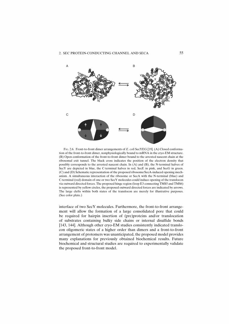
interface of two SecY molecules. Furthermore, the front-to-front arrange-ment will allow the formation of a large consolidated pore that couldbe required for hairpin insertion of (pre)proteins and/or translocationof substrates containing bulky side chains or internal disulfide bonds[143, 144]. Although other cryo-EM studies consistently indicated translo-con oligomeric states of a higher order than dimers and a front-to-frontarrangement of protomers was unanticipated, the proposed model providesmany explanations for previously obtained biochemical results. Futurebiochemical and structural studies are required to experimentally validatethe proposed front-to-front model.
A B
C D
X
FIG. 2.6. Front-to-front dimer arrangements of E. coli SecYEG [19]. (A) Closed conforma-
tion of the front-to-front dimer, nonphysiologically bound to mRNA in the cryo-EM structure.
(B) Open conformation of the front-to-front dimer bound to the arrested nascent chain at the
ribosomal exit tunnel. The black cross indicates the position of the electron density that
possibly corresponds to the arrested nascent chain. In (A) and (B), the N-terminal halves of
SecY are depicted in blue, the C-terminal halves in red, SecE in pink, and SecG in green.
(C) and (D) Schematic representation of the proposed ribosome/SecA-induced opening mech-
anism. A simultaneous interaction of the ribosome or SecA with the N-terminal (blue) and
C-terminal (red) domain of one or two SecY molecules could induce opening of the translocon
via outward directed forces. The proposed hinge region (loop E3 connecting TMS5 and TMS6)
is represented by yellow circles, the proposed outward directed forces are indicated by arrows.
The large clefts within both states of the translocon are merely for illustrative purposes.
(See color plate.)
2. SEC PROTEIN-CONDUCTING CHANNEL AND SECA 55
D. INDUCTION OF CONFORMATIONAL CHANGES IN SECYEG
Assuming that the proposed open conformation of dimeric SecYEGrepresents a physiologically active translocon, the question is how the ribo-some or SecA can induce opening of the channel. Interestingly, the ribosomeand SecA interact with similar regions of the translocon, suggesting that theymight share a common opening mechanism. The ribosome interacts withthe translocon via three distinct connections. In agreement with biochemicalstudies [145, 146], two connections are similarly formed by the pairs oflong cytoplasmic loops of SecY (C4 and C5, connecting TMS6 with TMS7and TMS8 with TMS9, respectively, black arrows in Figure 2.6C). The thirdconnection is mediated by the cytoplasmic loop of SecG and the N-terminustwo transmembrane segments of SecE (one of the white arrows inFigure 2.6C). SecA has been shown to interact with the C5 loop of SecY aswell (EvdS et al. submitted for publication), with SecG [147], and with theinterface between TMS4 and C3 of SecY (EvdS et al. submitted for publica-tion) that is in direct contact with SecG [127]. Importantly, the two regions ofinteraction are located in different domains of a single SecYEG protomer,and thus separation of the two SecY domains could be induced by a simulta-neous interaction with both of them (Figure 2.6C and D). In the front-to-front arrangement, separation of the two SecY domains mainly takes placeat the dimer interface, and thus opening of a single protomer will be directlytransmitted to the neighboring protomer.
It should be noted, however, that the features that mediate the thirdribosome–translocon connection (SecG/Secb and the SecE extension) arenot essential for viability or protein translocation [148, 149, 50]. Thus,ribosome-induced opening of the translocon might be primarily mediatedby the two C4/C5 connections, while the third connection plays an auxiliaryrole. This would explain the mere stimulatory role of Secb on posttransla-tional protein translocation [150]. The stimulatory role of SecG can beexplained similarly, but the SecA-induced opening mechanism differs inat least one aspect from the ribosome-induced opening mechanism, that isthe SecA ‘‘membrane insertion’’ cycle. The SecA interaction site in theN-terminal half of SecY (the TMS4–C3 interface) appears to be part ofthe region where SecA inserts at least partially into the translocon. SecG isin proximity of this region and might thus facilitate membrane cycling ofSecA [151, 152]. It seems unlikely however that SecG completely inverts itsmembrane topology during protein translocation via SecYEG as proposedpreviously [153], as topologically fixed SecG has been shown to be equallyactive as wild-type SecG [154]. The different conformations of SecG thatare observed in vitro most likely represent conformational changes withinthis highly dynamic region of the translocon.
56 ELI O. VAV DER SLUIS, ET AL.
E. THE ROLE OF THE PLUG
In addition to separation of the two SecY domains, the opening mecha-nism of the translocon is thought to involve displacement of the ‘‘plug’’domain formed by the E1 loop [16]. This proposal is based on the locationof the plug domain at the extracellular end of the pore region in the closedconformation of the channel, and the observation that it has the potential tobe cross-linked to the C-terminal region of SecE, located�20 A away [155].The mobile nature of the plug domain has been confirmed by moleculardynamics simulations [125] (Gumbart and Schulten, Biophysical Journal, inpress), homology modeling [119], and a cross-linking approach [156]. In thelatter study, it was shown that the plug domain is displaced during proteintranslocation, providing the first experimental evidence for its proposedfunction. An interesting observation that provides a novel hypothesis on themechanism of SecA-induced opening of the translocon was recently madewith a peptide scanning approach (EvdS et al., in preparation). It was shownthat SecA directly interacts with peptides derived from the plug domain,suggesting that displacement of the plug domain in bacteria might bedirectly induced by SecA.
VII. ConcludingRemarks
To summarize, our understanding of the molecular mechanism ofprotein translocation in bacteria has increased dramatically during thepast few years, and long held schematic models are slowly beginning totake shape on a detailed structural level. However, a ‘‘molecular movie’’ ofprotein translocation is not expected in the near future because of thetremendous complexity of the process. New insights have to be providedby a combination of structural, biophysical, and biochemical studies. Con-sidering the large amount of unresolved questions, research on the Secmachinery is expected to remain an exciting area in biology throughoutthe next decade.
REFERENCES
1. Pohlschroder, M., Prinz, W.A., Hartmann, E., and Beckwith, J. (1997). Protein transloca-
tion in the three domains of life: Variations on a theme. Cell 91:563–566.
2. Osborne, A.R., Rapoport, T.A., and Van den Berg, B. (2005). Protein translocation by
the Sec61/SecY channel. Annu Rev Cell Dev Biol 21:529–550.
3. Cao, T.B., and Saier, M.H. (2003). The general protein secretory pathway: Phylogenetic
analyses leading to evolutionary conclusions. Biochim Biophys Acta 1609:115–125.
2. SEC PROTEIN-CONDUCTING CHANNEL AND SECA 57
4. Ulbrandt, N.D., Newitt, J.A., and Bernstein, H.D. (1997). The E. coli signal recognition
particle is required for the insertion of a subset of innermembrane proteins.Cell 88:187–196.
5. Kalies, K.U., Gorlich, D., and Rapoport, T.A. (1994). Binding of ribosomes to the rough
endoplasmic reticulum mediated by the Sec61p-complex. J Cell Biol 126:925–934.
6. Pohlschroder, M., Hartmann, E., Hand, N.J., Dilks, K., and Haddad, A. (2005). Diversity
and evolution of protein translocation. Annu Rev Microbiol 59:91–111.
7. Rapiejko, P.J., and Gilmore, R. (1997). Empty site forms of the SRP54 and SR alpha
GTPases mediate targeting of ribosome-nascent chain complexes to the endoplasmic
reticulum. Cell 89:703–713.
8. Mason, N., Ciufo, L.F., and Brown, J.D. (2000). Elongation arrest is a physiologically
important function of signal recognition particle. EMBO J 19:4164–4174.
9. Luirink, J., and Sinning, I. (2004). SRP-mediated protein targeting: Structure and function
revisited. Biochim Biophys Acta 1694:17–35.
10. Wild, K., Halic, M., Sinning, I., and Beckmann, R. (2004). SRP meets the ribosome.
Nat Struct Mol Biol 11:1049–1053.
11. Hartl, F.U., Lecker, S., Schiebel, E., Hendrick, J.P., and Wickner, W. (1990). The binding
cascade of SecB to SecA to SecY/E mediates preprotein targeting to the E. coli plasma
membrane. Cell 63:269–279.
12. Brodsky, J.L., Goeckeler, J., and Schekman, R. (1995). BiP and Sec63p are required for
both co- and posttranslational protein translocation into the yeast endoplasmic reticulum.
Proc Natl Acad Sci USA 92:9643–9646.
13. Hunt, J.F., Weinkauf, S., Henry, L., Fak, J.J., McNicholas, P., Oliver, D.B., and
Deisenhofer, J. (2002). Nucleotide control of interdomain interactions in the conforma-
tional reaction cycle of SecA. Science 297:2018–2026.
14. Xu, Z., Knafels, J.D., and Yoshino, K. (2000). Crystal structure of the bacterial protein
export chaperone secB. Nat Struct Biol 7:1172–1177.
15. Zhou, J., and Xu, Z. (2003). Structural determinants of SecB recognition by SecA in
bacterial protein translocation. Nat Struct Biol 10:942–947.
16. VandenBerg,B.,Clemons,W.M., Jr.,Collinson, I.,Modis,Y.,Hartmann,E.,Harrison, S.C.,
and Rapoport, T.A. (2004). X-ray structure of a protein-conducting channel. Nature
427:36–44.
17. Sharma, V., Arockiasamy, A., Ronning, D.R., Savva, C.G., Holzenburg, A.,
Braunstein, M., Jacobs, W.R., Jr., and Sacchettini, J.C. (2003). Crystal structure of
Mycobacterium tuberculosis SecA, a preprotein translocating ATPase. Proc Natl Acad
Sci USA 100:2243–2248.
18. Beckmann, R., Spahn, C.M., Eswar, N., Helmers, J., Penczek, P.A., Sali, A., Frank, J., and
Blobel, G. (2001). Architecture of the protein-conducting channel associated with the
translating 80S ribosome. Cell 107:361–372.
19. Mitra, K., Schaffitzel, C., Shaikh, T., Tama, F., Jenni, S., Brooks, C.L., III, Ban, N., and
Frank, J. (2005). Structure of the E. coli protein-conducting channel bound to a translat-
ing ribosome. Nature 438:318–324.
20. Menetret, J.F., Neuhof, A., Morgan, D.G., Plath, K., Radermacher, M., Rapoport, T.A.,
and Akey, C.W. (2000). The structure of ribosome-channel complexes engaged in protein
translocation. Mol Cell 6:1219–1232.
21. Morgan, D.G., Menetret, J.F., Radermacher, M., Neuhof, A., Akey, I.V., Rapoport, T.A.,
and Akey, C.W. (2000). A comparison of the yeast and rabbit 80 S ribosome reveals the
topology of the nascent chain exit tunnel, inter-subunit bridges and mammalian rRNA
expansion segments. J Mol Biol 301:301–321.
22. de Keyzer, J., Van der Does, C., and Driessen, A.J.M. (2003). The bacterial translocase:
A dynamic protein channel complex. Cell Mol Life Sci 60:2034–2052.
58 ELI O. VAV DER SLUIS, ET AL.

23. Veenendaal, A.K., Van der Does, C., and Driessen, A.J.M. (2004). The protein-
conducting channel SecYEG. Biochim Biophys Acta 1694:81–95.
24. Vrontou, E., and Economou, A. (2004). Structure and function of SecA, the preprotein
translocase nanomotor. Biochim Biophys Acta 1694:67–80.
25. Urbanus, M.L., Scotti, P.A., Froderberg, L., Saaf, A., de Gier, J.W., Brunner, J.,
Samuelson, J.C., Dalbey, R.E., Oudega, B., and Luirink, J. (2001). Sec-dependent mem-
brane protein insertion: Sequential interaction of nascent FtsQ with SecY and YidC.
EMBO Rep 2:524–529.
26. Samuelson, J.C., Jiang, F., Yi, L., Chen, M., de Gier, J.W., Kuhn, A., and Dalbey, R.E.
(2001). Function of YidC for the insertion of M13 procoat protein in Escherichia coli:
Translocation of mutants that show differences in their membrane potential dependence
and Sec requirement. J Biol Chem 276:34847–34852.
27. Jia, L., Dienhart, M., Schramp,M., McCauley,M., Hell, K., and Stuart, R.A. (2003). Yeast
Oxa1 interacts with mitochondrial ribosomes: The importance of the C-terminal region of
Oxa1. EMBO J 22:6438–6447.
28. Szyrach, G., Ott, M., Bonnefoy, N., Neupert, W., and Herrmann, J.M. (2003). Ribosome
binding to the Oxa1 complex facilitates co-translational protein insertion in mitochon-
dria. EMBO J 22:6448–6457.
29. Duong, F., and Wickner, W. (1997). Distinct catalytic roles of the SecYE, SecG and
SecDFyajC subunits of preprotein translocase holoenzyme. EMBO J 16:2756–2768.
30. Duong, F., and Wickner, W. (1997). The SecDFyajC domain of preprotein translocase
controls preprotein movement by regulating SecA membrane cycling. EMBO J
16:4871–4879.
31. Kato, Y., Nishiyama, K., and Tokuda, H. (2003). Depletion of SecDF-YajC causes a
decrease in the level of SecG: Implication for their functional interaction. FEBS Lett
550:114–118.
32. Rain, J.C., Selig, L., De Reuse, H., Battaglia, V., Reverdy, C., Simon, S., Lenzen, G.,
Petel, F., Wojcik, J., Schachter, V., Chemama, Y., Labigne, A., et al. (2001). The protein-
protein interaction map of Helicobacter pylori. Nature 409:211–215.
33. Nouwen, N., and Driessen, A.J.M. (2002). SecDFyajC forms a heterotetrameric complex
with YidC. Mol Microbiol 44:1397–1405.
34. Nouwen, N., Van der Laan, M., and Driessen, A.J.M. (2001). SecDFyajC is not required
for the maintenance of the proton motive force. FEBS Lett 508:103–106.
35. Stenberg, F., Chovanec, P., Maslen, S.L., Robinson, C.V., Ilag, L.L., von Heijne, G., and
Daley, D.O. (2005). Protein complexes of the Escherichia coli cell envelope. J Biol Chem
280:34409–34419.
36. Lecker, S.H., Driessen, A.J.M., and Wickner, W. (1990). ProOmpA contains secondary
and tertiary structure prior to translocation and is shielded from aggregation by associa-
tion with SecB protein. EMBO J 9:2309–2314.
37. Gannon, P.M., Li, P., and Kumamoto, C.A. (1989). The mature portion of Escherichia
coli maltose-binding protein (MBP) determines the dependence of MBP on SecB for
export. J Bacteriol 171:813–818.
38. Fekkes, P., Van der Does, C., and Driessen, A.J.M. (1997). The molecular chaperone
SecB is released from the carboxy-terminus of SecA during initiation of precursor protein
translocation. EMBO J 16:6105–6113.
39. Shimizu, H., Nishiyama, K., and Tokuda, H. (1997). Expression of gpsA encoding biosyn-
thetic sn-glycerol 3-phosphate dehydrogenase suppresses both the LB-phenotype of a
secB null mutant and the cold-sensitive phenotype of a secG null mutant. Mol Microbiol
26:1013–1021.
2. SEC PROTEIN-CONDUCTING CHANNEL AND SECA 59
40. Delepelaire, P., and Wandersman, C. (1998). The SecB chaperone is involved in the
secretion of the Serratia marcescens HasA protein through an ABC transporter.
EMBO J 17:936–944.
41. Baars, L., Ytterberg,A.J., Drew,D.,Wagner, S., Thilo, C., VanWijk, K.J., and deGier, J.W.
(2005).Defining the role of theE. coli chaperoneSecBusing comparative proteomics. JBiol
Chem 281:10024–10034.
42. Knoblauch, N.T., Rudiger, S., Schonfeld, H.J., Driessen, A.J.M., Schneider-Mergener, J.,
and Bukau, B. (1999). Substrate Specificity of the SecB Chaperone. J Biol Chem
274:34219–34225.
43. Rajapandi, T., Dolan, K.M., and Oliver, D.B. (1991). The first gene in theEscherichia coli
secA operon, gene X, encodes a nonessential secretory protein. J Bacteriol 173:7092–7097.
44. Nakatogawa, H., and Ito, K. (2001). Secretion monitor, SecM, undergoes self-translation
arrest in the cytosol. Mol Cell 7:185–192.
45. Nakatogawa, H., and Ito, K. (2002). The ribosomal exit tunnel functions as a discriminat-
ing gate. Cell 108:629–636.
46. Nakatogawa, H., Murakami, A., Mori, H., and Ito, K. (2005). SecM facilitates translocase
function of SecA by localizing its biosynthesis. Genes Dev 19:436–444.
47. Chen, X., Xu, H., and Tai, P.C. (1996). A significant fraction of functional SecA is
permanently embedded in the membrane. SecA cycling on and off the membrane is not
essential during protein translocation. J Biol Chem 271:29698–29706.
48. Eichler, J., Rinard, K., and Wickner, W. (1998). Endogenous SecA catalyzes preprotein
translocation at SecYEG. J Biol Chem 273:21675–21681.
49. Schatz, P.J., Riggs, P.D., Jacq, A., Fath, M.J., and Beckwith, J. (1989). The secE gene
encodes an integral membrane protein required for protein export in Escherichia coli.
Genes Dev 3:1035–1044.
50. Schatz, P.J., Bieker, K.L., Ottemann, K.M., Silhavy, T.J., and Beckwith, J. (1991). One of
three transmembrane stretches is sufficient for the functioning of the SecE protein,
a membrane component of the E. coli secretion machinery. EMBO J 10:1749–1757.
51. Van der Sluis, E.O., and Driessen, A.J.M. (2006). Stepwise evolution of the Sec machin-
ery in Proteobacteria. Trends Microbiol 14:105–108.
52. Braunstein, M., Brown, A.M., Kurtz, S., and Jacobs, W.R., Jr. (2001). Two nonredundant
SecA homologues function in mycobacteria. J Bacteriol 183:6979–6990.
53. Lenz, L.L., and Portnoy, D.A. (2002). Identification of a second Listeria secA gene
associated with protein secretion and the rough phenotype.Mol Microbiol 45: 1043–1056.
54. Bensing, B.A., and Sullam, P.M. (2002). An accessory sec locus of Streptococcus gordonii
is required for export of the surface protein GspB and for normal levels of binding to
human platelets. Mol Microbiol 44:1081–1094.
55. Braunstein, M., Espinosa, B.J., Chan, J., Belisle, J.T., and Jacobs, R. (2003). SecA2
functions in the secretion of superoxide dismutase A and in the virulence of
Mycobacterium tuberculosis. Mol Microbiol 48:453–464.
56. Lenz, L.L., Mohammadi, S., Geissler, A., and Portnoy, D.A. (2003). SecA2-dependent
secretion of autolytic enzymes promotes Listeria monocytogenes pathogenesis. Proc Natl
Acad Sci USA 100:12432–12437.
57. Bensing, B.A., Gibson, B.W., and Sullam, P.M. (2004). The Streptococcus gordonii
platelet binding protein GspB undergoes glycosylation independently of export. J Bacter-
iol 186:638–645.
58. Bensing, B.A., Takamatsu, D., and Sullam, P.M. (2005). Determinants of the streptococ-
cal surface glycoprotein GspB that facilitate export by the accessory Sec system. Mol
Microbiol 58:1468–1481.
60 ELI O. VAV DER SLUIS, ET AL.

59. Takamatsu, D., Bensing, B.A., and Sullam, P.M. (2004). Four proteins encoded in the
gspB-secY2A2 operon of Streptococcus gordonii mediate the intracellular glycosylation
of the platelet-binding protein GspB. J Bacteriol 186:7100–7111.
60. Takamatsu, D., Bensing, B.A., and Sullam, P.M. (2004). Genes in the accessory sec locus
of Streptococcus gordonii have three functionally distinct effects on the expression of the
platelet-binding protein GspB. Mol Microbiol 52:189–203.
61. Chen, Q., Wu, H., and Fives-Taylor, P.M. (2004). Investigating the role of secA2 in
secretion and glycosylation of a fimbrial adhesin in Streptococcus parasanguis FW213.
Mol Microbiol 53:843–856.
62. Machata, S., Hain, T., Rohde, M., and Chakraborty, T. (2005). Simultaneous deficiency of
both MurA and p60 proteins generates a rough phenotype in Listeria monocytogenes.
J Bacteriol 187:8385–8394.
63. Tian, H., Boyd, D., and Beckwith, J. (2000). A mutant hunt for defects in membrane
protein assembly yields mutations affecting the bacterial signal recognition particle and
Sec machinery. Proc Natl Acad Sci USA 97:4730–4735.
64. Traxler, B., and Murphy, C. (1996). Insertion of the polytopic membrane protein MalF
is dependent on the bacterial secretion machinery. J Biol Chem 271:12394–12400.
65. Van der Laan, M., Nouwen, N., and Driessen, A.J.M. (2004). SecYEG proteoliposomes
catalyze the Dc-dependent membrane insertion of FtsQ. J Biol Chem 279:1659–1664.
66. Neumann-Haefelin, C., Schafer, U., Muller, M., and Koch, H.G. (2000). SRP-dependent
co-translational targeting and SecA-dependent translocation analyzed as individual
steps in the export of a bacterial protein. EMBO J 19:6419–6426.
67. Zito, C.R., and Oliver, D. (2003). Two-stage binding of SecA to the bacterial translocon
regulates ribosome-translocon interaction. J Biol Chem 278:40640–40646.
68. Karamyshev, A.L., and Johnson, A.E. (2005). Selective SecA association with signal
sequences in ribosome-bound nascent chains: A potential role for SecA in ribosome
targeting to the bacterial membrane. J Biol Chem 280:37930–37940.
69. Liebke, H.H. (1987). Multiple SecA protein isoforms in Escherichia coli. J Bacteriol
169:1174–1181.
70. Fekkes, P., de Wit, J.G., Van der Wolk, J.P., Kimsey, H.H., Kumamoto, C.A., and
Driessen, A.J.M. (1998). Preprotein transfer to the Escherichia coli translocase requires
the co-operative binding of SecB and the signal sequence to SecA. Mol Microbiol
29:1179–1190.
71. Economou, A., and Wickner, W. (1994). SecA promotes preprotein translocation by
undergoing ATP-driven cycles of membrane insertion and deinsertion. Cell 78:835–843.
72. Schiebel, E., Driessen, A.J.M., Hartl, F.U., and Wickner, W. (1991). Dm Hþ and ATP
function at different steps of the catalytic cycle of preprotein translocase. Cell 64:927–939.
73. Van der Wolk, J.P., de Wit, J.G., and Driessen, A.J.M. (1997). The catalytic cycle of the
Escherichia coli SecA ATPase comprises two distinct preprotein translocation events.
EMBO J 16:7297–7304.
74. Economou, A., Pogliano, J.A., Beckwith, J., Oliver, D.B., and Wickner, W. (1995). SecA
membrane cycling at SecYEG is driven by distinct ATP binding and hydrolysis events and
is regulated by SecD and SecF. Cell 83:1171–1181.
75. Osborne, A.R., Clemons, W.M., Jr., and Rapoport, T.A. (2004). A large conformational
change of the translocation ATPase SecA. Proc Natl Acad Sci USA 101:10937–10942.
76. Dekker, C., de Kruijff, B., and Gros, P. (2003). Crystal structure of SecB from Escherichia
coli. J Struct Biol 144:313–319.
77. Koonin, E.V., and Gorbalenya, A.E. (1992). Autogenous translation regulation by
Escherichia coli ATPase SecA may be mediated by an intrinsic RNA helicase activity
of this protein. FEBS Lett 298:6–8.
2. SEC PROTEIN-CONDUCTING CHANNEL AND SECA 61

78. Papanikou, E., Karamanou, S., Baud, C., Frank, M., Sianidis, G., Keramisanou, D.,
Kalodimos, C.G., Kuhn, A., and Economou, A. (2005). Identification of the preprotein
binding domain of SecA. J Biol Chem 280:43209–43217.
79. Kimura, E., Akita, M., Matsuyama, S., and Mizushima, S. (1991). Determination of a
region in SecA that interacts with presecretory proteins in Escherichia coli. J Biol Chem
266:6600–6606.
80. Woodbury, R.L., Hardy, S.J., and Randall, L.L. (2002). Complex behavior in solution of
homodimeric SecA. Protein Sci 11:875–882.
81. Akita, M., Shinaki, A., Matsuyama, S., and Mizushima, S. (1991). SecA, an essential
component of the secretory machinery of Escherichia coli, exists as homodimer. Biochem
Biophys Res Commun 174:211–216.
82. Dempsey, B.R., Economou, A., Dunn, S.D., and Shilton, B.H. (2002). The ATPase
domain of SecA can form a tetramer in solution. J Mol Biol 315:831–843.
83. Or, E., Navon, A., and Rapoport, T. (2002). Dissociation of the dimeric SecA ATPase
during protein translocation across the bacterial membrane. EMBO J 21:4470–4479.
84. Benach, J., Chou, Y.T., Fak, J.J., Itkin, A., Nicolae, D.D., Smith, P.C., Wittrock, G.,
Floyd, D.L., Golsaz, C.M., Gierasch, L.M., and Hunt, J.F. (2003). Phospholipid-induced
monomerization and signal-peptide-induced oligomerization of SecA. J Biol Chem
278:3628–3638.
85. Musial-Siwek, M., Rusch, S.L., and Kendall, D.A. (2005). Probing the affinity of SecA for
signal peptide in different environments. Biochemistry 44:13987–13996.
86. Bu, Z., Wang, L., and Kendall, D.A. (2003). Nucleotide binding induces changes in the
oligomeric state and conformation of SecA in a lipid environment: A small-angle
neutron-scattering study. J Mol Biol 332:23–30.
87. Bessonneau, P., Besson, V., Collinson, I., and Duong, F. (2002). The SecYEG preprotein
translocation channel is a conformationally dynamic and dimeric structure. EMBO J
21:995–1003.
88. Duong, F. (2003). Binding, activation and dissociation of the dimeric SecAATPase at the
dimeric SecYEG translocase. EMBO J 22:4375–4384.
89. Tziatzios, C., Schubert, D., Lotz, M., Gundogan, D., Betz, H., Schagger, H., Haase, W.,
Duong, F., and Collinson, I. (2004). The bacterial protein-translocation complex:
SecYEG dimers associate with one or two SecA molecules. J Mol Biol 340:513–524.
90. Jilaveanu, L.B., Zito, C.R., and Oliver, D. (2005). Dimeric SecA is essential for protein
translocation. Proc Natl Acad Sci USA 102:7511–7516.
91. de Keyzer, J., Van der Sluis, E.O., Spelbrink, R.E., Nijstad, N., de Kruijff, B., Nouwen, N.,
Van der Does, C., and Driessen, A.J.M. (2005). Covalently dimerized SecA is functional
in protein translocation. J Biol Chem 280:35255–35260.
92. Karamanou, S., Sianidis, G., Gouridis, G., Pozidis, C., Papanikolau, Y., Papanikou, E.,
and Economou, A. (2005). Escherichia coli SecA truncated at its termini is functional and
dimeric. FEBS Lett 579:1267–1271.
93. Randall, L.L., Crane, J.M., Lilly, A.A., Liu, G., Mao, C., Patel, C.N., and Hardy, S.J.
(2005). Asymmetric binding between SecA and SecB two symmetric proteins: Implica-
tions for function in export. J Mol Biol 348:479–489.
94. Or, E., Boyd, D., Gon, S., Beckwith, J., and Rapoport, T. (2005). The bacterial ATPase
SecA functions as a monomer in protein translocation. J Biol Chem 280:9097–9105.
95. Jilaveanu, L.B., and Oliver, D. (2006). SecA dimer cross-linked at its subunit interface is
functional for protein translocation. J Bacteriol 188:335–338.
96. Driessen, A.J.M. (1993). SecA, the peripheral subunit of the Escherichia coli precursor
protein translocase, is functional as a dimer. Biochemistry 32:13190–13197.
62 ELI O. VAV DER SLUIS, ET AL.

97. Ding, H., Hunt, J.F., Mukerji, I., and Oliver, D. (2003). Bacillus subtilis SecA ATPase
exists as an antiparallel dimer in solution. Biochemistry 42:8729–8738.
98. Ding, H., Mukerji, I., and Oliver, D. (2003). Nucleotide and phospholipid-dependent
control of PPXD and C-domain association for SecA ATPase. Biochemistry
42:13468–13475.
99. Ding, H., Mukerji, I., and Oliver, D. (2001). Lipid and signal peptide-induced conforma-
tional changes within the C-domain of Escherichia coli SecA protein. Biochemistry
40:1835–1843.
100. Chou,Y.T., Swain, J.F., andGierasch, L.M. (2002). Functionally significantmobile regions
of Escherichia coli SecA ATPase identified by NMR. J Biol Chem 277:50985–50990.
101. Ulbrandt, N.D., London, E., and Oliver, D.B. (1992). Deep penetration of a portion of
Escherichia coli SecA protein into model membranes is promoted by anionic phospholi-
pids and by partial unfolding. J Biol Chem 267:15184–15192.
102. Den Blaauwen, T., Fekkes, P., de Wit, J.G., Kuiper, W., and Driessen, A.J.M. (1996).
Domain interactions of the peripheral preprotein translocase subunit SecA. Biochemistry
35:11994–12004.
103. Schmidt, M., Ding, H., Ramamurthy, V., Mukerji, I., and Oliver, D. (2000). Nucleotide
binding activity of SecA homodimer is conformationally regulated by temperature and
altered by prlD and azi mutations. J Biol Chem 275:15440–15448.
104. Baud, C., Karamanou, S., Sianidis, G., Vrontou, E., Politou, A.S., and Economou, A.
(2002). Allosteric communication between signal peptides and the SecA protein DEAD
motor ATPase domain. J Biol Chem 277:13724–13731.
105. Vrontou, E., Karamanou, S., Baud, C., Sianidis, G., and Economou, A. (2004). Global
co-ordination of protein translocation by the SecA IRA1 switch. J Biol Chem 279(21):
22490–22497.
106. Story, R.M., Li, H., and Abelson, J.N. (2001). Crystal structure of a DEAD box protein
from the hyperthermophile Methanococcus jannaschii. Proc Natl Acad Sci USA
98:1465–1470.
107. Velankar, S.S., Soultanas, P., Dillingham, M.S., Subramanya, H.S., and Wigley, D.B.
(1999). Crystal structures of complexes of PcrA DNA helicase with a DNA substrate
indicate an inchworm mechanism. Cell 97:75–84.
108. Zhou, J., and Xu, Z. (2005). The structural view of bacterial translocation-specific chap-
erone SecB: Implications for function. Mol Microbiol 58:349–357.
109. Fekkes, P., de Wit, J.G., Boorsma, A., Friesen, R.H., and Driessen, A.J.M. (1999). Zinc
stabilizes the SecB binding site of SecA. Biochemistry 38:5111–5116.
110. Breukink, E., Nouwen, N., Van Raalte, A., Mizushima, S., Tommassen, J., and de
Kruijff, B. (1995). The C terminus of SecA is involved in both lipid binding and SecB
binding. J Biol Chem 270:7902–7907.
111. Dempsey, B.R., Wrona, M., Moulin, J.M., Gloor, G.B., Jalilehvand, F., Lajoie, G.,
Shaw, G.S., and Shilton, B.H. (2004). Solution NMR structure and X-ray absorption
analysis of the C-terminal zinc-binding domain of the SecA ATPase. Biochemistry
43:9361–9371.
112. Matousek, W.M., and Alexandrescu, A.T. (2004). NMR structure of the C-terminal
domain of SecA in the free state. Biochim Biophys Acta 1702:163–171.
113. Kimsey, H.H., Dagarag, M.D., and Kumamoto, C.A. (1995). Diverse effects of mutation
on the activity of the Escherichia coli export chaperone SecB. J Biol Chem
270:22831–22835.
114. Rajapandi, T., and Oliver, D. (1994). Carboxy-terminal region of Escherichia coli SecA
ATPase is important to promote its protein translocation activity in vivo. Biochem
Biophys Res Commun 200:1477–1483.
2. SEC PROTEIN-CONDUCTING CHANNEL AND SECA 63

115. Van Voorst, F., Van der Does, C., Brunner, J., Driessen, A.J.M., and de Kruijff, B. (1998).
Translocase-bound SecA is largely shielded from the phospholipid acyl chains. Biochem-
istry 37:12261–12268.
116. Dapic, V., and Oliver, D. (2000). Distinct membrane binding properties of N- and
C-terminal domains of Escherichia coli SecA ATPase. J Biol Chem 275:25000–25007.
117. Kinch, L.N., Saier, M.H., Jr., and Grishin, N.V. (2002). Sec61beta–a component of the
archaeal protein secretory system. Trends Biochem Sci 27:170–171.
118. Breyton, C., Haase, W., Rapoport, T.A., Kuhlbrandt, W., and Collinson, I. (2002). Three-
dimensional structure of the bacterial protein-translocation complex SecYEG. Nature
418:662–665.
119. Bostina, M., Mohsin, B., Kuhlbrandt, W., and Collinson, I. (2005). Atomic model of the
E. coli membrane-bound protein translocation complex SecYEG. J Mol Biol
352:1035–1043.
120. Veenendaal, A.K.J., Van der Does, C., and Driessen, A.J.M. (2002). The core of the
bacterial translocase harbors a tilted transmembrane segment 3 of SecE. J Biol Chem
277:36640–36645.
121. Beswick, V., Baleux, F., Huynh-Dinh, T., Kepes, F., Neumann, J.M., and Sanson, A.
(1996). NMR conformational study of the cytoplasmic domain of the canine Sec61 gamma
protein from the protein translocation pore of the endoplasmic reticulum membrane.
Biochemistry 35:14717–14724.
122. Plath, K., Mothes, W., Wilkinson, B.M., Stirling, C.J., and Rapoport, T.A. (1998). Signal
sequence recognition in posttranslational protein transport across the yeast ER
membrane. Cell 94:795–807.
123. Wang, L., Miller, A., Rusch, S.L., and Kendall, D.A. (2004). Demonstration of a specific
Escherichia coli SecY-signal peptide interaction. Biochemistry 43:13185–13192.
124. Plath, K., Wilkinson, B.M., Stirling, C.J., and Rapoport, T.A. (2004). Interactions
between Sec complex and prepro-alpha-factor during posttranslational protein transport
into the endoplasmic reticulum. Mol Biol Cell 15:1–10.
125. Tian, P., and Andricioaei, I. (2006). Size, motion and function of the SecY trans-
locon revealed by molecular dynamics simulations with virtual probes. Biophys J
90:2718–2730.
126. Mori, H., Tsukazaki, T., Masui, R., Kuramitsu, S., Yokoyama, S., Johnson, A.E.,
Kimura, Y., Akiyama, Y., and Ito, K. (2003). Fluorescence resonance energy transfer
analysis of protein translocase. SecYE from Thermus thermophilus HB8 forms a consti-
tutive oligomer in membranes. J Biol Chem 278:14257–14264.
127. Van der Sluis, E., Nouwen, N., and Driessen, A.J.M. (2002). SecY-SecY and SecY-SecG
contacts revealed by site-specific crosslinking. FEBS Lett 527:159–165.
128. Veenendaal, A.K.J., Van der Does, C., and Driessen, A.J.M. (2001). Mapping the sites of
interaction between SecY and SecE by cysteine scanning mutagenesis. J Biol Chem
276:32559–32566.
129. Kaufmann, A., Manting, E.H., Veenendaal, A.K.J., Driessen, A.J.M., and Van der
Does, C. (1999). Cysteine-directed cross-linking demonstrates that helix 3 of SecE is
close to helix 2 of SecY and helix 3 of a neighboring SecE. Biochemistry 38:9115–9125.
130. Scheuring, J., Braun, N., Nothdurft, L., Stumpf, M., Veenendaal, A.K., Kol, S., Van der
Does, C., Driessen, A.J.M., and Weinkauf, S. (2005). The oligomeric distribution of
SecYEG is altered by SecA and translocation ligands. J Mol Biol 354:258–271.
131. Nagamori, S., Nishiyama, K., and Tokuda, H. (2000). Two SecG molecules present in a
single protein translocation machinery are functional even after crosslinking. J Biochem
128:129–137.
64 ELI O. VAV DER SLUIS, ET AL.

132. Meyer, T.H., Menetret, J.F., Breitling, R., Miller, K.R., Akey, C.W., and Rapoport, T.A.
(1999). The bacterial SecY/E translocation complex forms channel-like structures similar
to those of the eukaryotic Sec61p complex. J Mol Biol 285:1789–1800.
133. Collinson, I., Breyton, C., Duong, F., Tziatzios, C., Schubert, D., Or, E., Rapoport, T., and
Kuhlbrandt, W. (2001). Projection structure and oligomeric properties of a bacterial core
protein translocase. EMBO J 20:2462–2471.
134. Manting, E.H., Van der Does, C., Remigy, H., Engel, A., and Driessen, A.J.M. (2000).
SecYEG assembles into a tetramer to form the active protein translocation channel.
EMBO J 19:852–861.
135. Hanein, D., Matlack, K.E., Jungnickel, B., Plath, K., Kalies, K.U., Miller, K.R.,
Rapoport, T.A., and Akey, C.W. (1996). Oligomeric rings of the Sec61p complex induced
by ligands required for protein translocation. Cell 87:721–732.
136. Woolf, P.J., and Linderman, J.J. (2003). Self organization of membrane proteins via
dimerization. Biophys Chem 104:217–227.
137. Scotti, P.A., Urbanus, M.L., Brunner, J., de Gier, J.W., von Heijne, G., Van der Does, C.,
Driessen, A.J.M., Oudega, B., and Luirink, J. (2000). YidC, the Escherichia coli homo-
logue ofmitochondrialOxa1p, is a component of the Sec translocase.EMBOJ 19:542–549.
138. Yahr, T.L., and Wickner, W.T. (2000). Evaluating the oligomeric state of SecYEG in
preprotein translocase. EMBO J 19:4393–4401.
139. Beckmann, R., Bubeck, D., Grassucci, R., Penczek, P., Verschoor, A., Blobel, G., and
Frank, J. (1997). Alignment of conduits for the nascent polypeptide chain in the
ribosome-Sec61 complex. Science 278:2123–2126.
140. Menetret, J.F., Hegde, R.S., Heinrich, S.U., Chandramouli, P., Ludtke, S.J., Rapoport, T.A.,
andAkey, C.W. (2005). Architecture of the ribosome-channel complex derived from native
membranes. J Mol Biol 348:445–457.
141. Morgan, D.G., Menetret, J.F., Neuhof, A., Rapoport, T.A., and Akey, C.W. (2002).
Structure of the mammalian ribosome-channel complex at 17A resolution. J Mol Biol
324:871–886.
142. Prinz, A., Behrens, C., Rapoport, T.A., Hartmann, E., and Kalies, K.U. (2000). Evolu-
tionarily conserved binding of ribosomes to the translocation channel via the large
ribosomal RNA. EMBO J 19:1900–1906.
143. Tani, K., Tokuda, H., andMizushima, S. (1990). Translocation of ProOmpA possessing an
intramolecular disulfide bridge into membrane vesicles of Escherichia coli. Effect of
membrane energization. J Biol Chem 265:17341–17347.
144. De Keyzer, J., Van der Does, C., and Driessen, A.J.M. (2002). Kinetic analysis of the
translocation of fluorescent precursor proteins into Escherichia coli membrane vesicles.
J Biol Chem 277:46059–46065.
145. Raden, D., Song, W., and Gilmore, R. (2000). Role of the cytoplasmic segments of
Sec61alpha in the ribosome-binding and translocation-promoting activities of the Sec61
complex. J Cell Biol 150:53–64.
146. Cheng, Z., Jiang, Y., Mandon, E.C., and Gilmore, R. (2005). Identification of cytoplasmic
residues of Sec61p involved in ribosome binding and cotranslational translocation. J Cell
Biol 168:67–77.
147. Nagamori, S., Nishiyama, K., and Tokuda, H. (2002). Membrane topology inversion of
SecG detected by labeling with a membrane-impermeable sulfhydryl reagent that causes
a close association of SecG with SecA. J Biochem (Tokyo) 132:629–634.
148. Nishiyama, K., Hanada, M., and Tokuda, H. (1994). Disruption of the gene encoding p12
(SecG) reveals the direct involvement and important function of SecG in the protein
translocation of Escherichia coli at low temperature. EMBO J 13:3272–3277.
2. SEC PROTEIN-CONDUCTING CHANNEL AND SECA 65

149. Finke, K., Plath, K., Panzner, S., Prehn, S., Rapoport, T.A., Hartmann, E., and
Sommer, T. (1996). A second trimeric complex containing homologs of the Sec61p
complex functions in protein transport across the ER membrane of S. cerevisiae.
EMBO J 15:1482–1494.
150. Kalies, K.U., Rapoport, T.A., and Hartmann, E. (1998). The beta subunit of the Sec61
complex facilitates cotranslational protein transport and interacts with the signal pepti-
dase during translocation. J Cell Biol 141:887–894.
151. Suzuki, H., Nishiyama, K., and Tokuda, H. (1998). Coupled structure changes of SecA
and SecG revealed by the synthetic lethality of the secAcsR11 and delta secG::kan double
mutant. Mol Microbiol 29:331–341.
152. Matsumoto, G., Mori, H., and Ito, K. (1998). Roles of SecG in ATP- and SecA-dependent
protein translocation. Proc Natl Acad Sci USA 95:13567–13572.
153. Nishiyama, K., Suzuki, T., and Tokuda, H. (1996). Inversion of the membrane topology of
SecG coupled with SecA-dependent preprotein translocation. Cell 85:71–81.
154. Van der Sluis, E.O., Van der Vries, E., Berrelkamp, G., Nouwen, N., andDriessen, A.J.M.
(2006). Topologically fixed SecG is fully functional. J Bacteriol 188:1188–1190.
155. Harris, C.R., and Silhavy, T.J. (1999). Mapping an interface of SecY (PrlA) and
SecE (PrlG) by using synthetic phenotypes and in vivo cross-linking. J Bacteriol
181:3438–3444.
156. Tam, P.C., Maillard, A.P., Chan, K.K., and Duong, F. (2005). Investigating the SecY plug
movement at the SecYEG translocation channel. EMBO J 24:3380–3388.
66 ELI O. VAV DER SLUIS, ET AL.