university of groningen effects of artificial dawn on ... · effects of artificial dawn on sleep...
TRANSCRIPT
University of Groningen
Effects of artificial dawn on sleep inertia, skin temperature, and the awakening cortisolresponsevan de Werken, Maan; Gimenez, Marina C.; de Vries, Bonnie; Beersma, Domien G. M.; vanSomeren, Eus J. W.; Gordijn, Marijke C. M.Published in:Journal of Sleep Research
DOI:10.1111/j.1365-2869.2010.00828.x
IMPORTANT NOTE: You are advised to consult the publisher's version (publisher's PDF) if you wish to cite fromit. Please check the document version below.
Document VersionPublisher's PDF, also known as Version of record
Publication date:2010
Link to publication in University of Groningen/UMCG research database
Citation for published version (APA):van de Werken, M., Gimenez, M. C., de Vries, B., Beersma, D. G. M., van Someren, E. J. W., & Gordijn, M.C. M. (2010). Effects of artificial dawn on sleep inertia, skin temperature, and the awakening cortisolresponse. Journal of Sleep Research, 19(3), 425-435. https://doi.org/10.1111/j.1365-2869.2010.00828.x
CopyrightOther than for strictly personal use, it is not permitted to download or to forward/distribute the text or part of it without the consent of theauthor(s) and/or copyright holder(s), unless the work is under an open content license (like Creative Commons).
Take-down policyIf you believe that this document breaches copyright please contact us providing details, and we will remove access to the work immediatelyand investigate your claim.
Downloaded from the University of Groningen/UMCG research database (Pure): http://www.rug.nl/research/portal. For technical reasons thenumber of authors shown on this cover page is limited to 10 maximum.
Download date: 12-08-2019
doi: 10.1111/j.1365-2869.2010.00828.x
Effects of artificial dawn on sleep inertia, skin temperature,
and the awakening cortisol response
MAAN VAN DE WERKEN 1 , MAR INA C . G IM ENEZ 1 , BONN IE DE VR IE S 1 ,
DOMIEN G . M . BEERSMA 1 , EUS J . W . VAN SOMEREN 2 , 3 and
MAR I JKE C . M . GORD I JN 1
1Department of Chronobiology, Center for Life Sciences, University of Groningen, The Netherlands, 2Department of Sleep and Cognition,
Netherlands Institute for Neuroscience, An Institute of the Royal Netherlands Academy of Arts and Sciences and 3Departments of Clinical
Neurophysiology, Neurology and Medical Psychology, VU Medical Center, Amsterdam, The Netherlands
Accepted in revised form 24 December 2009; received 21 July 2009
SUMMARY The effect of artificial dawn during the last 30 min of sleep on subsequent dissipation of
sleep inertia was investigated, including possible involvement of cortisol and thermo-
regulatory processes. Sixteen healthy subjects who reported difficulty with waking up
participated in random order in a control and an artificial dawn night. Sleep inertia
severity was measured by subjective ratings of sleepiness and activation, and by
performance on an addition and a reaction time task measured at 1, 15, 30, 45, 60, and
90 min after waking up at habitual wake up time at workdays. At all intervals, saliva
samples were collected for cortisol analysis. Sleep electroencephalogram was recorded
during the 30 min prior to waking up; core body temperature and skin temperatures
were recorded continuously until 90 min after waking up. Subjective sleepiness was
significantly decreased and subjective activation increased after waking up in the
artificial dawn condition as compared with control, in which lights were turned on at
waking up. These effects can be explained by effects of artificial dawn on skin
temperature and amount of wakefulness during the 30 min prior to the alarm. Artificial
dawn accelerated the decline in skin temperature and in the distal-to-proximal skin
temperature gradient after getting up. No significant effects of artificial dawn on
performance, core body temperature, and cortisol were found. These results suggest
that the physiology underlying the positive effects of artificial dawn on the dissipation
of sleep inertia involves light sleep and an accelerated skin temperature decline after
awakening.
k e y w o r d s artificial dawn, cortisol, human, light, skin temperature, sleep inertia
INTRODUCTION
During the period immediately after waking up people may
suffer from confusion, disorientation, sleepiness and groggi-
ness, and cognitive and physical performances may not be
optimal. This transitory process is called sleep inertia (Dinges,
1990; Kleitman, 1963; Tassi andMuzet, 2000). The severity and
duration of sleep inertia vary because of variations in sleep
architecture, sleep stage upon awakening, and circadian phase
(Scheer et al., 2008; Tassi and Muzet, 2000). Under natural
situations sleep inertia generates risks, in particular when
performance immediately upon awakening must be high, for
example when participating in traffic (Dinges and Kribbs, 1991;
Seminara and Shavelson, 1969). It is of great interest for
individuals and for society to understand the processes involved
in waking up and to test methods to reduce sleep inertia. One of
these methods is tested in this study, which experimentally
addresses the effects of artificial dawn prior to waking up.
Correspondence: Maan van de Werken, Department of Chronobiology,
University of Groningen, P.O. Box 14, 9750 AA Haren, The
Netherlands. Tel.: +31-0-50-3632026; fax: +31-0-50-3632148; e-mail:
J. Sleep Res. (2010) 19, 425–435 Sleep inertia
� 2010 European Sleep Research Society 425
Sleep inertia is seen upon awakening from various sleep
durations (Brooks and Lack, 2006; Jewett et al., 1999), and at
all times of day and night (Naitoh et al., 1993; Wilkinson and
Stretton, 1971). Immediately after waking up, sleep inertia
complaints are largest (Seminara and Shavelson, 1969). In the
first hour after awakening, subjective alertness and cognitive
performance rapidly improve, and sleep inertia slowly dissi-
pates in an asymptotic manner (Jewett et al., 1999).
The severity of sleep inertia is mainly assessed by its
intensity and duration (Tassi and Muzet, 2000). Reported
durations of sleep inertia vary from several minutes (Wilkinson
and Stretton, 1971) up to several hours (Naitoh, 1981). This
large variation can be a consequence of the applied types of
tests, for example, with high and low cognitive load, or the
methods of analysis (Achermann et al., 1995; Ferrara and De
Gennaro, 2000; Jewett et al., 1999; Muzet et al., 1995). Sleep
inertia is influenced by preceding sleep and can be quite severe.
After 8 h of normal sleep, the effects of sleep inertia are
reported to be modest and short-lived (Achermann et al., 1995;
Jewett et al., 1999), nevertheless cognitive performance imme-
diately after waking up is worse than after a night of total sleep
deprivation (Wertz et al., 2006).
Especially late chronotypes suffer from sleep inertia on a
daily basis (Roenneberg et al., 2003); because of the demands
of society they show a large discrepancy between obligatory
and preferred timing of sleep (Horne and Ostberg, 1976;
Roenneberg et al., 2003; Zavada et al., 2005), resulting in
so-called social jetlag (Wittmann et al., 2006). The increased
severity of sleep inertia of late chronotypes compared with
early chronotypes may originate from three sources. First,
their circadian phase in the morning is not optimal for high
performance. Second, sleep duration during workdays is short
because of late sleep onset and early wake up. Recovery sleep
after partial or total sleep deprivation is known to increase the
severity of sleep inertia (Balkin and Badia, 1988), possibly due
to increased amounts of slow-wave sleep (SWS; deep sleep) or
to an increased chance of waking up from SWS (Dinges, 1990).
Third, being sleepy in the morning motivates to get out of bed
as late as possible, shifting the interval of severe sleep inertia to
a period with high performance demands like commuting.
In this study, we test if artificial dawn reduces the severity of
sleep inertia in people who report having difficulty getting up
in the morning on workdays. We also investigate possible
physiological correlates of the induced effects. The process of
waking up in the morning coincides with a wide range of
physiological changes, among which changes in elelctroen-
cephalogram (EEG) spectrum (Tassi et al., 2006), thermoreg-
ulatory changes (Krauchi et al., 2004) and changes in cortisol
level (Aschoff, 1978). These changes might be associated with
the dissipation of sleep inertia. Changes in the distal-to-
proximal skin temperature gradient (DPG) have been shown
to correlate with sleepiness immediately after waking up
(Krauchi et al., 2004). Cortisol starts to increase during
the second half of the night and is typically characterized by
peak levels shortly after awakening (Edwards et al., 2001;
Wust et al., 2000). Waking up during the peak of the cortisol
rhythm results in a short-lasting further elevation of the
cortisol level (Edwards et al., 2001; Hucklebridge et al., 2005;
Wust et al., 2000); this elevation is called the awakening
cortisol response. Elevated plasma cortisol levels during the
second half of the night have been associated with an increase
in stage 1 sleep, movements, and wakefulness, supporting an
awakening effect of cortisol (Born et al., 1986; Fehm et al.,
1986). This was confirmed experimentally by direct adminis-
tration of cortisol during sleep (Born et al., 1989). Whether the
awakening cortisol response is related to sleep inertia com-
plaints is not known.
Light plays an important role in alerting the body (Cajochen
et al., 2000; Ruger et al., 2003), both during the day and
during the night (Phipps-Nelson et al., 2003; Ruger et al.,
2006). Furthermore, bright light after waking up in the
morning has been shown to increase cortisol levels in healthy
humans while light at other times of the day does not (Leproult
et al., 2001; Ruger et al., 2006; Scheer and Buijs, 1999).
Few studies test the effects of a dawn light signal in the early
morning on physiological and psychological parameters.
Dawn simulation that started during sleep increased the
awakening cortisol response (Thorn et al., 2004) and improved
sleep quality (Leppamaki et al., 2003) in healthy individuals. A
dawn signal has been shown to decrease depressive symptoms
in seasonal affective disorders (SAD; Avery et al., 1993, 2001;
Terman et al., 1989), and has been shown to be effective in
decreasing sleep inertia in patients with SAD and subsyndro-
mal SAD (Avery et al., 2002; Norden and Avery, 1993).
Dawn–dusk simulation light therapy has been shown to
improve sleep quality and to advance sleep timing in demented
elderly (Gasio et al., 2003).
This lab study involved two conditions in which only the
dawn period during the last 30 min of sleep was manipulated.
A condition with artificial dawn during sleep and light at
waking up was compared with a condition with light at waking
up only. The purpose of this experiment was to investigate the
effect of artificial dawn during sleep on sleep inertia and
physiological processes in people who regularly have to wake
up earlier than desired. Three aspects were investigated: sleep
architecture during the stimulus; body (core and skin)
temperature regulation; and cortisol production immediately
after waking up, in relation to sleepiness, activation, stress, and
performance measures.
MATERIALS AND METHODS
Subjects
Subjects were recruited by advertisements in public places and
at the University of Groningen. Sixteen healthy subjects [eight
men and eight women, mean age (±SD) 22.8 ± 4.6 years]
were selected, based on the following criteria: subjects had to be
between 18- and 36-years old, and live a regular life that
consisted of at least four working days a week. On these days,
they had to report that they need at least 60 min to fully
wake up in the morning [rated on the Munich Chronotype
426 M. van de Werken et al.
� 2010 European Sleep Research Society, J. Sleep Res., 19, 425–435

Questionnaire (MCTQ); Roenneberg et al., 2003]. This selec-
tion resulted in relatively late chronotypes: mean midsleep on
free days (MSF) (±SD) 5:53 ± 64 min (range from the same
age categories, mean MSF 4:25–5:23, Dutch population;
Zavada et al., 2005), who also had a relatively large �socialjetlag� mean (±SD), 1.88 ± 0.82 h (Wittmann et al., 2006). To
obtain a marker for circadian phase, MSF corrected for sleep
deficit accumulated over the workweek (Roenneberg et al.,
2007) was calculated: mean MSFsc (±SD) 5:23 ± 64 min.
Other subject characteristics (mean ± SD) were: timing of
lights off on workdays 23:48 ± 53 min; timing of alarm on
workdays 7:53 ± 54 min; time needed to fully wake up on
workdays 1.75 ± 0.87 h; phase angle betweenMSFsc and start
of artificial dawn 1.99 ± 0.76 h (range 0.77–3.43 h). Subjects
were healthy, did not suffer from (winter) depression (Beck
Depression Inventory-II, Dutch version, BDI-II_NL £ 8; Beck
et al., 1996, 2002) or sleep disorders, and did not use medica-
tion including sedatives, except for oral contraceptives (three
women), NuvaRing� (Organon, Oss, The Netherlands) (one
woman), hormone spiral (one woman) and copper spiral (one
woman). Shift workers and persons who had experienced
transmeridian flights within the last month were excluded from
the study. All subjects were born and raised in The Netherlands
so that they were fully able to understand the Dutch question-
naires. All subjects gave written informed consent and were
paid for their participation. The experimental protocol was
approved by the Medical Ethics Committee of the University
Medical Center Groningen.
Subjects were asked to keep a sleep–wake schedule for
workdays during the 7 days at home prior to participation,
and to take no naps during the experimental days. In most
cases, subjects would participate on the same day of the week
in the control and artificial dawn condition (maximum
difference 2 days). Drinking coffee or alcohol on the days of
participation was not allowed. All subjects reported to have
kept to this regime.
Experimental design
Subjects came to the human time isolation facility of the
Department of Chronobiology at the University of Groningen
on two occasions (control condition and experimental condi-
tion; Fig. 1), consisting of two nights each. Subjects were free to
go home during the day in between. Subjects stayed in
individual living and bedrooms with no information about time
of day. All rooms were completely dark, without windows. The
living room was lit by ceiling lighting resulting in an intensity of
300 lux measured at eye level in the direction of the computer
screen. During all four nights of the experiment subjects slept
according to their habitual sleep time on workdays.
Condition order was randomized, and there was a minimum
of 1 week between conditions. The first night of each condition
served as an adaptation night, which was not followed by a
testing period in the morning, and subjects were allowed to
leave after breakfast. The second night was either the control
or experimental night, followed by a testing period of 90 min
after which subjects were allowed to leave. Adaptation and
control nights did not have a period of artificial dawn prior to
wake up, instead, simultaneously with the audible alarm, the
light was switched on with an intensity of 300 lux, measured at
eye level in the direction of the Wake-up Light at 40 cm
distance (the Wake-up Light is modified in such a way that no
period of artificial dawn preceded the alarm; Philips DAP
B.V., Drachten, The Netherlands). The experimental night was
concluded with a 30-min period of artificial dawn, in which the
light increased (up to the maximum of 300 lux) before the
alarm and remained on after the audible alarm (normal Wake-
up Light; Fig. 5). Two Wake-up Lights were used, placed on
either side of the bed to make sure that the subjects were
exposed to the light. The Wake-up Light uses a 105-W light
bulb (HalogenA Pro, Philips Lighting, Eindhoven, The
Netherlands). Each night was ended by an audible alarm at
the habitual wake up time, which made a ticking sound. At the
same moment, a researcher entered the room to make sure that
the subjects got out of bed immediately. They subsequently
walked to their individual living room and stayed seated
behind the computer for the 90-min testing period. They were
allowed to visit the toilet once preferably between 15 and
30 min after the audible alarm. All recordings were carried out
between 16 January and 15 March 2007.
Measurements
Sleep inertia: subjective ratings and performance
All questionnaires and performance tasks were practiced twice
on each of the four evenings in the lab. Subjective ratings of
sleepiness [Karolinska Sleepiness Scale (KSS); Akerstedt and
Gillberg, 1990] were obtained at 1, 15, 30, 45, 60, and 90 min
after the alarm. Ratings on the KSS range from 1 to 9, with 1
meaning very alert and 9 meaning very sleepy. Subjective
ratings of activation and stress (two factors of the Thayer
Adjective check list; Thayer, 1967), with ratings ranging from
A:B:
= lights on (300 lux) = lights off = 30 min period of artificial dawn
N1
N2
N2
8:307:006:033:0023:0020:00
Artificial dawn night Testing period
5.543.50–4–7
Adaptation night
Control night Testing period
Figure 1. Scheme of the experimental design. Subjects arrived 3 h
before habitual bedtime. All tests and questionnaires were made twice
during this period to get familiar to them. The first night (N1) served as
an adaptation night. The second night (N2) was either the control
night or the experimental night, which ended in a 30-min period of
artificial dawn. The first test session started 1 min after waking up, the
last test session started 90 min after waking up. (A) Relative time to
habitual midsleep on workdays (h). (B) Accompanying example if
habitual sleep time on workdays is from 23:00 to 07:00 hours.
Effects of artificial dawn on sleep inertia, skin temperature and the awakening cortisol response 427
� 2010 European Sleep Research Society, J. Sleep Res., 19, 425–435

10 = minimal up to 40 = maximal, were obtained 1, 30, 60
and 90 min after the alarm. At 1, 30, 60, and 90 min after the
alarm two performance tasks were conducted. The first one
was an addition task in which subjects were asked to make as
many correct additions as possible within a 3-min period. All
additions consisted of two numbers of two digits. The second
task was a simple reaction time task in which during 2 min, 30
stimuli were presented with varying intervals. Subjects had to
respond by pressing the spacebar as quickly as possible. For
the calculation of the average reaction time, lapses (response
time > 500 ms) and anticipatory responses (respon-
se < 150 ms) were excluded.
Body temperature
Core body and skin temperatures were recorded throughout
the night at a rate of one sample per minute. Recording ended
immediately after the alarm following the adaptation nights,
and after the last testing periods following the control and
experimental night. Core body temperature was measured
continuously by a rectal probe and recorded with the online
wireless recording Puck Temperature Telemetry system
(Ambulatory Monitoring, Ardsley NY, USA). Skin tempera-
ture was measured using Ibuttons (DS1922L, Maxim Inte-
grated Products, Sunnyvale, CA, USA; resolution 0.0625 �C;for validation, see Marken Lichtenbelt et al., 2006) that were
placed on 11 locations: both hands (ventral part of left and
right wrist) and both feet (inner part of left and right foot, just
below the ankle bone); left and right infraclavicular region;
inner part of left and right thigh; inner part of left and right
calf; and one on the sternum. For the analysis distal skin
temperature was calculated by averaging the skin temperature
of both hands and feet, and proximal skin temperature was
calculated as the average temperature of the left and right
thigh, left and right infraclavicular region, and sternum using
the following formula (see Mitchell and Wyndham, 1969):
Proximal skin temperature ¼average thighþ average infraclavicular regionþsternumÞ=2ð
2
The data from the calf were excluded in calculating proximal
skin temperature because they appeared to represent interme-
diate values between those from distal and proximal locations.
The DPG was calculated as the difference between distal minus
proximal skin temperatures.
Only the last 30 min of sleep until 90 min after the alarm
will be compared between conditions for core body and skin
temperature. One male subject had to be excluded from the
analysis of skin temperature because data of some skin
temperature locations were missing.
Cortisol
Saliva samples for cortisol analysis were taken at 1, 15, 30, 45,
60 and 90 min after the alarm, using Salivettes� with a cotton
swab (Sarstedt B.V. Etten-Leur, The Netherlands). During the
90 min, eating and drinking (other than water) were not
allowed. A coated tube radioimmunoassay cortisol kit was
used to determine cortisol levels (Spectria, Orion Diagnostica,
Espoo, Finland). Each series from one individual was analyzed
within the same assay; sensitivity: 0.19 nmol L)1 (lower limit);
intra- and inter-assay variations: 3.9 and 6.7%, respectively.
Sleep EEG
Sleep EEGs were recorded during all four nights. EEG
derivations consisted of C3-A2, Fz-A1, in addition to two
electrooculogram (eye movements) and two electromyogram
(EMG; muscle tone) electrodes. The EEG recordings were
low-pass filtered at 30 Hz (24 dB oct)1) and digitized at a
sample rate of 128 Hz. Sleep stages were visually scored on 30-
s epochs according to the criteria defined by Rechtschaffen and
Kales (1968). For the present purpose, only the last half hours
of sleep in the control condition and the artificial dawn
condition were analyzed.
Statistical analysis
The differences over time of subjective ratings of sleepiness,
activation, stress and performance on an addition and
reaction time task were tested with a repeated-measures
anova, with two within factors (time and condition). The first
measurement after waking up was tested separately to check
whether already at this moment a difference could be observed
between the control and artificial dawn condition. Similar
statistics was used to test the pattern of cortisol concentration
over time.
To determine the effects of light condition on core body,
distal skin, proximal skin and DPG temperature profiles,
mixed effect regression analysis (also known as hierarchical or
multilevel analysis) were applied using MLwiN software
(Centre for Multilevel Modelling, Institute of Education,
London, UK). These analyses take into account the interde-
pendency of the data points inherent to the hierarchical
structure of the design, in our case the 1-min interval
sequential temperature measurements i that were nested within
days j, once more nested within participants k (Twisk, 2003).
Moreover, the software package allows for the definition of an
autocorrelated residual error data structure, which cannot be
neglected in frequently sampled temperature values. After
observation of the temperature curves and based on our
hypothesis, the following model equation was used to fit the
data:
Temperatureijk ¼ b0ijk þ b1 �Dawnijk þ b2� tpostijk
þ b3� Sqrt(tpost)ijk þ b4 �Dawn� tpostijk
þ b5�Wakefulnessijk þ b6 �Wakefulness
� tpostijk
where b0 represents the model intercept, b1 the main effect of
the artificial dawn condition (Dawn) as present from the start
428 M. van de Werken et al.
� 2010 European Sleep Research Society, J. Sleep Res., 19, 425–435

of dawn signal to the end of the 90-min postalarm period, b2
and b3 together represent the non-linear time course of the
decline in temperature after getting up (tpost indicates the
time since getting up), b4 the linearly increasing difference
between the artificial dawn and control temperature time
courses after getting up, b5 the main effect of the amount of
wakefulness during the 30 min prior to the audible alarm,
and b6 the effect of wakefulness prior to the alarm on the rate
of decline of temperature after getting up. Parameters b1
describe the main (time-independent) effect of artificial dawn,
and b4 its accelerating effect on the decline in temperatures
after getting up. The autocorrelation of residual errors was
included in the model as an exponentially decaying function
of the time interval between successive temperature measure-
ments. Maximum likelihood was used to estimate the
regression coefficients, which were tested for significance with
the z-test.
To determine if cortisol concentration during the period of
artificial dawn is already influenced, the difference in cortisol
concentration at 1 min after waking up was analyzed first,
using a paired samples t-test. The effect of artificial dawn over
time was analyzed using repeated-measures anova (both the
whole 90 min and the first 30 min based on the results found
by Thorn et al., 2004). The highest peak during the 90 min
after waking up was analyzed using a paired samples t-test,
and the difference in the timing of the highest peak in cortisol
concentration was analyzed using a �sign� test. For graphical
purposes only (Fig. 4), cortisol samples were normalized as
follows: all samples per subject were divided by the average of
all samples over both conditions of that subject and then
multiplied by 100%.
The differences between conditions for percentages of sleep
stages, sleep efficiency, first arousal, accumulation of wakeful-
ness, and final wake up time were tested with the Wilcoxon
matched-pairs signed rank test, and the difference in sleep
stage on final awakening with a chi-square test.
A mixed effect multiple regression analysis (MLwiN soft-
ware) was used to determine whether sleepiness and activation
scores could be predicted (other than by the time since alarm,
dawn and their interaction) by the physiological parameters
�momentary temperature� (core, distal, proximal, DPG),
�momentary cortisol� and �wake ⁄ sleep stage duration during
the final 30 min prior to the alarm� [movement time, W, S1–4,
rapid eye movement (REM)]. To determine if circadian phase
could explain sleepiness and activation scores, the phase angle
between MSFsc and start of artificial dawn was added to the
model. To investigate whether artificial dawn had a differential
effect on sleepiness and activation depending on circadian
phase, the interaction between dawn and circadian phase was
tested. By the use of the )2 · loglikelihood the most parsi-
monious model was selected using backward selection. Ancil-
lary analyses using forward selection led to identical results.
The significance of regression coefficients was tested with the
z-test.
Values are described as average ± SEM. All tests are
performed with a = 0.05, two-tailed.
RESULTS
Subjective ratings and performance
Sleepiness (KSS) was highest shortly after waking up and
decreased significantly during the following 90 min (Fig. 2a;
F5,11 = 14.11, P < 0.001), whereas subjective ratings of
activation (Thayer-activation) showed the opposite pattern
(Fig. 2b; F3,13 = 21.03, P < 0.001). Significantly lower levels
of sleepiness (F1,15 = 4.58, P < 0.05) and higher levels of
subjective activity (F1,15 = 7.58, P < 0.02) were found in the
90 min after artificial dawn compared with the same period in
the control condition. At the first time point after waking up,
neither sleepiness nor activation differed significantly between
the artificial dawn and control condition (sleepiness:
F1,15 = 0.32, NS; activation: F1,15 = 3.09, NS). There was
no significant interaction between condition and time, neither
for sleepiness (F5,11 = 1.44, NS) nor for activation
Time since alarm (min)
1 30 60 90
Su
bje
ctiv
e ac
tiva
tio
n
10
15
20
25
30
35
40
ControlArtificial dawn
Time since alarm (min)
1 15 30 45 60 90
Su
bje
ctiv
e sl
eep
ines
s
1
2
3
4
5
6
7
8
9
ControlArtificial dawn
(a)
(b)
Figure 2. Average (±SEM) subjective ratings in the control (open
symbols) and artificial dawn conditions (closed symbols) during the
90 min after the audible alarm for: (a) Sleepiness (KSS, 1 = low and
9 = high sleepiness); (b) Activation (Thayer Adjective check list,
10 = low and 40 = high activation) (n = 16).
Effects of artificial dawn on sleep inertia, skin temperature and the awakening cortisol response 429
� 2010 European Sleep Research Society, J. Sleep Res., 19, 425–435
(F3,13 = 0.79, NS), meaning that there was no significant
deceleration ⁄ acceleration of sleepiness and activation after
waking up between conditions. The subjective stress levels
(Thayer-stress) were very low (data not shown) and there was
no significant difference in pattern over time (F3,13 = 3.33,
NS). No significant main effect (F1,15 = 3.07, NS) or interac-
tion effect (F3,13 = 1.63, NS) over time was found between
conditions. The first stress rating after waking up did not differ
between the artificial dawn and control condition
(F1,15 = 0.08, NS).
Performance was measured by addition and simple reaction
time tasks. On the addition task, both the number of correct
additions and the total number of additions increased over the
90-min wake time after the audible alarm. The number of
correct additions at 1 min after waking up increased from
35.6 ± 2.3 to 43.2 ± 2.0 at 90 min after waking up
(F3,13 = 9.21, P < 0.01), and the total number of additions
increased from 37.0 ± 2.3 to 44.3 ± 2.1 (F3,13 = 12.73,
P < 0.001) with no significant differences between conditions
(correct additions: F1,15 = 0.11, NS; total additions:
F1,15 = 0.16, NS) and no significant interaction between
condition and time (correct additions: F3,13 = 1.55, NS; total
additions: F3,13 = 1.09, NS). At the first time point after
waking up, neither number of correct additions nor total
number of additions differed significantly between the artificial
dawn and control condition (correct additions: F3,13 = 0.55,
NS; total additions: F1,15 = 0.49, NS).
On the simple reaction time task, a significant reduction in
reaction time over time was found (average reaction time
1 min after waking up: 270.5 ± 5.0 ms, decreasing to
258.4 ± 3.7 ms at 90 min after waking up, F3,13 = 8.08,
P < 0.01), but no significant differences were found between
conditions (F1,15 = 1.37, NS), nor was there a significant
interaction effect between condition and time (F3,13 = 0.66,
NS). The first measurement after waking up did not differ
between the artificial dawn and control condition
(F1,15 = 0.00, NS).
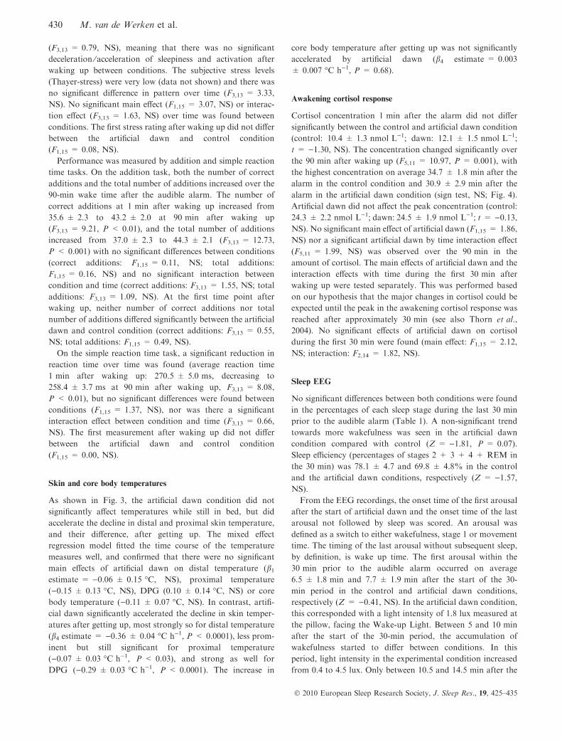
Skin and core body temperatures
As shown in Fig. 3, the artificial dawn condition did not
significantly affect temperatures while still in bed, but did
accelerate the decline in distal and proximal skin temperature,
and their difference, after getting up. The mixed effect
regression model fitted the time course of the temperature
measures well, and confirmed that there were no significant
main effects of artificial dawn on distal temperature (b1estimate = )0.06 ± 0.15 �C, NS), proximal temperature
()0.15 ± 0.13 �C, NS), DPG (0.10 ± 0.14 �C, NS) or core
body temperature ()0.11 ± 0.07 �C, NS). In contrast, artifi-
cial dawn significantly accelerated the decline in skin temper-
atures after getting up, most strongly so for distal temperature
(b4 estimate = )0.36 ± 0.04 �C h)1, P < 0.0001), less prom-
inent but still significant for proximal temperature
()0.07 ± 0.03 �C h)1, P < 0.03), and strong as well for
DPG ()0.29 ± 0.03 �C h)1, P < 0.0001). The increase in
core body temperature after getting up was not significantly
accelerated by artificial dawn (b4 estimate = 0.003
± 0.007 �C h)1, P = 0.68).
Awakening cortisol response
Cortisol concentration 1 min after the alarm did not differ
significantly between the control and artificial dawn condition
(control: 10.4 ± 1.3 nmol L)1; dawn: 12.1 ± 1.5 nmol L)1;
t = )1.30, NS). The concentration changed significantly over
the 90 min after waking up (F5,11 = 10.97, P = 0.001), with
the highest concentration on average 34.7 ± 1.8 min after the
alarm in the control condition and 30.9 ± 2.9 min after the
alarm in the artificial dawn condition (sign test, NS; Fig. 4).
Artificial dawn did not affect the peak concentration (control:
24.3 ± 2.2 nmol L)1; dawn: 24.5 ± 1.9 nmol L)1; t = )0.13,NS). No significant main effect of artificial dawn (F1,15 = 1.86,
NS) nor a significant artificial dawn by time interaction effect
(F5,11 = 1.99, NS) was observed over the 90 min in the
amount of cortisol. The main effects of artificial dawn and the
interaction effects with time during the first 30 min after
waking up were tested separately. This was performed based
on our hypothesis that the major changes in cortisol could be
expected until the peak in the awakening cortisol response was
reached after approximately 30 min (see also Thorn et al.,
2004). No significant effects of artificial dawn on cortisol
during the first 30 min were found (main effect: F1,15 = 2.12,
NS; interaction: F2,14 = 1.82, NS).
Sleep EEG
No significant differences between both conditions were found
in the percentages of each sleep stage during the last 30 min
prior to the audible alarm (Table 1). A non-significant trend
towards more wakefulness was seen in the artificial dawn
condition compared with control (Z = )1.81, P = 0.07).
Sleep efficiency (percentages of stages 2 + 3 + 4 + REM in
the 30 min) was 78.1 ± 4.7 and 69.8 ± 4.8% in the control
and the artificial dawn conditions, respectively (Z = )1.57,NS).
From the EEG recordings, the onset time of the first arousal
after the start of artificial dawn and the onset time of the last
arousal not followed by sleep was scored. An arousal was
defined as a switch to either wakefulness, stage 1 or movement
time. The timing of the last arousal without subsequent sleep,
by definition, is wake up time. The first arousal within the
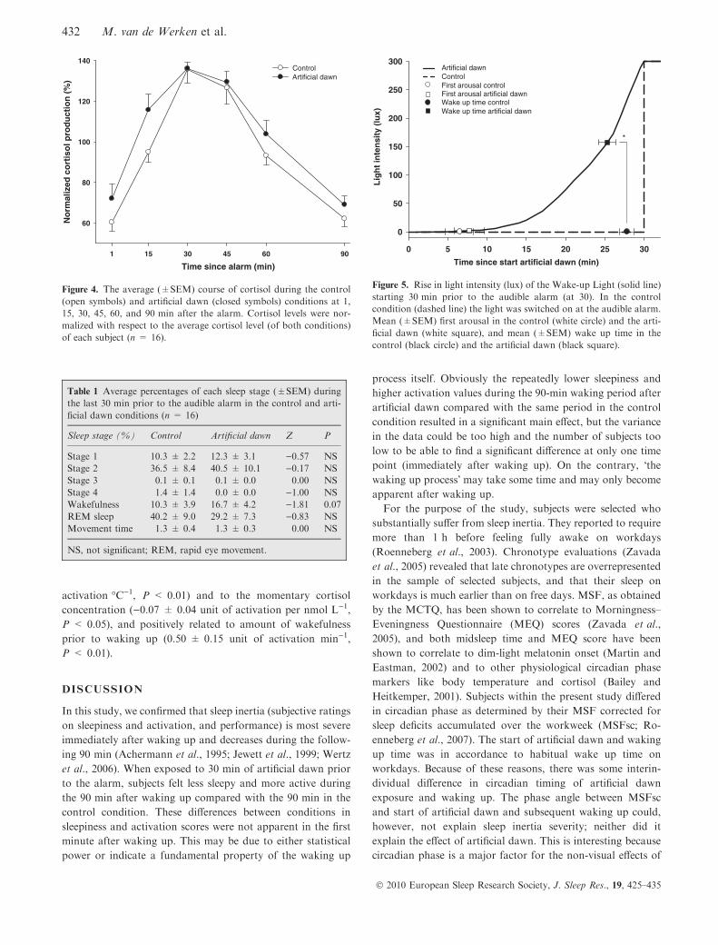
30 min prior to the audible alarm occurred on average
6.5 ± 1.8 min and 7.7 ± 1.9 min after the start of the 30-
min period in the control and artificial dawn conditions,
respectively (Z = )0.41, NS). In the artificial dawn condition,
this corresponded with a light intensity of 1.8 lux measured at
the pillow, facing the Wake-up Light. Between 5 and 10 min
after the start of the 30-min period, the accumulation of
wakefulness started to differ between conditions. In this
period, light intensity in the experimental condition increased
from 0.4 to 4.5 lux. Only between 10.5 and 14.5 min after the
430 M. van de Werken et al.
� 2010 European Sleep Research Society, J. Sleep Res., 19, 425–435

start of the 30-min period the cumulative amount of arousals
in the artificial dawn condition reached significance compared
with the control condition (two-tailed P-values between 0.04
and 0.09). The light intensity increased during this period from
5.4 to 17.8 lux. Most subjects fell asleep again after short
periods of wakefulness between 10.5 and 14.5 min after the
start of dawn. The final wake up time differed significantly
between conditions (Z = )2.59, P = 0.01), but the difference
was small: 2.2 ± 0.9 min before the alarm in the control; and
4.7 ± 1.1 min before the alarm in the artificial dawn condition
(Fig. 5).
Sleep stage on final awakening did not differ between
conditions (control: eight subjects REM sleep and eight
subjects Stage 2; artificial dawn: seven subjects REM sleep
and nine subjects Stage 2 (chi-square, NS).
Parameters explaining subjective ratings
To test what physiological parameters contributed to the
significant differences in sleepiness and activation between the
artificial dawn condition and the control condition, a mixed
effect multiple regression analysis was performed (see Section
�Materials and methods� for procedure).Sleepiness could best be explained by the following model:
Sleepinessijk ¼ b0ijk þ b1 �Distal temperatureijk
þ b2 �Wakefulnessijk þ b3 � Sqrt(tpost)ijk :
Sleepiness was positively related to momentary distal skin
temperature (b1 estimate = 0.31 ± 0.10 unit of sleepiness
�C)1, P < 0.01); negatively to the amount of wakefulness in
the half hour prior to the alarm (b2 estimate = )0.16 ± 0.05
unit of sleepiness min)1, P < 0.01); and to sqrt(tpost)
()0.14 ± 0.05 unit of sleepiness min)1, P < 0.01).
Activation could best be explained by the following model
equation:
Activationijk ¼ b0ijk þ b1 �Distal temperatureijk
þ b2 �Wakefulnessijk þ b3� Cortisolijk :
Activation was negatively related to the momentary
distal skin temperature (b1 estimate = )1.77 ± 0.13 unit of
Time since alarm (min)
Tem
per
atu
re (
°C)
30
31
32
33
34
35
36
ControlArtificial dawn Control fit Artificial dawn fit
Distal
–30 –15 0 15 30 45 60 75 90 –30 –15 0 15 30 45 60 75 90
–30 –15 0 15 30 45 60 75 90–30 –15 0 15 30 45 60 75 90
Time since alarm (min)
Tem
per
atu
re (
°C)
32
33
34
35
36
ControlArtificial dawn Control fit Artificial dawn fit
Proximal
Time since alarm (min)
Tem
per
atu
re (
°C)
–4
–3
–2
–1
0
ControlArtificial dawn Control fit Artificial dawn fit
DPG
Time since alarm (min)
Tem
per
atu
re (
°C)
36.0
36.5
37.0
37.5
38.0
ControlArtificial dawn Control fit Artificial dawn fit
Core
Figure 3. Average (±SEM) patterns of distal and proximal skin temperatures, of distal-to-proximal skin temperature gradient (DPG), and of core
body temperature starting 30 min prior to the audible alarm (at 0) until 90 min after the alarm in the control (circles, open symbols) and the
artificial dawn (squares, closed symbols) conditions. The lines represent the best-fitted curve through the data points (n = 15).
Effects of artificial dawn on sleep inertia, skin temperature and the awakening cortisol response 431
� 2010 European Sleep Research Society, J. Sleep Res., 19, 425–435
activation �C)1, P < 0.01) and to the momentary cortisol
concentration ()0.07 ± 0.04 unit of activation per nmol L)1,
P < 0.05), and positively related to amount of wakefulness
prior to waking up (0.50 ± 0.15 unit of activation min)1,
P < 0.01).
DISCUSSION
In this study, we confirmed that sleep inertia (subjective ratings
on sleepiness and activation, and performance) is most severe
immediately after waking up and decreases during the follow-
ing 90 min (Achermann et al., 1995; Jewett et al., 1999; Wertz
et al., 2006). When exposed to 30 min of artificial dawn prior
to the alarm, subjects felt less sleepy and more active during
the 90 min after waking up compared with the 90 min in the
control condition. These differences between conditions in
sleepiness and activation scores were not apparent in the first
minute after waking up. This may be due to either statistical
power or indicate a fundamental property of the waking up
process itself. Obviously the repeatedly lower sleepiness and
higher activation values during the 90-min waking period after
artificial dawn compared with the same period in the control
condition resulted in a significant main effect, but the variance
in the data could be too high and the number of subjects too
low to be able to find a significant difference at only one time
point (immediately after waking up). On the contrary, �thewaking up process� may take some time and may only become
apparent after waking up.
For the purpose of the study, subjects were selected who
substantially suffer from sleep inertia. They reported to require
more than 1 h before feeling fully awake on workdays
(Roenneberg et al., 2003). Chronotype evaluations (Zavada
et al., 2005) revealed that late chronotypes are overrepresented
in the sample of selected subjects, and that their sleep on
workdays is much earlier than on free days. MSF, as obtained
by the MCTQ, has been shown to correlate to Morningness–
Eveningness Questionnaire (MEQ) scores (Zavada et al.,
2005), and both midsleep time and MEQ score have been
shown to correlate to dim-light melatonin onset (Martin and
Eastman, 2002) and to other physiological circadian phase
markers like body temperature and cortisol (Bailey and
Heitkemper, 2001). Subjects within the present study differed
in circadian phase as determined by their MSF corrected for
sleep deficits accumulated over the workweek (MSFsc; Ro-
enneberg et al., 2007). The start of artificial dawn and waking
up time was in accordance to habitual wake up time on
workdays. Because of these reasons, there was some interin-
dividual difference in circadian timing of artificial dawn
exposure and waking up. The phase angle between MSFsc
and start of artificial dawn and subsequent waking up could,
however, not explain sleep inertia severity; neither did it
explain the effect of artificial dawn. This is interesting because
circadian phase is a major factor for the non-visual effects of
Time since alarm (min)
1 15 30 45 60 90
No
rmal
ized
co
rtis
ol p
rod
uct
ion
(%
)
60
80
100
120
140Control Artificial dawn
Figure 4. The average (±SEM) course of cortisol during the control
(open symbols) and artificial dawn (closed symbols) conditions at 1,
15, 30, 45, 60, and 90 min after the alarm. Cortisol levels were nor-
malized with respect to the average cortisol level (of both conditions)
of each subject (n = 16).
Time since start artificial dawn (min)
0 5 10 15 20 25 30
Lig
ht
inte
nsi
ty (
lux)
0
50
100
150
200
250
300
*
Artificial dawnControlFirst arousal controlFirst arousal artificial dawnWake up time controlWake up time artificial dawn
Figure 5. Rise in light intensity (lux) of the Wake-up Light (solid line)
starting 30 min prior to the audible alarm (at 30). In the control
condition (dashed line) the light was switched on at the audible alarm.
Mean (±SEM) first arousal in the control (white circle) and the arti-
ficial dawn (white square), and mean (±SEM) wake up time in the
control (black circle) and the artificial dawn (black square).
Table 1 Average percentages of each sleep stage (±SEM) during
the last 30 min prior to the audible alarm in the control and arti-
ficial dawn conditions (n = 16)
Sleep stage (%) Control Artificial dawn Z P
Stage 1 10.3 ± 2.2 12.3 ± 3.1 )0.57 NS
Stage 2 36.5 ± 8.4 40.5 ± 10.1 )0.17 NS
Stage 3 0.1 ± 0.1 0.1 ± 0.0 0.00 NS
Stage 4 1.4 ± 1.4 0.0 ± 0.0 )1.00 NS
Wakefulness 10.3 ± 3.9 16.7 ± 4.2 )1.81 0.07
REM sleep 40.2 ± 9.0 29.2 ± 7.3 )0.83 NS
Movement time 1.3 ± 0.4 1.3 ± 0.3 0.00 NS
NS, not significant; REM, rapid eye movement.
432 M. van de Werken et al.
� 2010 European Sleep Research Society, J. Sleep Res., 19, 425–435

light exposure. It suggests that the artificial dawn in this study
does not necessarily interact with the underlying circadian
system but rather has an acute effect upon physiological
processes around the moment of waking up irrespective of its
timing within the present (narrow) range of circadian phases.
In this study, it is shown that changes in skin blood flow and
associated changes in skin temperature parallel the sleep
inertia process; similar to how these thermoregulatory pro-
cesses are correlated to the evening increase of sleepiness and
the initiation of sleep after lights off in the evening. Sleep is
typically initiated when heat loss is maximal and usually occurs
during the circadian peak of skin temperature (Krauchi, 2007;
Van Someren, 2004), that is the major cause of the nocturnal
decline in core body temperature rhythm (Campbell and
Broughton, 1994; Krauchi, 2007; Van Someren et al., 2002;
Zulley et al., 1981). In the morning, the opposite occurs when
heat production is dominant over heat loss, resulting in an
increase in core body temperature (Krauchi, 2007). In this
study, subjects changed from supine to upright position after
waking up. This has caused strong masking effects in body
temperature on top of the waking up process during both the
control and artificial dawn conditions. Nevertheless, artificial
dawn prior to waking up did result in an accelerated decline in
distal and proximal skin temperatures after getting up, which
was also reflected in an accelerated decline in the DPG. This
suggests that vasoconstriction of skin blood vessels develops
faster after being exposed to artificial dawn prior to waking up
compared with the control condition. This could be explained
by direct activation of the sympathetic nervous system by light
(Saito et al., 1996; Scheer et al., 1999).
Fluctuations in distal skin temperature are always larger
than fluctuations in proximal skin temperature (Krauchi,
2007). Therefore, it is not surprising that in this study the
absolute effect of artificial dawn is larger in distal skin than
proximal skin regions. Another explanation could be that
artificial dawn interacts with the peripheral vasoconstriction
caused by the change in body position and that this effect is
stronger in distal skin regions.
During the 30 min of artificial dawn, there is little difference
in visually scored sleep stages compared with the last 30 min of
sleep in the control condition. The average timing of the first
arousal (either wakefulness, stage 1 or movement time) did not
differ between the artificial dawn and control condition.
Therefore, the relatively low light intensity in the artificial
dawn condition during the first 10 min, in which these arousals
occurred, did not wake up the subjects. However, during the
following 5 min the accumulation of arousals was steeper in
the artificial dawn condition than in the control condition.
This period also coincides with a substantial increase in light
intensity (from 5.4 to 17.8 lux). Short arousals (periods of
artifacts over EMG + EEG) occur regularly during the whole
sleep period (Dijk et al., 1987; Gordijn et al., 1999), and most
of them are not noticed by the person at all. Only some appear
to be associated with conscious awareness of being awake. It is
not known whether subjects were consciously aware of the
light during an arousal and whether this has contributed to the
observed changes after waking up. By asking the subjects
afterwards, they reported to have noticed the difference
between the artificial dawn and control conditions. It is also
unknown whether subjects opened their eyes during an
arousal. Only about 5% of light intensity comes through the
eyelids (Ando and Kripke, 1996), and if subjects opened their
eyes they were thus exposed to a much higher light intensity.
Abrupt awakenings are reported to worsen sleep inertia
complaints (Dinges, 1990; Dinges et al., 1981). A gradual way
of waking up, induced by artificial dawn, could thus reduce
sleep inertia complaints.
Indeed in our regression analysis the amount of wakeful-
ness during the 30 min prior to the alarm contributed both to
the decrease in sleepiness and to the increase in activation
after waking up. In addition to the amount of wakefulness
prior to the alarm, the decrease in distal skin temperature
after waking up contributed strongly to the model. Our data
confirm the close functional relationship between the dissipa-
tion of sleepiness and skin temperature, as was previously
reported in relation to DPG (Krauchi et al., 2004). It is
interesting that in our study distal skin temperature rather
than DPG or proximal skin temperature added significantly
to the model explaining sleepiness. Little is known about the
association of the regulation of skin blood flow and the
regulation of alertness upon waking up from sleep. Typically,
a 10-min nap does not induce the adverse effects of sleep
inertia upon awakening, and this may be because of insuffi-
cient time for thermoregulatory changes to occur (Brooks and
Lack, 2006). Further experiments are required to determine
whether skin temperature changes after waking up and the
dissipation of sleep inertia are causally related, as has been
shown to be feasible using mild skin temperature manipula-
tion while obtaining objective vigilance measures during
daytime (Fronczek et al., 2008; Raymann and Van Someren,
2007). This study is the first to show that manipulation of
sleep inertia by artificial dawn coincides with effects on skin
temperature.
Interestingly, but also unexpectedly, the effect of the
awakening cortisol response as included in the model on
activation appeared to be negative. This negative relationship
suggests that an increased awakening cortisol response has a
detrimental effect on the dissipation of sleep inertia. The exact
function of the awakening cortisol response is as yet unknown.
Although cortisol does show consistent responses to stress
(Kemeny, 2003), the morning increase in cortisol was not
associated to any detectable psychological stress in our study.
Previously, light after waking up has been shown to increase
cortisol production (Scheer and Buijs, 1999), and one study
showed a significantly higher awakening cortisol response with
the use of a dawn waking up system (Thorn et al., 2004). In
this study, we did not find an effect of the dawn signal on
cortisol. The discrepancy between our study and the study of
Thorn et al. (2004) can be explained by differences between the
control conditions used in both experiments. In the study of
Thorn et al. (2004) artificial dawn was tested against a normal
alarm clock (with no light). In our study light was turned on in
Effects of artificial dawn on sleep inertia, skin temperature and the awakening cortisol response 433
� 2010 European Sleep Research Society, J. Sleep Res., 19, 425–435

the control condition, together with the audible alarm at
waking up. This was performed to test whether dawn itself,
and not the light exposure after waking up, induced a
reduction of sleep inertia and an increase in cortisol. It is
possible that the light exposure immediately after the alarm in
both conditions may have increased cortisol to such an extent
that a possible additional effect of artificial dawn on cortisol
could not be detected.
CONCLUSION
People who require a large amount of time after awakening
before feeling fully alert can reduce their symptoms of sleep
inertia and the time until feeling fully awake by exposing
themselves to an artificial dawn signal. Skin temperatures
showed an accelerated decline after awakening in the artificial
dawn condition. A multiple regression analysis revealed that
the physiological background explaining the positive effects of
artificial dawn on the dissipation of sleep inertia involves light
sleep and an accelerated distal skin temperature decline after
awakening.
ACKNOWLEDGEMENTS
We thank the subject volunteers for their participation, Kurt
Krauchi (Psychiatric University Clinics Basel) for his advice on
our skin temperature protocol, and Mirre Simons and Martijn
Hessels for their general help and advice. Financial support
was obtained from Philips DAP B.V., CoC Vitality Care,
Drachten, The Netherlands and the sixth European Frame-
work project EUCLOCK (018741).
REFERENCES
Achermann, P., Werth, E., Dijk, D. J. and Borbely, A. A. Time course
of sleep inertia after nighttime and daytime sleep episodes. Arch.
Ital. Biol., 1995, 134: 109–119.
Akerstedt, T. and Gillberg, M. Subjective and objective sleepiness in
the active individual. Int. J. Neurosci., 1990, 52: 29–37.
Ando, K. and Kripke, D. F. Light attenuation by the human eyelid.
Biol. Psychiatry, 1996, 39: 22–25.
Aschoff, J. Circadiane rhythmen im endocrinen system. J. Mol. Med.,
1978, 56: 425–435.
Avery, D. H., Bolte, M. A., Dager, S. R. et al. Dawn simulation
treatment of winter depression: a controlled study. Am. J. Psychi-
atry, 1993, 150: 113–117.
Avery, D. H., Eder, D. N., Bolte, M. A. et al. Dawn simulation and
bright light in the treatment of SAD: a controlled study. Biol.
Psychiatry, 2001, 50: 205–216.
Avery, D. H., Kouri, M. E., Monaghan, K., Bolte, M. A., Hellekson,
C. and Eder, D. Is dawn simulation effective in ameliorating the
difficulty awakening in seasonal affective disorder associated with
hypersomnia? J. Affect. Disord., 2002, 69: 231–236.
Bailey, S. L. and Heitkemper, M. M. Circadian rhythmicity of cortisol
and body temperature: morningness–eveningness effects. Chronobi-
ol. Int., 2001, 18: 249–261.
Balkin, T. J. and Badia, P. Relationship between sleep inertia and
sleepiness: cumulative effects of four nights of sleep disrup-
tion ⁄ restriction on performance following abrupt nocturnal awak-
enings. Biol. Psychol., 1988, 27: 245–258.
Beck, A. T., Steer, R. A. and Brown, G. K. Manual for the Beck
Depression Inventory-II. Psychological Corporation, San Antonio,
1996.
Beck, A. T., Steer, R. A. and Brown, G. K. Beck Depression Inventory-
II. Dutch version. A. J. W. Van der Does, II (Ed) Swets Test
Publishers, Lisse, 2002.
Born, J., Kern, W., Bieber, K., Fehm-Wolfsdorf, G., Schiebe, M. and
Fehm, H. L. Night-time plasma cortisol secretion is associated with
specific sleep stages. Biol. Psychiatry, 1986, 21: 1415–1424.
Born, J., Spath-Schwalbe, E., Schwakenhofer, H., Kern, W. and
Fehm, H. L. Influences of corticotropin-releasing hormone, adre-
nocorticotropin, and cortisol on sleep in normal man. J. Clin.
Endocrinol. Metab., 1989, 68: 904–911.
Brooks, A. and Lack, L. A brief afternoon nap following nocturnal
sleep restriction: which nap duration is most recuperative? Sleep,
2006, 29: 831–840.
Campbell, S.S and Broughton, R.J. Rapid decline in body temperature
before sleep: fluffing the physiological pillow?. Chronobiol. Int., 1994,
11: 126–131.
Cajochen, C., Zeitzer, J. M., Czeisler, C. A. and Dijk, D. J. Dose–
response relationship for light intensity and ocular and electroen-
cephalographic correlates of human alertness. Behav. Brain Res.,
2000, 115: 75–83.
Dijk, D. J., Visscher, C. A., Bloem, G. M., Beersma, D. G. M. and
Daan, S. Reduction of human sleep duration after bright light
exposure in the morning. Neurosci. Lett., 1987, 73: 181–186.
Dinges, D. F. Are you awake? Cognitive performance and reverie
during the hypnopompic state. In: R. Bootzin, J. Kihistrom and D.
Schacter (Eds) Sleep and Cognition. American Psychological Asso-
ciation, Washington D.C, 1990: 159–175.
Dinges, D. F. and Kribbs, N. B. Performing while sleepy: effects of
experimentally induced sleepiness. In: T. H. Monk (Ed) Sleep,
Sleepiness and Performance. John Wiley and Sons, Chichester, UK,
1991: 97–128.
Dinges, D. F., Orne, E. C., Evans, F. J. and Orne, M. T. Performance
after naps in sleep-conducive and alerting environments. In: L. C.
Johnson, D. I. Tepas, W. P. Colquhoun and M. J. Colligan (Eds)
Biological Rhythms, Sleep and Shift Work. Spectrum, New York,
1981: 539–552.
Edwards, S., Clow, A., Evans, P. and Hucklebridge, F. Exploration of
the awakening cortisol response in relation to diurnal cortisol
secretory activity. Life Sci., 2001, 68: 2093–2103.
Fehm, H. L., Bieber, K., Benkowitsch, R., Fehm-Wolfsdorf, G., Voigt,
K. H. and Born, J. Relationships between sleep stages and plasma
cortisol: a single case study. Acta Endocrinol., 1986, 111: 264–270.
Ferrara, M. and De Gennaro, L. The sleep inertia phenomenon during
the sleep–wake transition: theoretical and operational issues. Aviat.
Space Environ. Med., 2000, 71: 843–848.
Fronczek, R., Raymann, R. J. E. M., Romeijn, N. et al. Manipulation
of core body and skin temperature improves vigilance and mainte-
nance of wakefulness in narcolepsy. Sleep, 2008, 31: 233–240.
Gasio, P. F., Krauchi, K., Cajochen, C. et al. Dawn–dusk simulation
light therapy of disturbed circadian rest–activity cycles in demented
elderly. Exp. Gerontol., 2003, 38: 207–216.
Gordijn, M. C. M., Beersma, D. G. M., Korte, H. J. and Van den
Hoofdakker, R. H. Effects of light exposure and sleep displacement
on dim light melatonin onset. J. Sleep Res., 1999, 8: 163–174.
Horne, J. A. and Ostberg, O. A self-assessment questionnaire to
determine morningness–eveningness in human circadian rhythms.
Int. J. Chronobiol., 1976, 4: 97–110.
Hucklebridge, F., Hussain, T., Evans, P. and Clow, A. The diurnal
patterns of the adrenal steroids cortisol and dehydroepiandrosterone
(DHEA) in relation to awakening. Psychoneuroendocrinology, 2005,
30: 51–57.
Jewett, M. E., Wyatt, J. K., Ritz-De Cecco, A., Khalsa, S. B., Dijk, D.
J. and Czeisler, C. A. Time course of sleep inertia dissipation in
human performance and alertness. J. Sleep Res., 1999, 8: 1–8.
434 M. van de Werken et al.
� 2010 European Sleep Research Society, J. Sleep Res., 19, 425–435

Kemeny, M. E. The psychobiology of stress. Curr. Dir. Psychol. Sci.,
2003, 12: 124–129.
Kleitman, N. Sleep and Wakefulness.2nd edn. University of Chicago
Press, Chicago, 1963.
Krauchi, K. The thermophysiological cascade leading to sleep initia-
tion in relation to phase of entrainment. Sleep Med. Rev., 2007, 11:
439–451.
Krauchi, K., Cajochen, C. and Wirz-Justice, A. Waking up properly: is
there a role of thermoregulation in sleep inertia? J. Sleep Res., 2004,
13: 121–127.
Leppamaki, S., Meesters, Y., Haukka, J., Lonnqvist, J. and Partonen,
T. Effect of simulated dawn on quality of sleep – a community-based
trial. BCM Psychiatry, 2003, 3: 14.
Leproult, R., Colecchia, E. F., L�Hermite-Baleriaux, M. and Van
Cauter, E. Transition from dim to bright light in the morning
induces an immediate elevation of cortisol levels. J. Clin. Endocrinol.
Metab., 2001, 86: 151–157.
Marken Lichtenbelt, W. D. v., Daanen, H. A. M., Wouters, L. et al.
Evaluation of wireless determination of skin temperature using
iButtons. Physiol. Behav., 2006, 88: 489–497.
Martin, S. K. and Eastman, C. I. Sleep logs of young adults with self-
selected sleep times predict the dim light melatonin onset. Chrono-
biol. Int., 2002, 19: 695–707.
Mitchell, D. and Wyndham, C. H. Comparison of weighting formulas
for calculating mean skin temperature. J. Appl. Physiol., 1969, 26:
616–622.
Muzet, A., Nicolas, A., Tassi, P., Dewasmes, G. and Bonneau, A.
Implementation of napping in industry and the problem of sleep
inertia. J. Sleep Res., 1995, 4: 67–69.
Naitoh, P. Circadian cycles and restorative power of naps. In: L. C.
Johnson, D. I. Tepas, W. P. Colquhoun and M. J. Colligan (Eds)
Biological Rhythms, Sleep and Shift Work. Spectrum, New York,
1981: 553–580.
Naitoh, P., Kelly, T. and Babkoff, H. Sleep inertia: best time not to
wake up? Chronobiol. Int., 1993, 10: 109–118.
Norden, M. J. and Avery, D. H. A controlled study of dawn
simulation in subsyndromal winter depression. Acta Psychiatr.
Scand., 1993, 88: 67–71.
Phipps-Nelson, J., Redman, J. R., Dijk, D. J. and Rajaratnam, S. M.
W. Daytime exposure to bright light, as compared to dim light,
decreases sleepiness and improves psychomotor vigilance perfor-
mance. Sleep, 2003, 26: 695–700.
Raymann, R. J. E. M. and Van Someren, E. J. W. Time-on-task
impairment of psychomotor vigilance is affected by mild skin
warming and changes with aging and insomnia. Sleep, 2007, 30: 96–
103.
Rechtschaffen, A. and Kales, A. A Manual of Standardized Terminol-
ogy, Techniques and Scoring System for Sleep Stages of Human
Subjects. Pub No. 204. National Institutes of Health, Washington
DC, 1968.
Roenneberg, T., Wirz-Justice, A. and Merrow, M. Life between clocks:
daily temporal patterns of human chronotypes. J. Biol. Rhythm,
2003, 18: 80–90.
Roenneberg, T., Kuehnle, T., Juda, M. et al. Epidemiology of the
human circadian clock. Sleep Med. Rev., 2007, 11: 429–438.
Ruger, M., Gordijn, M. C. M., Beersma, D. G. M., De Vries, B. and
Daan, S. Acute and phase-shifting effects of ocular and extraocular
light in human circadian physiology. J. Biol. Rhythms, 2003, 18:
409–419.
Ruger, M., Gordijn, M. C. M., Beersma, D. G. M., De Vries, B. and
Daan, S. Time-of-day-dependent effects of bright light exposure on
human psychophysiology: comparison of daytime and nighttime
exposure. Am. J. Physiol. Regul. Integ. Comp. Physiol., 2006, 290:
R1413–R1420.
Saito, Y., Shimizu, T., Takahashi, Y. et al. Effect of bright light
exposure on muscle sympathetic nerve activity in human. Neurosci.
Lett., 1996, 22: 135–137.
Scheer, F. A. J. L. and Buijs, R. M. Light affects morning salivary
cortisol in humans. J. Clin. Endocrinol. Metab., 1999, 84: 3395–3398.
Scheer, F. A. J. L., Van Doornen, L. J. and Buijs, R. M. Light and
diurnal cycle affect human heart rate: possible role for the circadian
pacemaker. J. Biol. Rhythms, 1999, 14: 202–212.
Scheer, F. A. J. L., Shea, T. J., Hilton, M. F. and Shea, S. A. An
endogenous circadian rhythm in sleep inertia results in greatest
cognitive impairment upon awakening during the biological night.
J. Biol. Rhythms, 2008, 23: 353–361.
Seminara, J. L. and Shavelson, R. J. Effectiveness of space crew
performance subsequent to sudden sleep arousal. Aerosp. Med.,
1969, 40: 723–727.
Tassi, P. and Muzet, A. Sleep inertia. Sleep Med. Rev., 2000, 4: 341–
353.
Tassi, P., Bonnefond, A., Engasser, O., Hoeft, A., Eschenlauer, R. and
Muzet, A. EEG spectral power and cognitive performance during
sleep inertia: the effect of normal sleep duration and partial sleep
deprivation. Physiol. Behav., 2006, 87: 177–184.
Terman, M., Schlager, D., Fairhurst, S. and Perlman, B. Dawn and
dusk simulation as a therapeutic intervention. Biol. Psychiatry, 1989,
25: 966–970.
Thayer, R. E. Measurement of activation through self-report. Psychol.
Rep., 1967, 20: 663–678.
Thorn, L., Hucklebridge, F., Esgate, A., Evans, P. and Clow, A. The
effect of dawn simulation on the cortisol response to awakening in
healthy participants. Psychoneuroendocrinology, 2004, 29: 925–930.
Twisk, J. W. R. Applied Longitudinal Data Analysis for Epidemiology.
Cambridge University Press, Cambridge, 2003.
Van Someren, E. J. W. Sleep propensity is modulated by circadian and
behavior-induced changes in cutaneous temperature. J. Therm. Biol.,
2004, 29: 437–444.
Van Someren, E. J. W., Raymann, R. J. E. M., Scherder, E. J.,
Daanen, H. A. and Swaab, D. F. Circadian and age-related
modulation of thermoreception and temperature regulation: mech-
anisms and functional implications. Ageing Rev. Res., 2002, 1: 721–
778.
Wertz, A. T., Ronda, J. M., Czeisler, C. A. and Wright, K. P. Effects
of sleep inertia on cognition. JAMA, 2006, 295: 163–164.
Wilkinson, R. T. and Stretton, M. Performance after awakening at
different times of night. Psychon. Sci., 1971, 23: 283–285.
Wittmann, M., Dinich, J., Merrow, M. and Roenneberg, T. Social
jetlag: misalignment of biological and social time. Chronobiol. Int.,
2006, 23: 497–509.
Wust, S., Wolf, J., Hellhammer, D. H., Federenko, I., Schommer, N.
and Kirschbaum, C. The cortisol awakening response – normal
values and confounds. Noise Health, 2000, 2: 79–88.
Zavada, A., Gordijn, M. C. M., Beersma, D. G. M., Daan, S. and
Roenneberg, T. Comparison of the Munich chronotype question-
naire with the Horne–Ostberg�s morningness–eveningness score.
Chronobiol. Int., 2005, 22: 267–278.
Zulley, J., Wever, R. and Aschoff, J. The dependence of onset and
duration of sleep on the circadian rhythm of rectal temperature.
Pflugers Arch., 1981, 391: 314–318.
Effects of artificial dawn on sleep inertia, skin temperature and the awakening cortisol response 435
� 2010 European Sleep Research Society, J. Sleep Res., 19, 425–435