university of groningen amyloidosis: hazenberg, …...serumamyloid p...
TRANSCRIPT

University of Groningen
Amyloidosis:Hazenberg, Bouke P. C.
Published in:RHEUMATIC DISEASE CLINICS OF NORTH AMERICA
DOI:10.1016/j.rdc.2013.02.012
IMPORTANT NOTE: You are advised to consult the publisher's version (publisher's PDF) if you wish to cite fromit. Please check the document version below.
Document VersionEarly version, also known as pre-print
Publication date:2013
Link to publication in University of Groningen/UMCG research database
Citation for published version (APA):Hazenberg, B. P. C. (2013). Amyloidosis: A Clinical Overview. RHEUMATIC DISEASE CLINICS OFNORTH AMERICA, 39(2), 323-345. https://doi.org/10.1016/j.rdc.2013.02.012
CopyrightOther than for strictly personal use, it is not permitted to download or to forward/distribute the text or part of it without the consent of theauthor(s) and/or copyright holder(s), unless the work is under an open content license (like Creative Commons).
Take-down policyIf you believe that this document breaches copyright please contact us providing details, and we will remove access to the work immediatelyand investigate your claim.
Downloaded from the University of Groningen/UMCG research database (Pure): http://www.rug.nl/research/portal. For technical reasons thenumber of authors shown on this cover page is limited to 10 maximum.
Download date: 16-02-2020

Amyloidosis: A Clinical Overview
Bouke P.C. Hazenberg, MD, PhD
KEYWORDS
� Systemic amyloidosis � Amyloid fibril � Protein misfolding � Precursor protein� Typing � Diagnosis � Treatment � Disease monitoring
KEY POINTS
� Amyloidosis is the name for some diseases caused by protein misfolding; 30 differentsoluble precursor proteins can aggregate and be deposited as insoluble amyloid fibrils.
� Amyloid deposition is localized or systemic; the 4 main types of systemic amyloidosis areAL (light chain), AA (inflammation), ATTR (hereditary and old age), and Ab2M (dialysis).
� Clinical management comprises proof of amyloid, systemic evidence, reliable typing,precursor assessment, severity of organ disease, choice of treatment, and plannedfollow-up.
� The precursor-product concept is the current basis of treatment, thereby aiming todecrease the levels of precursor proteins in serum to normal or undetectable values.Future clinical research will be directed at stopping amyloid deposition and increasingamyloid clearance.
� Protein misfolding is not only a characteristic of amyloidosis; it is also involved in manyother disabling cardiac and neurologic degenerative diseases that interfere with healthyaging.
INTRODUCTION
The description of the autopsy of a young man in 1639 by Nicolaes Fonteyn, a Dutchphysician and poet who lived in Amsterdam, was probably the first report of a patientwith systemic amyloidosis. Since then, lardaceous changes in enlarged organs, suchas the liver, spleen, heart, and kidneys, have drawn the attention of pathologists suchas Rokitansky. In 1854, Rudolph Virchow was one of the first to use the term amyloidfor this amorphous and hyaline change in tissue because of an iodine-staining reactionsimilar to that of starch (amylon; Greek for origin). Although it is now known thatamyloid has nothing to do with starch, the term amyloid is still in use today. Bennhold
Funding Sources: None.Conflict of Interest: Previous member of the Clinical Advisory Board of Neurochem; Chair of anExternal Safety Advisory Committee of GSK.DepartmentofRheumatology&Clinical Immunology,AA21,UniversityMedical CenterGroningen,University of Groningen, PO Box 30.001, 9700 RB Groningen, The NetherlandsE-mail address: [email protected]
Rheum Dis Clin N Am 39 (2013) 323–345http://dx.doi.org/10.1016/j.rdc.2013.02.012 rheumatic.theclinics.com0889-857X/13/$ – see front matter � 2013 Elsevier Inc. All rights reserved.
Hazenberg324
introduced Congo Red staining in 1922 as a useful method to identify amyloid in tissuespecimens. In 1927, Divry and Florkin described the characteristic green birefringencewhen Congo Red-stained amyloid was viewed under polarized light. In 1959, Cohenand Calkins detected the fibril nature of amyloid when viewed under the electronmicroscope.1
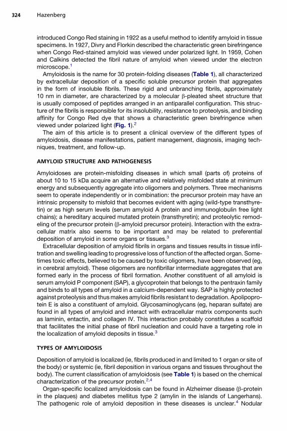
Amyloidosis is the name for 30 protein-folding diseases (Table 1), all characterizedby extracellular deposition of a specific soluble precursor protein that aggregatesin the form of insoluble fibrils. These rigid and unbranching fibrils, approximately10 nm in diameter, are characterized by a molecular b-pleated sheet structure thatis usually composed of peptides arranged in an antiparallel configuration. This struc-ture of the fibrils is responsible for its insolubility, resistance to proteolysis, and bindingaffinity for Congo Red dye that shows a characteristic green birefringence whenviewed under polarized light (Fig. 1).2
The aim of this article is to present a clinical overview of the different types ofamyloidosis, disease manifestations, patient management, diagnosis, imaging tech-niques, treatment, and follow-up.
AMYLOID STRUCTURE AND PATHOGENESIS
Amyloidoses are protein-misfolding diseases in which small (parts of) proteins ofabout 10 to 15 kDa acquire an alternative and relatively misfolded state at minimumenergy and subsequently aggregate into oligomers and polymers. Three mechanismsseem to operate independently or in combination: the precursor protein may have anintrinsic propensity to misfold that becomes evident with aging (wild-type transthyre-tin) or as high serum levels (serum amyloid A protein and immunoglobulin free lightchains); a hereditary acquired mutated protein (transthyretin); and proteolytic remod-eling of the precursor protein (b-amyloid precursor protein). Interaction with the extra-cellular matrix also seems to be important and may be related to preferentialdeposition of amyloid in some organs or tissues.3
Extracellular deposition of amyloid fibrils in organs and tissues results in tissue infil-tration and swelling leading to progressive loss of function of the affected organ. Some-times toxic effects, believed to be caused by toxic oligomers, have been observed (eg,in cerebral amyloid). These oligomers are nonfibrillar intermediate aggregates that areformed early in the process of fibril formation. Another constituent of all amyloid isserum amyloid P component (SAP), a glycoprotein that belongs to the pentraxin familyand binds to all types of amyloid in a calcium-dependent way. SAP is highly protectedagainst proteolysis and thusmakes amyloid fibrils resistant to degradation. Apolipopro-tein E is also a constituent of amyloid. Glycosaminoglycans (eg, heparan sulfate) arefound in all types of amyloid and interact with extracellular matrix components suchas laminin, entactin, and collagen IV. This interaction probably constitutes a scaffoldthat facilitates the initial phase of fibril nucleation and could have a targeting role inthe localization of amyloid deposits in tissue.3
TYPES OF AMYLOIDOSIS
Deposition of amyloid is localized (ie, fibrils produced in and limited to 1 organ or site ofthe body) or systemic (ie, fibril deposition in various organs and tissues throughout thebody). The current classification of amyloidosis (see Table 1) is based on the chemicalcharacterization of the precursor protein.2,4
Organ-specific localized amyloidosis can be found in Alzheimer disease (b-proteinin the plaques) and diabetes mellitus type 2 (amylin in the islands of Langerhans).The pathogenic role of amyloid deposition in these diseases is unclear.4 Nodular

Table 1Amyloid fibril proteins and their precursors in humans
FibrilProtein Precursor Protein
Systemic orLocalized
Acquired orHereditary Target Organs
AL Immunoglobulin lightchain
S, L A All organs except CNS
AH Immunoglobulin heavychain
S, L A All organs except CNS
Ab2M b2-microglobulin, wildtype
S A Musculoskeletal system
b2-microglobulin, variant S H ANS
ATTR Transthyretin, wild type S, L A Heart mainly in men,tenosynovium
Transthyretin, variants S H PNS, ANS, heart, eye,leptomeninges
AA (Apo) serum amyloid A S A All organs except CNS
AApoAI Apolipoprotein A I,variants
S H Heart, liver, kidney,PNS, testis, larynx(C-terminal variants),skin (C-terminalvariants)
AApoAII Apolipoprotein A II,variants
S H Kidney
AApoAIV Apolipoprotein A IV,wild type
S A Kidney medulla andsystemic
AGel Gelsolin, variants S H PNS, cornea
ALys Lysozyme, variants S H Kidney
ALect2 Leukocyte chemotacticfactor-2
S A Kidney, primarily
AFib Fibrinogen a, variants S H Kidney, primarily
ACys Cystatin C, variants S H PNS, skin
ABri ABriPP, variants S H CNS
ADana ADanPP, variants L H CNS
Ab Ab protein precursor,wild type
L A CNS
Ab protein precursor,variant
L H CNS
APrP Prion protein, wild type L A CJD, fatal insomniaPrion protein, variants L H CJD, GSS syndrome, fatal
insomnia
ACal (Pro)calcitonin L A C-cell thyroid tumors
AIAPP Islet amyloidpolypeptideb
L A Islets of Langerhans,insulinomas
AANF Atrial natriuretic factor L A Cardiac atria
APro Prolactin L A Pituitary prolactinomas,aging pituitary
AIns Insulin L A Iatrogenic, local injection
ASPC Lung surfactant protein L A Lung
AGal7 Galectin 7 L A Skin
ACor Corneodesmin L A Cornified epithelia, hairfollicles
(continued on next page)
Amyloidosis 325
Table 1(continued)
FibrilProtein Precursor Protein
Systemic orLocalized
Acquired orHereditary Target Organs
AMed Lactadherin L A Senile aortic, media
AKer Kerato-epithelin L A Cornea, hereditary
ALac Lactoferrin L A Cornea
AOaap Odontogenic ameloblast-associated protein
L A Odontogenic tumors
ASem1 Semenogelin 1 L A Vesicula seminalis
Abbreviations: ANS, autonomic nervous system; CJD, Creutzfeldt-Jakob disease; CNS, centralnervous system; GSS, Gerstmann-Straussler-Scheinker syndrome; PNS, peripheral nervous system.
a ADan is the product of the same gene as Abri.b Also called amylin.Data from Sipe JD, Benson MD, Buxbaum JN, et al. Amyloid fibril protein nomenclature: 2012
recommendations from the Nomenclature Committee of the International Society of Amyloidosis.Amyloid 2012;19:167–70.
Hazenberg326
localized amyloid is an incidental finding and can be present in the skin (not onlynodular but also macular amyloid and lichen amyloidosis), eyelid, conjunctiva, breast,larynx, bronchial tree, lung, and genitourinary tract. In most cases, low numbers ofclonal plasma cells can be detected in the biopsy sample. Surgery is usually the treat-ment of choice. Local recurrence is frequent and can again be treated surgically.5
Localized nodular skin amyloidosis is sometimes associated with Sjogren disease.6
In contrast to localized amyloidosis, systemic amyloidosis leads to serious signsand symptoms caused by progressive disease in organs and tissues. There aremany types of systemic amyloidosis (see Table 1), but 4 types are seen mostfrequently: AL, AA, ATTR, and Ab2M amyloidosis.4
AL amyloidosis is the most common type. This disease is caused by a clonal plasmacell dyscrasia; it often occurs as low-grade clonal disease, sometimes multiplemyeloma, and rarely non-Hodgkin lymphoma or Waldenstrom disease. The precursorof this type of amyloid is either lambda or kappa immunoglobulin free light chain. Clin-ical manifestations are diverse, such as cardiomyopathy, nephrotic syndrome, renalfailure, hepatomegaly, splenomegaly, orthostatic hypotension, diarrhea, intestinal
Fig. 1. A sample of abdominal subcutaneous fat aspirate containing amyloid deposits,stained with Congo Red. (A) Viewed in normal light, amyloid is stained red. Bar length is100 mm. (B) Viewed in polarized light, amyloid shows apple-green birefringence (collagenis bluish-gray).

Amyloidosis 327
pseudoobstruction, peripheral neuropathy, autonomic neuropathy, arthropathy, carpaltunnel syndrome (CTS), bleeding, adrenal dysfunction, goiter, pulmonary problems,weight loss, fatigue, malaise, and glossomegaly.7
The second most common type is AA amyloidosis. This disease is caused by long-standing inflammation, such as rheumatoid arthritis, inflammatory bowel disease,chronic infections (eg, tuberculosis, osteomyelitis, leprosy), and hereditary autoinflam-matory diseases (eg, familial Mediterranean fever, also called FMF). The precursor ofthis type is the HDL3-associated apolipoprotein serum amyloid A protein (SAA), anacute phase reactant. Signs of kidney disease, such as proteinuria (progressing tonephrotic syndrome) and loss of renal function (progressing to renal failure), areobserved most frequently (in about 90% of cases), followed at a distance by auto-nomic neuropathy, bowel involvement, splenomegaly, hepatomegaly, goiter, andcardiomyopathy.8,9
The third most common type is ATTR amyloidosis. The familial form of this disease iscaused by many autosomal dominantly inherited point mutations of the precursorprotein transthyretin (TTR). Transthyretin is an acronym for the transport protein ofthyroid hormone and retinol-binding protein. About 100 of these TTR mutations havebeen described, but most common is the TTR-Met30 mutation. Clinical manifestationsare predominantly peripheral and autonomic neuropathy, but also cardiomyopathy,renal failure, and eye involvement (vitreous opacities) are frequently observed. Some-times a severe cardiomyopathy is the initial presentation of the disease.10 There is alsoa nonfamilial acquired form of ATTR amyloidosis. In this disease of old-aged men(rarely women), nonmutated (wild-type) TTR can also act as an amyloid precursor bya still unknown mechanism. This wild-type ATTR amyloidosis (formerly called senilesystemic amyloidosis) is characterized by a slowly progressive cardiomyopathy,frequently associated with CTS, but without other neuropathy.11
The fourth type is Ab2M amyloidosis. This disease is caused by end-stage renaldisease in which highly increased serum levels of b2-microglobulin persist for yearsbecause b2-microglobulin is not effectively cleared by dialysis. The disease is charac-terized by high serum levels of b2-microglobulin and posttranslational modifications tobe deposited as amyloid fibrils in predominantly osteoarticular tissues. CTS andshoulder pain are among the first manifestations, followed by large periarticular cystsand sometimes by pathologic fractures, or a destructive spondylarthropathy.12,13
Recently, a hereditary Ab2M amyloidosis has been described, characterized by auto-nomic neuropathy and slowly progressive gastrointestinal symptoms.14
Other types of systemic amyloidosis are rare.15 However, in patients without a familyhistory of amyloid disease, a hereditary type of amyloidosis may be present.16 Inpatients with almost exclusive kidney disease, other types should be considered,such as fibrinogen a,17 lysozyme,18 apolipoprotein A I, apolipoprotein A II, apolipopro-tein A IV, and the recently described leukocyte chemotactic factor-2.19 In patients withalmost exclusive cardiac or neuropathic disease, the apolipoprotein A I type shouldnot be overlooked. In patients with hepatic amyloid, the lysozyme and apolipoproteinA I type can be present. Apolipoprotein A I can be present in the skin and larynx. Gel-solin is typically found in lattice corneal dystrophy.20
EPIDEMIOLOGY
Even in developed countries, few incidence data have been collected systemati-cally.21 The data from 1 study about the incidence of AL amyloidosis in OlmstedCounty, Minnesota, are still used. In this study, the overall age-adjusted and sex-adjusted 95% confidence interval was 5.1 to 12.8 per million patients per year.22 In

Hazenberg328
a recent Swedish study, the estimated incidence of AL amyloidosis was 3.2 per millionper year and AA amyloidosis 2.0 per million per year.23 The median age for AL and AAamyloidosis is between 55 and 60 years.24 The prognosis of untreated patients is poor,as noticed in older studies; median survival was 6 to 12 months for AL amyloidosis and3 to 4 years for AA amyloidosis.24 The estimated median survival of untreated patientswith hereditary ATTR amyloidosis is almost 10 years, although some patients maysurvive up to 15 years. An estimate of mortality is that 0.5 to 1.0 per 1000 personsdie in the United Kingdom because of AL amyloidosis.25 In most studies of patientswith the AL type, the proportion of men is somewhat higher (about 1.1–1.3) thanwomen. The reverse (more women than men) is observed for AA amyloidosis becauseof the high proportion of patients (mainly women) with underlying rheumatoid arthritis.In developing countries, the prevalence of AA amyloidosis is higher than AL amyloid-osis because of the higher prevalence of associated underlying infectious diseases.
PATIENT MANAGEMENT
A stepwise approach is useful for the clinical management of a patient with systemicamyloidosis (Box 1).26 If amyloidosis is suspected, the first step is to obtain histologicproof of amyloid, followed by a search for evidence of systemic amyloid deposition inthe patient. The next 2 steps are to determine the type of amyloid with confidence fol-lowed by detection and (in AA and AL) quantification of the precursor protein in theblood. A thoughtful clinical evaluation should lead to useful knowledge on the severityof organ involvement, associated risks, prognosis, and an overview of all treatmentoptions. The most effective treatment of the underlying precursor-producing processwith acceptable risks and side effects should then be discussed with the patient. In thefinal step, a proactive plan is made to evaluate the chosen treatment during follow-up.In this plan, the effects of treatment must be monitored by assessing serum precursorlevels and the amyloid load of the body. Although the amyloid load of the body cannotbe measured directly, it is indirectly reflected by the function and size of affectedorgans and by specific imaging techniques.
DETECTION OF AMYLOID
Detection of amyloid should start with a reasonable clinical suspicion of amyloidosis.Clinical suspicion may increase on finding unexplained signs such as proteinuria,organomegaly (liver, spleen, or tongue), right-sided cardiac failure and/or biventricularhypertrophic cardiac walls, orthostatic hypotension, peripheral axonal polyneuropathyor autonomic neuropathy (especially the combination of the 2), and malabsorption.4
Box 1
Clinical management of a patient with systemic amyloidosis
Definite proof of amyloid in tissue
Convincing evidence for systemic amyloid deposition
Unequivocal characterization of the type of amyloid
Detection and/or quantification of the serum precursor protein
Assessment of clinical severity of organ and tissue involvement
Balanced choice of the most effective treatment with lowest risks
Planned monitoring of the effect of treatment during follow-up

Amyloidosis 329
Sometimes, however, an alert pathologist performs a Congo Red stain after findingeosinophilic material in a tissue biopsy even without any clinical suspicion of amyloid.In that situation, the clinician has to deal with amyloid as a new and unsuspectedfinding.Amyloid is a tissue-based diagnosis. Therefore, the diagnosis of amyloid is based
on detecting its presence in tissue. The presence of amyloid is proved by a tissuespecimen showing positive for Congo Red stain and the characteristic apple-greenbirefringence in polarized light (see Fig. 1). Subcutaneous abdominal fat tissue iseasily accessible for this purpose and a video of such a fat aspiration procedure isavailable on the Web site at www.amyloid.nl.27 Ample fat tissue can also be obtainedby a simple surgical procedure.28 Fat tissue stained with Congo Red has high sensi-tivity for AL, AA, and hereditary ATTR (up to 90%) and high specificity (almost 100%) ifstained properly and viewed by experienced observers using a high-quality micro-scope with a good light source.29 Sensitivity of rectum tissue for these types is about80% and about 60% for bone marrow in AL. Although a biopsy of the affected organ(eg, kidney, liver, or heart) has the highest sensitivity (about 100%), it is recommendedto start with a biopsy of a clinically uninvolved site, such as fat tissue, rectum, bonemarrow, salivary gland, or gingiva, to avoid a biopsy of a vital organ and the associatedrisk of serious bleeding. In wild-type ATTR amyloidosis, the sensitivity of fat tissueanalysis is a bit lower, about 73%.30
New staining methods for amyloid have been developed, such as luminescentconjugated oligothiophenes (LCOs). The first results showed that these moleculesseem to bind to amyloid with higher sensitivity and greater selectivity than CongoRed, as determined by fluorescence microscopy and light polarization microscopy.Spectral profiles of tissue samples from 96 patients identified 3 nonoverlappingclasses, which were found to match AA, AL, and ATTR types.31 If these promisingdata can be confirmed by other investigators, this new staining technique mightlead to improved detection of amyloid.If amyloid has been found in a site specific for localized amyloidosis (genitourinary
tract, eyelid, conjunctiva, larynx, and so forth.) it is recommended to screen foramyloid in another site of the body, such as fat tissue, rectum, bone marrow, or sali-vary glands, before diagnosing localized amyloidosis. Systemic amyloidosis is diag-nosed if amyloid is present in 2 different sites of the body. There is consensus thatsystemic amyloidosis is also present if amyloid has been detected in only 1 site ofthe body in combination with a classic picture of amyloidosis (Table 2) at an alternatesite.32
TYPING OF AMYLOID WITH CONFIDENCE
After detection of amyloid, the specific type of amyloid should be characterized withconfidence. In most cases the type of amyloid can be assumed because of themedical history and clinical picture. Nevertheless, even in patients with strong clinicalevidence for a particular type of amyloid, it is still necessary to search for solidevidence of the specific type of amyloid involved because incorrect typing of amyloidcan have severe clinical consequences. The prognosis and treatment modalities differenormously among the 4 main types of systemic amyloidosis.The usual method of typing amyloid is by immunohistochemistry of a biopsy sample
using specific antibodies. In AA amyloidosis, this technique is sufficient providedsensitive and specific monoclonal antibodies are used. However, immunohistochem-istry is less reliable in ATTR amyloidosis and is frequently even useless to demonstrateAL amyloidosis.33,34 The absence of a positive family history does not exclude ATTR

Table 2Organ involvement: positive biopsy at an alternate sitea and a positive organ criterion
Organ Criterion
Kidney 24-h urine protein >0.5 g/d, predominantly albumin
Heart Echo: mean wall thickness >12 mm, no other cardiac cause
Liver Total liver span >15 cm in the absence of heart failure or alkalinephosphatase >1.5 times institutional upper limit of normal
Nerve Peripheral: clinical; symmetric lower extremity sensorimotorperipheral neuropathy
Autonomic: gastric-emptying disorder, pseudoobstruction, voidingdysfunction not related to direct organ infiltration
Gastrointestinal tract Direct biopsy verification with symptoms
Lung Direct biopsy verification with symptomsInterstitial radiographic pattern
Soft tissue Tongue enlargement, clinicalArthropathyClaudication, presumed vascular amyloidSkinMyopathy by biopsy or pseudohypertrophyLymph node (may be localized)Carpal tunnel syndrome
a Alternate sites available to confirm the histologic diagnosis of amyloidosis: fine-needle abdom-inal fat aspirate and/or biopsy of the minor salivary glands, rectum, or gingiva.
Data from Gertz MA, Comenzo R, Falk RH, et al. Definition of organ involvement and treatmentresponse in immunoglobulin light chain amyloidosis (AL): a consensus opinion from the 10th Inter-national Symposium on Amyloid and Amyloidosis, Tours, France, 18–22 April 2004. Am J Med2005;79:319–28.
Hazenberg330
amyloidosis, as demonstrated by several sporadic cases.16 Therefore, a TTRmutationhas to be confirmed by DNA analysis in ATTR amyloidosis. The exception to this rule iswild-type ATTR amyloidosis in which, by definition, a TTR mutation is absent.In patients with AL amyloidosis, an underlying monoclonal plasma cell dyscrasia
with overproduction of either lambda or kappa light chains can usually be detectedby investigating bone marrow (clonal dominance by immunophenotyping of plasmacells), urine (Bence Jones proteins, immunofixation of concentrated urine), and blood(M-protein, immunofixation, and, most important of all, by the free light chain assay).Detection of a monoclonal gammopathy of undetermined significance does notexclude other types than AL amyloidosis. The clinical pictures of ATTR amyloidosisand AL amyloidosis are sometimes quite similar, for example, in cases with polyneur-opathy, autonomic neuropathy, cardiomyopathy, or CTS. If such a clinical picture ispresent, it is not sufficient to detect the presence of a plasma cell dyscrasia; it isalso necessary to exclude a TTR mutation before reliably concluding AL amyloid-osis.16 In elderly men, the choice between wild-type ATTR and AL amyloidosis maybe hard to make if a slightly increased serum free light chain is detected in a patientwith cardiomyopathy as the sole disease manifestation.New proteomics techniques have been developed to chemically analyze the protein
composition of tissues. The application of proteomics seems to be a promising tool forreliable typing of amyloid.35–39 These techniques, with high sensitivity and high spec-ificity, are especially helpful to distinguish between AL and ATTR amyloid. One ofthese elegant techniques combines specific sampling by laser microdissection withthe analytical power of proteomic analysis based on tandem mass spectrometry.37

Amyloidosis 331
However, currently these sophisticated, expensive, and time-consuming techniquesare only available in highly specialized centers, so for the time being immunohisto-chemistry remains the standard procedure for typing of amyloid.40
DISEASE MANIFESTATIONS
Although amyloidosis must have been present for a long time (often more than a year,looking back to the start of the first symptoms), the disease goes unnoticed untilalarming symptoms appear relatively late in its course. Nonspecific complaints suchas fatigue and weight loss gradually appear and can be debilitating, but are oftennoticed only after disease progression leads to more specific signs, such as edema,dyspnea, bleeding, or orthostatic hypotension. A limited overview of the diversedisease manifestations of the main types of amyloidosis4,7–10,24 is presented in thissection.Renal disease is often seen in both AL and AA amyloidosis (in about 70%–90% of
cases) and rarely in ATTR amyloidosis. Sometimes the presentation is unremark-able as asymptomatic proteinuria, but often it appears dramatically as franknephrotic syndrome or severe renal failure. The insidious nature of amyloidosiskeeps the disease unnoticed for a while until edema appears as a first sign of thedisease.Edema, however, is also a presenting sign of cardiac disease. Signs and symptoms
of right-sided heart failure (edema, increased jugular venous pressure, third heartsound, and hepatomegaly) are often seen. The clinical picture is that of a constrictivecardiomyopathy, initially affecting the inflow of the heart more than the outflow. Theejection fraction and cardiac size on the chest radiograph usually remain in the normalrange for a long time. Hypotension is often a prominent feature. Rapidly progressivecardiac involvement is frequently part of the clinical picture of AL amyloidosis (in about40%–60% of cases), whereas slowly progressive cardiomyopathy is usually seen inATTR amyloidosis. Cardiomyopathy is infrequent (about 5% of cases) in AA amyloid-osis. A characteristic low-voltage pattern or a pseudoanteroseptal infarction pattern issometimes visible on the electrocardiogram. The combination of a low-voltage patternand thickened ventricular walls (left and right) on cardiac ultrasonography is pathogno-monic for an infiltrative cardiomyopathy such as amyloidosis. Accumulation of amyloidin the coronary arteries may lead to (often atypical) angina or infarction. Ventriculartachycardia is a frequent and dangerous complication of AL amyloidosis. Conductiondisturbances are often seen in ATTR amyloidosis and can necessitate insertion ofa pacemaker in the long term. Increased serum N-terminal pro-brain natriureticpeptide (NT-proBNP) and troponin-T concentrations can reveal asymptomatic cardiacinvolvement and help to assess associated risks before the start of any treatment.41
Midregional proadrenomedullin (MR-proADM) seems to be a new and powerful prog-nostic marker in AL amyloidosis, which may not only reflect cardiac dysfunction butalso widespread systemic disease, and can be combined with troponin-T to detectpatients at risk of early death.42
Hepatomegaly is rare in AA amyloidosis and is not seen in ATTR amyloidosis. It issometimes a presenting feature of AL amyloidosis, and the characteristic biochemicalprofile of intrahepatic cholestasis shows an increase in g-glutamine transpeptidase,followed by alkaline phosphatase and the bilirubin concentration. It is not alwayseasy to distinguish amyloid hepatomegaly from liver enlargement secondary to right-sided cardiac failure. Splenomegaly is seen in about 5% of patients with AL amyloid-osis and hyposplenism (identified by Howell-Jolly bodies or target cells) is presentin about 25%. Malabsorption, pseudoobstruction, ulceration, and gastrointestinal

Hazenberg332
bleeding are infrequent but severe manifestations of bowel involvement. Gastropare-sis, constipation, and diarrhea are more common and seem to be caused by auto-nomic neuropathy. Sometimes the clinical picture is worsened by bacterialovergrowth.Peripheral sensory polyneuropathy, with ascending symptoms of numbness, pares-
thesia, and pain, is frequently observed in AL and ATTR amyloidosis but is extremelyrare in AA amyloidosis. Autonomic neuropathy is seen in all types of amyloidosis but ismost frequent and severe in AL and ATTR amyloidosis; it can lead to orthostatic hypo-tension, impotence, bladder voiding disturbances, early sensation of fullness, nausea,vomiting, diarrhea, and constipation.Although CTS is a neuropathy of the median nerve and can therefore be part of
a generalized polyneuropathy, it is usually caused by entrapment caused by synovialthickening through amyloid. CTS can be a manifestation of amyloid arthropathy (withthe characteristic shoulder-pad sign and pseudoarthritis of the small hand joints andwrists) in AL and Ab2M amyloidosis. CTS is sometimes seen in wild-type ATTRamyloidosis. A diversity of other manifestations may be present in AL amyloidosis,such as waxy skin and skin nodules, easy bruising (vascular wall fragility), periorbitalpurpura (raccoon eyes), coagulation abnormalities caused by Factor X deficiency (asa result of increased removal by binding to amyloid), macroglossia (in about 20% ofcases) with indentations and submandibular swelling, dystrophic nails, taste distur-bance, hoarseness, jaw claudication, musculopathy (pseudohypertrophy or musculardystrophy), bladder bleeding, lymphadenopathy, subclinical hypothyroidism, andhypoadrenalism. Pulmonary amyloidosis is characterized by a reticulonodular patternon the chest radiograph and is rare. Pleural amyloidosis goes often undetected, butbecomes visible as rapidly progressive, relatively large, and diuretics-resistant pleuraleffusions, usually during concomitant cardiac failure. Vitreous opacities may bepresent, but only in ATTR amyloidosis in some of the TTR mutations. Central nervoussystem involvement is unusual. Meningeal amyloidosis is only seen in some mutationsin ATTR amyloidosis. Involvement of the pituitary gland has been described, but israre.43–45 Ischemic stroke may be seen and is often caused by an embolism derivedfrom the affected heart.46
IMAGING OF AMYLOIDOSIS
Amyloidoses are difficult diseases to diagnose. Their insidious appearance, the diverseways they present to manymedical specialists, the often severely affected organ func-tion at diagnosis and the dangerous combinations of vital organs affected all demandthat the treating physician obtains a clear overview of the amyloid disease. The clini-cian needs relevant information about the function and size of affected vital organs.This information is essential both for diagnosis and for disease monitoring duringfollow-up. Blood tests can be used to assess organ function, whereas imaging canbe used to assess size and function of organs. Ultrasonography of the abdomen, heart,and musculoskeletal system, magnetic resonance imaging (MRI) of the heart, bonescintigraphy, and SAP scintigraphy are all useful imaging techniques in selected cases.Ultrasonography, computed tomography (CT), and MRI are well-known techniques
to evaluate the size of the kidneys, liver, and spleen. Musculoskeletal ultrasonographyis useful in Ab2M amyloidosis.47 Cardiac ultrasonography is the method of choice toquickly investigate the presence of myocardial sparkling, restriction to diastolic filling,ejection fraction, and the thickness of the interventricular septum, left ventricularposterior wall, and right ventricular wall. MRI of the heart can show global gadoliniumlate enhancement in a subendocardial distribution that is highly sensitive and specific

Amyloidosis 333
for the identification of cardiac involvement.48 [123I]metaiodobenzylguanidine mayhelp to detect cardiac sympathetic denervation.49 Aprotinin scintigraphy has beenused successfully for the identification of cardiac involvement in AL amyloidosis.50
A disadvantage of this tracer, however, is the potential infectious risk, because it isa polypeptide derived from bovine lung tissue. Technetium Tc 99m pyrophosphateand diphosphonate sometimes bind to amyloid and have been used as imagingagents in amyloidosis, especially for detecting cardiac involvement in ATTR amyloid-osis (Fig. 2).51,52 A similar tracer (3,3-diphosphono-1,2-propanodicarboxylic acid) hasalso been shown to be useful for this purpose.53,54
SAP scintigraphy was developed in London by Hawkins and colleagues55 to detectand identify the distribution of amyloid in systemic amyloidosis. SAPbinds in a calcium-dependent way to all amyloid deposits. Scintigraphy with 123I-labeled SAP shows
Fig. 2. Technetium Tc 99m-methylene diphosphonate bone scintigraphy in a 31-year-oldman with ATTR amyloidosis. (A) Total body scan with increased cardiac uptake and softtissue uptake. The skeletal uptake is relatively diminished. (B) Coronal and (C) transversesingle photon emission-computed tomography/CT images of the heart showing increaseduptake in the ventricular walls.

Hazenberg334
specific uptake in the liver, spleen, kidneys, adrenals, bone marrow, and joints(Fig. 3).55–57 The heart cannot be visualized with this technique. Sensitivity of theSAP scan for AL and AA amyloidosis is about 90%, but it is only 48% for hereditaryATTR amyloidosis. Specificity is about 90%. SAP is isolated and purified from serumof healthy donors and the potential infectious risk is probably the major reason whythis useful technique is currently being used only in the United Kingdom and TheNetherlands. Measurement of 123I SAP retention in the body after 24 or 48 hoursprovides a rough quantitative estimate of the amyloid load in the patient.58,59 Recently,an amyloid fibril–specific monoclonal antibody, 11-1F4, has shown promising results indetecting AL amyloidosiswhen used as a tracer in positron emission tomography/CT.60
TREATMENT
The current basis for treatment is the so-called precursor-product concept. Thecentral idea of this concept is that further growth of amyloid deposits will stop whenthe supply of necessary precursors is stopped. Thus, it is important to diagnoseamyloidosis early and start treatment as early as possible to stabilize the diseaseand prevent ongoing progression.
Fig. 3. Total body SAP scintigraphy (anterior and posterior images) of a 60-year-old womanwith systemic amyloidosis (probably AL type) with increased uptake (11) in the liver, spleen,and kidneys and (1) bone marrow. Minor nonspecific uptake can be seen in the parotidglands, nasopharynx and stomach.

Amyloidosis 335
Treatment for AA amyloidosis is aimed at decreasing SAA serum levels to normalbasal values (<3 mg/L).8 If this level can be reached and maintained at less than10 mg/L, the 10-year survival rate increases to 90%; when the SAA levels are morethan 10 mg/L, this figure is less than 40%.61 The only way to achieve a normal basalserum value of SAA is by complete suppression or eradication of the underlyingchronic inflammatory disease. This can be realized in patients with infectiousdiseases such as tuberculosis, leprosy, recurrent pulmonary infections, and osteomy-elitis through eradication of the infection by antibiotic treatment sometimes combinedwith surgery. The treatment of chronic inflammatory diseases such as rheumatoidarthritis, ankylosing spondylitis, psoriatic arthritis, and Crohn disease has improveddramatically in the last decades. Because of the introduction of more effectiveantiinflammatory drugs such as methotrexate and biologics, especially those directedagainst tumor necrosis factor (TNF) and interleukin-1 (IL-1), effective suppression ofSAA to low or even normal serum concentrations has become a realistic goal. Inaddition, the autoinflammatory diseases such as FMF, TNF-receptor associated peri-odic syndromes, the hyper-IgD syndrome, and cryopyrin-associated periodicsyndromes often respond well to some of these biologics, especially anakinra, whichis directed against IL-1.62 A promising new biologic is tocilizumab, an anti-IL6receptor antibody that directly suppresses the production of C-reactive protein andSAA by the liver.63
Treatment for AL amyloidosis is aimed at eradicating the underlying plasma celldyscrasia by chemotherapy, and return the abnormally increased level of kappa orlambda free light chain in the blood to the normal range.64 High-dose melphalan(HDM) followed by autologous stem cell transplantation (ASCT) in eligible patientshas shown considerable benefits.65 Median survival in this low-risk group, the onesillegible for ASCT, was 4.6 years. However, 1 randomized clinical trial questionedthe favorable results of HDM followed by ASCT.66 Meanwhile, many more studies ofnovel drugs, such as thalidomide, bortezomib, lenalidomide, pomalidomide, andMLN9708, have shown clear effects, often with the best effects in combination withdexamethasone.67 In a review by Gatt and Palladini,67 a state-of-the-art treatmentschedule is presented for low-risk, intermediate-risk and high-risk patients. Thesurvival of responding patients has increased and recent reports of patient cohortson long-term survival are encouraging. However, early deaths due to advanced,irreversible cardiac dysfunction at presentation remain a huge unsolved problem.Median survival of such untreated patients with advanced cardiac involvement is 3to 6 months and does not really change with treatment. The debate concerning themost effective and least dangerous treatment regimens will probably continue forthe next few years. The concept of striving for normalization of the free light chaininvolved is still unchallenged and this normalization seems to result in actual regres-sion of the amount of amyloid in tissue, as has been shown in fat tissue.68
Until recently, the only treatment for patients with hereditary ATTR amyloidosis wasliver transplantation with the aim of removing the source of 99% of the mutated TTR inthe circulation.69 However, this approach is not always successful because ATTRamyloid sometimes progresses in the heart after liver transplantation (Fig. 4). Forthis reason, patients with late disease onset (often men with cardiomyopathy) andnon–TTR-Met30 mutations are less suitable for liver transplantation.69,70 The amyloidfibril composition also seems to predict progressive cardiomyopathy after liver trans-plantation.71 The 10-year survival of the TTR-Met30 patients after liver transplantationis currently about 85%.69
In Ab2M amyloidosis, high-flux membranes and adsorption columns have beenstudied in hemodialysis in an attempt to lower the b2M serum concentrations. Kidney

Fig. 4. A 51-year-old woman with hereditary ATTR amyloidosis who received a liver trans-plant (Liver Tx) 2 years after diagnosis. Progressive cardiomyopathy reflected by an increaseof NT-proBNP (black circles), mean left ventricular wall size (black diamonds), and rightventricular wall size (white diamonds). The dotted lines represent the upper limits of theright ventricular wall diameter (5 mm) and left ventricular wall diameter (12 mm).
Hazenberg336
transplantation, however, remains the treatment of choice. After transplantation, theamyloidosis seems to stabilize; the b2M serum levels decrease to normal, bone painand stiffness decrease, the cystic lesions do not increase further in size, but regressionof amyloid deposits has not been reported.72
Apart from treating the underlying precursor-producing process, it is also importantto provide supportive treatment for decreased organ function caused by amyloiddeposition. Involvement of more than 1 vital organ frequently results in a mixture ofserious problems. It is often difficult and sometimes impossible to find an acceptablesolution for all problems. To get the best results of supportive treatment for the indi-vidual patient with amyloidosis, it is necessary that all medical specialists involvedcollaborate closely and that 1 specialist coordinates the collective efforts.
DISEASE MONITORING
Maintaining a good overview of the effect of treatment is important for such an intan-gible disease as systemic amyloidosis. The accumulation of amyloid is expected tostop after successful elimination of the precursor supply and the tissue itself tries toremove amyloid. Repeated measurements are useful to monitor the effect (or lackof effect) of treatment. Two different processes need to be monitored (Box 2).The first process is that of the underlying production of the precursor proteins SAA,
free kappa or lambda light chain, and (mutated) TTR in AA, AL, and ATTR amyloidosis,respectively. After successful treatment, the SAA levels should decrease and remain
Box 2
Monitoring of systemic amyloidosis during follow-up using a set of core data. Not all items are
always indicated; the choice depends on type of amyloid and patient characteristics
Frequent (3–6 times per year):Precursor SAA and CRP (AA)
Kappa or lambda free light chain, M-protein, Bence Jones in urine (AL)Serum Urea, creatinine, albumin
Total bilirubin, alkaline phosphatase, gamma glutamyl transpeptidaseN-terminal pro-brain natriuretic peptide, troponin-T
Urine Endogenous creatinine clearanceProtein/24 h, protein/creatinine ratio
Infrequent (once per year, two years, or if indicated):Precursor Bone marrow biopsy (AL) to verify complete response or suspicion of relapseImaging Echocardiography (ventricular wall thicknesses, ejection fraction)
Abdominal echography (sizes of liver, spleen, and kidneys)Diphosphonate scintigraphy (ATTR)SAP scintigraphy (AA and AL)
Function ECG, Holter monitoring for 24 hTissue Amyloid grade (or amyloid protein concentration) in subcutaneous fat tissue
Amyloidosis 337
at less than 3 mg/L continuously, the free kappa and lambda levels and the kappa/lambda ratio should definitely decrease within the reference ranges, and mutantTTR should no longer be detectable in the blood.The second process is that of amyloid accumulation, the so-called clinical amyloid
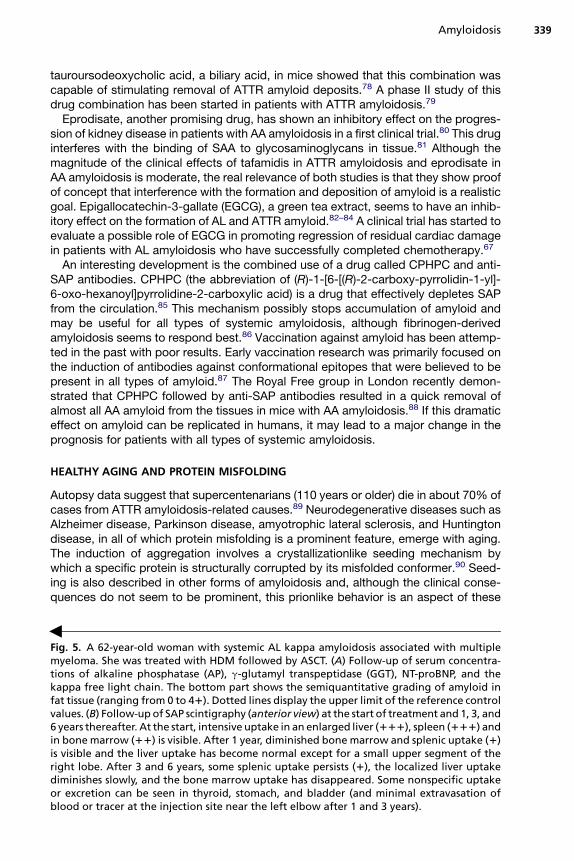
load. Quantitative clinical abnormalities are available for monitoring this clinical amyloidload, such as serum albumin, alkaline phosphatase, bilirubin, NT-proBNP, troponin,creatinine clearance, proteinuria, ventricular wall thickness, ejection fraction, conduc-tion and rhythm, heart rate variability, Ewing battery results of autonomic function, andthe sizes of enlarged organs, such as the liver, spleen, and kidneys. Imaging techniquesand subcutaneous fat tissue samples can be used for monitoring (Fig. 5). Ata consensus meeting in Tours in 2004, a set of response criteria for systemic ALamyloidosiswas acceptedby the international amyloidosis community,32with the addi-tion of some modifications after the last consensus meeting in Groningen in 2012.67
CURRENT PERSPECTIVES
The precursor-product concept helps in understanding why the current treatmentaimed at normalization of the precursor protein is important to prevent further accu-mulation of amyloid. New developments in this area include antisense73 and RNAi74
treatment. Both treatments suppress TTR production by the liver resulting in lowTTR serum concentrations. Most of the current research, however, has changed itsfocus to the development of new drugs to stabilize precursor proteins, interfere withamyloid deposition, or stimulate amyloid removal.New drugs are under investigation for ATTR amyloidosis, such as diflunisal and tafa-
midis, that stabilize the conformation of TTR in the circulation and interfere with depo-sition of amyloid. Both diflunisal and tafamidis stabilize the TTR tetramer in bloodin vitro and inhibit the tetramer in breaking up into amyloidogenic dimers and mono-mers.75 A clinical trial of tafamidis has recently been published and shows an inhibitoryeffect on the progression of polyneuropathy in patients with ATTR amyloidosis.76 Aclinical trial of diflunisal comprising 130 patients was completed at the end of 2012,so the results are expected soon.77 A study of doxycyclin in combination with

Hazenberg338
=
Amyloidosis 339
tauroursodeoxycholic acid, a biliary acid, in mice showed that this combination wascapable of stimulating removal of ATTR amyloid deposits.78 A phase II study of thisdrug combination has been started in patients with ATTR amyloidosis.79
Eprodisate, another promising drug, has shown an inhibitory effect on the progres-sion of kidney disease in patients with AA amyloidosis in a first clinical trial.80 This druginterferes with the binding of SAA to glycosaminoglycans in tissue.81 Although themagnitude of the clinical effects of tafamidis in ATTR amyloidosis and eprodisate inAA amyloidosis is moderate, the real relevance of both studies is that they show proofof concept that interference with the formation and deposition of amyloid is a realisticgoal. Epigallocatechin-3-gallate (EGCG), a green tea extract, seems to have an inhib-itory effect on the formation of AL and ATTR amyloid.82–84 A clinical trial has started toevaluate a possible role of EGCG in promoting regression of residual cardiac damagein patients with AL amyloidosis who have successfully completed chemotherapy.67
An interesting development is the combined use of a drug called CPHPC and anti-SAP antibodies. CPHPC (the abbreviation of (R)-1-[6-[(R)-2-carboxy-pyrrolidin-1-yl]-6-oxo-hexanoyl]pyrrolidine-2-carboxylic acid) is a drug that effectively depletes SAPfrom the circulation.85 This mechanism possibly stops accumulation of amyloid andmay be useful for all types of systemic amyloidosis, although fibrinogen-derivedamyloidosis seems to respond best.86 Vaccination against amyloid has been attemp-ted in the past with poor results. Early vaccination research was primarily focused onthe induction of antibodies against conformational epitopes that were believed to bepresent in all types of amyloid.87 The Royal Free group in London recently demon-strated that CPHPC followed by anti-SAP antibodies resulted in a quick removal ofalmost all AA amyloid from the tissues in mice with AA amyloidosis.88 If this dramaticeffect on amyloid can be replicated in humans, it may lead to a major change in theprognosis for patients with all types of systemic amyloidosis.
HEALTHY AGING AND PROTEIN MISFOLDING
Autopsy data suggest that supercentenarians (110 years or older) die in about 70% ofcases from ATTR amyloidosis-related causes.89 Neurodegenerative diseases such asAlzheimer disease, Parkinson disease, amyotrophic lateral sclerosis, and Huntingtondisease, in all of which protein misfolding is a prominent feature, emerge with aging.The induction of aggregation involves a crystallizationlike seeding mechanism bywhich a specific protein is structurally corrupted by its misfolded conformer.90 Seed-ing is also described in other forms of amyloidosis and, although the clinical conse-quences do not seem to be prominent, this prionlike behavior is an aspect of these
Fig. 5. A 62-year-old woman with systemic AL kappa amyloidosis associated with multiplemyeloma. She was treated with HDM followed by ASCT. (A) Follow-up of serum concentra-tions of alkaline phosphatase (AP), g-glutamyl transpeptidase (GGT), NT-proBNP, and thekappa free light chain. The bottom part shows the semiquantitative grading of amyloid infat tissue (ranging from 0 to 41). Dotted lines display the upper limit of the reference controlvalues. (B) Follow-up of SAP scintigraphy (anterior view) at the start of treatment and 1, 3, and6 years thereafter. At the start, intensive uptake in an enlarged liver (111), spleen (111) andin bone marrow (11) is visible. After 1 year, diminished bone marrow and splenic uptake (1)is visible and the liver uptake has become normal except for a small upper segment of theright lobe. After 3 and 6 years, some splenic uptake persists (1), the localized liver uptakediminishes slowly, and the bone marrow uptake has disappeared. Some nonspecific uptakeor excretion can be seen in thyroid, stomach, and bladder (and minimal extravasation ofblood or tracer at the injection site near the left elbow after 1 and 3 years).
Hazenberg340
protein-misfolding diseases that certainly deserves serious attention.91,92 In commoncardiac diseases, such as pathologic cardiac hypertrophy and dilated and ischemiccardiomyopathies, misfolded proteins have a direct causative role.93 Protein misfold-ing is not only a characteristic of some obscure amyloid diseases but is a muchbroader phenomenon that is involved in many other degenerative diseases closelyassociated with aging.94 Therefore, a better understanding of the nature and patho-genesis of all types of amyloidosis may also increase our knowledge of these commonand disabling degenerative diseases that interfere so much with healthy aging.
ACHIEVEMENTS
The amyloidoses are fascinating representatives of a new disease category of protein-misfolding diseases. Hundred years ago it was a mystery why so different diseases asmultiple myeloma and tuberculosis could end up with organs massively filled withapparently similar amyloid. Often, however, amyloidosis appeared in a patient withoutany clear cause, and was therefore thought to be primary. Occasionally the diseasewas found in succeeding generations of a family. But diagnostic possibilities improvedsteadily. The iodine sulphuric staining test was replaced by metachromatic stains,such as Congo red, in order to better identify amyloid in tissue. It was the introductionof biopsies that enabled clinicians to diagnose amyloid during life, although biopsieswere used restrictively until the mid twentieth century. But the major breakthroughcame with the work of Pras in 1968, who succeeded in isolating amyloid fibrils byextraction with distilled water.95 Since that time the amyloid protein became acces-sible for chemical analysis. The amyloid proteins were chemically characterized,amyloid precursor proteins were detected in blood, and many DNA mutations wereconnected to familial types of amyloidosis.The current classification of amyloidosis is in a way a monument of the recent
analytical and clinical research. Nowadays a patient suffering from systemic amyloid-osis can be diagnosed and typed rapidly with minimal discomfort. The patientundergoes a clinical assessment consisting of blood analyses, function tests, andimaging techniques that yields a quick overview of the severity of organ diseaseand associated risks. Treatment possibilities range widely from several biologicmodifiers, novel cytostatic drugs, or drugs specifically designed for amyloidosis totransplantation of liver or bone marrow. Monitoring the effects of treatment duringfollow-up has improved and enables the clinician to assess early whether or not theamyloidosis responds to the chosen treatment.
CURRENT CHALLENGES
Despite these favorable developments, however, the prognosis remains grim for manypatients suffering from systemic amyloidosis. The reasons are manifold, but thecurrent main challenges are lack of awareness and loss of precious time in waitingfor - often ineffective - precursor elimination.Lack of awareness leads to late detection and far advanced disease at presentation,
often severe cardiac disease or multi-organ disease. Doctors should think of amyloid-osis - or their computers should suggest it - if key symptoms for the diagnosis arepresent. This is especially true if more symptoms than one are present. Typical keysymptoms are right sided cardiac failure, proteinuria or renal failure, organ swelling(of tongue, myocardium, liver, or spleen), polyneuropathy, or autonomic neuropathythat are all unaccounted for. In many such cases looking for an elevated serum freelight chain and performing an abdominal fat aspiration may speed up the diagnosisby months.

Amyloidosis 341
Waiting until the precursor has decreased to normal levels is sometimes impossible,because the disease progresses meanwhile. Many patients with severe cardiac ALamyloidosis have not had any profit of improved treatment, because they die fromdisease progression in the first months after diagnosis. And even sometimes theamyloidosis progresses despite successful reduction or even elimination of theprecursor. This is frequently observed in some types of ATTR amyloidosis after livertransplantation, because wild-type TTR becomes deposited in the heart instead ofmutant TTR. There is a need for reliable methods to measure the actual productionand deposition of amyloid. And treatment tools need to be developed that stopamyloid deposition immediately after detection as well as additional tools that effec-tively help to remove amyloid from the body. So, although much has been achievedin the last fifty years, there is much left for fundamental and clinical researchers toimprove the prospects of patients suffering from these deadly, but also fascinatingamyloid diseases.
ACKNOWLEDGMENTS
I would like to thank Johan Bijzet BSc and Andor Glaudemans MD for their kindsupport in obtaining material for the figures.
REFERENCES
1. Kyle RA. Amyloidosis: a convoluted story. Br J Haematol 2001;114:529–38.2. Sipe JD, Benson MD, Buxbaum JN, et al. Amyloid fibril protein nomenclature:
2012 recommendations from the Nomenclature Committee of the InternationalSociety of Amyloidosis. Amyloid 2012;19:167–70.
3. Merlini G, Bellotti V. Molecular mechanisms of amyloidosis. N Engl J Med 2003;349:583–96.
4. Falk RH, Comenzo RL, Skinner M. The systemic amyloidoses. N Engl J Med 1997;337:898–909.
5. Westermark P. Localized AL amyloidosis: a suicidal neoplasm? Ups J Med Sci2012;117:244–50.
6. Meijer JM, Schonland SO, Palladini G, et al. Sjogren’s syndrome and localizednodular cutaneous amyloidosis: coincidence or a distinct clinical entity? ArthritisRheum 2008;58:1992–9.
7. Kyle RA, Gertz MA. Primary systemic amyloidosis: clinical and laboratory featuresin 474 cases. Semin Hematol 1995;32:45–59.
8. Lachmann HJ, Goodman HJ, Gilbertson JA, et al. Natural history and outcome insystemic AA amyloidosis. N Engl J Med 2007;356:2361–71.
9. Hazenberg BP, van Rijswijk MH. Clinical and therapeutic aspects of AA amyloid-osis. Baillieres Clin Rheumatol 1994;8:661–90.
10. Connors LH, Lim A, Prokaeva T, et al. Tabulation of human transthyretin (TTR)variants, 2003. Amyloid 2003;10:160–84.
11. Dungu JN, Anderson LJ, Whelan CJ, et al. Cardiac transthyretin amyloidosis.Heart 2012;98:1546–54.
12. Drueke TB, Massy ZA. Beta2-microglobulin. Semin Dial 2009;22:378–80.13. Heegaard NH. Beta(2)-microglobulin: from physiology to amyloidosis. Amyloid
2009;16:151–73.14. Valleix S, Gillmore JD, Bridoux F, et al. Hereditary systemic amyloidosis due to As-
p76Asn variant b2-microglobulin. N Engl J Med 2012;366:2276–83.15. Benson MD. Ostertag revisited: the inherited systemic amyloidoses without
neuropathy. Amyloid 2005;12:75–87.

Hazenberg342
16. Lachmann HJ, Booth DR, Booth SE, et al. Misdiagnosis of hereditary amyloidosisas AL (primary) amyloidosis. N Engl J Med 2002;346:1786–91.
17. Gillmore JD, Lachmann HJ, Rowczenio D, et al. Diagnosis, pathogenesis, treat-ment, and prognosis of hereditary fibrinogen A alpha-chain amyloidosis. J AmSoc Nephrol 2009;20:444–51.
18. Sattianayagam PT, Gibbs SD, Rowczenio D, et al. Hereditary lysozyme amyloid-osis – phenotypic heterogeneity and the role of solid organ transplantation.J Intern Med 2012;272:36–44.
19. Larsen CP, Walker PD, Weiss DT, et al. Prevalence and morphology of leukocytechemotactic factor 2-associatedamyloid in renal biopsies.Kidney Int 2010;77:816–9.
20. Kiuru S. Gelsolin-related familial amyloidosis, Finnish type (FAF), and its variantsfound worldwide. Amyloid 1998;5:55–66.
21. Simms RW, Prout MN, Cohen AS. The epidemiology of AL and AA amyloidosis.Baillieres Clin Rheumatol 1994;8:627–34.
22. Kyle RA, Linos A, Beard CM, et al. Incidence and natural history of primarysystemic amyloidosis in Olmsted County, Minnesota, 1950 through 1989. Blood1992;79:1817–22.
23. Hemminki K, Li X, Forsti A, et al. Incidence and survival in non-hereditaryamyloidosis in Sweden. BMC Public Health 2012;12:974.
24. Janssen S, van Rijswijk MH, Meijer S, et al. Systemic amyloidosis: a clinicalsurvey of 144 cases. Neth J Med 1986;29:376–85.
25. Pepys MB. Pathogenesis, diagnosis and treatment of systemic amyloidosis.Philos Trans R Soc Lond B Biol Sci 2001;356:203–10.
26. Hazenberg BP, van Gameren II, Bijzet J, et al. Diagnostic and therapeuticapproach of systemic amyloidosis. Neth J Med 2004;62:121–8.
27. Bijzet J, van Gameren II, Hazenberg BP. Fat tissue analysis in the management ofpatients with systemic amyloidosis. In: Picken MM, Dogan A, Herrera GA, editors.Amyloid and related disorders: surgical pathology and clinical correlations. NewYork: Humana Press; 2012. p. 191–207.
28. Westermark P. Subcutaneous adipose tissue biopsy for amyloid protein studies.Methods Mol Biol 2012;849:363–71.
29. van Gameren II, Hazenberg BP, Bijzet J, et al. Diagnostic accuracy of subcuta-neous abdominal fat tissue aspiration for detecting systemic amyloidosis andits utility in clinical practice. Arthritis Rheum 2006;54:2015–21.
30. Ikeda S, Sekijima Y, Tojo K, et al. Diagnostic value of abdominal wall fat padbiopsy in senile systemic amyloidosis. Amyloid 2011;18:211–5.
31. Nilsson KP, Ikenberg K, Aslund A, et al. Structural typing of systemic amyloidosesby luminescent-conjugated polymer spectroscopy. Am J Pathol 2010;176:563–74.
32. Gertz MA, Comenzo R, Falk RH, et al. Definition of organ involvement and treat-ment response in immunoglobulin light chain amyloidosis (AL): a consensusopinion from the 10th International Symposium on Amyloid and Amyloidosis,Tours, France, 18-22 April 2004. Am J Med 2005;79:319–28.
33. Kebbel A, Rocken C. Immunohistochemical classification of amyloid in surgicalpathology revisited. Am J Surg Pathol 2006;30:673–83.
34. Satoskar AA, Efebera Y, Hasan A, et al. Strong transthyretin immunostaining:potential pitfall in cardiac amyloid typing. Am J Surg Pathol 2011;35:1685–90.
35. Murphy CL, Wang S, Williams T, et al. Characterization of systemic amyloiddeposits by mass spectrometry. Methods Enzymol 2006;412:48–62.
36. Lavatelli F, Perlman DH, Spencer B, et al. Amyloidogenic and associated proteinsin systemic amyloidosis proteome of adipose tissue. Mol Cell Proteomics 2008;7:1570–83.

Amyloidosis 343
37. Vrana JA, Gamez JD, Madden BJ, et al. Classification of amyloidosis by lasermicrodissection and mass spectrometry-based proteomic analysis in clinicalbiopsy specimens. Blood 2009;114:4957–9.
38. Lavatelli F, Vrana JA. Proteomic typing of amyloid deposits in systemic amyloid-oses. Amyloid 2011;18:177–82.
39. Brambilla F, Lavatelli F, Di Silvestre D, et al. Reliable typing of systemic amyloid-oses through proteomic analysis of subcutaneous adipose tissue. Blood 2012;119:1844–7.
40. Picken MM. Amyloidosis-where are we now and where are we heading? ArchPathol Lab Med 2010;134:545–51.
41. Dispenzieri A, Gertz MA, Kyle RA, et al. Serum cardiac troponins and N-terminalpro-brain natriuretic peptide: a staging system for primary systemic amyloidosis.J Clin Oncol 2004;22:3751–7.
42. Palladini G, Barassi A, Perlini S, et al. Midregional proadrenomedullin (MR-proADM) is a powerful predictor of early death in AL amyloidosis. Amyloid 2011;18:216–21.
43. Ozdemir D, Dagdelen S, Erbas T, et al. Amyloid goiter and hypopituitarism ina patient with systemic amyloidosis. Amyloid 2011;18:32–4.
44. Ishihara T, Nagasawa T, Yokota T, et al. Amyloid protein of vessels in leptome-ninges, cortices, choroid plexuses, and pituitary glands from patients withsystemic amyloidosis. Hum Pathol 1989;20:891–5.
45. Erdkamp FL, Gans RO, Hoorntje SJ. Endocrine organ failure due to systemicAA-amyloidosis. Neth J Med 1991;38:24–8.
46. Zubkov AY, Rabinstein AA, Dispenzieri A, et al. Primary systemic amyloidosis withischemic stroke as a presenting complication. Neurology 2007;69:1136–41.
47. Kay J, Benson CB, Lester S, et al. Utility of high-resolution ultrasound for the diag-nosis of dialysis-related amyloidosis. Arthritis Rheum 1992;35:926–32.
48. Dubrey SW, Hawkins PN, Falk RH. Amyloid diseases of the heart: assessment,diagnosis, and referral. Heart 2011;97:75–84.
49. Noordzij W, Glaudemans AW, van Rheenen RW, et al. Iodine-123 labelled meta-iodobenzylguanidine for the evaluation of cardiac sympathetic denervation inearly stage amyloidosis. Eur J Nucl Med Mol Imaging 2012;39:1609–17.
50. Aprile C, Marinone G, Saponaro R, et al. Cardiac and pleuropulmonary ALamyloid imaging with technetium-99m labelled aprotinin. Eur J Nucl Med 1995;22:1393–401.
51. Janssen S, Piers DA, van Rijswijk MH, et al. Soft-tissue uptake of 99mTc-di-phosphonate and 99mTc-pyrophosphate in amyloidosis. Eur J Nucl Med 1990;16:663–70.
52. Wechalekar K, Ng FS, Poole-Wilson PA, et al. Cardiac amyloidosis diagnosedincidentally by bone scintigraphy. J Nucl Cardiol 2007;14:750–3.
53. Rapezzi C, Quarta CC, Guidalotti PL, et al. Role of (99m)Tc-DPD scintigraphy indiagnosis and prognosis of hereditary transthyretin-related cardiac amyloidosis.JACC Cardiovasc Imaging 2011;4:659–70.
54. Kristen AV, Haufe S, Schonland SO, et al. Skeletal scintigraphy indicates diseaseseverity of cardiac involvement in patients with senile systemic amyloidosis. Int JCardiol 2011. http://dx.doi.org/10.1016/j.ijcard.2011.06.123.
55. Hawkins PN, Lavender JP, Pepys MB. Evaluation of systemic amyloidosis by scin-tigraphy with 123I-labeled serum amyloid P component. N Engl J Med 1990;323:508–13.
56. Jager PL, Hazenberg BP, Franssen EJ, et al. Kinetic studies with iodine-123-labeled serum amyloid P component in patients with systemic AA

Hazenberg344
and AL amyloidosis and assessment of clinical value. J Nucl Med 1998;39:699–706.
57. Hazenberg BP, van Rijswijk MH, Piers DA, et al. Diagnostic performance of 123I-labeled serum amyloid P component scintigraphy in patients with amyloidosis.Am J Med 2006;119:355.e15–24.
58. Hawkins PN, Richardson S, MacSweeney JE, et al. Scintigraphic quantificationand serial monitoring of human visceral amyloid deposits provide evidence forturnover and regression. Q J Med 1993;86:365–74.
59. Hazenberg BP, van Rijswijk MH, Lub-de Hooge MN, et al. Diagnostic perfor-mance and prognostic value of extravascular retention of 123I-labeled serumamyloid P component in systemic amyloidosis. J Nucl Med 2007;48:865–72.
60. Wall JS, Kennel SJ, Stuckey AC, et al. Radioimmunodetection of amyloid depositsin patients with AL amyloidosis. Blood 2010;116:2241–4.
61. Gillmore JD, Lovat LB, Persey MR, et al. Amyloid load and clinical outcome in AAamyloidosis in relation to circulating concentration of serum amyloid A protein.Lancet 2001;358:24–9.
62. Caorsi R, Federici S, Gattorno M. Biologic drugs in autoinflammatory syndromes.Autoimmun Rev 2012;12:81–6.
63. Hakala M, Immonen K, Korpela M, et al. Good medium-term efficacy of tocilizu-mab in DMARD and anti-TNF-a therapy resistant reactive amyloidosis. AnnRheum Dis 2013;72:464–5.
64. Gertz MA, Lacy MQ, Dispenzieri A, et al. Effect of haematological responseon outcome of patients undergoing transplantation for primary amyloidosis:importance of achieving a complete response. Haematologica 2007;92:1415–8.
65. Skinner M, Sanchorawala V, Seldin DC, et al. High-dose melphalan and autolo-gous stem-cell transplantation in patients with AL amyloidosis: an 8-year study.Ann Intern Med 2004;140:85–93.
66. Jaccard A, Moreau P, Leblond V, et al. High-dose melphalan versus melphalanplus dexamethasone for AL amyloidosis. N Engl J Med 2007;357:1083–93.
67. Gatt ME, Palladini G. Light chain amyloidosis 2012: a new era. Br J Haematol2013;160:582–98.
68. van Gameren II, van Rijswijk MH, Bijzet J, et al. Histological regression of amyloidin AL amyloidosis is exclusively seen in patients with a complete response ofserum free light chain. Haematologica 2009;94:1094–100.
69. Wilczek HE, Larsson M, Ericzon BG, et al. Long-term data from the Familial Amy-loidotic Polyneuropathy World Transplant Registry (FAPWTR). Amyloid 2011;18(Suppl 1):193–5.
70. Okamoto S, Zhao Y, Lindqvist P, et al. Development of cardiomyopathy after livertransplantation in Swedish hereditary transthyretin amyloidosis (ATTR) patients.Amyloid 2011;18:200–5.
71. Gustafsson S, Ihse E, Henein MY, et al. Amyloid fibril composition as a predictorof development of cardiomyopathy after liver transplantation for hereditary trans-thyretin amyloidosis. Transplantation 2012;93:1017–23.
72. Yamamoto S, Gejyo F. Historical background and clinical treatment of dialysis-related amyloidosis. Biochim Biophys Acta 2005;1753:4–10.
73. Ackermann EJ, Guo S, Booten S, et al. Clinical development of an antisensetherapy for the treatment of transthyretin-associated polyneuropathy. Amyloid2012;19(Suppl 1):43–4.
74. Barros SA, Gollob JA. Safety profile of RNAi nanomedicines. Adv Drug Deliv Rev2012;64:1730–7.

Amyloidosis 345
75. Hammarstrom P, Wiseman RL, Powers ET, et al. Prevention of transthyretin amyloiddisease by changing protein misfolding energetics. Science 2003;299:713–7.
76. Coelho T, Maia LF, Martins da Silva A, et al. Tafamidis for transthyretin familialamyloid polyneuropathy: a randomized, controlled trial. Neurology 2012;79:785–92.
77. Berk JL, Suhr OB, Sekijima Y, et al. The diflunisal trial: study accrual and drugtolerance. Amyloid 2012;19(Suppl 1):37–8.
78. Cardoso I, Martins D, Ribeiro T, et al. Synergy of combined doxycycline/TUDCAtreatment in lowering Transthyretin deposition and associated biomarkers:studies in FAP mouse models. J Transl Med 2010;8:74.
79. Obici L, Cortese A, Lozza A, et al. Doxycycline plus tauroursodeoxycholic acidfor transthyretin amyloidosis: a phase II study. Amyloid 2012;19(Suppl 1):34–6.
80. Dember LM, Hawkins PN, Hazenberg BP, et al. Eprodisate for the treatment of AAamyloidosis. N Engl J Med 2007;356:2349–60.
81. Inoue S, Hultin PG, Szarek WA, et al. Effect of poly(vinylsulfonate) on murine AAamyloid: a high-resolution ultrastructural study. Lab Invest 1996;74:1081–90.
82. Hunstein W. Epigallocathechin-3-gallate in AL amyloidosis: a new therapeuticoption? Blood 2007;110:2216.
83. Mereles D, Buss SJ, Hardt SE, et al. Effects of the main green tea polyphenolepigallocatechin-3-gallate on cardiac involvement in patients with AL amyloid-osis. Clin Res Cardiol 2010;99:483–90.
84. Kristen AV, Lehrke S, Buss S, et al. Green tea halts progression of cardiac trans-thyretin amyloidosis: an observational report. Clin Res Cardiol 2012;101:805–13.
85. Pepys MB, Herbert J, Hutchingson WL, et al. Targeted pharmacological deple-tion of serum amyloid P component for treatment of human amyloidosis. Nature2002;417:254–9.
86. Gillmore JD, Tennent GA, Hutchinson WL, et al. Sustained pharmacologicaldepletion of serum amyloid P component in patients with systemic amyloidosis.Br J Haematol 2010;148:760–7.
87. Hrncic R, Wall J, Wolfenbarger DA, et al. Antibody-mediated resolution of lightchain-associated amyloid deposits. Am J Pathol 2000;157:1239–46.
88. Bodin K, Ellmerich S, Kahan MC, et al. Antibodies to human serum amyloidP component eliminate visceral amyloid deposits. Nature 2010;468:93–7.
89. Coles LS, Young RD. Supercentenarians and transthyretin amyloidosis: the nextfrontier of human life extension. Prev Med 2012;54(Suppl):S9–11.
90. Walker LC, LeVine H 3rd. Corruption and spread of pathogenic proteins in neuro-degenerative diseases. J Biol Chem 2012;287:33109–15.
91. Westermark GT, Westermark P. Prion-like aggregates: infectious agents in humandisease. Trends Mol Med 2010;16:501–7.
92. Solomon A, Richey T, Murphy CL, et al. Amyloidogenic potential of foie gras. ProcNatl Acad Sci U S A 2007;104:10998–1001.
93. Willis MS, Patterson C. Proteotoxicity and cardiac dysfunction – Alzheimer’sdisease of the heart? N Engl J Med 2013;368:455–64.
94. Schnabel J. Protein folding: the dark side of proteins. Nature 2010;464:828–9.95. Pras M, Schubert M, Zucker-Franklin D, et al. The characterization of soluble
amyloid prepared in water. J Clin Invest 1968;47:924–33.