university of groningen a potential strategy to treat … chapter 6 well-established carrier that...
TRANSCRIPT

University of Groningen
A potential strategy to treat liver fibrosisGonzalo Lázaro, Teresa
IMPORTANT NOTE: You are advised to consult the publisher's version (publisher's PDF) if you wish to cite fromit. Please check the document version below.
Document VersionPublisher's PDF, also known as Version of record
Publication date:2006
Link to publication in University of Groningen/UMCG research database
Citation for published version (APA):Gonzalo Lázaro, T. (2006). A potential strategy to treat liver fibrosis: Drug targeting to hepatic stellate cellsapplying a novel linker technology. s.n.
CopyrightOther than for strictly personal use, it is not permitted to download or to forward/distribute the text or part of it without the consent of theauthor(s) and/or copyright holder(s), unless the work is under an open content license (like Creative Commons).
Take-down policyIf you believe that this document breaches copyright please contact us providing details, and we will remove access to the work immediatelyand investigate your claim.
Downloaded from the University of Groningen/UMCG research database (Pure): http://www.rug.nl/research/portal. For technical reasons thenumber of authors shown on this cover page is limited to 10 maximum.
Download date: 03-03-2019

Chapter6Local inhibition of liver fi brosis by specifi c delivery of a PDGF kinase inhibitor to hepatic stellate cells
Submitted for publication.
1,Teresa Gonzalo, 1Leonie Beljaars, 1Marja van de Bovenkamp, 1Anne-Miek van Loenen, 1Catharina Reker-Smit, 1Dirk K.F. Meijer, 2Marie Lacombe, 2Frank Opdam, 3György Kéri, 3László Orfi , 1Klaas Poelstra, 1,4Robbert J Kok.
1Department of Pharmacokinetics and Drug Delivery, Groningen University Institute for Drug Exploration, University of Groningen, The Netherlands;
2Kreatech Biotechnology B.V., Amsterdam;
3Department of Medical and Pharmaceutical Chemistry, Semmelweis University, Budapest, Hungary;
4Department of Pharmaceutics, Utrecht University, The Netherlands.

126
Local inhibition of liver fibrosis by targeting a PDGF kinase inhibitor to stellate cells
Abstract
Liver fibrosis is characterized by an excessive proliferation and activation of Hepatic
Stellate Cells (HSC). Platelet-derived growth factor (PDGF-BB) is the most potent
mitogen for HSC and inhibition of PDGF signalling via specific delivery of a PDGF
kinase inhibitor to HSC might therefore be an attractive strategy to counteract the
progression of liver fibrosis. The HSC-selective carrier M6PHSA was equipped with a
PDGF receptor tyrosine kinase inhibitor (PTKI, an imatinib derivative) by means of a
novel platinum-based linker (ULSTM).
Culture-activated rat HSC and precision-cut liver slices from fibrotic rats were incubated
with PTKI-M6PHSA and fibrosis markers were evaluated by quantitative RT-PCR. The
gene expression of α-smooth muscle actin and α1-(I)-procollagen were reduced by 50%
in both in vitro systems after treatment with PTKI-M6PHSA (0.1 mg/ml, corresponding to
10µM of PTKI) and free PTKI showed similar effects.
Next, we examined the homing and antifibrotic effects of PTKI-M6PHSA in bile duct
ligated (BDL) rats. Male Wistar rats at day 10 after BDL were injected intravenously with a
single dose of 3.3 mg/kg of PTKI-M6PHSA and compared with non-treated BDL rats.
PTKI-M6PHSA was detected in the liver 2h after administration in a non-parenchymal
distribution pattern. The antifibrotic effects of PTKI-M6PHSA were analysed by Sirius
Red and α-smooth muscle actin stainings. Both parameters were reduced 24h and 48h
after the single dose of PTKI-M6PHSA (p<0.01, p<0.05. resp), in comparison with non-
treated rats. We subsequently conducted a multiple dose study administering PTKI-
M6PHSA to BDL rats and untargeted PTKI, where we found no effect of either
treatment.
In summary, PTKI-M6PHSA showed an antifibrogenic effect in cultured HSC and
fibrotic liver slices. This effect was also demonstrated in vivo after direct targeting of the
PDGFR kinase inhibitor to activated HSC during liver fibrosis after single administration.
Upon longer treatment however, PDGF kinase inhibition can not block the fibrotic

127
Chapter 6
process. We therefore conclude that delivery of a PDGF-kinase inhibitor to HSC is a
promising technology to attenuate liver fibrogenesis, although other activating pathways
may need to be inhibited in parallel.
Theoretical
synthesis ratio
PTKI-ULS: protein
Coupling ratio
PTKI : protein
Yield of synthesis#
PTKI-M6PHSA
10:1
8 : 1 (±1.9)
84%
A
0
100
200
300
400
500
600
700
800
900
10 15 20 25 30
PTKI-M 6PHSA
M 6PHSA
NHCl
O
NH
N NCH3
NPt
2+M6PHSA carrier
UV
280
nm
(m
AU
)
MonoQ anion exchange
UV
280
nm
(m
AU
)
0
200
400
600
800
00
1200
1400
00
9 10 11 12 13
HSAM6PHSAPTKI-M6PHSA
16B
10
Size exclusion chromatography
Time (min) Time (min)
Figure 1. Characteristics of PTKI-M6PHSA drug targeting conjugate.
A. Synthesis of PTKI-M6PHSA. PTKI was conjugated via the platinum based linker ULS to the
stellate cell selective carrier M6PHSA. Coordination bonds between drug-linker and linker-carrier are
depicted as dotted lines. ¶ PTKI/M6PHSA coupling ratio was determined by HPLC after competitive
displacement of the drug with potassium thiocyanate (KSCN) at 80°C for 24h. # Based in the BCA
assay or protein concentration. Values obtained from three independent PTKI-M6PHSA synthesized
conjugates. B. MonoQ anion exchange chromatography confirmed that the charge of the protein was not
affected. Size exclusion chromatography showed the monomeric composition of PTKI-M6PHSA.
M6PHSA: mannose-6-phosphate modified human serum albumin; ULS: Universal Linkage System.
Chapter 6
128
Local inhibition of liver fibrosis by targeting a PDGF kinase inhibitor to stellate cells
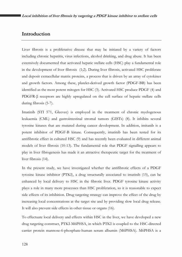
Introduction
Liver fibrosis is a proliferative disease that may be initiated by a variety of factors
including chronic hepatitis, virus infections, alcohol drinking, and drug abuse. It has been
extensively documented that activated hepatic stellate cells (HSC) play a fundamental role
in the development of liver fibrosis (1;2). During liver fibrosis, activated HSC proliferate
and deposit extracellular matrix proteins, a process that is driven by an array of cytokines
and growth factors. Among these, platelet-derived growth factor (PDGF-BB) has been
identified as the most potent mitogen for HSC (3). Activated HSC produce PDGF (4) and
PDGFR-β receptors are highly upregulated on the cell surface of hepatic stellate cells
during fibrosis (5-7).
Imatinib (STI 571, Gleevec) is employed in the treatment of chronic myelogenous
leukaemia (CML) and gastrointestinal stromal tumors (GISTs) (8). It inhibits several
tyrosine kinases that are mutated during cancer development. In addition, imitanib is a
potent inhibitor of PDGF-B kinase. Consequently, imatinib has been tested for its
antifibrotic effect in cultured HSC (9) and has recently been evaluated in different animal
models of liver fibrosis (10-13). The fundamental role that PDGF signalling appears to
play in liver fibrogenesis has made it an attractive therapeutic target for the treatment of
liver fibrosis (14).
In the present study, we have investigated whether the antifibrotic effects of a PDGF
tyrosine kinase inhibitor (PTKI), a drug structurally associated to imatinib (15), can be
enhanced by local delivery to HSC in the fibrotic liver. PDGF tyrosine kinase activity
plays a role in many more processes than HSC proliferation, so it is reasonable to expect
side effects of its inhibition. Drug targeting strategy can improve the effect of the drug by
increasing local concentrations at the target site and by providing slow local drug release.
It will also prevent side effects in other tissue or organs (16).
To effectuate local delivery and effects within HSC in the liver, we have developed a new
drug targeting construct, PTKI-M6PHSA, in which PTKI is coupled to the HSC-directed
carrier protein mannose-6-phosphate-human serum albumin (M6PHSA). M6PHSA is a

129
Chapter 6
well-established carrier that binds to the M6P/IGFII receptor on HSC and accumulates
rapidly and extensively in the liver of fibrotic rats (17). To conjugate PTKI to M6PHSA,
we have employed a novel type of platinum linker chemistry called ULSTM (Universal
Linker System)(18). ULS allows stable coupling of drug molecules to proteins based on
the formation of a platinum-ligand coordination bond (19). Application of this novel
linker technology was essential since it appears to be straightforward and reliable for
linking PTKI molecules to the carrier, allowing high synthesis yields in a relatively simple
approach. Second, the resulting PTKI-ULS-M6PHSA conjugates display a unique
behavior of slow release of drug molecules during a period of days within the designated
target cells.
In the present study, we describe the development of PTKI-M6PHSA and its impact on
liver fibrogenesis in vitro and in vivo. Culture of HSC and fibrotic liver slices were employed
as in vitro systems to prove the antifibrotic effect of PTKI-M6PHSA. In addition, PTKI-
M6PHSA was tested in a model of liver fibrosis to study the distribution and effects on
the development of liver fibrosis.
Chapter 6

130
Local inhibition of liver fibrosis by targeting a PDGF kinase inhibitor to stellate cells
BDL
day 10
control saline
BDL
day 11
control saline
BDL
day 12
control saline
BDL
day 10
+PTKI-M6PHSA
BDL
day 11
+PTKI-M6PHSA
BDL
day 12
+PTKI-M6PHSA
Number of rats 5 5 5 3 5 5
Body weight (g) 250.2 ± 46.7 311.2 ± 14.7 273.4 ± 16.4 280.7 ± 10.1 301.4 ± 14.1 306.4 ± 13.5
Liver/body ratio (g/g) 0.055 ± 0.009 0.062 ± 0.007 0.065 ± 0.004 0.061 ± 0.002 0.060 ± 0.006 0.065 ± 0.009
Dose Saline Saline Saline 3.3mg/kg 3.3mg/kg 3.3mg/kg
Bilirubin (umol/l) 183.7 ± 35.8 178.0 ± 48.2 228.5 ± 18.7 252.3 ± 55.9 136.0 ± 10.0
183.5 ± 27.9
ALT (U/l) 86.3 ±30.9 79.8 ±46.1 79.3 ±11.2 96.5 ± 4.9 72.7 ±10.6 81.0 ±17.0
Alkaline Phosphatase
(U/l) 391.2 ±46.4 476.0 ±162.6 358.3 ±234.1 458.0 ±11.2 426.0±101.3 511.3±121.4
Table 1. Dose regimens, animal data and biochemical parameters from PTKI-M6PHSA single injection study. Data are shown as mean ± SD. BDL, bile duct ligated animals; ALT, alanine aminotransferase.

131
Chapter 6
Materials and methods
Materials
The Protein Tyrosine Kinase Inhibitor (PTKI, 4-Chloro-N-[4-methyl-3-(4-pyridin-3-yl-
pyrimidin-2-ylamino)-phenyl]-benzamide) was kindly provided by György Kéri (Vichem
Chemie Research Ltd., Budapest, Hungary). M6PHSA was prepared as described
previously (19). Cis-[Pt(ethylenediamine)nitrate-chloride] (cisULS) was prepared as
previously described (19).
Synthesis of PTKI-ULS-M6PHSA
PTKI-ULS was synthesized and purified by Kreatech Biotechnology (Amsterdam, The
Netherlands). In brief, PTKI (7.2 µmol, 3 mg; 10 mg/ml in DMF) was mixed with an
equimolar amount of cisULS (7.2 µmol, 2.4 mg; 20 mM in DMF). The reaction mixture
was heated at 37°C for 24h after which consumption of the starting material was
monitored by analytical HPLC. An additional amount of cisULS was added (0.5
equivalent, 3.6 µmol) and the reaction was continued for 48h at 37°C. The crude mixture
was concentrated under reduced pressure and dissolved in methanol (600 µl). The crude
product was purified by preparative HPLC and the collected peaks of the main product
were taken to dryness under reduced pressure. The resulting white solid was treated with
water to remove anorganic salts and dried. Yield: 0.9 mg (20%). Mass spectrometry
analysis confirmed the presence of the 1:1 PTKI-ULS species.
1H NMR of PTKI (CD3OD): δH 2.33 (s, 3H, CH3), 7.26 (d, J = 8.28 Hz, 1H, CCH3CH),
7.37 (m, 2H, CHCl), 7.52 (m, 3H, N(CH)2CCCH), 7.93 (d, J = 8.60 Hz, 2H, CHCHCl),
8.22 (s, 1H, NHCCHC), 8.47 (d, J = 5.23 Hz, 1H, CHNCNH), 8.64 (m, 2H, CH(CH)2C
and CHCHCNH), 9.29 (s, 1H, NCHC) ppm.
1H NMR of PTKI-ULS (CD3OD): δH 2.27 (m, 3H, CH3), 2.66 (m, 2H, CH2), 2.74 (m,
2H, CH2), 7.15 (m, 3H, CHCCH3 and CHCl), 7.47 (m, 3H, N(CH)2CCCH), 7.84 (d, J =
5.23 Hz, 1H, CHCHCNH), 7.89 (m, 2 H, CHCHCl), 8.39 (d, J = 3.95 Hz, 1H,
CHNCNH) ppm.
Chapter 6

132
Local inhibition of liver fibrosis by targeting a PDGF kinase inhibitor to stellate cells
Mass spectrometry of PTKI-ULS (ESI+): calculated mass: 705 (m/z); detected masses:
706 [M]+, 688 [M-Cl-+OH-]+, 670 [M-Cl--H+]+, 669 [M-Cl--H+]+, 651 [M-Cl--Cl-+OH-
+H]+.
HPLC analysis: Separations were performed on a Luna2 C18 column that was maintained
at 40°C. The mobile phase consisted of a binary solvent system of triethylammonium
acetate (100mM pH 5.0):acetonitrile 90:10 (solvent A) and triethylammonium acetate
(100mM pH 5.0):acetonitrile 70:30 (solvent B). The column was eluted at a flow rate of
1.1 mL/min. Compounds were eluted at a stepwise gradient (0%B from 0-4 min; 0-46%B
from 4-17 min; 46-100%B from 17-19 min; 100%B from 19-25 min; 100-0%B from 25-
27 min; 0%B from 27-34 min). PTKI eluted at 21.2 min (60.8%B) and PTKI-ULS eluted
at 11.5 min (26.5%B).
PTKI-ULS was conjugated to M6PHSA according to a general protocol that has been
described elsewhere for the synthesis of Pentoxifylline-ULS-M6PHSA (19). Briefly,
PTKI-ULS (143 nmol, 1.6 mg that was dissolved in DMF/H2O at 6.7 mg/ml) was added
in 10-fold molar excess to M6PHSA (14.3 nmol, 10 mg, dissolved in 1 ml of 20 mM
tricine/NaNO3 buffer pH 8.3). The pH was checked and adjusted to pH 8 if necessary.
The mixture was incubated overnight at 37°C, and dialysed against PBS at 4°C. The final
product was sterilized by filtration via a 0.2 µm filter and stored at -20°C. Protein content
was assessed by the BCA assay (Pierce, Rockford, IL, USA). PTKI-M6PHSA and
M6PHSA were analyzed by size-exclusion chromatography and anion exchange
chromatography as described before (17) to verify that coupling of PTKI-ULS did not
alter the properties of the M6PHSA protein. The amount of PTKI coupled to M6PHSA
was analyzed by isocratic HPLC after competitive displacement of the drug by overnight
incubation at 80°C with excess of potassium thiocyanate (KSCN, 0.5M in PBS). Elutions
were performed on a Waters system (Waters, Milford, MA, USA) equipped with a 5 µm
Hypersil BDS C8 column (250x4.6 mm, Thermoquest Runcorn, UK), a thermostated
column oven operated at 40°C and an UV detector operated at 269 nm. The mobile phase
consisted of acetonitrile/water/trifluoracetic acid (40/60/0.1, pH 2) at a flow rate of 1.0
ml/min with a sensitivity of 0.01. Retention times: PTKI: 7 min; PTKI-ULS: 5 min.

133
Chapter 6
Figure 2. Activated HSC incubated with PTKI-M6PHSA.
A. Effect of PTKI-M6PHSA and PTKI on HSC cell viability, as determined by Alamar Blue viability
assay. Indicated concentrations reflect the platinum content of the conjugate, or equivalent amounts
cisplatin, PTKI or M6PHSA. Cultured HSC were incubated for 24h with the compounds (*P<0.01).
A. Effect of PTKI-M6PHSA and PTKI on HSC cell viability, as determined by Alamar Blue viability
assay. Indicated concentrations reflect the platinum content of the conjugate, or equivalent amounts
cisplatin, PTKI or M6PHSA. Cultured HSC were incubated for 24h with the compounds (*P<0.01).
B. Activated HSC gene expression after incubation with PTKI-M6PHSA, PTKI and M6PHSA.
Concentrations denote the amount of PTKI (10 µM) or the corresponding amount of M6PHSA carrier
(0.1mg/ml). Gene expressions levels were normalized to the expression of GAPDH and subsequently
normalized to the relative expression of control cells (*P<0.01 and #P<0.05).
B. Activated HSC gene expression after incubation with PTKI-M6PHSA, PTKI and M6PHSA.
Concentrations denote the amount of PTKI (10 µM) or the corresponding amount of M6PHSA carrier
(0.1mg/ml). Gene expressions levels were normalized to the expression of GAPDH and subsequently
normalized to the relative expression of control cells (*P<0.01 and
Activated HSC
Activated HSC
cisPlatinum 100µM
cisPlatinum 100µM
PTKI PTKI -M6PHSA -M6PHSA
100µM 100µM PTKI PTKI 100µM 100µM
M6PHSA 100µM
M6PHSA 100µM
Stel
late
cel
l via
bilit
y (A
lam
ar b
lue)
St
ella
te c
ell v
iabi
lity
(Ala
mar
blu
e)
**
0
20
40
60
80
100
120A
B
0,0
0,2
0,4
0,6
0,8
1,0
1,2
1,4
1,6
HSCcontrol PTKI-M6PHSA 10µM(0.1mg/ml)
PTKI 10uM M6PHSA 0.1mg/ml
GAPDH TIMP-1 PDGFR-B Collagen 1a2 SMA
*
#
**
Rel
ativ
e ge
ne e
xpre
ssio
n
#P<0.05).
Chapter 6

134
Local inhibition of liver fibrosis by targeting a PDGF kinase inhibitor to stellate cells
Cells
Hepatic stellate cells were isolated from male Wistar rat livers by the pronase-collagenase
method followed by a density centrifugation on a 12% Nycodenz gradient according to
Geerts et al (20;21)(Hepatol 1998). The isolated stellate cell fraction was cultured in 6 well-
plates (Corning) in Dulbecco’s Modified Eagle’s Medium (Gibco, Life technologies Ltd.)
supplemented with 10% fetal calf serum, 100 U/ml penicillin and 100 mg/ml
streptomycin in a 5% CO2 humidified atmosphere at 37°C. Cells were split after 3 days
and cultured until day 10 to obtain the activated HSC phenotype, and these culture
activated HSC were used for the experiments described below.
Cell viability studies
Activated HSC (10,000 cells/well seeded in 96 well-plates, Corning) were washed with
serum-free medium and incubated for 24h in medium supplemented with 100 µM PTKI,
PTKI-M6PHSA (1mg/ml, corresponding to 100 µM of PTKI) or M6PHSA (1mg/ml).
During the last 2h of the incubations, Alamar blue reagent (Serotec, Oxford, UK) was
added in a proportion of 10% of the volume per well. Viability of the cells was determined
fluorimetrically according to the supplier’s instructions.
Effects on gene expression
The potential antifibrotic activity of PTKI-M6PHSA and PTKI was evaluated in cultured
HSC and in precision-cut liver slices of fibrotic rat livers.
Activated HSC were incubated as described above with PTKI (10 µM), PTKI-M6PHSA
(0.1mg/ml, corresponding to 10 µM of PTKI), or M6PHSA (0.1 mg/ml) for 24h, after
which they were processed for RNA analysis as described below. A total of four
independent experiments were performed in HSC cultures from four different Wistar
Male rats.
Precision-cut liver slices were prepared as described elsewhere (22;23). Briefly, precision-
cut liver slices (8mm diameter, 250µm) from fibrotic livers of BDL3 rats (week 3 after
BDL) were prepared using a Krumdieck tissue slicer and stored in University of
Winconsin preservation solution (UW) on ice until further use.

135
Chapter 6
0,0
0,2
0,4
0,6
0,8
1,0
1,2
1,4
BDL liver slice + PBS PTKI-M6PHSA 10µM(0.1mg/ml)
PTKI 10uM M6PHSA 0.1mg/ml
GAPDH TIMP-1 PDGFR-B Collagen 1a2 SMA
*#
* *
#
**
Rel
ativ
ege
neex
pres
sion
Figure 3. Effect on gene expression of PTKI-M6PHSA on BDL3 liver slices.
Concentrations denote the amount of PTKI (10 µM) or the corresponding amount of M6PHSA carrier
(0.1mg/ml). Gene expression levels of control slices were normalized versus GAPDH and subsequently
normalized versus the expression levels in control fibrotic liver slices (*P<0.01 and #P<0.05).
Slices were preincubated for 2 h in William’s medium E (Gibco, Life technologies Ltd.,
Paisley, Scotland, UK) supplemented with D-glucose (25mM) and gentamycin (50 mg/ml)
and saturated with 95% O2, 5% CO2 at 37°C. Slices were transferred into fresh medium
and incubated individually in six-well plates with PTKI-M6PHSA (0.1 mg/ml,
corresponding to 10 µM of PTKI), PTKI (10 µM) or M6PHSA (0.1 mg/ml). After 24 h
of incubation, slices were snap-frozen in liquid nitogen (real-time PCR analysis). Each
measurement was performed on three liver slices from the same liver and the experiment
was repeated on livers from three different BDL3 rats.
Chapter 6

136
Local inhibition of liver fibrosis by targeting a PDGF kinase inhibitor to stellate cells
Animal experiments
All animal studies were approved by the local committee for care and use of laboratory
animals at Groningen University, and were performed according to strict governmental
and international guidelines on animal experimentation. Animals had free access to tap
water and standard lab chow and were housed in a 12h/12h light/dark cycle. All the
animals included in these studies were monitored by analysis of body weight (BW) and
biochemical parameters reflecting liver functions like serum bilirubin levels, AST, ALT,
AP and gamma-GT levels. These analyses were performed at the University Medical
Center Groningen by standard biochemical procedures.
BDL model
Liver fibrosis was induced in male Wistar rats (250 g, Harlan, Zeist, The Netherlands) by
bile duct ligation as described previously (24). Briefly, rats were anesthesized with
isoflurane (2% isoflurane in 2:1 O2/N2O, 1 L/min) (Abbot Laboratories Ltd.,
Queensborough, UK). After midline laparotomy, the common bile duct was ligated by
double ligature with 4-0 silk and transected between the two ligations. Animals were
allowed to recover and carefully observed until final sacrifice at the end of the
experiments.
Distr bution study i
At day 10 after BDL, rats received a single intravenous injection of PTKI-M6PHSA (3.3
mg/kg, corresponding to 150 µg PTKI/kg). Control animals were injected with an
equivalent volume of the vehicle (saline, 250 µl). Animals were sacrificed 2h post injection
of the compounds. Organs were harvested and processed for immunohistochemical
detection of the conjugate as described below.
Single dose effect study
At day 10 after BDL, rats received a single intravenous injection of 3.3 mg/kg PTKI-
M6PHSA or saline. Animals were sacrificed day 11 or 12 post injection of the
compounds. Blood and organs were harvested and processed for analysis of serum
markers, RNA isolation and immunohistochemical analysis as described below.

137
Chapter 6
A PTKI-M6PHSA after 2h Control saline
100µM 100µM
100µM 100µM
100µM 100µM
Figure 4. Localization of PTKI-M6PHSA in different organs and liver.
Immunostaining with Anti-HSA of different organs from rats treated with PTKI-M6PHSA after
2hours of PTKI-M6PHSA single administration. A. liver; B. heart; C. kidney; D. lung; E. spleen.
Immunostaining with Anti-HSA of liver tissue from rats treated with PTKI-M6PHSA at different time
points after single administration: control saline; 2h; 24h; 48h. Magnification, 20x.
Multiple dose study
Starting at day 10 after BDL, rats received four daily intravenous injections of PTKI-
M6PHSA (3.3 mg/kg, corresponding to 150 µg PTKI/kg), free PTKI (150 µg/kg) or
saline. PTKI was dissolved first in DMSO at a concentration of 15 mg/ml and
subsequently diluted to a concentration of 0.25 mg/ml with Hydroxy propyl-B-
cyclodextrin (ENCAPSIN, Janssen Biotech 30-222-55) 20% w/v (pH adjusted to 3.0 with
1M HCl). The final dosage of DMSO was less than 0.05%. Animals were treated again
every 24h, and received a total of 4 doses until they were sacrificed at day14 after BDL
(24h post injection of the last dose). A control group of animals was sacrificed at day 10,
Chapter 6

138
Local inhibition of liver fibrosis by targeting a PDGF kinase inhibitor to stellate cells
i.e. just before start of the treatments. Blood and organs were harvested and processed
similar as for single dose effect studies.
i
BDL day 10
control saline
BDL day 14
control saline
BDL day 14
+ PTKI-M6PHSA
BDL day 14
+ PTKI
Number of rats 5 6 5 6
Body weight (g) 250.2 ± 46.7 290.4 ± 9.6 273.4 ± 16.4 280.7 ± 10.1
Liver/body ratio (g/g)
0.055 ± 0.009 0.060 ± 0.009 0.063 ± 0.011 0.060 ± 0.004
Dose saline saline
150 ug/kg (PTKI conc) or
3.3mg/kg (prot conc)
150 ug/kg
Bilirubin (umol/l)
183.7 ± 35.8 152.0 ± 14.2 187.4 ± 35.7 156.5 ± 32.8
ALT (U/l) 86.3 ±30.9 118.5 ±46.9 117.6 ±32.3 121.3 ±34.2
Alkaline Phosp
(U/l) 391.2 ±46.4 405.2 ±46.0 478.6 ±132.9 410.0 ±91.2
Table 2. Dose regimens, animal data and biochem cal serum parameters from
PTKI-M6PHSA multiple administration study. Data are shown as mean ± SD. BDL, bile
duct animals.
RNA isolation and gene expression analysis
Total RNA was isolated from cells or liver slices using an RNA isolation kit (Qiagen
GmbH, Hilden,Germany) according to the manufacturer’s procedures. Isolation of RNA
from liver tissue was performed using Trizol (manufacturer). The amount of RNA was
estimated with a Nanodrop system (ND-1000 Spectrophotometer, NanoDrop
Technologies, Wilmington, DE). cDNA was synthesized from similar amounts of RNA

139
Chapter 6
using Superscript III first strand synthesis kit (Invitrogen life technology, Carlsbad, CA).
The reverse transcriptase reaction was performed for 5min at 65°C, 10 min at 25°C, 50
min at 50°C and 5 min at 85°C with random primers. Quantitative Real-time RT-PCR was
performed in duplicate on a ABI 7900HT system (Applied Biosystems, Foster City, CA)
using SYBR green primers. 1µl of cDNA solution corresponding to approximately 0.5µg
was added in each PCR reaction as well as 10µl of SYBR Green PCR Master Mix (Applied
Biosystems) and the forward and reverse primers (50 µM). For each sample, 1 µl of
cDNA was mixed with 0.4 µl of each gene-specific primer (50 µM), 0.8 µl DMSO, 8.4 µl
water and 10 µl SYBR Green PCR Master Mix. The housekeeping gene Glyceraldehyde-3-
phosphate dehydrogenase (GAPDH) was used as a reference gene and for normalization
of the other genes, while ultrapure water was used as a negative control.
The following primers were used to investigate antifibrotic responses: rat smooth muscle
α actin forward, 5'-GACACCAGGGAGTGATGGTT -3’, reverse, 5'-
GTTAGCAAGGTCGGATGCTC-3’ (product size 202 bp); rat collagen type 1 a1
(collagen 1 a1) forward, 5’-AGCCTGAGCCAGCAGATTGA-3’, reverse, 5’-
CCAGGTTGCAGCCTTGGTTA-3’, (product size 145 bp); rat PDGFR-β forward, 5'-
TGTTCGTGCTATTGCTCCTG -3’, reverse, 5'-TCAGCACACTGGAGAAGGTG -3’,
(product size 201 bp); rat timp-1 forward, 5'-GAGAGCCTCTGTGGATATGT -3’,
reverse, 5'-CAGCCAGCACTATAGGTCTT -3’, (product size 334 bp); and rat GAPDH
forward 5’-CGCTGGTGCTGAGTATGTCG-3’, reverse, 50-CTGTG
GTCATGAGCCCTTCC-30, (product size 179 bp). PCR reactions consisted of 40 cycles
(denaturation 15 s 95°C, annealing 15 s 56°C, extension 40 s 72°C). The formation of
single products was confirmed by analyzing the dissociation step at the end of each PCR
reaction.
Analysis of hepatic gene expression
RNA was isolated from frozen liver samples using an RNA isolation kit (Qiagen GmbH,
Hilden,Germany). Quantitative PCR was performed as described above with primers for
rat collagen α1 (II), smooth muscle α-actin, PDGFR-β, tissue inhibitor metalloproteinase
type 1 (TIMP-1) and GAPDH (Applied Biosystems, Foster City, CA). Real-time PCR was
Chapter 6

140
Local inhibition of liver fibrosis by targeting a PDGF kinase inhibitor to stellate cells
carried out on an ABI PRISM 7900HT. Data were analyzed with the SDS 2.1 software
program (Applied Biosystems). The relative amount of the designated PCR product was
calculated by the comparative threshold cycle (CT) method and referred to control
treatment.
Immunohistochemical analyses
Cryostat sections (4 µm) of liver, heart, kidney, lung and spleen were acetone-fixed and
stained for the presence of the PTKI-M6PHSA conjugate with an antibody directed
against HSA (Cappel ICN Biomedicals, Zoetermeer, The Netherlands) as described
elsewhere (24).
The degree of hepatic fibrosis was estimated as the percentage of area of each section
(4µm) stained positive with picro Sirius Red in saturated picric acid (both from Sigma).
The amount of fibrogenic myofibroblasts was estimated by measuring the percentage of
area stained with anti-smooth muscle α actin (αSMA, Sigma, Gillingham, UK) in 10
randomly selected high power fields per sample. An optic microscope (Olympus BX40)
connected to a high-resolution camera (Olympus Camedia C-5050 zoom) was used for
morphometric assessment of percentage of area with positive staining. Microphotographs
were taken at an original magnification of 4x10. The fibrotic area per liver section was
quantified by morphometric analysis of the sections using the Image J software package
(NIH, Bethesda, ML, USA) and images were captured following automatic white balance
and light intensity equilibration with a 40 × magnification objective and digitized as RGB
24-bit. After shading correction and interactive thresholding, the selected positive pixels
were measured. The positive area was the sum of the area of positive pixels per liver
biopsy. Results were calculated as the average area of positive pixels per liver biopsy and
divided into each group of animal treatment.
Statistical analysis
Results are expressed as the mean of at least three independent experiments ± SD, unless
otherwise indicated. Statistical analysis was performed with an unpaired Student’s t-test
and differences were considered significant at p<0.05.

141
Chapter 6
Results
Synthesis and characterization of PTKI-M6PHSA conjugate
When observing the structure of PTKI (Figure 1A) or the related kinase inhibitor
imitanib, it is clear that the structure is mainly composed of aromatic rings lacking
functional groups that can be used for drug linking purposes. In general, drug conjugation
protocols often start with the preparation of a drug-linker adduct at carboxyl, hydroxyl,
thiol or primary amino groups. Since this is not possible for PTKI, we applied a novel
type of platinum-based drug linker that conjugates the drug via a coordinative linkage at
one of the pyridyl nitrogens (Figure 1A). HPLC analysis and mass spectrometry indicated
complete derivatization of the drug into the drug-ULS 1:1 product. The PTKI-ULS
adduct was subsequently conjugated to M6PHSA and the final high-molecular weight
product was extensively purified by dialysis, which also removed free PTKI-ULS
molecules not linked to the carrier (HPLC analysis, data not shown). Summarizing the two
reaction steps, an overall yield of 84% was reached for the synthesis of PTKI-M6PHSA
(Figure 1A). An average of eight PTKI-ULS molecules was coupled per M6PHSA, as
determined by HPLC after release of the drug from the carrier. Conjugation of PTKI to
M6PHSA did not change the charge or size features of M6PHSA, as assessed by anion-
exchange chromatography and size exclusion chromatography, respectively (Figure 1B).
Effect of PTKI-M6PHSA on cultured stellate cells
The use of ciplatinum as a linker may introduce platinum-associated effects in the drug
targeting preparation. We therefore evaluated PTKI-ULS-M6PHSA for its effect on HSC
viability. In contrast to free cisplatin, which clearly displayed toxic effects, PTKI-
M6PHSA, PTKI and M6PHSA did not reduce the viability of HSC after 24h treatment
(Figure 2A).
To examine the potential antifibrotic effects of PTKI-M6PHSA conjugate and non-
conjugated PTKI on activated HSC, we investigated the expression of fibrosis related
genes in cultured HSC by quantitative RT-PCR. PTKI-M6PHSA downregulated the
Chapter 6

142
Local inhibition of liver fibrosis by targeting a PDGF kinase inhibitor to stellate cells
expression of α-SMA and collagen α1 (II) by 80% and 60% respectively, and PTKI
reduced the expression of the same genes with a similar potency (figure 2B). In contrast,
M6PHSA did not affect the expression of these genes in HSC. The other genes that were
examined, PDGFR-β or TIMP-1, were not significantly reduced by either treatment.
Effect of PTKI-M6PHSA on BDL3 liver slices
In addition to cultured cells, we also used another in vitro system, which encompasses all
liver cell types (hepatocytes, HSC, Kupffer cells and liver endothelial cells) in the context
of their natural environment and extracellular matrix (22). We incubated precision-cut
slices from fibrotic BDL rat livers (BDL3 slices) with PTKI-M6PHSA, PTKI or
M6PHSA and measured the impact of the compounds on gene expression by real-time
PCR.
Interestingly, PTKI-M6PHSA and PTKI had similar effects in liver-slices treatment and
cultured HSC (Figure 3). A significant suppression of collagen α1 (II), α-smooth muscle
actin, PDGFR-β and TIMP-1 mRNA levels by PTKI (80%; 70%; 60%, p<0.01 and 50%,
p<0.05, respectively) and PTKI-M6PHSA (70%; 70%, p<0.01; 40%, p<0.05, respectively)
was found after evaluation of mRNA expression levels.
Animal studies
Table 1 shows the animal characteristics after a single injection study of PTKI-M6PHSA
or vehicle. Administration of PTKI-M6PHSA did not affect the body weight of the
treated BDL rats versus non-treated. None of the biochemical parameters analyzed were
significantly changed.
Distr bution of PTKI-M6PHSA in f brotic ratsi i
Previous studies in our laboratory have demonstrated that M6PHSA binds to and is taken
up by activated HSC in the fibrotic liver. Typically, about 60% of the injected dose
accumulated in the fibrotic liver already within 10 min after administration, in a non-
parenchymal distribution pattern (17). The present study confirms this accumulation of
PTKI-M6PHSA in the liver 2h after administration (Figure 4A), while the construct was
undetectable in other organs like heart, kidney, lung and spleen. (Figure 4B-E). As shown

143
Chapter 6
in Figure 4A (right panel), the staining in the liver reflects non-parenchymal cell
distribution, with hardly any accumulation of the product in the hepatocytes. These results
suggest that the coupling of PTKI-ULS did not alter the distribution of the HSC-selective
carrier in vivo.
0
5000
10000
15000
20000
25000
30000
BDL + saline BDL + PTKI-M6PHSA
Sir
ius
Red
stai
ning
: ar
ea s
tain
ed p
er li
ver
bios
y
bdl day 10 bdl day 11bdl day 12
#
*
A
t
B
BD
L da
y 11
B
DL
day
12
Figure 5. Effect of PTKI-M6PHSA single adminis ration to BDL rats on the
deposition of collagen in the liver, as assessed by Sirius Red staining.
A. Quantification of the area with Sirius Red staining in rat liver specimens (n = 25, mean±sd ttest
between saline and drug treated groups:*P<0.01 and #P<0.05 vs. BDL saline control). B. Liver
sections were processed for immunohistochemistry and then stained with Sirius Red. Rats receiving saline
(left column) showed a marked deposition of collagen, as assessed with Sirius Red, which co-localized with
areas with active fibrogenesis. However, rats treated with PTKI-M6PHSA (right column) showed
Chapter 6
144
Local inhibition of liver fibrosis by targeting a PDGF kinase inhibitor to stellate cells
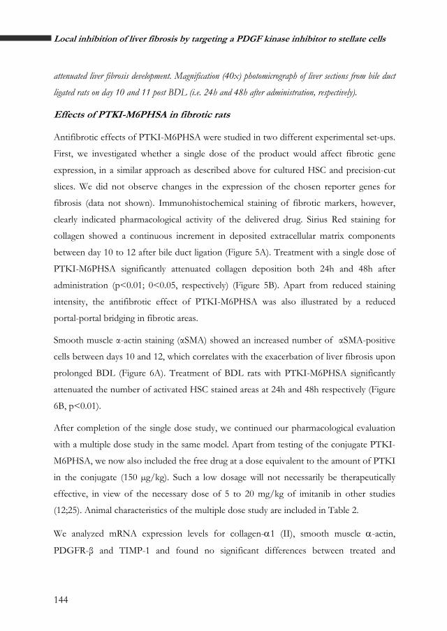
attenuated liver fibrosis development. Magnification (40x) photomicrograph of liver sections from bile duct
ligated rats on day 10 and 11 post BDL (i.e. 24h and 48h after administration, respectively).
fiEffects of PTKI-M6PHSA in brotic rats
Antifibrotic effects of PTKI-M6PHSA were studied in two different experimental set-ups.
First, we investigated whether a single dose of the product would affect fibrotic gene
expression, in a similar approach as described above for cultured HSC and precision-cut
slices. We did not observe changes in the expression of the chosen reporter genes for
fibrosis (data not shown). Immunohistochemical staining of fibrotic markers, however,
clearly indicated pharmacological activity of the delivered drug. Sirius Red staining for
collagen showed a continuous increment in deposited extracellular matrix components
between day 10 to 12 after bile duct ligation (Figure 5A). Treatment with a single dose of
PTKI-M6PHSA significantly attenuated collagen deposition both 24h and 48h after
administration (p<0.01; 0<0.05, respectively) (Figure 5B). Apart from reduced staining
intensity, the antifibrotic effect of PTKI-M6PHSA was also illustrated by a reduced
portal-portal bridging in fibrotic areas.
Smooth muscle α-actin staining (αSMA) showed an increased number of αSMA-positive
cells between days 10 and 12, which correlates with the exacerbation of liver fibrosis upon
prolonged BDL (Figure 6A). Treatment of BDL rats with PTKI-M6PHSA significantly
attenuated the number of activated HSC stained areas at 24h and 48h respectively (Figure
6B, p<0.01).
After completion of the single dose study, we continued our pharmacological evaluation
with a multiple dose study in the same model. Apart from testing of the conjugate PTKI-
M6PHSA, we now also included the free drug at a dose equivalent to the amount of PTKI
in the conjugate (150 µg/kg). Such a low dosage will not necessarily be therapeutically
effective, in view of the necessary dose of 5 to 20 mg/kg of imitanib in other studies
(12;25). Animal characteristics of the multiple dose study are included in Table 2.
We analyzed mRNA expression levels for collagen-α1 (II), smooth muscle α-actin,
PDGFR-β and TIMP-1 and found no significant differences between treated and

145
Chapter 6
untreated animals of study. Also collagen deposition and α- SMA expression was not
different between those groups (Table 3).
t
l
0
5000
10000
15000
20000
25000
30000
BDL + saline BDL + PTKI-M6PHSA
SM
A st
aini
ng:
area
sta
ined
per
live
r bio
sy bdl day 10 bdl day 11bdl day 12
**
A
B
BD
L da
y 11
B
DL
day
12
Figure 6. Effect of PTKI-M6PHSA single adminis ration to BDL rats on the
accumulation of myofibrob asts and activated HSC, as assessed by smooth muscle
α-actin expression (αSMA).
A. Quantification of the area with αSMA staining in rat liver specimens (n = 25, mean±sd ttest
between saline and drug treated groups:*P<0.01 vs. BDL saline control).
B. Liver sections were processed for immunohistochemistry and then stained with anti-αSMA antibody.
Rats receiving saline (left panels) showed a marked accumulation of αSMA-positive cells, which co-
localized with areas with active fibrogenesis. However, rats treated with PTKI-M6PHSA (right panels)
showed less quantity of αSMA-positive cells. Magnification (40x) photomicrograph of liver sections from
bile duct ligated rats on day 10 and 11 post BDL (i.e. 24h and 48h after administration, respectively)
Chapter 6

146
Local inhibition of liver fibrosis by targeting a PDGF kinase inhibitor to stellate cells
Discussion
Liver fibrosis is in principle a reversible process in which the stellate cells have been
identified as the key fibrogenic cells (26). PDGF is the most potent mitogen for HSC in
vitro, and it plays an important role in the transformation of HSC into myofibroblast-like
cells in vivo (27). Targeting the PDGF signalling cascade therefore represents a promising
antifibrotic approach. In the present study, we employed a kinase inhibitor with a closely
related structure to imatinib (Gleevec, Novartis). The antifibrogenic properties of the
latter well-known kinase inhibitor have already been described (10;12). On the other hand,
concerns have been raised in relation to the effectiveness of imatinib as an antifibrogenic
drug (13), especially in later stages of fibrosis. The new product PTKI-M6PHSA was
designed to effectively and specifically deliver the inhibitor to HSC, in order to improve
its antifibrotic properties. In the present study, we demonstrate the antifibrogenic effects
of PTKI-M6PHSA in two different and validated in vitro systems and in the BDL model
of liver fibrosis.
We confirmed that PTKI showed a similar antifibrotic potential as imatinib by incubating
the drug with culture-activated HSC and precision-cut liver slices. In both test systems,
PTKI potently inhibited the gene expression of fibrotic markers. The observed effect is
presumably attributed to the blockade of PDGF’s profibrogenic and mitogenic actions
since the growth factor is synthesized in situ by the activated HSC in culture or in slices
(28). Furthermore, PTKI is a very potent inhibitor of PDGFR-kinase with an IC50 of 10
nM (15). We also found that PTKI reduced PDGFR-β and TIMP-1 expression. TIMP-1
expression is increased during liver fibrosis and plays an important role in liver
fibrogenesis by modulating extracellular matrix remodelling (29). More importantly,
PTKI-M6PHSA had similar pharmacological effects as PTKI in activated HSC and
fibrotic liver slices. These results suggest that the drug is effectively released from the
carrier during the incubation period, and is capable of inhibiting the PDGF-R kinase
pathway.

147
Chapter 6
Having established the antifibrotic potential of the conjugate, we confirmed in vivo homing
of PTKI-M6PHSA from the circulation to the fibrotic liver. Similar to findings with other
drug-M6PHSA conjugates (19;30) we detected its presence in non-parenchymal cells by
anti-HSA immunostaining. These findings suggest that PTKI-M6PHSA is mainly
delivered within the fibrotic organ in BDL rats and therefore is associated with HSC.
Assuming that PTKI-M6PHSA behaves similar to other drug targeting preparations that
encompass the ULS linker, we can expect a slow release of PTKI during days rather than
hours, providing continuous local drug levels (19) (Prakash, SB-LZM paper2006,
Temming RGD-PEG-SB-HSA paper2006).
Several approaches for a blockade of the PDGF signaling have been investigated as a
strategy to block the ongoing liver fibrotic process. Some papers showed that monoclonal
antibodies directed against the extracellular domain of the PDGF receptor can prevent
binding of the PDGF ligand, thereby inhibiting mitogenic signaling (31;32). Consistent
with this, Kinnman and colleagues (9) described that imatinib inhibited PDGF-BB
induced proliferation of HSC in vitro, but a short-term treatment in vivo did not completely
blunt HSC proliferation 48 hours after BDL. In a different strategy, Borkham-Kamphorst
and colleagues produced a soluble antifibrogenic protein drug, sPDGFRB, and observed a
reduction in ECM deposition after daily administration fourteen days after BDL (33).
According to Neef and colleagues (13) this effect of imatinib is limited to the early phase
of fibrogenesis, due to the lack of efficiency in advanced liver injury, i.e., 3 to 4 weeks
after BDL. In contrast to this latter observtions, there is another study where a
pronounced inhibition of SMA-positive cells is found after 8 weeks of imatinib
administration (12). These authors found a marked attenuation of the liver fibrosis in the
pig-serum induced model of liver fibrosis, characterized by a slow progression of
fibrogenesis, resembling the human situation. Thus, it might be a relevant therapy in
patients with early stages of liver fibrosis.
Our strategy differs completely from the above listed studies in which the free drug
imatinib is used (10;12). We employed a carrier, M6PHSA that is internalized by the target
HSC and releases the kinase inhibitor inside the cells.
Chapter 6

148
Local inhibition of liver fibrosis by targeting a PDGF kinase inhibitor to stellate cells
After single administration, PTKI-M6PHSA markedly reduced collagen deposition
together with a suppression of αSMA-positive cells number which reflects activated HSC
(34). In contrast to the effects on protein levels, PTKI-M6PHSA administered to BDL
animals did not affect the gene expression of the fibrotic markers. This discrepancy either
reflects different sensitivity of the assays, or corresponds to a greater reduction of collagen
and αSMA at the protein level than at gene expression level. This divergence has
previously been described in experiments investigating the inhibition of hepatic fibrosis
(35), and might be due to post-transcriptional regulation of collagen expression in cultured
HSC (36).
The observation that antifibrotic effects are discernable even 48h after a single dose
suggests that effective PTKI levels are present during a prolonged period of time. In
previous studies with other ULS-based conjugates, we have observed a slow drug release
pattern from the ULS linker that may afford continuous drug release during a period of
days (19). The proposed mechanism is competitive displacement of the drug by S-donor
ligands like glutathione and ligands containing sulfur atoms (e.g. methionine) or aromatic
nitrogens (nucleotides, histidine). After binding of PTKI-M6PHSA to HSC and its
internalization, the release of PTKI from the conjugate is a critical point for exerting the
inhibitory effect on PDGF-R kinase. The release of PTKI may occur in the lysosomes, the
compartment to which M6PHSA is routed after internalization (37). Hence, a continued
release of PTKI from the conjugate would be available and a sustained blockade of
PDGF kinase activity may be achieved. In the present study, PTKI-M6PHSA produced a
significant effect on the fibrotic process 24 to 48 hours after its administration to BDL
rats. These results are in good agreement with the previously hypothesized release kinetics
of our drug-conjugates. Furthermore, it suggests the possibility of employing PTKI-
M6PHSA as a depot from which the drug is released for a prolonged period of time.
Our data are in good agreement with the studies of Neef and colleagues. Yet, after
multiple dose administration of PTKI-M6PHSA, the collagen and αSMA protein
expression was not decreased while the results mentioned above show that the process of
liver fibrosis development is clearly decreased by a single dose of PTKI-M6PHSA. How

149
Chapter 6
can this unexpected lack of effect after repeated administration of our drug-targeting
conjugate be explained? We propose that this may be due to the activation of alternative
cytokine pathways that compensates for the inhibition of PDGF-R cascade (38).
Parameter analyzed
BDL day 10 + control
saline
BDL day 14 + control
saline
BDL day 14 + PTKI-M6PHSA
BDL day 14 + PTKI
GAPDH 1.1 ± 0.4 1.3 ± 0.6 0.8 ± 0.2 1.3 ± 0.4
α-SMA 0.8 ± 0.4 1.1 ± 0.7 1.2 ± 0.3 1.1 ± 0.3
Collagen
0.8 ± 0.3 1.3 ± 0.3 1.2 ± 0.4 1.2 ± 0.5
Gene expression
(fold induction)
PDGF Receptor 1.0 ± 0.3 3.2 ± 2.4 3.2 ± 1.9 2.6± 0.6
Sirius Red staining ++ +++ +++ +++
Protein expression
(staining intensity)
αSMA staining
++ +++ +++ +++
Table 3. Effect of PTKI-M6PHSA multiple administration to BDL rats. Data are
shown as mean ± SD.
Redundancy of regulatory pathways is a common feature in the fibrotic process.
Especially if the causative agent of liver fibrosis is not removed, the specific inhibition of
PDGF-R on the hepatic stellate cells may temporarily affect the proliferation of HSC and
collagen deposition, as we observed after single injection of PTKI-M6PHSA, but not after
prolonged administration of PDGF-R inhibitor. Long-term administration of PTKI-
M6PHSA may allow enough time for the activation of alternative pathways, such as TGF-
Chapter 6

150
Local inhibition of liver fibrosis by targeting a PDGF kinase inhibitor to stellate cells
β, that may compensate for the inhibition of PDGF cascade (13). Albeit other
compensatory mechanisms are also possible, this would also suggest that the combined
elimination of more than one pathway could lead to an effective inhibition of hepatic
fibrosis (33). For example, simultaneous inhibition of PDGF and TGF-β cascade might
be more appropriate strategy to attenuate the process of liver fibrosis. Recent experiments
explaining this approach showed that it was more effective than inhibition of either
pathway alone (39). A TGF-β receptor kinase inhibitor has been described and cell-
selective delivery of this construct to the kidney is now being examined in our laboratory
(Prakash, manuscript in preparation). Consequently, targeting both cascades would be an
eligible approach in future investigations.
In conclusion, the data presented in the present study provide evidence that PTKI-
M6PHSA is effective in vitro and specifically taken up by non-parenchymal cells in the
liver, during fibrosis. Single dose administration of PTKI-M6PHSA proved to be an
effective antifibrotic strategy in vivo, although prolonged administration did not reveal
significant effects. Cell-selective elimination of multiple signalling pathways may provide a
tool to design novel strategies.
Acknowledgements
This study was supported by grants from SenterNovem (TSGE1083), NWO Science
Netherlands (R 02-1719, 98-162). Jan Visser and Jai Prakash (University of Groningen,
The Netherlands) are kindly acknowledged for assistance in HPLC analysis and colleagues
at Kreatech for critical reading of the manuscript.

151
Chapter 6
Reference List
1. Tsukada,S., Parsons,C.J., and Rippe,R.A. 2006. Mechanisms of liver fibrosis. Clin. Chim. Acta 364:33-60.
2. Friedman,S.L. 1999. The virtuosity of hepatic stellate cells. Gastroenterology 117:1244-1246.
3. Pinzani,M. 2002. PDGF and signal transduction in hepatic stellate cells. Front Biosci. 7:d1720-d1726.
4. Wong,L., Yamasaki,G., Johnson,R.J., and Friedman,S.L. 1994. Induction of b-platelet-derived growth factor receptor in rat hepatic lipocytes during cellular activation in vivo and in culture. J. Clin. Invest.1563-1569.
5. De Bleser,P.J., Jannes,P., Van Buul-Offers,S.C., Hoogerbrugge,C.M., Van Schravendijk,C.F.H., Niki,T., Rogiers,V., Van den Brande,J.L., Wisse,E., and Geerts,A. 1995. Insulinlike growth factor-II/mannose 6-phosphate receptor is expressed on CCl4-exposed rat fat-storing cells and facilitates activation of latent transforming growth factor-b in cocultures with sinusoidal endothelial cells. Hepatol 21:1429-1437.
6. Weiner,J.A., Chen,A., and Davis,B.H. 2000. Platelet-derived growth factor is a principal inductive factormodulating mannose 6-phosphate/insulin-like growth factor-II receptorgene expression via a distal E-box in activated hepatic stellate cells. Biochem. J. 345 Pt 2:225-231.
7. Bachem,M.G., Meyer,D., Schäfer,W., Riess,U., Melchior,R., Sell,K.M., and Gressner,A.M. 1993. The response of rat liver perisinusoidal lipocytes to polypeptide growth regulator changes with their transdifferentiation into myofibroblast-like cells in culture. J. Hepatol. 18:40-52.
8. von,M.M. 2005. Targeted therapy with imatinib: hits and misses? J. Clin. Oncol. 23:8-10.
9. Kinnman,N., Goria,O., Wendum,D., Gendron,M.C., Rey,C., Poupon,R., and Housset,C. 2001. Hepatic stellate cell proliferation is an early platelet-derived growth factor-mediated cellular event in rat cholestatic liver injury. Lab Invest 81:1709-1716.
10. Buchdunger,E., Cioffi,C.L., Law,N., Stover,D., Ohno-Jones,S., Druker,B.J., and Lydon,N.B. 2000. Abl protein-tyrosine kinase inhibitor STI571 inhibits in vitro signal transduction mediated by c-kit and platelet-derived growth factor receptors. J. Pharmacol. Exp. Ther. 295:139-145.
11. Buchdunger,E., Zimmermann,J., Mett,H., Meyer,T., Muller,M., Regenass,U., and Lydon,N.B.
1995. Selective inhibition of the platelet-derived growth factor signal transduction pathway by a protein-tyrosine kinase inhibitor of the 2-phenylaminopyrimidine class. Proc. Natl. Acad. Sci. U. S. A 92:2558-2562.
12. Yoshiji,H., Noguchi,R., Kuriyama,S., Ikenaka,Y., Yoshii,J., Yanase,K., Namisaki,T., Kitade,M., Masaki,T., and Fukui,H. 2005. Imatinib mesylate (STI-571) attenuates liver fibrosis development in rats. Am. J. Physiol Gastrointest. Liver Physiol 288:G907-G913.
13. Neef,M., Ledermann,M., Saegesser,H., Schneider,V., Widmer,N., Decosterd,L.A., Rochat,B., and Reichen,J. 2006. Oral imatinib treatment reduces early fibrogenesis but does not prevent progression in the long term. J. Hepatol. 44:167-175.
14. Bataller,R., and Brenner,D.A. 2005. Liver fibrosis. J. Clin. Invest 115:209-218.
15. Zimmermann,J., Buchdunger,E., Mett,H., Meyer,T., and Lydon,N.B. 1997. Potent and selective inhibitors of the abl-kinase:phenylamino-pyrimidine (PAP) derivatives. Bioorganic & Medicinal Chemistry Letters 7:187-192.
16. Beljaars,L., Meijer,D.K., and Poelstra,K. 2002. Targeting hepatic stellate cells for cell-specific treatment of liver fibrosis. Front Biosci. 7:e214-e222.
17. Beljaars,L., Molema,G., Weert,B., Bonnema,H., Olinga,P., Groothuis,G.M.M., Meijer,D.K.F., and Poelstra,K. 1999. Albumin modified with mannose 6-phosphate: A potential carrier for selective delivery of antifibrotic drugs to rat and human hepatic stellate cells. Hepatol 29:1486-1493.
18. van Gijlswijk,R.P., Talman,E.G., Janssen,P.J., Snoeijers,S.S., Killian,J., Tanke,H.J., and Heetebrij,R.J. 2001. Universal Linkage System: versatile nucleic acid labeling technique. Expert. Rev. Mol. Diagn. 1:81-91.
19. Gonzalo,T., Talman,E.G., van,d., V, Temming,K., Greupink,R., Beljaars,L., Reker-Smit,C., Meijer,D.K., Molema,G., Poelstra,K. et al 2006. Selective targeting of pentoxifylline to hepatic stellate cells using a novel platinum-based linker technology. J. Control Release 111:193-203.
20. Beljaars,L., Weert,B., Geerts,A., Meijer,D.K.F., and Poelstra,K. 2003. The preferential homing of a platelet derived growth factor receptor-recognizing macromolecule to fibroblast-like cells in fibrotic tissue. Biochem. Pharmacol. 66:1307-1317.
21. Geerts,A., Niki,T., Hellemans,K., De Craemer,D., Van den Berg,K., Lazou,J.-M., Stange,G., Van de Winkel,M., and De Bleser,P.J.
Chapter 6

152
Local inhibition of liver fibrosis by targeting a PDGF kinase inhibitor to stellate cells
1998. Purification of rat hepatic stellate cells by side scatter-activated cell sorting. Hepatol 27:590-598.
22. Olinga,P., Merema,M.T., de Jager,M.H., Derks,F., Melgert,B.N., Moshage,H., Slooff,M.J., Meijer,D.K., Poelstra,K., and Groothuis,G.M. 2001. Rat liver slices as a tool to study LPS-induced inflammatory response in the liver. J. Hepatol. 35:187-194.
23. van de,B.M., Groothuis,G.M., Draaisma,A.L., Merema,M.T., Bezuijen,J.I., van Gils,M.J., Meijer,D.K., Friedman,S.L., and Olinga,P. 2005. Precision-cut liver slices as a new model to study toxicity-induced hepatic stellate cell activation in a physiologic milieu. Toxicol. Sci. 85:632-638.
24. Beljaars,L., Poelstra,K., Molema,G., and Meijer,D.K.F. 1998. Targeting of sugar- and charge-modified albumins to fibrotic rat livers: the accessibility of hepatic cells after chronic bile duct ligation. J. Hepatol. 29:579-588.
25. Neef,M., Ledermann,M., Saegesser,H., Schneider,V., Widmer,N., Decosterd,L.A., Rochat,B., and Reichen,J. 2006. Oral imatinib treatment reduces early fibrogenesis but does not prevent progression in the long term. J. Hepatol. 44:167-175.
26. Friedman,S.L. 2003. Liver fibrosis -- from bench to bedside. J. Hepatol. 38 Suppl 1:S38-S53.
27. Pinzani,M., Milani,S., Grappone,C., Weber,F.L., Gentilini,P., and Abboud,H.E. 1994. Expression of Platelet-Derived Growth Factor in a Model of Acute Liver Injury. Hepatol 19:701-707.
28. Pinzani,M. 2002. PDGF and signal transduction in hepatic stellate cells. Front Biosci. 7:d1720-d1726.
29. Iredale,J.P., Benyon,R.C., Pickering,J., McCullen,M., Northrop,M., Pawley,S., Hovell,C., and Arthur,M.J.P. 1998. `Mechanisms of spontaneous resolution of rat liver fibrosis - Hepatic stellate cell apoptosis and reduced hepatic expression of metalloproteinase inhibitors. J. Clin. Invest. 102:538-549.
30. Greupink,R., Bakker,H.I., Reker-Smit,C., van Loenen-Weemaes,A.M., Kok,R.J., Meijer,D.K.F., Beljaars,L., and Poelstra,K. 2005. Studies on the targeted delivery of the antifibrogenic compound mycophenolic acid to the hepatic stellate cell. J. Hepatol. 43:884-892.
31. Heldin,C.-H. 1997. Simultaneous induction of stimulatory and inhibitory signals by PDGF. FEBS Lett. 410:17-21.
32. Shulman,T., Sauer,F.G., Jackman,R.M., Chang,C.N., and Landolfi,N.F. 1997. An antibody reactive with domain 4 of the platelet-derived growth factor beta receptor allows BB binding while inhibiting proliferation by impairing receptor dimerization. J. Biol. Chem. 272:17400-17404.
33. Borkham-Kamphorst,E., Herrmann,J., Stoll,D., Treptau,J., Gressner,A.M., and Weiskirchen,R. 2004. Dominant-negative soluble PDGF-beta receptor inhibits hepatic stellate cell activation and attenuates liver fibrosis. Lab Invest 84:766-777.
34. Olaso,E., and Friedman,S.L. 1998. Molecular regulation of hepatic fibrogenesis. J. Hepatol. 29:836-847.
35. Yata,Y., Gotwals,P., Koteliansky,V., and Rockey,D.C. 2002. Dose-dependent inhibition of hepatic fibrosis in mice by a TGF-beta soluble receptor: implications for antifibrotic therapy. Hepatology 35:1022-1030.
36. Stefanovic,B., Hellerbrand,C., Holcik,M., Briendl,M., Aliebhaber,S., and Brenner,D.A. 1997. Posttranscriptional regulation of collagen alpha1(I) mRNA in hepatic stellate cells. Mol. Cell Biol. 17:5201-5209.
37. Beljaars,L., Olinga,P., Molema,G., De Bleser,P., Geerts,A., Groothuis,G.M.M., Meijer,D.K.F., and Poelstra,K. 2001. Characteristics of the hepatic stellate cell-selective carrier mannose 6-phosphate modified albumin (M6P28-HSA). Liver 21:320-328.
38. Kinnman,N., Hultcrantz,R., Barbu,V., Rey,C., Wendum,D., Poupon,R., and Housset,C. 2000. PDGF-mediated chemoattraction of hepatic stellate cells by bile duct segments in cholestatic liver injury. Lab Invest 80:697-707.
39. Yoshiji,H., Kuriyama,S., Noguchi,R., Ikenaka,Y., Yoshii,J., Yanase,K., Namisaki,T., Kitade,M., Yamazaki,M., Asada,K. et al 2006. Amelioration of liver fibrogenesis by dual inhibition of PDGF and TGF-beta with a combination of imatinib mesylate and ACE inhibitor in rats. Int. J. Mol. Med. 17:899-904.


2
costa da mote
boektitel
In ancient times, Romans called the Galician coast in Spain along the n
tip of the Iberia
orthwestern
n Peninsula " Finisterrae" (End of the Land), and in modern
osta da Morte, Finisterre, La Coruña
times this coastal area is known as "Costa da Morte" (The Dead Coast).
Severe storms have destroyed many ships over the centuries.
C
Galicia, España.