university of california fall 2014 berkeley … 7, number 1 ... write my first message for the...
TRANSCRIPT

NINETY YEARS OF VISION SCIENCE RESEARCH
UNIVERSITY OF CALIFORNIA VOL. 7, NO. 1 | FALL 2014
m a g a z i n eBerkeley Optometry
ADVANCING EDUCATION AND RESEARCH AT BERKELEY OPTOMETRY

dean’s messageEXECUTIVE EDITOR
Lawrence Thal
MANAGING EDITOR
Kellie Rogers
EDITOR
LeeAnn Pickrell
ART DIRECTOR AND PROJECT MANAGER
Tom Sharp
WRITERS/CONTRIBUTORS
John Fiorillo, Karsten Gronert, Calista
Ming, Monica Rodriguez, Michelle Wong
DESIGN
ContentWorks, Inc.
Published by Berkeley Optometry,
University of California, Berkeley,
Development and Alumni Relations
Phone: 510-642-2622, Fax: 510-643-6583
Send comments, letters, class notes
submissions, and change of address
or e-mail to:
Submit Berkeley Optometry gifts online
at: givetocal.berkeley.edu/
makeagift/optometry
or mail to:
University of California, Berkeley
Berkeley Optometry Fund
P.O. Box 774
Berkeley, CA 94701-0774
510-642-1877
Fall 2014
Volume 7, Number 1
© 2014 Regents of the University of
California
Berkeley Optometry is a trademark of
University of California, Berkeley,
Berkeley Optometry.
Front cover design by ContentWorks, Inc.
Back cover photo courtesy of Grad Images.
I arrived at the School of Optometry, University of California, Berkeley, nearly six months ago; and
what a six months it has been. I am delighted to be here and delighted to have this opportunity to
write my first message for the Berkeley Optometry Magazine. Although it would be premature to
make any definitive announcements with regard to the direction I see us headed over the next ten years,
it seems like a good time to share some of my observations so far.
I have been overwhelmed by the support that Berkeley Optometry enjoys from its alumni. Over the
last month alone I have experienced a record-breaking alumni weekend with over 500 attendees at the
Saturday evening event, hosted a raucous reception at the Academy meeting in Denver, and enjoyed a
strident Optometry performance for the inaugural “Big Give”. I have been humbled by our students;
their intelligence, humility, sense of humor and intuitive sense of responsibility. The profession has a
great future in their hands. I have also been astonished by the talents and accomplishments of our faculty
and staff; they run the busiest, biggest and best clinics, offer exceptional student services, attract extraor-
dinary levels of research funding, and passionately teach to the highest possible standards.
I have discovered the joys of a summer concert at the Greek
Theater, a peaceful walk through Memorial Glade and past the
Wickson redwoods, the spectacle and emotion of college football,
enjoyed the chimes of the Campanile, and have felt fortunate to
have helped celebrate the 50th anniversary of the Free Speech
Movement. There is certainly no shortage of culture and stimulus
on this extraordinary campus.
It is well known that Berkeley Optometry historically has
been, and remains today, the top optometry school in the world.
No matter what metric you choose – qualifications of students,
national board results, scholarships and awards, quality of faculty,
quality of research, federal funding of vision research, or clinical
experience of our students—Berkeley is at the top. No other school
can boast the awards our faculty has achieved (e.g., 16 Prentice
Medals). These are all reasons why our faculty and students chose
to come here. These are some of the reasons that I am so proud to
be here.
It took very little time to realize some of the challenges facing
the School, problems we must overcome to maintain our position
of leadership. As we embark on a renovation of the ground floor of Old Minor Hall, it should be noted
that this will be the first substantial upgrade to this part of the School since we occupied the building
in 1949. Our clinic is majestic in its scope, efficiency and accomplishments, but does so in what is now
a wholly inadequate facility compared to its opening day. When you walk through our labs, offices,
teaching rooms and clinics in New Minor Addition, you note that little has been upgraded since the
building was completed in 1978. The elevators are unreliable, the ducting no longer circulates air, the
student facilities are cramped, the décor is tired, and the University is without capital and unable to
help. This is a significant challenge, but essential to address if we are to attract the next generation of
top faculty, staff and students.
I have been lucky to know (or know of) many of the legends from our past (Meredith Morgan, Mer-
ton Flom, Morton Sarver, Irving Fatt, Robert Mandell, Monroe Hirsch, Ian Bailey, Anthony Adams,
Gerald Westheimer, Elwin Marg, Jay Enoch, Lawrence Stark, Russell and Karen DeValois, Clifton
Schor, Ralph Freeman, Sir Colin Blakemore, Ken Polse, Arthur Jampolsky, the list goes on). Who are
the future legends? How will we attract and recruit them to Berkeley? This is our challenge, but also the
greatest of opportunities!
How we address these issues and maintain our standards of excellence, will be a substantial part of
our strategic plan moving forward. I look forward to working with you to solve these problems, make
the most of our opportunities, and to having Berkeley Optometry emerge as an even greater institution.
This will be a truly collaborative effort. Please stay tuned as we develop our plans and strategies. I look
forward to getting to know you, discovering your communities, hearing your stories and celebrating
our future achievements with you.
JOHN G. FLANAGAN, OD, PhD, FAAODean, School of Optometry Professor of Optometry and Vision Science

3 Albert Reinke, Oakland optometrist and university lecturer, in 1932 with his experimental perimeter as he tests peripheral vision.
humanitarian efforts
school support
on-campus research stay connected
UNIVERSITY OF CALIFORNIA VOL. 7, NO. 1 | FALL 2014
m a g a z i n eBerkeley Optometry
optometry.berkeley.edu | 1
3 VISION SCIENCE AT BERKELEY By John Fiorillo and Karsten Gronert
14 VOSH NICARAGUA By Monica Rodriguez and Calista Ming
16 LOCAL OUTREACH By Michelle Wong
18 CAMPAIGN SUMMARY
19 CAMPAIGN FOR BERKELEY BY THE NUMBERS
20 A FISCAL SNAPSHOT
21 OPTOMETRY ASSOCIATES OF THE BENJAMIN IDE WHEELER SOCIETY
22 THE MEREDITH W. MORGAN SOCIETY
23 DONOR PROFILE: DR. JOY OHARA AND KOK LOONG LYE
24 NEWS & NOTES
26 AWARDS
28 STUDENT SPOTLIGHT
30 FACULTY FOCUS
32 CLASS NOTES
36 ALUMNI FOCUS
38 HALL OF FAME
42 STUDENT AFFAIRS
43 CONTINUING EDUCATION
45 CLINIC ADMINISTRATOR TEAM

2 | BERKELEY OPTOMETRY FALL 2014
IT IS HARD TO BELIEVE that it has been nearly nine years since I retired from my private practice and was asked to join the Berkeley Optometry team, to lead the recently completed campaign for Berkeley Optometry. I had thought then that I would serve until Dean Levi retired after he completed 10 years as dean. As it happened, Dean Levi served 13 years; and my time as Assistant Dean also was extended. It is now time to begin my delayed retirement, but not without taking this opportunity to thank all of you for making this experience one of the most enjoyable and gratifying of my life. The time has flown, and my time in this position has been extraordinarily rewarding.
It has been a privilege to serve Berkeley Optometry in a number of positions since 1978: 1) as a clinical professor member of the adjunct faculty; 2) as a lecturer teaching in the practice management curriculum; and 3) as Assistant Dean for Development and External Relations. I am particularly grateful to the University for the once-in-a-lifetime opportunity to help lead, as part of the Campaign Management Team, the recently completed and enormously successful “Campaign for Berkeley.” All the goals accomplished here over the duration of this campaign and the groundwork that is in place for future success are due to the outstanding efforts and dedication of our excellent staff and faculty, as well as the philanthropic support of our alumni, students and friends of the school. I am honored to have served with each of these groups.
I am leaving Berkeley Optometry with a much broader and more profound appreciation for all that Cal and Berkeley Optometry are and for all that they promise to be. I have developed a genuine appreciation and respect for the thousands of alumni, faculty, staff and students who contribute daily to our institution and who do so in many diverse ways. Change is good for any organization; Berkeley Optometry is no exception. While I certainly will miss seeing so many of you regularly and working daily with the outstanding staff we have assembled, as I told my patients when I retired from private practice, I look forward (finally) to spending more time traveling with family and friends. I am especially appreciative for the opportunity I have had, in some small way, to “pay back” for the opportunities afforded me as a result of that very fortunate day in 1971 when I received my acceptance letter to be part of the Class of 1975 of Berkeley Optometry!
Again, thank you all for helping to deepen my love and appreciation for this amazing institution. Our alumni and friends will play an ever-increasing role in the ongoing success of our school, and I am extremely proud to have played a small part in that effort. Esther and I will take away countless fond memories, and we are deeply indebted to those reading this message for enriching our lives.
editor’s message
LAWRENCE THAL, OD, MBA, FAAO
Assistant Dean, Development & Alumni RelationsSchool of [email protected]

on-campus research
optometry.berkeley.edu | 3
F OR NINETY YEARS after the
founding of Berkeley Optometry in
1923, faculty, scientists and alumni
have been conducting groundbreaking
clinical and basic vision science research.
Their studies have ranged from the early
days of optical instrument design and
pioneering contact lens research to the
complex multidisciplinary investigations that
characterize the modern era. Today’s vision
scientists work in such diverse fields of
basic science as visual neuroscience and
perception, spatial navigation, psychophysics,
computational vision, molecular and cell
biology, neurophysiology, bioengineering,
robotics, biochemistry, contact lenses,
refractive development, ocular surface
biochemistry, ocular infection, ocular
diseases and immunology. An important
goal for many research groups in the Vision
Science Program is the application of basic
science discoveries to the development of
studies and clinical trials in humans. These
“translational research” efforts are an
essential pipeline for developing new clinical
diagnoses and treatments.
Early Years of Clinical Research
Instrument design was a key component of early research
in optometry. In 1928 Frederick Mason (clinical faculty) made
one of the first haploscopes in the U.S. for the study of
binocular vision, and in the 1930s he refined its functionality by
introducing a “synchrohaploscope” to treat strabismus. Albert
Reinke (’26), lecturer in optometry, designed a perimeter in
1932 to test visual fields and to obtain data for assessing general
health through ocular manifestations of systemic diseases.
Early contact lens design was advanced by Dr. Edward
Goodlaw’s (’34) research confirming that contact lenses were
barriers to anterior oxygen supply (1946). He and Dr. Solon
Braff (’37) also helped design the first corneal contact lens
(patented by Kevin Tuohy in 1948).
In 1959, Professor Elwin Marg (OD ’40, PhD ’50), the first
to graduate from Berkeley Optometry’s Physiological Optics
Graduate Program (now called the Vision Science Program),
designed the Mackay-Marg Tonometer (with R. Stuart Mackay,
electrical engineer, UC Berkeley). The instrument enabled
optometrists to measure intraocular pressure without topical
anesthetics (legally off-limits to optometrists at the time),
initiating a significant advance for the optometry profession by
broadening its scope of practice.
Research During the Middle Years
VISUAL ANATOMY
Professor Gordon Walls, PhD, was the first vision scientist with-
out an OD degree appointed to the tenured Optometry faculty
(1947–62). He was a great synthesizer of scientific knowledge—
his The Vertebrate Eye & Its Adaptive Radiation (1942) remains
a classic text. Dr. Walls helped excite interest in vision science
and advance the Physiological Optics Graduate Program by
publishing papers on subjects such as the fundamentals of
vision, color vision, ocular dominance and physiological optics.
He was also a legendary lecturer and ambassador for vision
Vision Science at BerkeleyBy John Fiorillo and Karsten Gronert
on-campus research
primate subcortical brain, developing methods now widely
used or adapted in laboratories worldwide. He showed that
transformation of the color signal was derived from three types
of broadly selective cone photoreceptors into an opponent
color organization, so that certain neurons might be activated
by the light of one color but inhibited by a different color. His
research validated a theory supporting the perception of four
unique hues by a fundamentally trichromatic system, which
had been debated for a century. Often in collaboration with
Professor Karen De Valois (Psychology and Vision Science),
he also focused on early stages of spatial vision, in particular
how perception varies with the resolution of perceived objects.
They discovered that cortical cells were tuned to specific limited
bands of spatial frequency and orientation. Their influential
book Spatial Vision (1988) summarized more than two decades
of research.
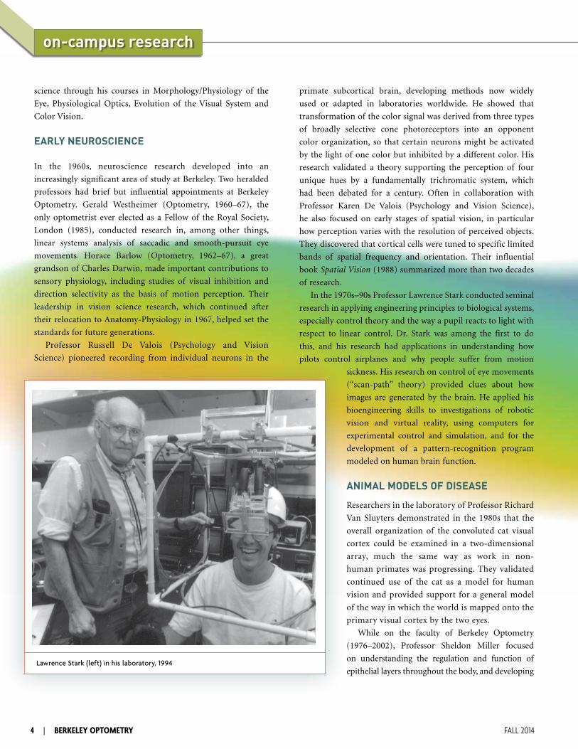
In the 1970s–90s Professor Lawrence Stark conducted seminal
research in applying engineering principles to biological systems,
especially control theory and the way a pupil reacts to light with
respect to linear control. Dr. Stark was among the first to do
this, and his research had applications in understanding how
pilots control airplanes and why people suffer from motion
sickness. His research on control of eye movements
(“scan-path” theory) provided clues about how
images are generated by the brain. He applied his
bioengineering skills to investigations of robotic
vision and virtual reality, using computers for
experimental control and simulation, and for the
development of a pattern-recognition program
modeled on human brain function.
ANIMAL MODELS OF DISEASE
Researchers in the laboratory of Professor Richard
Van Sluyters demonstrated in the 1980s that the
overall organization of the convoluted cat visual
cortex could be examined in a two-dimensional
array, much the same way as work in non-
human primates was progressing. They validated
continued use of the cat as a model for human
vision and provided support for a general model
of the way in which the world is mapped onto the
primary visual cortex by the two eyes.
While on the faculty of Berkeley Optometry
(1976–2002), Professor Sheldon Miller focused
on understanding the regulation and function of
epithelial layers throughout the body, and developing
science through his courses in Morphology/Physiology of the
Eye, Physiological Optics, Evolution of the Visual System and
Color Vision.
EARLY NEUROSCIENCE
In the 1960s, neuroscience research developed into an
increasingly significant area of study at Berkeley. Two heralded
professors had brief but influential appointments at Berkeley
Optometry. Gerald Westheimer (Optometry, 1960–67), the
only optometrist ever elected as a Fellow of the Royal Society,
London (1985), conducted research in, among other things,
linear systems analysis of saccadic and smooth-pursuit eye
movements. Horace Barlow (Optometry, 1962–67), a great
grandson of Charles Darwin, made important contributions to
sensory physiology, including studies of visual inhibition and
direction selectivity as the basis of motion perception. Their
leadership in vision science research, which continued after
their relocation to Anatomy-Physiology in 1967, helped set the
standards for future generations.
Professor Russell De Valois (Psychology and Vision
Science) pioneered recording from individual neurons in the
4 | BERKELEY OPTOMETRY FALL 2014
Lawrence Stark (left) in his laboratory, 1994
animal models of retinal disease to help establish therapeutic
interventions. At the National Eye Institute (NIH) since 2002,
Dr. Miller has continued and expanded his research from Berkeley,
investigating plasma membrane proteins and intracellular signaling
pathways that mediate human retinal pigment epithelial (RPE) cell
physiology and its interactions with retinal photoreceptors. His
work has led to further pre-clinical animal models of disease,
including retinal re-attachment and choroidal neovascularization
in age-related macular degeneration. His lab identified micro
RNAs enriched in human RPE, compared to adjacent retina and
choroid, showing how they help maintain tight junction integrity,
RPE immune responses and epithelial phenotype. Miller’s lab
also identified 154 genes that distinguish native human RPE from
practically all other cells.
CONTACT LENSES
The heyday of contact lens research took place in the 1960s–80s,
when Berkeley Optometry was virtually the center of the contact
lens universe. Professor Irving Fatt (Engineering and Optometry),
a research chemist and petroleum engineer, began collaborating
with Optometry faculty in 1963 when he applied his expertise
in fluid flow through porous media to seminal investigations
regarding oxygen supply to the cornea (oxygen transmissibility).
Professor Fatt and Professor Richard Hill were the first to
quantify oxygen uptake in living human cornea (1963), the
genesis for decades of related cornea and contact lens research,
including the first corneal deswelling studies in humans (Fatt and
Professor Emeritus Robert Mandell) and the research of Professor
Emeritus Kenneth Polse on quantitative markers for corneal
function. Professor Fatt also invented an oxygen sensor (1976) for
measuring oxygen permeability/transmissibility of contact lenses.
Professor Polse, in collaboration with Professor Joseph Bonanno,
later developed a technique to measure in vivo corneal pH, which
led to pioneering work for a safe and accurate measurement
(fluorometry), confirming that corneal pH (corneal acidosis)
increased during contact lens wear and thereby altered corneal
endothelial morphology. Professor Polse’s experiments were
among the earliest clinical patient-based trials in translational
vision research.
LOW VISION AND VISION WITH AGING
In the 1960s–70s, clinical faculty Edwin Mehr (’41) and Allan
Freid (’52) advanced understanding and training in low vision
at Berkeley and published their influential book Low Vision Care
(1975). Professor Emeritus Ian Bailey joined the faculty in 1976,
a year after creating the famed Bailey-Lovie LogMAR chart for
optometry.berkeley.edu | 5
testing visual acuity (VA), the gold standard for scoring VA in
clinical research. A seminal paper in 1982 showed that visual acuity
was a poor predictor of mobility performance and that contrast
sensitivity and visual fields were far more important. More recently,
in 2012, Professor Bailey and colleagues introduced the Berkeley
Rudimentary Vision Test for clinical assessment of ultra low vision.
In the 1980s Professor Gunilla Haegerström-Portnoy
conducted research on visual function with age-related
maculopathy, and by 1992 she focused much of her research on
vision and aging, an area in need of expanded investigation at a
time when virtually the only well-researched information about
people over 70 years of age was visual acuity. She explored aging
in relation to visual and physical functional ability (including
reading, mobility and vehicular driving ability), development
of refractive errors, low-contrast vision function and face
recognition in the elderly.
New Era of Vision Science ResearchContemporary multidisciplinary vision science research has
focused on two broad categories: the exploration of neural
functioning and development of the visual system; and the
discovery of mechanisms, diagnostics and new treatments for
ocular diseases, as well as defining how ocular surface health
is maintained, employing experimental models and clinical
research. Integration of these two areas and interactions with the
UCB School of Optometry Clinical Research Center provides a
unique framework for translational research. Researchers in the
Vision Science Program are composed of faculty in Optometry
and affiliated faculty in other schools and departments on
the UC Berkeley campus. These include Bioengineering,
Molecular & Cell Biology, Psychology, Public Health and
Computer Science, as well as vision science researchers affiliated
with the Helen Wills Neuroscience Institute, which integrates
multi-disciplinary neuroscience faculty across the University,
including some Optometry faculty.
VISUAL PERCEPTION AND COMPUTER DESIGN AND MODELING
Professor Theodore Cohn conducted innovative research in
signal detection theory and its real-world applications during
the 1990s and 2000s. He was intrigued by the brain’s ability
to detect weak signals from noisy environments and was
among the first to apply the principle of signal detection to

6 | BERKELEY OPTOMETRY FALL 2014
on-campus research
physiological systems. Professor Cohn succeeded in quantifying
observations, showing that information transmission and its
reliability depended not only on the strength of the electrical
response but also on the reproducibility of the response.
Visual input often arrives in a noisy and discontinuous stream,
owing to head and eye movements, occlusion, lighting changes
and other factors. Yet the physical world is generally stable;
objects and physical characteristics rarely change spontaneously.
How does the human visual system capitalize on continuity in
the physical environment over time? The laboratory of Professor
David Whitney (Psychology) has found that visual perception
in humans is serially dependent, using both prior and present
input to inform perception at the present moment. In effect,
there is a “visual smoothing” whereby our perception of things
is influenced by what we saw up to 15 seconds before, resulting
in a time-averaged composite. Using an orientation judgment
task, researchers found that even when visual input changed
randomly over time, perceived orientation was strongly and
systematically biased toward recently seen stimuli. Furthermore,
the strength of this bias was modulated by attention and tuned
to the spatial and temporal proximity of successive stimuli. These
results reveal a serial dependence in perception characterized by
a spatiotemporally tuned, orientation-selective operator—which
the Whitney laboratory calls a “continuity field”—that may
promote visual stability over time.
How do humans see in three dimensions and what sort
of technological adaptations can we apply to enhance this
ability? Even though retinal images are two dimensional, we
perceive the three-dimensional structure of our environment.
Vision accomplishes this by using a set of depth cues and prior
expectations about the environment. An important cue is
stereopsis, the perception of depth from differences in images
from the two eyes. Researchers in the laboratory of Professor
Martin Banks have investigated perceptual distortions and
visual discomfort that occur when people view stereoscopic
displays, discovering that those problems can be minimized by
using different display and graphics technologies.
The laboratory of Professor Maneesh Agrawala (Electrical
Engineering and Computer Science) has investigated how
cognitive design principles can be used to improve the
effectiveness of visual displays, with the goal of discovering
design models for both interactive and automated design tools
for human-computer interactions.
Computer-aided geometric design and modeling has been
a main interest of Professor Brian Barsky (Computer Science).
Display devices such as mobile phones, tablets, laptops and
computer monitors can be difficult to see for those with vision
problems. Professor Barsky and his students and colleagues
are developing displays that correct vision and enable viewing
of displays. The system relies on either the patient’s spectacle
prescription, in simple cases, or measurements of the patient’s
optical aberrations, in more complex cases. The content on the
display device is digitally modified so that when it is viewed,
it will appear in sharp focus to the particular user. In addition
to correcting common optical problems
such as near-sightedness, far-sightedness
and astigmatism, Professor Barsky’s current
research is also investigating more challenging
vision deficiencies, or “higher order
aberrations,” which are impossible to correct
with eyeglasses. This research could potentially
give patients with such challenging vision
disabilities the ability to see these displays for
the first time.
Neurons in the visual cortex are selective
to local shape features such as orientation and
spatial frequency. Until recently it was not
understood how or if these feature-selective
properties could be quantitatively explained
by a theory of information processing. The
aforementioned Horace Barlow hypothesized
that neurons try to encode sensory
information by exploiting redundancies or
statistical dependencies arising from the
Photographs taken in the Banks Laboratory show how changes in lens focal length affect
interpretation of the same 3D scene.

optometry.berkeley.edu | 7
natural environment. One form of this hypothesis proposes that
neurons attempt to form a “sparse code” in which only a small
fraction of neurons are active at any given moment. Researchers
in the laboratory of Professor Bruno Olshausen are simulating
model neural circuits that adapt to natural image statistics to
form a sparse representation of visual input. The results make
predictions about the types of receptive fields that may be found
in the highly over-complete populations of neurons in layer 4 of
the primary visual cortex. Such representations are now widely
employed in branches of engineering for image processing and
object-recognition applications.
FUNCTIONAL MAGNETIC RESONANCE IMAGING (fMRI)
Understanding how the brain works and decoding its structure
and the function of the visual system is a primary interest
of Professor Jack Gallant (Psychology). Predictive models of
brain activity are critical for the long-term advancement of
neuroscience and medicine. The research program in the Gallant
laboratory integrates three distinct approaches: neuroscience
experiments involving both classical electrophysiology and
noninvasive functional magnetic resonance imaging (fMRI);
statistical analysis using methods adapted from nonlinear system
identification and nonlinear regression; and theoretical modeling.
Computational encoding models that can
predict brain activity have many potential
practical applications in medicine and beyond.
These models might also provide a new tool
for neurological evaluation and diagnosis, and
they can be inverted to decode brain activity,
providing a direct and principled way to do
“brain reading” and to build brain-machine
interfaces and neural prosthetics.
The laboratory of Professor Ralph
Freeman has explored behavioral and
neurophysiological studies of basic visual
function, neural organization of binocular
vision and neurometabolic coupling in
relation to fMRI. In its extended research on
the physiological organization of binocular
vision, Freeman’s lab first developed an
original stimulation approach of binocular
presentation, using phase-varying gratings to
yield a detailed and quantitative assessment
of binocular interaction. They identified
specific linear and nonlinear elements of
binocular organization for the two major cell classes in the visual
cortex. Using binocular interaction patterns, they investigated
mechanisms of visual deprivation, demonstrating that binocular
tests are required to show the full range of visual function
in a deprived system, whereas effects are not evident during
monocular tests. Freeman’s lab also developed an important and
wide-ranging theory of stereoscopic depth discrimination based
on a version of the energy model, used previously to describe
other visual functions. Experimental tests confirmed that
predictions from the model were nearly entirely consistent with
the collected data. In other research an innovative technique was
used to obtain detailed temporal and spatial analysis of receptive
field organization in the central visual pathway. In more recent
work, Professor Freeman has studied the basis of noninvasive
neural imaging by use of a combined sensor that provides
simultaneous co-localized measurements of oxygen and neural
activity in a specific region of the cerebral cortex. Studies in this
area have led to an increased understanding of signals generated
in fMRI.
Noninvasive fMRI technology localizes many aspects of
perception to small regions of the brain, but a major limitation
is its reliance on blood flow, with a temporal resolution on the
order of seconds. Researchers in the laboratory of Professor
Stanley Klein combined fMRI with electroencephalography
(EEG), bringing temporal resolution down to milliseconds,
An observer in the Klein laboratory views a satellite image scene while looking for
specific targets (photo 2008)

8 | BERKELEY OPTOMETRY FALL 2014
on-campus research
along with a new approach to EEG localization that for some
stimuli matches the spatial resolution of fMRI. Klein’s laboratory
tracked the mechanisms responsible for eye movements plus
the flow of brain activity in response to detecting a target. The
pattern of EEG responses, together with eye movement plus
pupil responses, detected substantially more targets than using
only the observer’s button press, with only a minor increase in
false-alarm rate that could be eliminated with a double pass
method while maintaining a higher hit rate and speeding up the
detection process.
Directing spatial attention to a specific location facilitates
the perception of objects and patterns at that location, but the
brain mechanisms of this phenomenon are unknown. In the
early 2000s, researchers in the laboratory of Professor Michael
Silver began using fMRI to measure differences in brain activity
when a visual stimulus is attended versus when attention is
directed away from the stimulus. They found that attending
to the stimulus not only increases the strength of the brain’s
response to that stimulus, but also reduces the strength of slow
internally generated fluctuations in brain activity. Surprisingly,
they discovered that the enhancement of perception by attention
is not correlated with effects of attention on the brain’s response
to the stimulus. Rather, it is the suppression of internally
generated fluctuations in brain activity that is associated with
improved perception by attention. A better understanding of
the brain mechanisms for attentional facilitation of perception
will be useful for improving visual-attention training paradigms
and for developing clinical treatments for attention disorders.
THE RETINAProfessor Marla Feller (Neurobiology/Molecular & Cellular
Biology) has investigated the mechanisms that guide the
assembly of visual circuits during development of the visual
system. Her laboratory has studied how immature retinal circuits
generate retinal waves—highly patterned spontaneous activity
in the immature retina—and what role this activity plays in the
development of the visual system. They have been using a variety
of physiologic imaging and modeling approaches to elucidate
how the synaptic mechanisms for generating spontaneously
correlated signals evolve as the retina reaches its mature wiring
state. They have also investigated maturation of circuits in the
retina that detect motion in the visual field. These direction-
selective circuits are intact at the onset of vision, indicating that
the precise retinal circuitry mediating direction selectivity is
“wired-up” prior to normal visual experience.
Professor Austin Roorda has developed advanced
ophthalmoscopes for clinical and basic science. The primary
tool in his research laboratory is the adaptive optics scanning
laser ophthalmoscope (AOSLO), a system that can provide
views of the retina in a living human eye on a microscopic scale,
and which Roorda has had a hand in inventing. On the clinical
side, he uses the AOSLO to better understand eye disease. He
is involved in clinical trials to evaluate new
treatments for retinal degeneration. On the
basic science side, Roorda uses the AOSLO and
other tools to visualize, track and target light
stimulation to individual cone photoreceptors
in an effort to answer long-standing questions
about human spatial and color vision.
Researchers in the laboratory of Professor
Emeritus Anthony Adams have discovered
that tiny delays in retinal nerve transmission
may indicate future diabetic retinopathy.
Using multifocal electroretinogram (mfERG),
they can predict future location-specific
retinopathy along with healthy patches of
retina. In 2011 they showed for the first time
that these neuroretinal delay measures are
predictive of the onset of retinopathy in eyes
that had no prior retinopathy. These early
neural changes in the retina caused by diabetes
have significant implications for patient
care and management of eye complications.
Ophthalmologists and optometrists, able
Professor Roorda aligning the optics in an adaptive optics scanning laser ophthalmoscope, a
system he and his lab designed and built for recording microscopic images of the living human
retina (photo 2008)

optometry.berkeley.edu | 9
to gauge both the severity of neural dysfunction and the
likelihood of future site-specific local retinopathy, might use
this information to stage appropriate and timely interventions.
A holy grail in vision science is the restoration of visual
function to the blind. The laboratory of Professor Richard
Kramer (Neurobiology/Molecular & Cellular Biology) is
pursuing a simple drug treatment for bestowing light-sensitivity
onto blind retinas, without requiring genetic modification.
Retinitis pigmentosa (RP) and age-related macular degeneration
(AMD) are degenerative blinding diseases caused by the death
of rods and cones, leaving the remainder of the retina intact
but unable to respond to light. The Kramer lab has discovered
a class of photo-switch molecules that confer light-sensitivity
on ion channels in “downstream” retinal neurons, allowing
remote control of action potential firing with light. Studies
on blind mice afflicted with RP show that photo-switches can
restore both electrophysiological and behavioral responses to
ordinary daylight. Pupillary constriction and visual learning
can be brought back, indicating reconstitution of light-triggered
signaling through brain circuits. Ongoing studies are aimed
at identifying the optimal photo-switch that provides safe,
effective and long-lasting vision restoration.
Gene therapies may one day offer great breakthrough
treatments for certain human eye diseases involving the retina.
In 2012 researchers in the laboratory of Professor John Flannery
helped engineer a virus to carry healthy genes through the
vitreous humor to reach photoreceptor cells
in the retina, thus avoiding a riskier procedure
that delivers therapeutic viruses by way of
a needle piercing the retina. The researchers
sifted through hundreds of millions of newly
engineered viruses to identify one they called
a 7M8, which had the right properties to
penetrate the cells of the retina. When tested
in a healthy primate, 7M8 delivered genes to
the retina and the fovea—a hard-to-penetrate
region of the retina responsible for fine-scale
vision. The ultimate aim is to cure two types
of hereditary blindness: X-linked retinoschisis
(which only affects boys) and Leber’s congenital
amaurosis. The accompanying picture
illustrates this process—the green is glowing
proof that the virus successfully delivered its
genetic cargo into the photoreceptor cells of
the retina. Although early, this step is significant
in the development of new therapies that will
restore sight to people with both inherited and
age-related forms of blindness.
Cross section from retina of non-human primate shows GFP expression in all
layers, including photoreceptors in outer retina, after intravitreal injection of
7M8. Cell nuclei are labeled in blue and laminin is labeled in red (Flannery Lab).
Synaptic terminals of rods and cones in the fovea of lizard retina. Fluorescent chemicals are used
to measure the release of neurotransmitters from the terminals as cells respond to light. The
Kramer lab uses chemical agents as probes, both to measure and manipulate activity in the retina.

10 | BERKELEY OPTOMETRY FALL 2014
on-campus research
AUTOIMMUNE DISEASE, INFECTION, INFLAMMATION AND DRY EYE
Research into the effects of infection and inflammation at the
systemic or cellular level has also been an active area of vision
science investigation. Complex regulation of healthy inflammation
(a self-resolving response to disease and bodily injury) involves
control of inflammation by classes of lipid signals (specialized
pro-resolving mediators, SPM) that remove spent leukocytes,
balance pro-inflammatory circuits and control immune function.
Dysregulated inflammatory circuits cause tissue damage and
chronic inflammation and initiate autoimmune responses that are
key features of many ocular diseases. To develop new treatments
for disease-causing inflammation, the laboratory of Professor
Karsten Gronert employs mass-spectrometry-based lipid mediator
bio-informatics to define molecular mechanisms and regulation
of SPM and identify novel therapeutic targets that are essential
for executing healthy immune responses. The long-term goal is
to develop “resolution pharmacology” to counterbalance essential
pro-inflammatory circuits, resolve tissue leukocytes, drive healing
and nerve regeneration, and control immune system activation.
Many physical disorders are associated with lymphatic
dysfunction, including inflammatory and immune diseases,
transplant (tissue or organ) rejection and cancer metastasis.
Lymphatic and blood vessels are not normally present in avascular
structures like the cornea, but they are induced under disease
conditions, after inflammatory, infectious, immunogenic,
traumatic, chemical or toxic insult. Lymphatic vessels can enhance
high-volume delivery of antigens and immune cells, and accelerate
disease progression and transplant rejection, with rates as high as
50–90 percent, irrespective of treatment modalities. Researchers
in the laboratory of Professor Lu Chen have demonstrated that a
number of factors play critical roles in corneal lymphatic growth
and maturation. Molecular blockade of these factors reduces
corneal inflammation and promotes transplant survival, holding
the promise of developing new pharmaceutical strategies for
lymphatic-related disorders, both inside and outside the eye.
Why does contact lens wear raise the risk of corneal infection?
Researchers in the laboratory of Professor Suzanne Fleiszig have
shown that tear fluid acts on corneal cells to trigger defenses
against bacterial adhesion and penetration. Anti-microbial activity
also involves keratin-derived antimicrobial peptides (KDAMPs),
small fragments from structural proteins, such as cytokeratin 6A,
that protect against bacteria, including virulent Pseudomonas
aeruginosa. If contact lens wear interrupts tear fluid flow, it
may suppress the production of bactericidal proteins, thereby
promoting bacterial adaptation to host defenses. Such discoveries
could lead to new antibiotics or methods in preventing some
ocular as well as non-ocular infections.
Dry eye disease is a common (5–10% of the U.S. population)
debilitating disorder characterized by ocular irritation, light
sensitivity and fluctuating vision. Despite important insights
in defining the pathologic features associated with dry eye,
the underlying mechanisms that cause and perpetuate the
disease are not well defined. Dry eye has no cure, and the
most effective current therapy is topical steroids, which have
significant side effects limiting long-term use. Research led by
Professor Nancy McNamara has revealed several key steps in the
immunopathogenesis of dry eye
disease that have led to the
discovery of novel and targeted
approaches to treat the
disorder. Her group described
the functional role of
pro-inflammatory cytokine,
interleukin-1 (IL-1), in pro-
voking ocular surface damage
in dry eye disease. In follow-up
studies, they demonstrated the
beneficial therapeutic effects of
the IL-1 receptor anatagonist,
Anakinra, for dry eye when
applied directly to the eye.
Interestingly, Anakinra is an
FDA-approved medication for
the treatment of rheumatoid
(L) Lymphatic (red) and blood vessels (green) in inflamed cornea. (R) Lymphatic vessels with lymphatic valves (red)
in conjunctiva (Chen Lab).

optometry.berkeley.edu | 11
arthritis that is now undergoing clinical studies as a dry eye
therapy. More recently, Dr. McNamara’s group collaborated
on research that demonstrated the therapeutic benefits of tear
replacement therapy using lacritin, a naturally occurring pro-
secretory and mitogenic glycoprotein in tears. This collection of
mechanism-based studies has led to a better understanding of
the molecular patterns that contribute to pathological alteration
of the chronically inflamed eye and, through further clinical
translation, may provide relief to millions of Americans who
suffer from the debilitating symptoms of dry eye disease.
CATARACTS, GENETICS AND EYE DEVELOPMENT
A majority of people will develop age-related cataracts (lens
opacities) as the crystallin proteins in the lenses of their eyes
aggregate, leading to clouded vision. From the early 2000s, genetic
and developmental research in the laboratory of Professor Xiaohua
Gong has revealed novel molecular and cellular components
that play critical roles in lens development, homeostasis and
transparency, as well as in the formation of cataracts. A more
complete understanding of the mechanistic functions of these
newly discovered genes, proteins and metabolites will not only
improve our knowledge of lens biology, but also potentially help
to delay or even avoid age-related cataract formation and thereby
provide a better quality of life.
AMBLYOPIA, MYOPIA, OPTICAL ABERRATIONS AND VISUAL SYSTEM PLASTICITYWhile amblyopia in children can be treated successfully through
occlusion therapy—putting a patch over the “good eye” to force
the brain to use the weaker “lazy eye”—few options have been
available for adults. Previously, if the disorder was not corrected in
childhood (a critical window of development in the visual cortex),
damage was thought to be irreversible after approximately age
eight. In 2011 researchers in the laboratory of Professor Dennis
Levi discovered that playing video games could help improve the
vision of adults with amblyopia. They used an action video game
requiring subjects to shoot at targets, plus a non-action game
requiring users to construct something while they wore patches
over their good eyes. Participants showed a marked improvement
in visual acuity and 3D depth perception after spending just 40
hours playing off-the-shelf video games. This study was the first to
demonstrate that practicing visual tasks through video game play
is useful for improving blurred vision in adults with amblyopia.
Myopia, with its associated risk of sight-threatening
pathologies such as retinal detachment, aculopathies,
glaucoma and cataracts, now represents a major public
health problem worldwide. The Berkeley Myopia Research
Group, headed by Professor Christine Wildsoet, has been
researching why some individuals are more susceptible to
myopia than others, which aspects of visual experience
derail eye growth and what role illumination plays, which
molecular signals and genes are involved in regulating eye
growth and outer sclera, and which patients are most likely
to benefit from novel contact lens therapies to control
myopia (such as orthokeratology). The research group has
applied advanced electronic and bioengineering technologies,
including wearable light sensors and biopolymers, toward
developing new optical, pharmacological and bioengineered
treatments for myopia control.
Two-photon microscopy of mouse corneal epithelium shows Pseudomonas aeruginosa adhesion (left) and traversal (right) (glowing green areas) at 4 hrs and
8 hrs, respectively, after inoculation with bacteria. A normal cornea would not show adhesion or traversal (Fleiszig Lab).

12 | BERKELEY OPTOMETRY FALL 2014
on-campus research
Before joining the Berkeley Optometry faculty in 2011,
Professor Maria Liu collaborated with Professor Wildsoet in
myopia research. Since then, Dr. Liu has continued to investigate
optical contributions to myopia development and their
application for clinical myopia control. More specifically, her
research emphasizes understanding the mechanisms by which
complex optical environments affect emmetropization and
ocular growth. Clinically, this research has led to the application
of novel contact lens designs such as multifocal contact lenses
and orthokeratology lenses as anti-myopia treatments for
children and adolescents.
In keratoconus, corneal irregularity causes increased lower-
and higher-order optical aberrations (LOAs and HOAs) that
are different between the two eyes. HOAs, unlike LOAs, are
not correctable with eyeglasses (spectacles, SP), but are only
correctable using contact lenses (CL). The irregular optics
can cause difficulty with binocular vision and impaired
depth perception. Since 2011, researchers in the laboratory
of Professor Emeritus Clifton Schor have investigated the
influence of irregular corneal optics on the extent/nature of
potential impairment by comparing stereo depth performance
between SP and CL wear. Their goal is to develop clinical/optical
strategies that can account for the potentially greater effects of
HOAs on binocular, rather than on monocular, visual outcomes.
Contrary to the long-held assumption that many visual
functions become stabilized after the critical period of visual
development ends (during the first few years of life), researchers
in Professor Susana Chung’s laboratory discovered in 2011 that
experience-dependent plasticity is present in the visual system even
in the seventh or eighth decade of life. Thus visual functions can be
modified in response to experiences throughout life. Specifically,
people who develop bilateral central vision loss (whose leading
cause is age-related macular degeneration) can develop a retinal
location outside their dysfunctional macular area to serve as their
‘‘new fovea’’ and thereby use it as a reference locus for oculomotor
and visual tasks. The presence of this experience-dependent
plasticity offers exciting opportunities to adopt perceptual learning
as a rehabilitative strategy for improving visual functions for
patients who suffer from central vision loss.
CLINICAL RESEARCH AND HUMAN TEAR FILMHealthy human tear film is a highly complex, multilayered
biological colloid system that is delicately balanced and regulated
by tear glands to maintain optimal function and stability during
blink cycles. Dry eye, a common ocular ailment, is most frequently
associated with unstable tear film. In 2010 Professor Meng
C. Lin and colleagues introduced a novel technique to examine
the biophysical properties of human tear lipids and identify
Stereoscopic view with less contrast in R-eye view to reduce suppression of dominant eye, checked at intervals by having the patient shoot at a grating
patch (seen in L-eye view) whenever the lines appeared vertical (Levi Lab)
optometry.berkeley.edu | 13
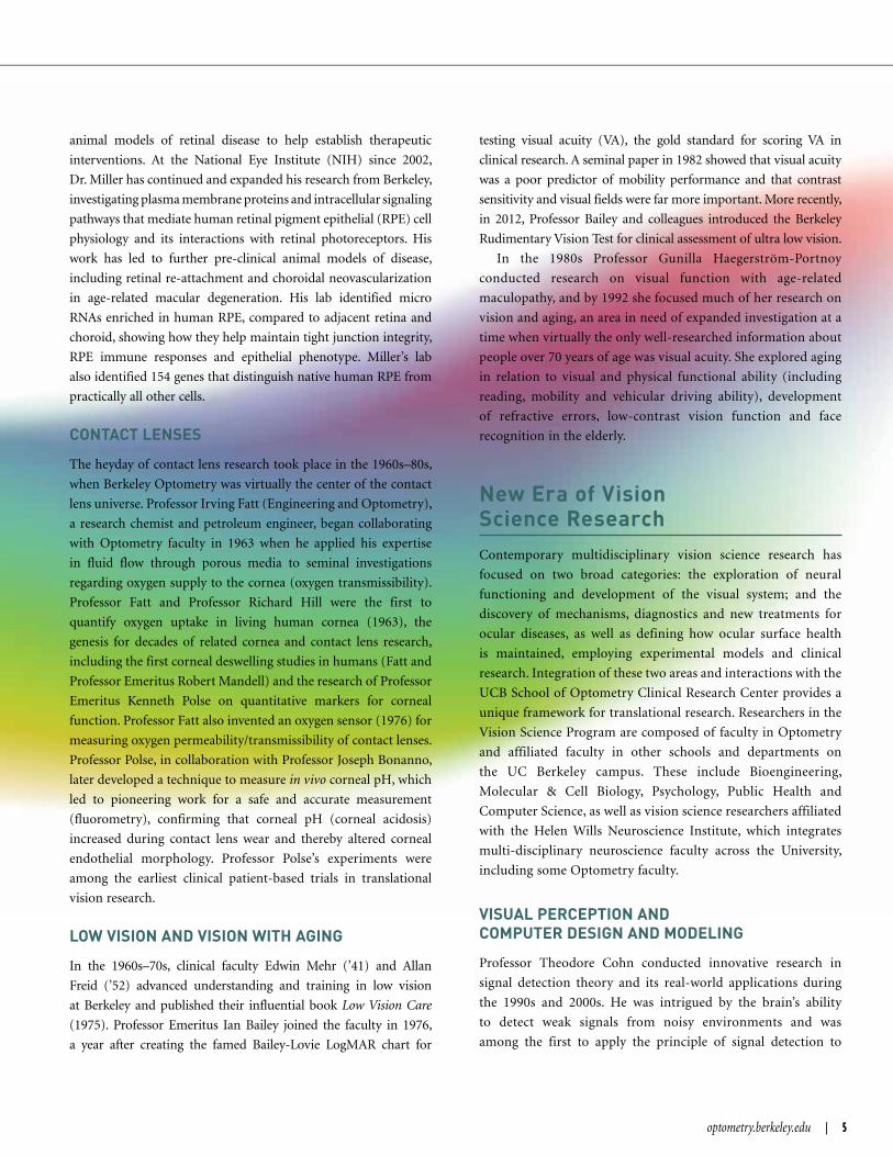
mechanisms in tear film stability and breakup. By using a sessile
captive-bubble apparatus to create thick lipid films with an
ultra-small amount (< 1 μg) of human tear lipids extracted from
whole tear samples, they identified dynamic and equilibrium
interfacial properties of thick lipid films deposited at the air-water
interface under conditions closely mimicking in vivo human-
eye conditions. Professor Lin and Dr. Tatyana Svitova (research
chemist, UC Berkeley) found that interfacial rheological and
dynamic properties of human tear lipids vary between racial/
ethnic groups. These variations correspond to clinically observable
differences in tear film stability between the groups. This technique
also helps to explain how contact-lens-care solutions affect the
biophysics of human tear lipids and provides insight into possible
etiologies of contact-lens-induced dry eye.
The laboratory of Professor Clayton Radke (Chemical
Engineering) uses surface and colloid science technology
to apply modern spectroscopic tools, along with molecular
theory and simulation, and continuum transport and reaction
engineering, for the development of quantitative descriptions
of interfacial behavior important to technology development.
Included among the areas of research is the study of the
dynamics and stability of thin films, such as human tear film,
which has a bearing on contact lens coating and design. ■
Sessile captive-bubble apparatus in Professor Lin’s laboratory, used to
create thick lipid films from extracts of human tear lipids (photo 2010)
The goal of the multidisciplinary graduate program in Vision Sci-
ence is to train graduates in the subfields that constitute Vision
Science. These fields include visual neuroscience and perception,
spatial navigation, psychophysics, computational vision, molecular
and cell biology, neurophysiology, bioengineering, robotics, bio-
chemistry, contact lenses, refractive development, ocular surface
biochemistry, ocular infection, ocular disease and immunology.
Forty-six Vision Science faculty members originate from eleven
different schools and departments on the UC Berkeley campus
(Optometry, Psychology, Public Health, Molecular & Cell Biology,
Neurobiology, Neuroscience, Infectious Disease and Immunology,
Bioengineering, Computer Science, Electrical Engineering and
Chemical Engineering).
The interdisciplinary program in Vision Science has been in
existence for 68 years. There are currently 39 pre-doctoral students
engaged in studies leading to the PhD in Vision Science as well as
five post-OD/MD students working toward a PhD. In addition,
there are 55 postdoctoral fellows training in the laboratories of
faculty in Vision Science. Primary funding for the graduate program
comes from the National Institutes of Health (NIH) and National
Eye Institute (NEI). State-of-the-art, cutting-edge research in the
program is supported by over $11 million annually in individual
faculty research support funds, which includes 40 grants from the
NIH, plus a shared NEI Core Research Grant that has been awarded
to the Vision Science Program for the last 32 years. PhD Graduate
student training, stipends and fees are supported primarily by the
individual investigator NIH research grants and an NEI Training
Grant that has been awarded to the program for the last 37 years.
Our mission is to attract outstanding trainees who will develop
independent, productive and notable vision research careers. Of the
more than 211 trainees who have received research degree training
in Vision Science (almost all PhDs), as of 2013, the vast majority
established highly successful careers as vision researchers — many
as faculty with independent funding in prestigious colleges and uni-
versities, or as researchers in private-sector institutions worldwide.
THE UC BERKELEY VISION SCIENCE GRADUATE PROGRAM

THE BERKELEY GROUP consisted of
1 third-year, 17 second-year and 3 first-
year optometry students. Although several
students were part of last year’s mission to
Nicaragua, for most students, this was their
first time participating in a VOSH clinic.
The mission leaders were Calista Ming ’16
and Monica Rodriguez-Bayes ’16, who
worked closely with the director of VOSH
Connecticut to make the trip a success for
all involved. This was VOSH Connecticut’s
sixteenth year of service to San Juan del Sur,
Nicaragua, and along with Berkeley VOSH’s
help, this team was the largest ever.
Over the clinic’s four days of operation,
the group saw 3,129 patients and dispensed
over 1,800 prescription eyeglasses, includ-
ing single vision, multifocal and readers,
most of which were donated by VOSH
humanitarian efforts
14 | BERKELEY OPTOMETRY FALL 2014
VOSH NICARAGUA
Jill Lobinger (’16) assesses the health of the patient’s eyes under the supervision of Dr. Michelle Ming. (Photo courtesy of Sheena Songpiriyakij.)
Team of volunteer optometrists, opticians, nurses and students from VOSH Connecticut and Berkeley Optometry (Photo courtesy of Sheena Songpiriyakij.)
F rom January 3 through 10, twenty-one students from Berkeley Optometry’s Volunteer
Optometric Services to Humanity (VOSH) program and one Berkeley undergraduate
student joined eight optometrists from VOSH Connecticut and traveled to San Juan del
Sur, Nicaragua, to provide eye care services to those in need.By Monica Rodriguez ’16
and Calista Ming ’16

Connecticut. Every patient also received a pair
of sunglasses, and donated pharmaceuticals were
used to treat patients with glaucoma, blepharitis,
dry eye and conjunctivitis. The yearly clinic pro-
vides continuous care to the Southern Nicara-
guan population and remains their main source
of eye care. Most of the patients seen in the clinic
were bused in from surrounding rural areas, and
many of these patients waited up to five hours to
see a doctor or student clinician.
The clinic was located at a local elementary
school, which consisted of six exam rooms that
had one to two optometrists, two to four optom-
etry students and several translators. The students
performed an entire exam, including retinoscopy,
trial frame refraction, and direct and indirect
ophthalmoscopy. A large percentage of the patient
population was presbyopic, and many had pin-
gueculas, pterygium and dry eye. The students
had an opportunity not only to improve their
optometric skills but also to see pathologies not
common in the United States such as coloboma,
advanced cataracts and toxoplasmosis.
The trip was a huge success and several stu-
dents are already planning to go on next year’s
trip! This mission provides Berkeley Optometry
students with an invaluable learning experi-
ence as well as the communities of Southern
Nicaragua with much needed services. Berkeley
Optometry continues to have a strong relation-
ship with VOSH Connecticut and looks forward
to working with them next year!
Each morning, hundreds of patients lined up outside of the clinics waiting to be seen. (Photo courtesy of Britney Kitamata-Wong.)
optometry.berkeley.edu | 15
Anna Harding (’16) takes the case history for a young girl prior to her exam. (Photo courtesy of Anna Harding.)
Berkeley Optometry students pose with the final count—number of patients seen: 3,129! (Photo courtesy of Shreya Malli.)

16 | BERKELEY OPTOMETRY FALL 2014
MANY OF THESE events are organized by
community leaders—politicians, churches and local
doctors—and serve to increase access to healthcare and
meet the visual needs of low-income and underserved
populations.
Berkeley Optometry students are also involved in
educational outreach. Volunteers visit elementary
schools to teach students about ocular anatomy
and binocular vision. Kids take a look inside the
eye as optometry students deconstruct a model eye
and discuss the functions of each part of the eye.
Students learn about eye dominance and the
breadth of visual fields and observe some optical
illusions. Optometry students also address common
eye diseases such as diabetic retinopathy, cataracts,
macular degeneration and glaucoma, and attempt
to demonstrate these conditions with glasses
manipulated to simulate visual changes associated
with each condition.
If you’re interested in volunteering as an optometrist,
or would like to request student volunteers for your
event, please contact [email protected].
16 | BERKELEY OPTOMETRY FALL 2014
humanitarian efforts
LOCAL OUTREACH
B erkeley Optometry faculty and students are frequently involved in local community
outreach events. Students routinely participate in vision screenings, which include
assessment of visual functions and ocular health under the supervision of a licensed
optometrist.
By Michelle Wong ’16
Shannon Lee (’16) (front) and Yangdi Chen (’16) (back) assessing confrontational visual fields at the Diabetes Health Event at John Muir Hospital.
Christina Belter (’16) and Erica Perlman-Hensen (’16) demonstrate pupillary responses to elementary school students.

optometry.berkeley.edu | 17optometry.berkeley.edu | 17
“Our students walked away with lots of knowledge
about eyes and ocular disease! Each student has
a different concept that they learned and each
was excited about learning from you experts!
Thank you so much for inspiring our students!”
—WASHINGTON ELEMENTARY
SCHOOL SCIENCE TEACHER
“[Thanks to] your group from UC Berkeley for
spending an entire day doing vision screening for
the Oakland community at our Health Fair. You
are setting a great example for our youth that
community humanitarian services can greatly
benefit the city at large. Thank you again.”
—OAKLAND CHINATOWN LIONS CLUB
Berkeley Optometry students participate in a vision screening under the supervision of Dr. Darlene Fong (’88). Pictured from left to right: (back row) Monica Chernoguz (’16), Narges Kalantarian (’17), Emmeline Cruz (’17), Tu-Anh Nguyen (’16), Alina Pechko (’17), Dr. Darlene Fong (’88), Craig Wong, (front row) Rebecca Lee (’16), Carol Chang (’16), Tara Pahlevan (’16).
Berkeley Optometry students visit Washington Elementary School and teach fifth graders about the eye. Pictured from left to right: (back row) Alex Chow (’16), Annie Lee (’17), Timoteo Flores (’17), Ashley Kadakian (’17), Carol Chang (’16), Britney Kitamata-Wong (’16), Arie Wong (’16), (front row) Michelle Wong (’16), Sandra Harpster (’17).
Berkeley Optometry students, Foresight Pre-optometry students, and Dr. Kathleen Low (’84) participate in Oakland Chinatown Lions Club Health Fair. Pictured from left to right: (back row) Andrew Ta (’15), Alice Han*, Armaeiti Chami*, Salena Thong*, Peter Tien (’16), Tracy Wang (’16), Britney Kitamata-Wong (’16), (front row) Jennifer Truong (’15), Jennifer Ding (’17), Cindy Lam*, Peter Wu (’16), Michelle Wong (’16), Dr. Kathleen Low (’84), Emily Mann. (*Foresight Pre-optometry Club)

18 | BERKELEY OPTOMETRY FALL 2014
school support
CAMPAIGN SUMMARY
BERKELEY OPTOMETRY HAS completed its portion of the “Campaign for Berkeley,” which had as its goal $3.1B and which was met in December 2013. The School of Optometry started its portion of this campaign, a $20M campaign, two years after the campus began. However, Berkeley Optometry has completed its goal, as well, simultaneously with the campus campaign. Some highlights of this campaign include raising 1) $2M for an endowed chair, 2) nearly $10M for research, 3) $2M for graduate fellowships, and 4) over $3M for program support. During the campaign, more than $1M also was contributed to the annual fund.
But perhaps the gift that most signified this campaign was a gift from students. The Berkeley Optometry Student Association donated $250,000, which the University Chancellor was delighted to match. That $500,000 endowment provides tuition assistance to nearly every student enrolled in the program at some point during the four-year curriculum. The UC Optometric Student Association (UCOSA) donation holds the record as the largest-ever contribution by current students to the Berkeley campus. (This has been followed more recently with an additional $40,000 gift from current students!)
For decades, the UCOSA had been tucking away proceeds from student dues, bake and clothing sales. Britta Hansen, former president of UCOSA, explains, “We wanted … it to go to the greater good of the University rather than sitting in our account.” “Getting a gift like that from current students just blew me away,” said Dennis Levi, Dean of Optometry at the time.
Largely as the result of the generosity of faculty and students, the School of Optometry now has 70 endowments for student support.
The completion of this campaign is truly a significant chapter in the school’s history. This is the first campaign for the school where there truly was equal participation from alums, friends, students, faculty and staff. Despite the worries from some that the goals for this campaign were much too lofty, it is tremendously gratifying to see how all of our constituents came together to reach this $20M milestone. Ninety-two cents from every dollar raised directly supports the school or our students. Quantifying development costs expended shows that each dollar contributed to the Berkeley Optometry campaign cost only 8 cents! (If one excludes research gifts, that return on investment changes to 15 cents for every dollar donated.) For a more in-depth summary, see “Campaign by the Numbers” on the next page.
Berkeley Optometry Concludes $20M Fundraising Campaign

optometry.berkeley.edu | 19
Third-and fourth-year students celebrate with donors the awards for tuition assistance provided by endowments created by faculty, staff, students, friends and alums.

20 | BERKELEY OPTOMETRY FALL 2014
school support
Berkeley OptOmetry—A FiscAl snApshot
OF THE SCHOOL’S nearly $32 mi l l ion
a nnua l revenue a nd ca mpus suppor t
funding, only $ 5 mi l l ion comes f rom
the state of Ca l i fornia. This has changed
dramat ica l ly f rom earl ier days, when nearly the ent ire
cost of educat ion at the Universit y of Ca l i fornia was
supported by the state.
Resea rch f u nd i ng i nc ludes f u nds f rom pr ivate
indust r y, federa l government agencies such as t he
Nat iona l Inst itutes of Hea lth (NIH), Depar tment of
Defense (DOD), Nat iona l Science Foundat ion (NSF),
the state, the Universit y of Ca l i fornia and var ious non-
prof it organizat ions.
Other sources include cont inuing educat ion revenue,
cont rac t a nd g ra nt overhead f u nd s , shor t-ter m
investment pool (STIP) income and endowments, and
other miscel laneous funds.
Professiona l degree supplementa l fees (PDST) are
pa id by students and are designated by Universit y
pol icy to be a l located as two-thirds for the support of
the school and one-third for student awards. Note that
no tuit ion comes direct ly to Berkeley Optometr y, as
tuit ion goes to the Berkeley Graduate Div ision and the
Universit y.
Genera l f unds a re f unds prov ided by t he s tate .
Berkeley Optomet r y receives s tate f unds on ly for
sa lar ies pa id to ladder-rank facu lt y.
Student support includes funds prov ided to students
in the form of depar tmenta l student a id and fel lowships,
awa rds f rom t ra i n i ng g ra nt s a nd pa id posit ions
(research, teaching and federa l work-study).
Facult y and sta f f compensat ion (sa lar ies and benef its)
include pay ments for academic, research and cl inica l
facu lt y and sta f f.
Berekeley Optometry Revenue SourcesFiscal Year 2013–2014
� Optometry Student Fees (13%)
� Contracts and Grants (28%)
� Campus Support Funding (24%)
� Clinic and CE Revenue (32%)
� Private Gifts for Current Use (3%)
13%3%
28%
24%
32%
Berekeley Optometry ExpendituresFiscal Year 2013–2014
� Student Aid and Fellowships (7%)
� Supplies, Materials, Equipment and Other Operating Expenses (32%)
� Faculty and Staff Compensation (61%)
61%32%
7%

Anthony and Elna Adams *
Adi D. Adins ’68
Otto Anderson ’39 †
Norma and John Austin ’52 †
Charles Bailey ’82
Ian and Valerie Bailey
Robert Benn ’54 †
Roy Black ’52 †
Karen Walker-Brandreth, ’68 and Roy Brandreth ’53 †
Charles Brown ’47 * †
Bettina Bruckman †
Maxine † and William Burlington ’40 †
Lily and Collin Chu ’71
Doris Sze and Stephen Chun ’74
Jeanette and Robert Cibull ’57
Allen Coe ’50 †
Jack Cohen ’52
Barbara and Theodore Cohn †
Charles Conrad ’40 †
Lawrence Creasey ’58
Henry Leibee and Bonne Curtis-Leibee ’69
John Daly ’52
Scott Daly ’83
Herbert Elefant ’39
Dianna Ellerbeck
Roselyn and Weylin Eng ’65 *
Steven Ngin † Charmaine Eng-Ngin ’63
Jay and Rebekah Enoch
Joseph Farrington ’51 * †
Bernard Feldman ’52
Pamela P. Fong ’77
Allan Freid ’52
Marcia and Lee Goldstein ’66
Edward I. Goodlaw ’34 †
Michael Harris ’65 *
Frank Johnson, Jr. ’51 * †
Barbara and Marshall Kamena ’65
Suga † and Henry Kawahara ’46 * †
Roderick J. Keener ’71
Cynthia and Jeffrey Ko ’73
Aman I. Kumar
David Leibel ’48 †
Dennis and Marilyn Levi
Joyce and A. Saul Levine ’52
Jeffrey Lieberman ’89
Aloha T. † and Edward R. Ligon ’48 †
Henry Linker ’48
Warren LoPresti ’51
Jimmy Low ’52
Edwin Mehr ’41 †
Meredith Morgan, Jr. ’34 * †
Thomas Nagy ’49
Lillian † and Benjamin Nerenberg ’41 * †
Maurice Newman ’52
Winston Nielsen ’49 †
Joy Ohara ’10 and Kok Loong Lye
Leonard Osias ’48
Paul Peng ’86
Herman Popiul ’52
Jeanette and Edward Revelli ’77
Donald Sarver ’71
Dorothy Bates Searles †
Charles Seger ’48 †
Elliott Shane †
Leonard Shenkan ’44 * †
Sylvia † and Irvin Silberstein ’42 †
Curtis Simmons ’82
Bette and Mervyn Simon ’37 * †
Richard Simsarian ’54
Branna and Irving Sisenwein
Roberta Smith †
Harry Springer ’42 †
Lawrence Stern ’62
Eleanor Sweigert †
Jenny and Ernest Takahashi ’68
Betty and Bernhardt Thal ’48 * †
Esther and Lawrence Thal ’75
Charlotte Tlachac ’78
Bryan Vanesian ’89
Lesley Walls ’68 and Mary Ann Keverline-Walls ’67
Sheldon Wechsler ’52 †
Simon P. Williams ’51 †
William Wong ’73
* Founding member† Deceased
O p t O m e t r y A s s O c i A t e s O f t h e B e n j A m i n i d e W h e e l e r s O c i e t y
Many people Make their final gift to Berkeley Optometry their most significant,
by including Berkeley Optometry in their estate planning.
The Optometry Associates of the Benjamin Ide Wheeler Society recognizes individuals
who have developed an estate plan which will benefit Berkeley Optometry. Planned gifts
provide philanthropic support that is essential in enabling the School of Optometry to
remain a leader in optometric education and research.
optometry.berkeley.edu | 21

22 | BERKELEY OPTOMETRY FALL 201422 | BERKELEY OPTOMETRY FALL 2014
school support
Dean’s Circle ($1,000 and up)AnonymousAlcon Laboratories, Inc.Thomas A. Aller ’83Barton Eye AssociatesBay Area Optometric Council
(BAOC)Berkeley On-Line Lectures
and Demonstrations (BOLD)Jeffrey A. Calmere ’88 and
Linda K. Hur-Calmere ’90Tony Chahine ’95Gerald D. Chan ’75 and
Lisa E. Moon ’76Collin S. Chu ’71 and Lily F. ChuWarren V. De Haan ’66Davida J. Dong-Leong ’78 and
Richard G. Leong ’78Eyexam of CaliforniaGenentech, Inc.Johnson & JohnsonAkio KanaiPaul A. Kiyan ’75 and
Beverly N. KiyanStanley A. KleinWarren I. LoPresti ’51Edward Low ’74Seymour C. Marcuse, Jr. TrustStephen A. MarcuseHoward Pflug ’47Edward J. Revelli ’77 and
Jeanette RevelliSamsung Electronics America, Inc.Ernest K. Takahashi ’69 and
Jenny M. TakahashiLawrence S. Thal ’75 and
Esther G. ThalCorina Van de Pol ’90 PhD ’99Vision One Credit Union Vision Service Plan (VSP)Vision West, Inc. (VWI)Mark E. Winston ’82Shinichi YamadeRandall W. Yumori ’80
Berkeley Optometry Annual Fund Donors $250+ (July 1, 2013 to June 30, 2014)The MerediTh W. Morgan SocieTy
Class year denotes year of OD degree.
William Kent Dorrance Trust Sherri M. Egashira ’92L. Edward Elliott ’65 and
Terry E. ElliottCharmaine M. Eng ’63Thomas H. Eves ’54 and
Diane K. EvesCraig K. Hisaka ’72Jane M. Kadoya ’80Dennis M. Levi and Marilyn LeviMarin Community FoundationJeffrey S. McFeely ’85 and
Eeva Maria K. McFeelyJohn J. McNally ’79 and
Heike McNallyGabriela McTyier ’04Thomas H. Murphy ’78Thomas R. Nagy ’49 and
Vicie M. NagyMelvin O’NealOptometry Care Santa BarbaraPower Integrations, Inc. Marina L. Rocchi ’11John G. Rosten ’77 and
Susan J. RostenDonna Simonian ’07Marvin L. Smotrich ’62 and
Mehrey D. SmotrichPamela Theriot ’01Tri Valley OptometryMann D. Trinh ’07Ana P. Vargas ’06Betty Wang ’12Thomas W. Wing ’74Andrew B. Wong ’06 and
Karen G. Wong ’06Joseph D. Wong ’74 and
Catherine WongJean A. Wrightnour ’88
Benefactor ($500–$999)Melinda A. Cano-Howes ’99 and
Ronald G. HowesJoan D. CostelloEdward O. France, Jr. ’76Lee A. Goldstein ’66Tem R. Gronquist ’97 and
Joanne GronquistMonroe J. Hirsch TrustRichard C. Hom ’75Shawn HooperNadine I. Howell ’82 and
Steven L. Howell ’82Brian Levy ’76Donald E. Louie ’65 and
Midori U. LouieRicoh Innovations, Inc.Nicolette J. Sacco-Brown ’87Donald D. Schmidt ’66 and
Georgenia M. SchmidtSteven H. Schwartz ’79Lois M. Shiozawa ’81Betty ThalTina P. Tsai ’99Louise Vogelsberg ’56 and
Frederick E. VogelsbergGregory J. Ward Nora Weissman Letitia Y. Yee ’97Wayne W. Zimmerman ’73 and
Susan L. Zimmerman
Partner ($250–$499)Adair OptometryTodd M. Adair ’92 and Felecia AdairJeffrey L. Azus ’86 and
Alice Ng Azus ’87Eric BassChristopher R. Cabrera ’75Shao-Pin Chen and Li-Li ChenDistinctive Eyes Optometry

DONOR PROFILE
optometry.berkeley.edu | 23
M ING POW WAS diagnosed with retinitis
pigmentosa at an early age and was
legally blind by her mid-20s. Without
formal low vision optometric care in Malaysia,
Ming Pow learned to adapt to her vision loss
without the aid of any electronic aids or magnifiers.
She developed remarkable memory and sensory
ability—among other handy skills, she would be
able to “smell” who was in the room and memorize
numerous telephone numbers.
Despite her vision loss, Ming Pow had a successful
career of more than 30 years as one of the first
female stockbrokers in Malaysia. She was actively
engaged in the community and supported a number
of Buddhist foundations throughout her life. Drawing
inspiration from Ming Pow’s ability to overcome her
vision loss, Joy attended Berkeley Optometry from
2006–2010 and after graduation was a resident at
the low vision program at the University of Houston
College of Optometry.
Lye and his father have seen the value of being
community-minded through Ming Pow and know
first-hand the challenges of adjusting to living with
an individual with visual impairment. With the
encouragement of Joy, they established the Ming
Pow Low Fund in 2010, which aims to aid graduating
optometry students who are inspired to study and
specialize in low vision.
Dr. Joy Ohara ’10 and Kok Loong Lye have had a longstanding connection to UC Berkeley. Lye met Joy while she was finishing her Chemistry BS in 2005 and they soon fell in love. They then moved on to study Public Policy and Optometry, respectively–both at Berkeley. In 2008, in the middle of their graduate studies, Lye’s mother Ming Pow Low passed away from breast cancer.

David Kirschen OD ’72, PhD ’77, is the first Berkeley Optometry alum to be
awarded a World Series ring! As team optometrist for the Boston Red Sox,
he received his 2013 World Series ring in recognition of the critical role that
exceptional vision plays in sports performance at the professional level. He
has worked with the Red Sox since 2004, during which time they have won
three World Series championships. Prior to 2004 the team had gone 86 years
without a championship. As a major pioneer in sports vision, Dr. Kirschen’s
innovative approach and techniques have made a key impact in improving
the performance of athletes such as Stephen Drew whose post-season batting
average was .080 prior to seeing Dr. Kirschen and his partner Dr. Daniel Laby;
afterward he finished the series batting two for four, including a home run!
Dr. Kirschen is winner of the American Optometric Association’s 2009 Sports
Vision Optometrist of the Year award and has worked with other professional
teams such as the Los Angeles Dodgers, Los Angeles Kings, Boston Celtics,
Chicago Cubs, Cleveland Indians, Houston Astros, New York Mets and
St. Louis Cardinals for over 20 years. He is currently Chief of Binocular
Vision and Orthoptic Services at the Jules Stein Eye Institute at the UCLA
David Geffin School of Medicine. He also serves as Professor Emeritus at the
Southern California College of Optometry and has been in private practice in
Brea, California, for 42 years.
David Kirschen OD ’72, PhD ’77, received a World Series Ring!
UC Berkeley, Vision Science alumni, Sir Colin Blakemore has been granted
an honorary title of knighthood. At the age of 70, Dr. Blakemore “has been
recognized for his research and for communicating the importance of often
controversial science.” Dr. Blakemore’s research has focused on many aspects
of vision, development, plasticity of the brain and neurodegenerative disease.
He has sought to define the developmental errors that underlie cognitive
disorders, such as autism, dyslexia and schizophrenia. Dr. Blakemore also
championed the communication of science and engagement with the public
on controversial and challenging aspects. In 1976, at the age of 32, he was
the youngest person to give the BBC Reith Lectures for which he presented
a series of six talks entitled Mechanics of the Mind, and has subsequently
presented or contributed to hundreds of radio and television broadcasts. He
has been an outspoken defender of animal research and an advocate for many
other issues including classification of drugs and genetically modified foods.
Sir Colin Blakemore PhD ’68, was knighted.
24 | BERKELEY OPTOMETRY FALL 2014
NEWS & NOTES
stay connected

AUSTIN ROORDA—GUGGENHEIM FELLOWSHIPAustin Roorda, Chair, Vision Science Graduate Group and Professor of
Optometry and Vision Science, is one of eight UC Berkeley faculty who
have been awarded 2014 John Simon Guggenheim Memorial Foundation
Fellowships. Guggenheim Fellowships are awarded for “impressive
achievement in the past and exceptional promise for future accomplishments”
and provide funding to further the recipient’s work in fields ranging from
the natural sciences to the creative arts.
BERKELEY OPTOMETRY STUDENTS MAKE $40K GIFTOn July 3 current and former Berkeley Optometry students presented a
$40,000 gift to Berkeley Optometry’s new dean, John G. Flanagan. With
matching funds made available by the Chancellor, the total impact of the
University of California Optometric Student Association gift is $80,000
toward the renovation of Minor Hall. The check presentation ceremony
highlighted the important partnership between the school’s students—past
and present—and its leadership. Dean Flanagan commended the students
for their contributions, both academic and financial, and emphasized their
role in defining the future of Berkeley Optometry.
optometry.berkeley.edu | 25
NOA MEETINGBerkeley Optometry hosted the annual Board Meeting of the National
Optometric Association (NOA) in October. The NOA has been a leader in
the cause of improving the quality and accessibility of eye care in minority
and other historically underserved communities. Increasing the number
of minority eye care practitioners, through student recruitment and career
placement, has been the NOA’s historical focus. Those attending the meetings
and dinner, hosted by Dean John Flanagan, were honored to hear first-hand the
history of the NOA from co-founder Dr. C. Clayton Powell. Originally from
Atlanta, Georgia, Dr. Powell served as the NOA’s first president from 1969 to
1974. The timing for Berkeley Optometry to host the NOA Board Meeting could
not have been better as current optometry students have also reestablished
the school’s relationship with the National Optometric Student Association
(NOSA) and are excited to help execute the mission of the organization.
Shown above right are students Maria Cardenas ’17 and Angel Barajas ’17 with NOA co-founder Dr. C. Clayton Powell.
Shown at left with Dean John Flanagan are members of the NOA Board of Directors and members of the Berkeley Optometry faculty and staff who helped host the NOA for their annual meeting at Berkeley Optometry.

26 | BERKELEY OPTOMETRY FALL 2014
AWARDS
stay connected
26 | BERKELEY OPTOMETRY FALL 2014
The Michael G. Harris Family Award for Excellence in Optometric Education—Edward J. Revelli ’77
This award is presented by the American Optometric Foundation (AOF) to an optometric educator who has demonstrated ongoing and consistent excellence in the education of optometry students and/or the advancement of optometric education. Ed has been a tireless educator who has been dedicated to providing the best instruction and training to optometry students for many years. Along the way he has also enhanced the clinical skills and expertise of faculty in new and expanding areas of the profession. He has received great respect from not only his students, but also his peers.
2014 Ezell Club Fellowship to honor Anthony “Tony” Adams
The American Optometric Foundation honored former Ezell Fellow Dr. Anthony “Tony” Adams with a named William C. Ezell Fellowship at its recent meeting in Denver, Colorado. A long-time supporter of the foun-dation, Dr. Adams is the Editor-in-Chief of Optometry & Vision Science, former Berkeley Optometry dean and past president of both the American Academy of Optometry and the American Optometric Foundation.
The flagship program of the American Optometric Foundation (AOF), Ezell Fellowships have been awarded for over 60 years. These fellowships support graduate students enrolled in a full-time program of study and training in vision-related research that leads to a master’s or PhD degree.
Foundation Fighting Blindness Grants
Foundation Fighting Blindness Research grants have been awarded to Austin Roorda, John Flannery and Richard Kramer, all vision scientists at UC Berkeley. These awards recognize the investigators’ outstanding prog-ress in research that advances sight-saving treatments and cures.
Dr. Roorda’s project is titled “AOSLO: Detecting Retinal Degeneration Before Vision is Lost.” The adaptive optics laser scanning ophthalmo-scope (AOSLO) is similar to a powerful microscope that enables retinal researchers to see structural changes in the retina well before vision is lost from a retinal disease. That power can enable researchers to more quickly determine if a treatment is working in a clinical trial. Dr. Roorda is performing studies of AOSLO to correlate changes in the retina (e.g., loss of photoreceptors) with changes in vision.
Optogenetics is a relatively new field of research, which involves the delivery of genes or chemicals to restore light sensitivity to a highly degenerated retina. In essence, this type of treatment holds promise for
restoring vision in people with advanced disease and may do so regard-less of the genetic defect causing the disease. Dr. Flannery is developing an optogenetic gene therapy for bipolar cells in the retina, which survive for many people with significant retinal degeneration. Given these cells’ adjacency to photoreceptors, he believes bipolar cells have the potential to provide meaningful vision when treated with an optogenetic therapy. He will test the effectiveness of four gene therapies, each delivering a different light-sensing protein to bipolar cells.
Dr. Kramer will study variations of a synthetic compound called AAQ as an optogenetic treatment in ganglion cells. Previous studies showed that injections of AAQ restored vision in mice, but only in very bright light. Dr. Kramer will be engineering compounds to work in dimmer light. And, because injections of AAQ have worked only temporarily, Dr. Kramer will evaluate microspheres, small biodegradable particles, to provide sustained release of the compounds.
American Optometric Association Legend Award—Kenneth Polse ’68
Professor Emeritus Kenneth A. Polse will receive a Legend Award from the Contact Lens and Cornea Section of the American Optometric Association in June. He was recognized for his contributions to the culture of contact lenses through leadership and dedication. The Legends Award was created to distinguish individuals who have demonstrated intellectual depth and have a passion about contact lenses, the cornea or refractive technology.
American Academy of Optometry Life Fellow Award—Michael Harris ’65
Michael G. Harris was awarded Life Fellowship status in the American Academy of Optometry after presses had already rolled for last year’s magazine. The distinction of Life Fellowship was created to provide recognition to those Fellows who, through long-time membership in the Academy, have rendered distinguished service to the science and art of optometry.
2014 Fry Medal from The Ohio State College of Optometry—Gerald Westheimer
On September 16, 2014, the actual centennial date of optometry at The Ohio State University, Berkeley Professor Gerald Westheimer received the 14th Fry Medal, awarded to individuals who have truly distinguished themselves with long and exemplary records of vision research.

optometry.berkeley.edu | 27optometry.berkeley.edu | 27
Dr. Westheimer trained in and practiced optometry in Sydney, Australia, before going to Ohio State for his PhD study with Dr. Glenn Fry. He was on the faculty of The Ohio State University School of Optometry until com-ing to Berkeley in 1960. His teaching and research accomplishments have been widely recognized. He is the only optometrist to have been elected to the Royal Society of London. He is the recipient of ophthalmology’s Proctor Medal, the Academy of Optometry’s Prentice Medal, and is a fel-low of the American Academy of Arts and Sciences.
Hoya Student Vision Grant and Scholarship—Susannah Lee ’15
Susannah Lee received the 2014 Hoya Student Vision Grant and Scholarship. Third- and fourth-year students from each school and college of optometry compete for this award, which is presented dur-ing an awards luncheon during the annual meeting of the American Optometric Association. Susannah is the first Berkeley Optometry recipient of this award.
Vistakon Award of Excellence in Contact Lens Patient Care—Jeffry Wu ’14
This award recognizes outstanding fourth-year student clinicians who have demonstrated excellence in contact lens patient care during their optometric education. The award is given to a graduating student from each school and college of optometry in North America. Jeffry Wu is this year’s recipient from Berkeley Optometry.
J. Pat Cummings Scholarship— Krystal Vanichsarn ’15
This award is conferred annually to a second- or third-year optometry student at each of the North American schools and colleges of optometry who best demonstrates the ideal eye-care standards of practice, achieve-ment in both academic performance and extracurricular activities, and par-ticipation with other professional pursuits such as involvement with patients through internships, community service and other volunteer activities.
Douglas W. Hopkins Primary Care Residency Awards—Gloria Chow ’12 and Mark Landig ’13
The Douglas Hopkins Residency Award is intended to promote the practice and development of the field of primary care optometry by pro-
viding incentive and support to talented optometric residents who dem-onstrate a passion and commitment to practice, research and education in primary care. The award was established by the Primary Care Section of the American Academy of Optometry in 2012 to honor Dr. Hopkins who was a leader within the Primary Care Section of the Academy and who passed away unexpectedly in 2007. Gloria is the second recipient of this award. Mark is a primary care/geriatric resident at the West Los Angeles Veterans Affairs Health Care Center.
Gloria Chow is shown with Stephen Chun ’74, one of Gloria’s former instructors at Berkeley Optometry.
Stephen Chun ’74 (back right) is shown with the Douglas Hopkins family: Mrs. Roberta Hopkins top row and daughter front row. Also shown with Dr. Chun are Drs. Ann Clark, Jeff Magun and Linda Casser—all co-founders of the Primary Care Section of the American Academy of Optometry.
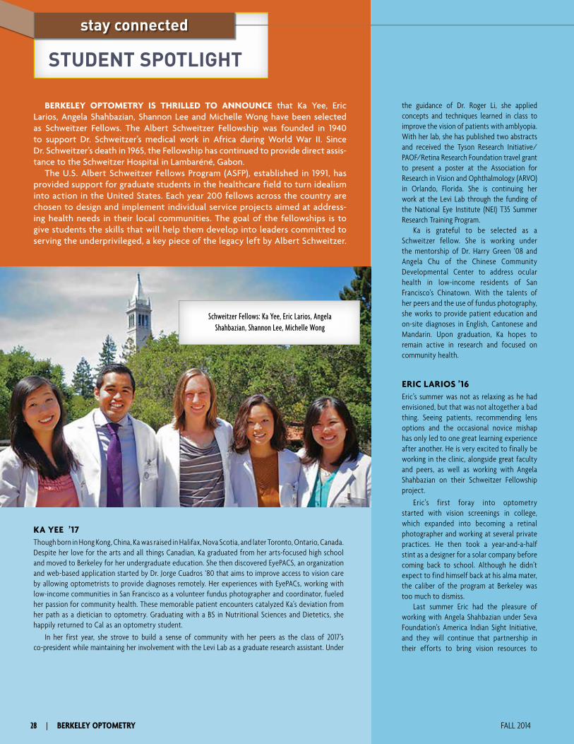
the guidance of Dr. Roger Li, she applied concepts and techniques learned in class to improve the vision of patients with amblyopia. With her lab, she has published two abstracts and received the Tyson Research Initiative/PAOF/Retina Research Foundation travel grant to present a poster at the Association for Research in Vision and Ophthalmology (ARVO) in Orlando, Florida. She is continuing her work at the Levi Lab through the funding of the National Eye Institute (NEI) T35 Summer Research Training Program.
Ka is grateful to be selected as a Schweitzer fellow. She is working under the mentorship of Dr. Harry Green ‘08 and Angela Chu of the Chinese Community Developmental Center to address ocular health in low-income residents of San Francisco’s Chinatown. With the talents of her peers and the use of fundus photography, she works to provide patient education and on-site diagnoses in English, Cantonese and Mandarin. Upon graduation, Ka hopes to remain active in research and focused on community health.
ERIC LARIOS ’16Eric’s summer was not as relaxing as he had envisioned, but that was not altogether a bad thing. Seeing patients, recommending lens options and the occasional novice mishap has only led to one great learning experience after another. He is very excited to finally be working in the clinic, alongside great faculty and peers, as well as working with Angela Shahbazian on their Schweitzer Fellowship project.
Eric’s first foray into optometry started with vision screenings in college, which expanded into becoming a retinal photographer and working at several private practices. He then took a year-and-a-half stint as a designer for a solar company before coming back to school. Although he didn’t expect to find himself back at his alma mater, the caliber of the program at Berkeley was too much to dismiss.
Last summer Eric had the pleasure of working with Angela Shahbazian under Seva Foundation’s America Indian Sight Initiative, and they will continue that partnership in their efforts to bring vision resources to
BERKELEY OPTOMETRY IS THRILLED TO ANNOUNCE that Ka Yee, Eric Larios, Angela Shahbazian, Shannon Lee and Michelle Wong have been selected as Schweitzer Fellows. The Albert Schweitzer Fellowship was founded in 1940 to support Dr. Schweitzer’s medical work in Africa during World War II. Since Dr. Schweitzer’s death in 1965, the Fellowship has continued to provide direct assis-tance to the Schweitzer Hospital in Lambaréné, Gabon.
The U.S. Albert Schweitzer Fellows Program (ASFP), established in 1991, has provided support for graduate students in the healthcare field to turn idealism into action in the United States. Each year 200 fellows across the country are chosen to design and implement individual service projects aimed at address-ing health needs in their local communities. The goal of the fellowships is to give students the skills that will help them develop into leaders committed to serving the underprivileged, a key piece of the legacy left by Albert Schweitzer.
Schweitzer Fellows: Ka Yee, Eric Larios, Angela Shahbazian, Shannon Lee, Michelle Wong
STUDENT SPOTLIGHT
stay connected
KA YEE ’17Though born in Hong Kong, China, Ka was raised in Halifax, Nova Scotia, and later Toronto, Ontario, Canada. Despite her love for the arts and all things Canadian, Ka graduated from her arts-focused high school and moved to Berkeley for her undergraduate education. She then discovered EyePACS, an organization and web-based application started by Dr. Jorge Cuadros ‘80 that aims to improve access to vision care by allowing optometrists to provide diagnoses remotely. Her experiences with EyePACs, working with low-income communities in San Francisco as a volunteer fundus photographer and coordinator, fueled her passion for community health. These memorable patient encounters catalyzed Ka’s deviation from her path as a dietician to optometry. Graduating with a BS in Nutritional Sciences and Dietetics, she happily returned to Cal as an optometry student.
In her first year, she strove to build a sense of community with her peers as the class of 2017’s co-president while maintaining her involvement with the Levi Lab as a graduate research assistant. Under
28 | BERKELEY OPTOMETRY FALL 2014

undergraduate, receiving a bachelor’s degree in Molecular & Cell Biology. Michelle started exploring optometry as a career option after taking Dr. Richard Van Sluyter’s freshman seminar on the anatomy of the human eye, and Dean Emeritus Jay Enoch’s sophomore seminar on eye care in rural India. Michelle gained more knowledge about optometry through the pre-optometry club, Foresight, where she heard about the profession, different optometric programs, and engaged with Berkeley Optometry faculty and students over lunches and workshops.
Michelle eventually decided on optom-etry as her career after volunteering in the community with EyePACS, Project Vision, and Prevent Blindness Northern California, and seeing what an impact she could make in people’s lives by restoring their vision. Michelle ultimately chose Berkeley Optometry for the rigorous curriculum, high volume of patient encounters, and the tight-knit community of students and faculty.
As a graduate student, Michelle has continued her commitment to service. She has coordinated volunteer opportunities for students at local health fairs and vision screenings, as well as educational outreach events at nearby elementary schools. She also volunteered with VOSH to provide eye care to over 3,000 residents of San Juan Del Sur and neighboring cities in Nicaragua, and she looks forward to entering clinic to care for patients and grow in her optometric career.
As 2014–2015 Albert Schweitzer Fellows, Michelle and Shannon Lee plan to offer vision screenings at Monument Crisis Center in Concord, California. Monument Crisis Center supports over 14,000 households, many low income. Monument Crisis Center strives to alleviate poverty by providing services such as grocery distribution, workshops on language and career development, and assistance for insurance enrollment. Monument Crisis Center also offers health exams through RotaCare and dental screenings through local student volunteers. Michelle is excited to expand the health offerings of Monument Crisis Center by providing a vision component to address the visual needs of these patients and to educate patients on the importance of eye and systemic health.
students and their parents in the Oakland school districts. They are both very excited to begin on their Schweitzer project and help these communities with services that they would not have otherwise.
With his career starting to take hold, Eric is very enthusiastic about his future and looks forward to growing his skills and knowledge as a clinician, both in and out of the classroom.
ANGELA SHAHBAZIAN ’16Angela is excited about seeing patients as a third-year student clinician. When she decided several years ago to leave her career in website/media management to begin a new career in optometry, she knew that the satisfaction of helping patients with their real-life visual needs would far outweigh the sacrifices of returning to school. Now that she is beginning to experience the fruits of her labor, she cannot wait to dive more into her studies so she can provide her patients with the best optometric care possible.
As a returning student, balancing the demands of family life and home life has been challenging. But Angela has found inspiration in her fellow students who continually amaze her with their commitment, professionalism and enthusiasm. She has also been thrilled to meet so many students with similar goals of increasing access to eye care, and has been able to collaborate with fellow student Eric Larios on related projects.
She and Eric conducted research for the Seva Foundation during the initial stages of their American Indian Sight Initiative. She is looking forward to expanding on this work next year. Through their Schweitzer Fellowship project, they will increase access to eye care for students and their families in underprivileged parts of Oakland.
Angela looks forward to many adventures in optometry as her career continues, and she is thankful to her family, particularly her husband, for the opportunity to grow and learn every day in this profession.
SHANNON LEE ’16Raised in San Jose, California, Shannon loved science, but never enjoyed the dissection
labs in class. Oddly enough, her first experience with a cow’s eye in 6th grade was the only dissection activity that she was thoroughly fascinated by. In high school she accompanied doctors to rural China to help North Korean refugees; witnessing these individuals use their profession to help others sparked a passion in Shannon to do the same. She majored in human biology at the University of California, San Diego, in hopes of pursuing a health profession career. Her experience of studying abroad in Singapore and traveling all over Southeast Asia solidified her passion for traveling and helping the global community. After shadowing an optometrist upon graduation and realizing her fascination for eyes, she began to pursue a career in optometry by working for a local optometrist while completing her prerequisite course work.
At Berkeley Optometry, she was the vice president for the Volunteer Optometric Services to Humanity (VOSH), Berkeley chapter, and served on the Student Life Committee in her second year. Though many hours of planning were involved, she enjoyed coordinating events and boosting the morale of students by planning fun activities. She grabbed the opportunities to combine her love for optometry and travel by participating in a VOSH trip to Nicaragua in 2012 and accompanying an optometrist and two other optometry students to Mexico in 2013. She will be helping the local community as an Albert Schweitzer Fellow this year. She and her partner, Michelle Wong, will be working with Monument Crisis Center in Concord, California, by offering vision screenings to low-income individuals. Through this project, she hopes to create more awareness about being proactive about one’s eye health. Shannon looks forward to all the potential learning experiences ahead—from patient encounters to all the opportunities to try delicious food with her optometry colleagues.
MICHELLE WONG ’16Michelle was born and raised in San Leandro, California. She attended UC Berkeley as an
optometry.berkeley.edu | 29

30 | BERKELEY OPTOMETRY FALL 2014
SHIRIN BAREZ, MDShirin Barez came to Berkeley as an undergraduate and quickly became intrigued by the complex-
ity of the visual system. This interest led her to enter the UC Berkeley School of Optometry, where she worked with Professor Lawrence Stark, a pioneer in the field of bioengineering, on a model of the pupil response mechanism. She received a master’s degree in Physiological Optics. While at UCBSO she successfully ran for an elected seat on the Associated Students of University of California (ASUC) senate, representing the professional schools on campus. Dr. Barez received her medical degree from the George Washington University School of Medicine.
Following her interest in basic science and ocular disease, she spent a year at the National Eye Institue studying the immunology of retinoblastoma. She completed her residency in ophthalmology at the George Washington University Medical Center and a two-year vitreoretinal fellowship at the Devers Eye Institute in Portland, Oregon. Upon returning to the Bay Area to begin private practice in the East Bay, Dr. Barez joined the clinical faculty at Berkeley Optometry in 1997, where she currently is a clinical professor. In addition to her teaching and clinical responsibilities, she remains active in clinical research. A decade-long collaboration with former Dean Anthony Adams’ Diabetes Study has resulted in numerous abstracts and publications.
Dr. Barez has served as an advisor to and qualifying exam committee member for a number of PhD students in the Vision Science Program. She is senior consultant to EyePACS diabetic screening program and provides treatment for many patients with diabetic retinopathy who would otherwise not have access to treatment for this potentially blinding disease. Dr. Barez is a diplomate of the American Board of Ophthalmology and a fellow of the American Academy of Ophthalmology. She has been named as one of the East Bay’s top doctors by Oakland Magazine. Dr. Barez enjoys organic gardening, hiking, photography and spending time with family and friends.
CLAUDIA C. RUEGG, OD ’11Claudia Ruegg joined the full-time clinical faculty in 2013 as an assistant clinical professor and
works at various on- and off-campus clinics. She supervises third- and fourth-year clinicians at UC Berkeley and the Alameda County Medical Center in Oakland, with a focus on primary care and ocular disease, particularly diabetes and glaucoma. As the clinical supervisor at our new satellite clinic at the UC Santa Cruz Student Health Center, she performs direct patient care and supervises fourth-year students. She is enjoying the rewards and challenges of starting a new clinic from the ground up. Located on campus, the Student Health Center serves UCSC undergraduate and graduate students, and optometry is a well-integrated part of the healthcare team. As a mentor at numerous Glaucoma Grand Rounds, Dr. Ruegg has provided continuing education to many optometrists in the process of glaucoma certification.
Dr. Ruegg grew up in the South Bay and later moved to Los Angeles to obtain a BS in Psychobiology from UCLA in 2007. Subsequently she completed her OD degree at UC Berkeley in 2011. She attributes much of her ocular disease experience to her residency training at the Albuquerque VA hospital in New Mexico where she was mentored by Michael Sullivan-Mee, OD and focused on ocular disease manage-ment and integration with systemic disease, including vascular, infectious and neurologic conditions. She also completed a year-long glaucoma research project to investigate novel use of the Heidelberg Spectralis OCT in glaucoma detection. Her work was subsequently published in the American Journal of Ophthalmology in 2013 and the project is still on-going. After residency she moved to Washington to join the medical staff at the Spokane VA hospital, providing direct patient care as well as supervising fourth-year interns from Pacific University. Dr. Ruegg was excited at the opportunity to move back to California to join the excellent faculty at UC Berkeley and continue teaching. She enjoys seeing her stu-dents grow as clinicians and expand their knowledge of ocular disease and evidence-based medicine.
Dr. Ruegg loves spending time with friends, exploring the Bay Area’s culinary scene and traveling internationally, especially to Europe to visit her family and cultivate her European roots.
FACULTY FOCUS
stay connected

optometry.berkeley.edu | 31
CHRISTINA S. WILMER, OD ’96Chris Wilmer was born in Ojai, California, and has a continuing connection to the beauty of the Southern
California Mountains and the Ojai Valley. Most of her school age years were spent living and playing in Long Beach before moving to Santa Cruz to attend college. Dr. Wilmer received her BA in Biology from the University of California at Santa Cruz and attended UC Berkeley Optometry for her OD degree. She com-pleted her optometry residency training at the San Francisco VA Medical Center in primary care optometry.
Dr. Wilmer joined the clinical faculty part-time in 1997 and quickly jumped in full-time by the end of that calendar year. Her early clinical teaching was in primary care, contact lenses and vision screen-ings, and she was one of the first faculty members to teach in the eye clinic at San Francisco State University. In 1998 she became Chief of the Tang Eye Center, housed in the University Health Center. The emphasis on anterior segment disease and urgent care consultations at Tang was the catalyst for her interest in this area of eye care.
Residency education has been a major focus of Dr. Wilmer’s career here at the school. In 1999 she became Chief Mentor of the Primary Care Residency Program. In 2001 Dr. Wilmer was appointed to the position of Director of Residency Programs where she provides guidance and oversight to the affiliated optometry residency programs located in the Fresno, Palo Alto, Reno and San Francisco VA Medical Centers.
In addition to her activities at the school, Dr. Wilmer is a frequent lecturer locally, regionally and nationally, on a number of topics with the emphasis on treatment and management of anterior segment disease and injury. She is the Chair Elect for the Anterior Segment Section of the American Academy of Optometry. She is also a member of the Disease Examination Development Committee for the National Board of Examiners in Optometry.
This year, Dr. Wilmer’s contributions to Berkeley Optometry have taken a new focus. She became Associate Dean of Clinics on July 1, 2014. Her spouse Miesje and two sons Kai (8) and Rory (6) are incredibly supportive of her decision to take on this new role. When Dr. Wilmer is not thinking about optometry, she is coaching her sons on the soccer field, mountain biking, skiing, hiking and traveling with her adventurous family. This April, she achieved two of her long-term goals: she was appointed Clinical Professor at UCB and completed a 100-mile mountain bike ride on the White Rim Trail in Canyonlands, Utah, with her son Kai.
CARL JACOBSEN, OD ’92Carl Jacobsen is a graduate of Berkeley Optometry and a Fellow in the American Academy
of Optometry. After completing a residency in hospital-based optometry at the Veteran’s Administration Medical Center in San Francisco, he returned to UC Berkeley to manage a group of sub-specialty ophthalmologists as Chief of the Medical Eye Services Clinic.
He works with a group of four sub-specialty doctors: retinal, glaucoma, corneal and oculo-plastics. His duties at Berkeley Optometry are many and include patient care, clinical instruction, ocular photography and classroom lectures. He is also Chief Mentor for the ocular disease resi-dent training program. Dr. Jacobsen has received the school’s prestigious Roy Brandreth Teaching Award on several occasions and has entertained several graduating classes as their Faculty Commencement Speaker. He is an active member of the Optometric Glaucoma Society (OGS), serving on the executive board as chair of the OGS e-journal/newsletter committee. One of his teaching colleagues says of Carl, “His combination of clinical knowledge, sound judgment and interpersonal skills make him a perfect clinician!”
In his spare time, Dr. Jacobsen can be found riding one of his three motorcycles around the Bay Area and beyond.

32 | BERKELEY OPTOMETRY FALL 2014
CLASS NOTES
stay connected
41 Morrie Kirschen
enjoyed a nostalgic visit
back to the Berkeley campus on
May 31, 2014. With his son, DaviD Kirschen OD ’72, PhD ’77, he
walked through Sather Gate and up
the hill to Minor Hall. David notes
that it was great listening to all of
his father’s stories about when he
was a student at Berkeley and all the
changes to the campus he noticed
since he had been there last, “not
bad for a man 95 years old.”
58 JiM Lawson joined
the military as a Second
Lieutenant directly out of school.
Jim says, “I got patriotic and
volunteered because otherwise I
would have been drafted as a
private.” After four years in the
service, he left as a Captain and
purchased a private practice in
Klamath Falls, Oregon. “I practiced
solo (nobody would put up with
me) for 40 years and retired 12
years ago.”
He and his wife travel
extensively and own a townhouse
apartment on the Caribbean
island of Bonaire. They stay there
three months at a time, twice a
year for a total of six months each
year, scuba diving nearly every
day. His hobbies have always been
music and sailing. He plays his
guitar and sings whenever he can
find a captive audience. He has a
31-foot racing trimaran in Oregon
and a 14-foot racing Sunfish on
Bonaire. As his wife Chris says, “It’s
a tough life, but somebody has to
do it.” When asked if he misses
his optometric practice, he says,
“Well, I miss the good patients, but
not the bad patients—been there,
done that for over 40 years—time
to do something else!”
63 Dan sanDers ’63,
ernie TaKahashi
’68, MiKe harris ’65 and
Larry ThaL ’75 were available
for consults on the 2013 Alumni
Association trip to Vietnam and
Cambodia.
65 Opto Bears cheered the
Cal Bears to a 59–56
victory over Colorado in double
overtime on September 27, 2014.
Shown from right to left are
weyLin eng ’65, Jenny Takahashi,
ernie TaKahashi ’68, Professor
Janaki Bakhle, Chancellor Nicholas
Dirks, Kathy Dumbleton, PaM Fong ’77, Dean John Flanagan,
Rose Eng, Esther Thal and Larry ThaL ’75.
“Yes, Michael, we did tell you
we would come back as soon as
you yelled—but we got hungry
and forgot.” MichaeL harris
says he enjoyed the last Alumni
Trip to Vietnam and Cambodia,
despite the python!
68 ernie TaKahashi
and his wife, Jenny,
explored the 12th-century temple
at Angkor Wat during the last
Alumni trip to Vietnam and
Cambodia.
74 sTePhen chun has a
new passion—dragon
boat racing! His Berkeley-based
team, Dragon Max, won a bronze
medal at the World Dragon Boat
Championships in Ravenna, Italy,
in September. Steve is shown here
after his crew won a gold medal
in the San Francisco International
Dragon Boat Festival (senior
division) on Treasure Island. A few
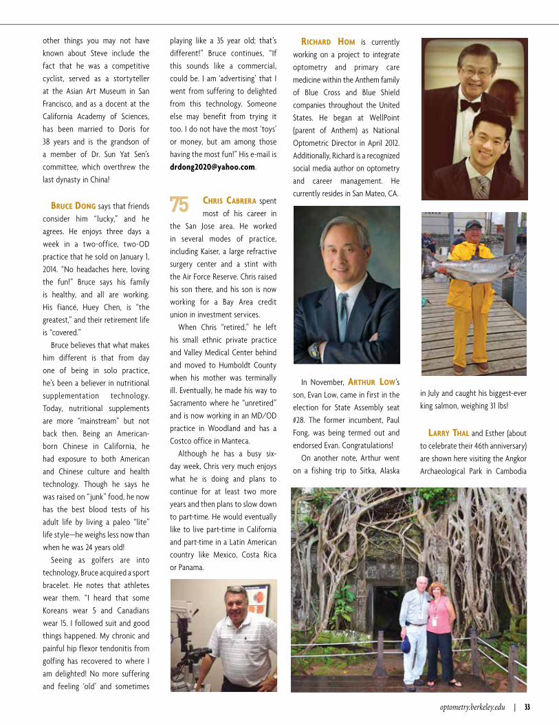
optometry.berkeley.edu | 33
other things you may not have
known about Steve include the
fact that he was a competitive
cyclist, served as a stortyteller
at the Asian Art Museum in San
Francisco, and as a docent at the
California Academy of Sciences,
has been married to Doris for
38 years and is the grandson of
a member of Dr. Sun Yat Sen’s
committee, which overthrew the
last dynasty in China!
Bruce Dong says that friends
consider him “lucky,” and he
agrees. He enjoys three days a
week in a two-office, two-OD
practice that he sold on January 1,
2014. “No headaches here, loving
the fun!” Bruce says his family
is healthy, and all are working.
His fiancé, Huey Chen, is “the
greatest,” and their retirement life
is “covered.”
Bruce believes that what makes
him different is that from day
one of being in solo practice,
he’s been a believer in nutritional
supplementation technology.
Today, nutritional supplements
are more “mainstream” but not
back then. Being an American-
born Chinese in California, he
had exposure to both American
and Chinese culture and health
technology. Though he says he
was raised on “junk” food, he now
has the best blood tests of his
adult life by living a paleo “lite”
life style—he weighs less now than
when he was 24 years old!
Seeing as golfers are into
technology, Bruce acquired a sport
bracelet. He notes that athletes
wear them. “I heard that some
Koreans wear 5 and Canadians
wear 15. I followed suit and good
things happened. My chronic and
painful hip flexor tendonitis from
golfing has recovered to where I
am delighted! No more suffering
and feeling ‘old’ and sometimes
playing like a 35 year old; that’s
different!” Bruce continues, “If
this sounds like a commercial,
could be. I am ‘advertising’ that I
went from suffering to delighted
from this technology. Someone
else may benefit from trying it
too. I do not have the most ‘toys’
or money, but am among those
having the most fun!” His e-mail is
75 chris caBrera spent
most of his career in
the San Jose area. He worked
in several modes of practice,
including Kaiser, a large refractive
surgery center and a stint with
the Air Force Reserve. Chris raised
his son there, and his son is now
working for a Bay Area credit
union in investment services.
When Chris “retired,” he left
his small ethnic private practice
and Valley Medical Center behind
and moved to Humboldt County
when his mother was terminally
ill. Eventually, he made his way to
Sacramento where he “unretired”
and is now working in an MD/OD
practice in Woodland and has a
Costco office in Manteca.
Although he has a busy six-
day week, Chris very much enjoys
what he is doing and plans to
continue for at least two more
years and then plans to slow down
to part-time. He would eventually
like to live part-time in California
and part-time in a Latin American
country like Mexico, Costa Rica
or Panama.
richarD hoM is currently
working on a project to integrate
optometry and primary care
medicine within the Anthem family
of Blue Cross and Blue Shield
companies throughout the United
States. He began at WellPoint
(parent of Anthem) as National
Optometric Director in April 2012.
Additionally, Richard is a recognized
social media author on optometry
and career management. He
currently resides in San Mateo, CA.
In November, arThur Low’s
son, Evan Low, came in first in the
election for State Assembly seat
#28. The former incumbent, Paul
Fong, was being termed out and
endorsed Evan. Congratulations!
On another note, Arthur went
on a fishing trip to Sitka, Alaska
in July and caught his biggest-ever
king salmon, weighing 31 lbs!
Larry ThaL and Esther (about
to celebrate their 46th anniversary)
are shown here visiting the Angkor
Archaeological Park in Cambodia

34 | BERKELEY OPTOMETRY FALL 2014
stay connected
during the most recent Optometry
Alumni trip. Larry is retiring for the
third time at year end (Air Force,
private practice and Berkeley
Optometry). Esther retired more
than a year ago and has many
more trips planned. Daughter
Danielle and husband Ben live
in New Orleans where they met
while going to Tulane. They both
completed MBAs and are “off the
payroll.” Middle daughter Nicole
has a scholarship to Tulane Law
School and is nearly “off the
payroll.” Arielle, the youngest, is in
her second year of medical school
at George Washington University,
Washington, DC—no scholarships
to be had and a while to go yet,
so “still on the payroll.” Larry’s
mother, widow of Bernhardt
Thal ’48, is 95, doing very well and
making all the Thals quite proud!
76 In January 2014,
DaviD hsieh earned
a master’s of science degree
in systems engineering from
George Washington University,
Washington, DC. In April, he
renewed his Project Management
Professional (PMP) certification.
80 Congratulations to
(pictured left to right)
ranDaLL yuMori and DonaLD MaTsuMoTo whose office was
named Optometry Giving Sight
(OGS) National Practice of the Year.
Optometry Giving Sight, a
global fundraising organization that
specifically targets the prevention
of blindness and impaired vision
due to uncorrected refractive error,
has honored Pacific EyeCare and its
doctors, Randall and Donald, as the
National Practice of the Year for
2013. Drs. Yumori and Matsumoto
have tirelessly given and sponsored
fundraising efforts for OGS to help
fund OGS’ efforts to train local eye-
care professionals, establish vision
centers for sustainability and deliver
eye care and low-cost glasses.
OGS was created by the World
Council of Optometry, the Brien
Holden Vision Institute Foundation
and the International Agency for
the Prevention of Blindness (IAPB)
in 2003 to mobilize resources from
the global optometric community
to help eliminate refractive error
blindness and low vision. OGS
supports the goals of VISION
2020—the global initiative to
eliminate preventable blindness
sponsored by IAPB and the World
Health Organization (WHO).
82 Don MuTTi OD ’82,
PhD ’92, received The
Ohio State Alumni Association
Award for Distinguished Teaching.
85 vengu LaKshMianrayanan,
PhD ’85, just completed a yearlong
sabbatical as a visiting professor of
physics and electrical engineering
at the University of Michigan, Ann
Arbor. Last year, he was awarded
the Esther Hoffman Beller medal
of the Optical Society of America
for contributions to optical science
and engineering. He serves on
the Steering Committee of the
International Year of Light, declared
to be in 2015 by the United Nations.
His eldest daughter, Molly, is
currently finishing an MS in
ethnomusicology at Memorial
University in Canada, and his second
daughter, Kelly, is a second-year
student in software engineering at
the University of Waterloo.
86 Pictured from left to
right are MiKe Boggs,
BLaKe Kuwahara , Karin Meng and JeFF azus celebrating
Mike’s birthday in San Francisco’s
North Beach.
89 cLauDia Bear is
currently Managing
Optometrist at the Kaiser
Permanente South Bay Medical
Center, which includes 18 optom-
etrists working in 4 locations.
In her spare time, she conducts
parent-training workshops for her
daughters’ charter schools, along
with exhibiting paintings and artist
books in several art shows.
90 corina van De PoL
joined the faculty at
Marshall B. Ketchum University
(Southern California College
of Optometry) as an Assistant
Professor in June 2014. She is now
teaching Geometric and Physical
Optics to first-year students.

optometry.berkeley.edu | 35
92 This summer carL JacoBsen completed
a 3,300-mile solo motorcycle
trip to Colorado. He has recently
expanded his teaching duties to
include work as a motorcycle riding
instructor for Moto-U, a local riding
school. He lives in an El Cerrito
home with a stunning view, a lovely
wife and a grumpy cat.
93 Randall Lum moved
his practice to a new
shopping center in South San
Francisco, Westborough Square.
This move will be the last of his
career as he has up to a 25-year
lease. It is his second move since
purchasing the practice in 1998.
The grand opening was scheduled
for October!
Randall has two children at
St. Ignatius High School in San
Francisco. Matthew is in the class
of 2016, and Carolyn is in the class
of 2018.
98 KrisTine eng ,
weyLin eng ’65 and
their family attended the showing
of The Price Is Right in Los Angeles.
Kristine notes that it was quite
the “Eng Adventure,” as Shannon
Eng (Kristine’s sister and Weylin’s
daughter) was a contestant…
and won!
06 Classmates sara chiu
and chrisToPher gee purchased the practice of
retiring MarK McaDaMs ’74 in
Dublin, CA. The grand opening
of Iron Horse Optometric Group
was scheduled for October. You
can follow the updates on their
Facebook page at www.facebook .com/ironhorseoptometry.
They are pictured in their office
(which used to be a tile store).
10 TiFFany chan was
recently promoted
to Assistant Professor of
Ophthalmology in the Vision
Rehabilitation Service at the
Wilmer Eye Institute, Johns
Hopkins School of Medicine.
In MeMorIaM
Sherwyne r. Bakar ’61
CurtiS Benton ’42
CharleS F. Brown ’47
william e. Dore ’56
John r. GriFFin ’58
winiFreD hirSCh
roBert r. reCtor ’51
VinCent G. SanChez ’90
Peter V. SiGnorotti ’49
ShelDon m. Smith ’58
(wiFe oF Former Dean, monroe hirSCh)

36 | BERKELEY OPTOMETRY FALL 2014
ALUMNI FOCUS
stay connected
AFTER UNDERGRADUATE WORK at the University
of California, Santa Barbara, Dr. Zadnik received all
three of her academic degrees from the University
of California, Berkeley, School of Optometry (BS ’80,
OD ’82, PhD ’92). After optometry school and before
graduate school, she worked in the Department of
Ophthalmology at the University of California, Davis, full-
time, ultimately serving on the faculty there for 14 years.
In 1996 Dr. Zadnik joined the faculty at The Ohio State
University College of Optometry where she became a full
professor and the first and only Glenn A. Fry Professor
in Optometry and Physiological Optics. She has chaired
the university’s Biomedical Sciences Institutional Review
Board for 10 years. As Associate Dean for a decade,
Dr. Zadnik was responsible for both the professional and
graduate programs and research. In June of 2014 she was
named the fifth (and first female) dean of The Ohio State
University’s College of Optometry.
Dr. Zadnik has an impressive list of academic and
professional achievements. Very early in her career, she
attended the American Academy of Optometry meeting
and found the organization of her heart. She quickly
became a Fellow and Diplomat in Cornea and Contact
Lenses. While ascending to chair the Section on Cornea
and Contact Lenses, she served as Chair of the Academy’s
Research Committee. In 1995 Dr. Zadnik received the
American Optometric Foundation’s Glenn A. Fry Award,
in recognition of the quality, significance, impact and
relevance to optometry of her research contributions.
After many years of service on the Board of Directors, she
served the Academy as President in 2010–2012.
Dr. Zadnik has served as Chair of the American
Optometric Association’s (AOA) Council on Research since
Dr. Karla Zadnik has been clear about her life’s purpose since she was 16 years old. Her paternal grandfather, Dr. William J. Henry, an optometrist in Ohio, had a huge influence on her. Shortly after her grandfather’s passing, Dr. Zadnik decided to follow his professional path and become an optometrist. While she originally planned a career in the private practice of optometry, life presented her with mentors and opportunities that led her on a path to being one of the most prolific optometric researchers of our time.
Dr. Karla Zadnik OD ’82, PhD ’92 was named to the National Optometry Hall of Fame.

optometry.berkeley.edu | 37
2000. She is especially proud of the Council’s Summer
Research Institutes, held every other summer since 1988
and co-sponsored by the AOA and the Academy, that have
resulted in an estimated $78 million in extramural funding
for optometric research.
Her record of leadership is impressive. Dr. Zadnik served
on the National Eye Institute’s (NEI) National Advisory
Eye Council in 2000–04, based on her research success.
She chaired the Collaborative Longitudinal Evaluation of
Keratoconus Study, the first multicenter study based in
optometry that was funded by the NEI. Simultaneously,
she and Don Mutti (OD ’82, PhD ’92) and Emeritus Dean
Tony Adams initiated the Orinda Longitudinal Study of
Myopia, which became the Collaborative Longitudinal
Evaluation of Ethnicity and Refractive Error Study, a
20-year, multi-center project to develop predictors for
juvenile-onset myopia.
Dr. Zadnik has served the optometry profession well.
For her outstanding accomplishments and significant
contributions to the profession, Dr. Zadnik was recently
honored as an inductee of the National Optometry Hall
of Fame. She is well known among her colleagues as
an accomplished researcher, gifted educator and skilled
administrator, yet her most remarkable attribute is her
energetic personality and selfless concern for others;
she has hosted countless faculty members from all over
the country over lobster served in her Ohio backyard.
In fact, when her husband, Kurt Zadnik, managing editor
of Optometry and Vision Science, sees someone at an
optometric gathering and the person says, “I’ve had lobster
at your house,” he replies drily, “Hasn’t everyone?”
Dr. Zadnik notes her own career “has been highlighted
by a series of mentors,” and she is a sought-after mentor
herself, especially by young students and optometrists
who are navigating their own personal and professional
juggling acts. Her experiences raising daughters Andra (29,
working in international film distribution at Lionsgate in
Los Angeles) and Nina Marie (24, a preschool teacher in
Powell, Ohio) give her lots of stories to tell about having
it all, leaning in, and just getting through the day. Building
on the legacies of her own mentors—Mark Mannis, Tony
Adams and Mert Flom (OD ’51, PhD ’57)—she is excited to
take The Ohio State University College of Optometry to
new heights.
Out of Ohio State’s five deans, three are graduates of Berkeley Optometry. From left to right: Karla Zadnik OD ’82, PhD ’92, Frederick Hebbard MS ’51, PhD ’57 and Richard Hill OD ’58, PhD ’61.

38 | BERKELEY OPTOMETRY FALL 2014
HALL OF FAME
stay connected
In honor of those alumni and retired faculty whose contributions to the profession of optometry, through service, teaching or research have been so monumental as to be universally
recognized, the school has dedicated the “Hall of Fame.” These individuals have made contributions that distinguish the University of California, Berkeley, School of Optometry above all others!
BERKELEY OPTOMETRY CELEBRATED its third induction to its Hall of Fame at a gala on February 22. Being inducted this year were Tony Adams, Colin Blakemore, Darrell Carter, Ted Cohn, Ed Elliott, Ralph Freeman, Mike Harris, Don Korb, Sheldon Miller, Don Mitchell, Tom Peters, Larry Stark and Karen Walker-Brandreth. The formal presentation started with a brief and humorous introduction by Assistant Dean Larry Thal, who with former Clinical Professor Craig Hisaka, originally conceived of the Hall of Fame to recognize not only those who have had such an important historical role at Berkeley Optometry, but also for whom future students should reflect upon as role models.
Larry and Craig had noted that with discussions with students in Primary Care Clinic regarding the heritage of our profession that Chuck Segar (past AOA president and HOF member) was thought to be a famous guitar player (probably confused with Robert Clark Segar and the Silver Bullet Band), that Monroe Hirsch (former Dean and HOF Charter member) was confused with the famous football player, Elroy “Crazy Legs” Hirsch from the University of Wisconsin, and that Allan Freid (another HOF Charter member and at the time a professor and chair of the COA Legislative Committee), was thought to be Alan Freed, the founder of “Rock n’ Roll.”
As a result Larry and Craig assigned each student in the Practice Management class that they taught a biography. The biographies were quite good and students enjoyed doing them, especially the interviews
of those who were still living. Larry took them to Dean Adams and out of this project came the HOF.
Tony appointed Meredith Morgan to chair the first committee and asked Larry to serve as secretary. Of course he would not pass up an opportunity to work with Meredith on such a project. He told the story of his first meeting where he met Meredith outside of what was then the contact lens clinic at the top of the stairwell near the dean’s office. At that time, the bronze bust of Meredith was there. Meredith arrived shortly before Larry, and as he came up the stairs from the clinic he heard a fourth-year student ask Meredith if he could help him. Meredith asked if he looked like he needed help and the student responded that he thought Meredith looked lost. Meredith asked the student if he knew who he was, which he didn’t. Just as Larry reached the top of the stairs to try and bail him out, he saw the student look over at the bronze,
back at Meredith, back at the bronze and then turn as red as a pomegranate.
What has evolved is not yet perfect but that is the challenge moving forward. Meredith Morgan established a precedent that to avoid politics in the selection process, if the nominee was a faculty member that he or she was not eligible until retired. After Executive Vice Chancellor and Provost George Bresslauer thanked Dean Dennis Levi for his tenure and great service, Dennis and Bob Mandell introduced each Hall of Fame inductee. (Complete descriptions are available on the Berkeley Optometry website.) Among the remarks that were particularly memorable are the following:
Hall of Fame 2014 inductees (pictured from left to right): Donald Korb, OD; Tony Adams, OD, PhD; Darrell Carter, OD, PhD ’57 (seated); Donald E. Mitchell, PhD; Colin Blakemore, PhD ’68; Karen Walker-Brandreth, OD ’68; Michael G. Harris, OD ’65, Ed Elliott, OD ’65; Sheldon S. Miller, PhD.
George Bresslauer and Master of Ceremonies Dennis Levi
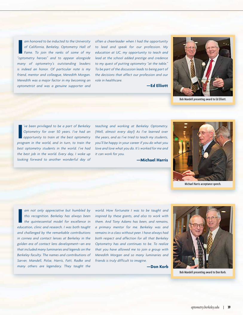
optometry.berkeley.edu | 39
I am honored to be inducted to the University
of California, Berkeley, Optometry Hall of
Fame. To join the ranks of some of my
“optometry heroes” and to appear alongside
many of optometry’s outstanding leaders
is indeed an honor. Of particular note is my
friend, mentor and colleague, Meredith Morgan.
Meredith was a major factor in my becoming an
optometrist and was a genuine supporter and
often a cheerleader when I had the opportunity
to lead and speak for our profession. My
education at UC, my opportunity to teach and
lead at the school added prestige and credence
to my quest of putting optometry “at the table.”
To be part of the discussion leads to being part of
the decisions that affect our profession and our
role in healthcare.
—Ed Elliott
Bob Mandell presenting award to Ed Elliott.
I ’ve been privileged to be a part of Berkeley
Optometry for over 50 years. I’ve had an
opportunity to train at the best optometry
program in the world, and in turn, to train the
best optometry students in the world. I’ve had
the best job in the world. Every day, I woke up
looking forward to another wonderful day of
teaching and working at Berkeley Optometry.
(Well, almost every day!) As I’ve learned over
the years, and as I’ve tried to teach my students,
you’ll be happy in your career if you do what you
love and love what you do. It’s worked for me and
it can work for you.
—Michael Harris
Michael Harris acceptance speech.
I am not only appreciative but humbled by
this recognition. Berkeley has always been
the quintessential model for excellence in
education, clinic and research. I was both taught
and challenged by the remarkable contributions
in cornea and contact lenses at Berkeley in the
golden era of contact lens development—an era
that included many luminaries and legends on the
Berkeley faculty. The names and contributions of
Sarver, Mandell, Polse, Harris, Fatt, Radke and
many others are legendary. They taught the
world. How fortunate I was to be taught and
inspired by these giants, and also to work with
them. And Tony Adams has been, and remains,
a primary mentor for me. Berkeley was and
remains in a class without peer. I have always had
both respect and affection for all that Berkeley
Optometry has and continues to be. To realize
that you have allowed me to join a group with
Meredith Morgan and so many luminaries and
friends is truly difficult to imagine.
—Don Korb Bob Mandell presenting award to Don Korb.

40 | BERKELEY OPTOMETRY FALL 2014
HALL OF FAME
stay connected
40 | BERKELEY OPTOMETRY FALL 2014
T he School of Optometry and the Berkeley campus
have benefited each one of us in unique ways
and I feel privileged and very lucky to share with
all of you this particular and very special cycle of life.
I would like to begin by emphasizing what I believe all of us
understand—the School of Optometry is a relatively unique
entity on the Berkeley campus.
There are a variety of different reasons for this and
I’d like to point out one of them. It has to do with the
different ways we can carry out our scientific mission. The
history of the School of Optometry encompasses a critical
association with two important ways of thinking about how
science should be done. For example, there is the notion of
“pure” science. What we usually mean by that is basic or
laboratory-based research that is reductionist in nature
and usually hypothesis driven. In contrast with that form of
thinking and behaving, is science for “use.”
Science for use is sometimes referred to as translational
science or in the biomedical world—clinical science. There
is a dynamic tension that has historically existed between
those two ways of behaving. That tension has been noticed
at least since the time of Aristotle. Some think that those
two ways of thinking about science are really disparate and
are necessarily separated by long periods of gestation.
I think, however, there are clear examples to show that is
not necessarily the case and that those two ways of doing
science can be encompassed together. An example is the
recent spectacular advances in stem cell biology that have
been made in the last eight years or so.
They have demonstrated that human adult somatic
cells can be reprogrammed into their pluripotent state,
practically equivalent to embryonic stem cells. Those
induced pluripotent stem cells can be then differentiated
into stable authentic adult cells of almost any kind. These
cells have been used in a wide variety of investigations to
significantly deepen our understanding of mechanisms of
human disease in organs
throughout the body.
These basic science
advances have led to
fundamentally important
discoveries in human
developmental biology.
Whole organs from
throughout the body
(including the eye) have
been generated by a single
population of these cells.
This outcome has provided the basis for fundamentally
important clinical advances in regenerative medicine
(transplantation) and in the development of small molecule
therapeutic interventions against a growing number of
diseases. What this work shows is that concomitant clinical
and basic work can expand, via positive feedback in both
directions, at an edifyingly rapid pace.
This example illustrates one aspect of science,
sometimes called translational, that embodies separate
ways of thinking. Unfortunately, each of these separate
ways of thinking are often exaggerated/disparaged in the
competition for research dollar support.
Here the point of my comments is to illustrate how
clinically driven (science for use) advances serve to drive the
basic science part of the equation by causing an enormous
increase in our understanding of human developmental
biology.
I believe that the embodiment of pure science and science
for use is a critically important aspect of the Berkeley
Optometry mission, along with teaching and service. These
pillars honor the highest goals of this University, of the
biomedical community, and of the public that we all serve.
—Sheldon Miller
Dennis Levi presenting award to Sheldon Miller.

optometry.berkeley.edu | 41
As a former graduate student here at Berkeley I have to
single out the counsel provide by my supervisor, Gerald
Westheimer, who taught me true scientific rigor.
Toward the end of my graduate training, Gerald provided me with
wise advice that set me on the career path that followed. He suggested
that I should borrow an old practice from my Australian roots,
called squatters rights, which would allow me to broaden my future
career prospects by obtaining a postdoctoral position in a field like
psychology. After a while he suggested that people may not question
my qualifications for an academic position in this field. This strategy
worked extremely well as I obtained a position in the Department of
Psychology (now the Department of Psychology & Neuroscience) at
Dalhousie University in Halifax NS, Canada.
It has not all been a bed of roses at Dalhousie; I remember early
on an incident triggered by the high teaching loads associated
with psychology. I was required each morning to teach three
different classes in four hours, and on the second day of the new
term I mixed up the four-digit numbers of the lecture room and
turned up at the wrong room. The
philosophy students in the room
were expecting a lecture on logic
but instead I began to describe the
scotopic luminosity curve.
Either because of the subject
matter or my use of colored chalk,
they became very attentive and
silent. Ten minutes into the class an
elderly student (as I thought) who
was the true professor for the class
(philosophy profs are habitually
late), entered the room and began to yell at me. I told him to be quiet
and wait until later to ask a question. Eventually I realized that I was
in the wrong room and beat a hasty retreat that was accompanied by
money changing hands as students settled their bets as to the length
of time it would take for me to realize my error.
—Donald Mitchell
Dennis Levi presenting award to Donald Mitchell.
T hey say that no one is ever sorry on their deathbed that
they spent too little time at the office, but in the case of
my father, he wished until his last moment that he was
able to work. He loved his work, his great good fortune at getting
paid for doing what he loved, and his colleagues and graduate
students from all around the world, who filled our home during my
childhood.—Elizabeth Stark Powers
on behalf of her father, Larry Stark
I am truly honored and deeply touched by
this recognition. I have a brief story to
share with you. Often, over the last few
years, in the middle of night, when it was dark
and quiet, and when I let my mind wander, I
would have a recurrent vision. It was an image of
my photograph hanging in “The Hall” with the
picture of my late husband, Roy.
Then, in the light of day, I’d tell myself to
“snap out of it” and “quit dreaming,” because the
reality was that such an event was never going
to happen. Well, tonight, from the bottom of my
heart, I must thank you for making my dream
come true, for making this my reality
—Karen Walker-Brandreth
Bob Mandell presenting award to Karen Walker-Brandreth.
I
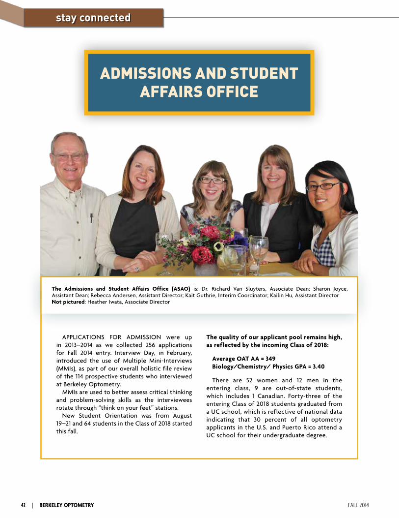
ADMISSIONS AND STUDENT AFFAIRS OFFICE
APPLICATIONS FOR ADMISSION were up in 2013–2014 as we collected 256 applications for Fall 2014 entry. Interview Day, in February, introduced the use of Multiple Mini-Interviews (MMIs), as part of our overall holistic file review of the 114 prospective students who interviewed at Berkeley Optometry.
MMIs are used to better assess critical thinking and problem-solving skills as the interviewees rotate through “think on your feet” stations.
New Student Orientation was from August 19–21 and 64 students in the Class of 2018 started this fall.
The quality of our applicant pool remains high, as reflected by the incoming Class of 2018:
Average OAT AA = 349Biology/Chemistry/ Physics GPA = 3.40
There are 52 women and 12 men in the entering class, 9 are out-of-state students, which includes 1 Canadian. Forty-three of the entering Class of 2018 students graduated from a UC school, which is reflective of national data indicating that 30 percent of all optometry applicants in the U.S. and Puerto Rico attend a UC school for their undergraduate degree.
The Admissions and Student Affairs Office (ASAO) is: Dr. Richard Van Sluyters, Associate Dean; Sharon Joyce, Assistant Dean; Rebecca Andersen, Assistant Director; Kait Guthrie, Interim Coordinator; Kailin Hu, Assistant DirectorNot pictured: Heather Iwata, Associate Director
stay connected
42 | BERKELEY OPTOMETRY FALL 2014

optometry.berkeley.edu | 43
Each year the Berkeley Optometry Continuing Education Office certifies over 15,000 continuing education hours for optometrists practicing in California and other states, making it the largest program at an accredited optometry school in North America. Highlights include the Berkeley Practicum, Morgan Symposium and Berkeley Glaucoma Day.
Directed by Patsy Harvey, OD, MPH ’81, and coordinated by Danni Peck, MBA, CMP, the
Continuing Education Office seeks to provide a variety of high-quality courses for optometrists as they fulfill their requirements of licensure renewal set by the California State Board of Optometry.
Other CE programs provided by our clinical faculty, Vision Science faculty and the Office of Development and Alumni Relations include the annual Berkeley Optometry CE Conference and Reunion Celebration, Glaucoma Grand Rounds and the Resident Forum.
UC Berkeley Optometry’s Lectures and Demonstrations (UC-BOLD) will be rolling out a new and continuing series of COPE-approved online Continuing Education programs. We will be offering programs covering a wide-variety of TPA-certified topics, including various lectures on the diagnosis and treatment of ocular and systemic diseases. We will also have programs dedicated to enhancing knowledge about new developments in ophthalmic materials and advancements in contact lenses.
UC-BOLD’s goal is not only to provide the convenience, efficiency and financial advantages of delivering content over the internet, but also to continually utilize and integrate technology to provide an unmatched quality of education, both through superlative content and through user experience and interactive content.
For more information please visit the UC-BOLD website: www.berkeleyce.org
ONLINE CONTINUING EDUCATION
Dean John Flanagan, OD, PhD, will be the Keynote Speaker and present the Karen Walker-Brandreth Lecture at the Berkeley Practicum on January 17-19, 2015.
His lecture, “Glaucoma: What We Know, How We Should Practice, and What the Future May Hold,” will present valuable information and insight on glaucoma diagnosis and management, as well as possible directions for the future.
The remainder of the 26th Annual Berkeley Practicum will feature a wide range of exciting topics as well as the return of its third keynote speaker, Jonathan Horton, MD, PhD, Professor of Ophthalmology, Neurology and Physiology at UC San Francisco. Dr. Horton will present “My Approach to Vision Loss,” his methodology for investigating vision loss at points along the visual pathway with examples and cases involving the photoreceptor layer to the visual cortex.
Additional presentations will include “Nutrition: Looking Beyond the Golden Arches” with nutrition specialist, Kimberly Reed, OD. Exploring a prevalent disease and recent findings of its ocular manifestations will be “Alzheimer’s Disease: Causes, Treatments and Ocular Manifestations” by Patsy Harvey, OD, MPH. Ocular disease presentations will include “Early Detection of Glaucoma–Clinical Clues,” “Glaucoma Pharmacology A–Z,” “High Risk Refractive Surgery: Complications and Solutions,” and our interactive series “Grand Rounds: What Would You Do?”
The Berkeley Practicum will be held at the DoubleTree Hotel, Berkeley Marina. More information may be found at optometry.berkeley.edu/ce and online registration is available at regonline.com/UCBSO_BP2015.
DEAN FLANAGAN TO KEYNOTE BERKELEY PRACTICUM
CONTINUING EDUCATION
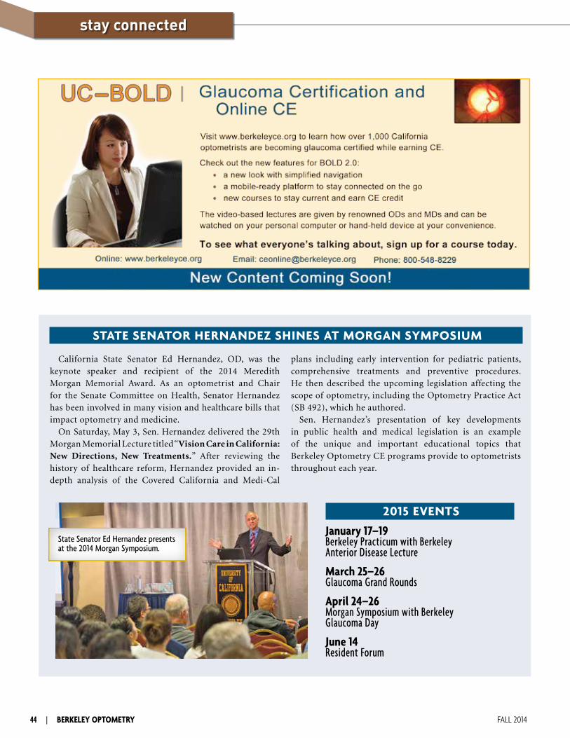
44 | BERKELEY OPTOMETRY FALL 2014
The Meredith W. Morgan University of California Eye Center
stay connected
California State Senator Ed Hernandez, OD, was the keynote speaker and recipient of the 2014 Meredith Morgan Memorial Award. As an optometrist and Chair for the Senate Committee on Health, Senator Hernandez has been involved in many vision and healthcare bills that impact optometry and medicine.
On Saturday, May 3, Sen. Hernandez delivered the 29th Morgan Memorial Lecture titled “Vision Care in California: New Directions, New Treatments.” After reviewing the history of healthcare reform, Hernandez provided an in-depth analysis of the Covered California and Medi-Cal
plans including early intervention for pediatric patients, comprehensive treatments and preventive procedures. He then described the upcoming legislation affecting the scope of optometry, including the Optometry Practice Act (SB 492), which he authored.
Sen. Hernandez’s presentation of key developments in public health and medical legislation is an example of the unique and important educational topics that Berkeley Optometry CE programs provide to optometrists throughout each year.
STATE SENATOR HERNANDEZ SHINES AT MORGAN SYMPOSIUM
January 17–19 Berkeley Practicum with Berkeley Anterior Disease Lecture
March 25–26 Glaucoma Grand Rounds
April 24–26 Morgan Symposium with Berkeley Glaucoma Day
June 14 Resident Forum
2015 EVENTS
State Senator Ed Hernandez presents at the 2014 Morgan Symposium.
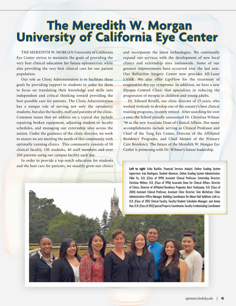
optometry.berkeley.edu | 45
THE MEREDITH W. MORGAN University of California
Eye Center strives to maintain the goals of providing the
very best clinical education for future optometrists while
also providing the very best clinical care for our patient
population.
Our role as Clinic Administrators is to facilitate these
goals by providing support to students in order for them
to focus on translating their knowledge and skills into
independent and critical thinking toward providing the
best possible care for patients. The Clinic Administration
has a unique role of serving not only the optometry
students, but also the faculty, staff and patients of the clinic.
Common issues that we address on a typical day include
repairing broken equipment, adjusting student or faculty
schedules, and managing our externship sites across the
nation. Under the guidance of the clinic director, we work
to ensure we are meeting the needs of this community with
optimally running clinics. This community consists of 50
clinical faculty, 130 students, 40 staff members and over
200 patients using our campus facility each day.
In order to provide a top-notch education for students
and the best care for patients, we steadily grow our clinics
and incorporate the latest technologies. We continually
expand our services with the development of new local
clinics and externship sites nationwide. Some of our
greatest improvements have occurred over the last year.
Our Refractive Surgery Center now provides All-Laser
LASIK. We also offer LipiFlow for the treatment of
evaporative dry eye symptoms. In addition, we have a new
Myopia Control Clinic that specializes in reducing the
progression of myopia in children and young adults.
Dr. Edward Revelli, our clinic director of 23 years, who
worked tirelessly to develop one of the country’s best clinical
training programs, recently retired. After searching for over
a year, the School proudly announced Dr. Christina Wilmer
’96 as the new Associate Dean of Clinical Affairs. Her many
accomplishments include serving as Clinical Professor and
Chief of the Tang Eye Center, Director of the Affiliated
Residency Programs, and Chief Mentor of the Primary
Care Residency. The future of the Meredith W. Morgan Eye
Center is promising with Dr. Wilmer’s future leadership.
The Meredith W. Morgan University of California Eye Center
Left to right: Erika Racklin, Financial Services Analyst, Online Grading System Supervisor; Irais Rodriguez, Student Absences, Online Grading System Administrator; Vikki Yu, O.D. (Class of 1999) Assistant Clinical Professor, Externship Director; Christina Wilmer, O.D. (Class of 1996) Associate Dean for Clinical Affairs, Director of Clinics, Director of Affiliated Residency Programs; Kerri Yoshiyama, O.D. (Class of 2004) Assistant Clinical Professor, Assistant Clinic Director; Tom Michelsen, Clinic Administrative Office Manager, Building Coordinator for Minor Hall Addition; Linh Le, O.D. (Class of 2011) Clinical Faculty, Faculty/Student Schedules Manager; and Anney Han, O.D. (Class of 2013) Special Projects Coordinator, Faculty Credentialing Coordinator

UNIVERSITY OF CALIFORNIA, BERKELEYSCHOOL OF OPTOMETRY302 Minor Hall # 2020Berkeley, California 94720-2020
ADDRESS SERVICE REQUESTED
Nonprofit Organization U.S. Postage Paid
University of California, Berkeley
The Class of 2014