unit 3 (chapter 5) - unit 3 (chapter 5) - the integumentary system and body membranes presented by...
TRANSCRIPT

Unit 3 (Chapter 5) Unit 3 (Chapter 5) - - The
Integumentary System and Body
Membranes
Presented by Dawn Duran, Presented by Dawn Duran, PT, MHS,CSCSPT, MHS,CSCS
Adjunct Professor, Kaplan Adjunct Professor, Kaplan UniversityUniversity
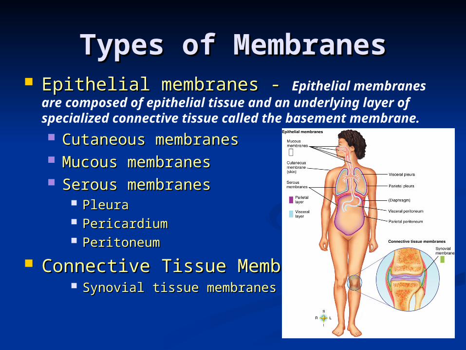
Types of MembranesTypes of Membranes Epithelial membranes - Epithelial membranes - Epithelial membranes are
composed of epithelial tissue and an underlying layer of specialized connective tissue called the basement membrane.
Cutaneous membranesCutaneous membranes Mucous membranes Mucous membranes Serous membranes Serous membranes
Pleura Pleura PericardiumPericardium PeritoneumPeritoneum
Connective Tissue MembranesConnective Tissue Membranes Synovial tissue membranesSynovial tissue membranes

CLASSIFICATION OF BODY MEMBRANESCLASSIFICATION OF BODY MEMBRANES
Epithelial membranesEpithelial membranesThe cutaneous membrane refers The cutaneous membrane refers to the skin, which is the to the skin, which is the primary organ of the primary organ of the integumentary system. integumentary system. It is the largest organ of the It is the largest organ of the bodybody
Composes ~15% of total body Composes ~15% of total body weight in most individuals.weight in most individuals.

CLASSIFICATION OF BODY MEMBRANESCLASSIFICATION OF BODY MEMBRANES
Mucous membranesMucous membranes line body cavities that open up to the outside, such as the digestive tract.
The linings of the respiratory, reproductive, and urinary tracts are examples of mucous membranes.
Other examples: eyes, nose, mouth vagina, Other examples: eyes, nose, mouth vagina, anus anus
Produce mucus, a thick secretion that Produce mucus, a thick secretion that keeps the membranes soft and moistkeeps the membranes soft and moist
Another purpose of mucus is to catch Another purpose of mucus is to catch debris, and not allow it to enter the debris, and not allow it to enter the body. It helps to keep out invading body. It helps to keep out invading pathogens. pathogens.

CLASSIFICATION OF BODY MEMBRANESCLASSIFICATION OF BODY MEMBRANES
Serous MembranesSerous Membranes Line the cavities that do not open to Line the cavities that do not open to the outside worldthe outside world
Examples include the heart, kidneys, spleen, and lungs.
Composed of two distinct layers of Composed of two distinct layers of tissuetissue
Epithelial sheet is a thin layer of simple Epithelial sheet is a thin layer of simple squamous epitheliumsquamous epithelium
Connective tissue layer forms a thin, gluelike Connective tissue layer forms a thin, gluelike basement membrane that holds and supports basement membrane that holds and supports epithelial cellsepithelial cells
In other words, In other words, sserous membranes are double membranes. They may become inflamed due to infection.

CLASSIFICATION OF BODY MEMBRANESCLASSIFICATION OF BODY MEMBRANES
Double layered – the serous membrane is a Double layered – the serous membrane is a single, continuous sheet that covers two single, continuous sheet that covers two different surfacesdifferent surfaces
Visceral membraneVisceral membrane covers the surface of organs covers the surface of organs found in body cavitiesfound in body cavities
A serous membrane that covers the organs found in a body cavity is referred to as the visceral portion.
Parietal membraneParietal membrane lines body cavities lines body cavities A serous membrane that lines the walls of a body cavity is referred to as the parietal
portion.
Serous fluid is in the cavity between themSerous fluid is in the cavity between them This fluid prevents friction when movement This fluid prevents friction when movement occursoccurs
Friction would create a build-up of heatFriction would create a build-up of heat

Serous MembranesSerous Membranes
Pleura = A serous membrane lining the walls of the thoracic cavity and covering the lungs.
The parietal layer of the pleura The parietal layer of the pleura adheres to the wall of the chest and adheres to the wall of the chest and the visceral layer is attached to the the visceral layer is attached to the lung. With every breath we take the lung. With every breath we take the two layers slide past each other with two layers slide past each other with little friction, making it easier for little friction, making it easier for us to breathe. us to breathe.
Peritoneum = serous membrane lining the walls of the abdominal cavity and covering the organs in that cavity.

Synovial MembranesSynovial Membranes
Synovial membranes are connective Synovial membranes are connective tissue membranes tissue membranes They do not contain epithelial componentsThey do not contain epithelial components They They line the spaces between bones and line the spaces between bones and jointsjoints that move as well as the lining of that move as well as the lining of bursa sacs that are found between many bursa sacs that are found between many moving body partsmoving body parts
Produce a lubricant called Produce a lubricant called synovial fluidsynovial fluid Synovial membranes and synovial fluid Synovial membranes and synovial fluid help to reduce frictionhelp to reduce friction

Disorders of Body Disorders of Body MembranesMembranes
DiseasesDiseases Pleurisy—inflammation of the serous Pleurisy—inflammation of the serous membranes that line the chest cavity and membranes that line the chest cavity and cover the lungscover the lungs
Pain is caused by irritation and Pain is caused by irritation and friction as the lungs rub against the friction as the lungs rub against the walls of the chest cavitywalls of the chest cavity
Peritonitis—inflammation of the serous Peritonitis—inflammation of the serous membranes in the abdominal cavity that membranes in the abdominal cavity that line the walls and cover the abdominal line the walls and cover the abdominal organsorgans
A possible complication of appendicitis is inflammation of the serous membrane that lines the abdominal cavity is and is termed peritonitis.
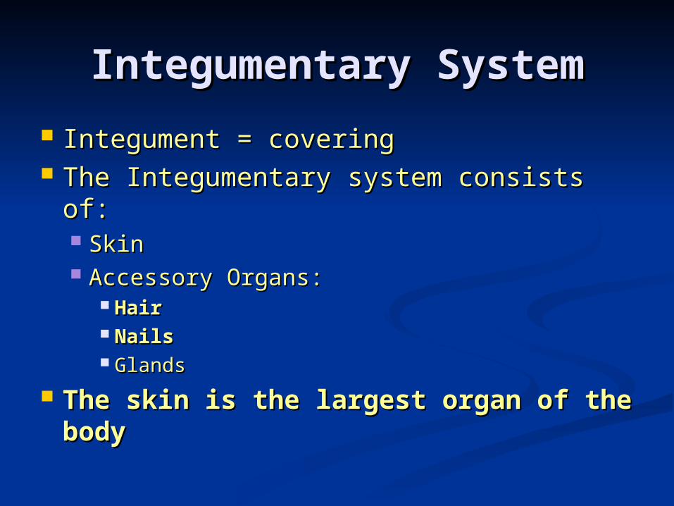
Integumentary SystemIntegumentary System
Integument = coveringIntegument = covering The Integumentary system consists The Integumentary system consists of:of: SkinSkin Accessory Organs:Accessory Organs:
HairHair NailsNails GlandsGlands
The skin is the largest organ of The skin is the largest organ of the bodythe body

QUESTION:QUESTION:
What are the What are the functions of functions of the skin?the skin?

Functions of the SkinFunctions of the Skin
Protection from injuriesProtection from injuries Acts as a barrier and regulates Acts as a barrier and regulates what enters/leaves body.what enters/leaves body.
Regulates body temperature.Regulates body temperature. Synthesizes and stores vitamin Synthesizes and stores vitamin DD
Sensory functions/sense organ Sensory functions/sense organ activityactivity

Temperature Regulation Temperature Regulation
The skin helps with controlling The skin helps with controlling our body temperature by our body temperature by regulating sweat secretion and regulating sweat secretion and by regulating the flow of blood by regulating the flow of blood close to the body surface. close to the body surface. Blood vessels to the skin constrict when the
body needs to conserve heat.

Functions of the SkinFunctions of the Skin
Sense organ activitySense organ activity Skin functions as an enormous Skin functions as an enormous sense organsense organ
Receptors serve as receivers for Receptors serve as receivers for the body, keeping it informed of the body, keeping it informed of changes in its environmentchanges in its environment We have specialized receptors for We have specialized receptors for light touch, pressure, pain, heat and light touch, pressure, pain, heat and cold. cold.

Functions of the SkinFunctions of the Skin Protection—first line of defenseProtection—first line of defense
Against infection by microbesAgainst infection by microbes Against fluid loss Against fluid loss
Keratin is a tough waterproof protein that gives the outer layer of the skin some protection and helps to prevent dehydration.
Against ultraviolet rays from sunAgainst ultraviolet rays from sun Melanin is responsible for absorbing harmful ultraviolet radiation
from the sun before it reaches the deeper tissues of the integumentary system.
Against harmful chemicalsAgainst harmful chemicals Against cuts and tearsAgainst cuts and tears

Structures of the SkinStructures of the Skin
3 main layers from superficial 3 main layers from superficial to deep:to deep: EpidermisEpidermis DermisDermis Subcutaneous/Subcutaneous/
HypodermisHypodermis

Structures of the SkinStructures of the Skin
1) Epidermis1) EpidermisEpi- Epi- derm/o derm/o -is-isaboveabove skinskin structurestructure
““structure above the skin”structure above the skin”a. a. The outermost and primary layer of the The outermost and primary layer of the skin.skin. b. The stratum corneum is the tough b. The stratum corneum is the tough outer/superficial layer of the epidermis. outer/superficial layer of the epidermis. This layer contains keratin. This layer contains keratin. c. c. The innermost layer of the epidermis that that has cells that continually reproduce with new cells moving toward the surface stratum germinativum.d. The epidermis is avascular.

Structures of the SkinStructures of the Skin
2) Dermis2) DermisDerm/oDerm/o -is-isSkinSkin structurestructure
““true skin”true skin”a. thick, connective tissue layer a. thick, connective tissue layer with collagen and elastic fibers, with collagen and elastic fibers, epithelial tissue, smooth muscle epithelial tissue, smooth muscle tissue, nervous tissue and bloodtissue, nervous tissue and bloodb. The dermis lies directly above b. The dermis lies directly above the subcutaneous layer. the subcutaneous layer. c. The dermis is thicker than the c. The dermis is thicker than the epidermis. epidermis.
Dermis Dermis The dermal papilla forms ridges and grooves The dermal papilla forms ridges and grooves
that make up the fingerprints on the tips of the that make up the fingerprints on the tips of the fingers. fingers.
The purpose of skin ridges on our fingers and The purpose of skin ridges on our fingers and toes is to improve our grip when using tools or toes is to improve our grip when using tools or walking barefoot. walking barefoot.
Loss of elasticity due to decreased numbers of elastic fibers contributes to wrinkles and aging. The dermis contains an abundance of elastic fibers.
Subcutaneous injections of medicines are meant to reach the hypodermis.

Sensory Structures of Sensory Structures of DermisDermis
Receptors of the integumentary system are found in the dermis.
Deep touch/pressure on the skin Deep touch/pressure on the skin surface/vibration: Pacinian corpusclessurface/vibration: Pacinian corpuscles
Light touch/pressure: Meisner’s corpusclesLight touch/pressure: Meisner’s corpuscles Warm temperature: Free nerve endingsWarm temperature: Free nerve endings Cold temperature: Free nerve endingsCold temperature: Free nerve endings Pain: Free nerve endingsPain: Free nerve endings

Structure of the Skin, Structure of the Skin, cont. cont.
3) Subcutaneous tissue/Hypodermis3) Subcutaneous tissue/Hypodermis
Sub-Sub- cutane/ocutane/o -us-us
BelowBelow skinskin structurestructure
““structure below the skin”structure below the skin”
a. a. The hypodermis consists of adipose tissue which provides insulation from extremes of heat and cold as well as a source of stored energy. It can be used as a food source in some situations. It also provides a cushion of protection from injury.

You should be able to You should be able to label this diagram, found label this diagram, found
on p 103.on p 103.

QUESTION:QUESTION:
What are some of What are some of the accessory the accessory structures of structures of
the skin?the skin?

ANSWER:ANSWER:
HairHair Nails (produced by the epidermis)Nails (produced by the epidermis) Specialized Sense Organs (found in the Specialized Sense Organs (found in the dermis)dermis)
GlandsGlands
The skin itself is the main organ of the The skin itself is the main organ of the integumentary system. integumentary system.
The integumentary system also The integumentary system also INCLUDESINCLUDES the skin, hair and specialized sense the skin, hair and specialized sense organs. organs.

Accessory Structures of Accessory Structures of the skinthe skin
Hair Hair The fine, soft hair of fetus and newborn The fine, soft hair of fetus and newborn called called lanugolanugo
Hair growth requires epidermal tube-like Hair growth requires epidermal tube-like structurestructure called called hair follicle hair follicle (Fig 5.5, p (Fig 5.5, p 107)107)
Hair growth begins in the papilla when Hair growth begins in the papilla when cells of the epidermal layer grown down cells of the epidermal layer grown down into the dermis, forming a small tube into the dermis, forming a small tube called a hair folliclecalled a hair follicle
Hair root lies hidden in follicle; visible Hair root lies hidden in follicle; visible part of the hair is called the shaftpart of the hair is called the shaft
Alopecia is the term for hair lossAlopecia is the term for hair loss Arrector piliArrector pili - - specialized smooth muscle specialized smooth muscle that produces “goose bumpsthat produces “goose bumps” and causes ” and causes hair to stand up straight when we become hair to stand up straight when we become frightened or coldfrightened or cold

Accessory Structures of Accessory Structures of the skinthe skin
NailsNails (Figure 5-6, p 108) (Figure 5-6, p 108) Produced by epidermal cells over Produced by epidermal cells over terminal ends of fingers and toesterminal ends of fingers and toes
Visible part called nail bodyVisible part called nail body Root lies in a groove and is hidden by Root lies in a groove and is hidden by
cuticlecuticle Crescent-shaped area nearest root Crescent-shaped area nearest root called called llunulaunula
Nail bed may change color with change Nail bed may change color with change in blood flow as it is abundant in in blood flow as it is abundant in blood vesselsblood vessels


Accessory Structures of Accessory Structures of the skinthe skin
Skin glandsSkin glands TypesTypes
Sweat or sudoriferous Sweat or sudoriferous ((apocrine apocrine and eccrine)and eccrine)
SebaceousSebaceous
Accessory Structures of Accessory Structures of the skinthe skin
Sweat or sudoriferous glandsSweat or sudoriferous glands TypesTypes
Eccrine sweat glandEccrine sweat gland Most numerous, important, and widespread of Most numerous, important, and widespread of the sweat glands. There are more sweat the sweat glands. There are more sweat glands than oil glands in one square inch glands than oil glands in one square inch of skin. of skin.
Produce perspiration or sweat, which flows Produce perspiration or sweat, which flows out through pores on skin surface. When out through pores on skin surface. When this evaporates, the body loses heat. this evaporates, the body loses heat.
Function throughout life and Function throughout life and assist in body assist in body heat regulationheat regulation as well as elimination of as well as elimination of waste products such as ammonia and uric waste products such as ammonia and uric acidacid

Accessory Structures of Accessory Structures of the skinthe skin
Apocrine sweat glandApocrine sweat gland Found primarily in axilla and Found primarily in axilla and around genitaliaaround genitalia
Secrete a thicker, milky Secrete a thicker, milky secretion quite different from secretion quite different from eccrine perspirationeccrine perspiration
Breakdown of secretion by skin Breakdown of secretion by skin bacteria produces odorbacteria produces odor
These glands enlarge and begin These glands enlarge and begin to function at pubertyto function at puberty

Accessory Structures of Accessory Structures of the skinthe skin
Sebaceous glandSebaceous gland Secrete oil/sebum to lubricate hair and Secrete oil/sebum to lubricate hair and skinskin
Level of secretion increases during Level of secretion increases during adolescenceadolescence
Amount of secretion regulated by sex Amount of secretion regulated by sex hormoneshormones
Sebum accunulation in sebaceous gland Sebum accunulation in sebaceous gland ducts forms white pimples, and sebum ducts forms white pimples, and sebum often darkens when exposed to air, often darkens when exposed to air, forming a blackheadforming a blackhead
Acne vulgaris is the term for Acne vulgaris is the term for inflammation of sebaceous gland ductsinflammation of sebaceous gland ducts
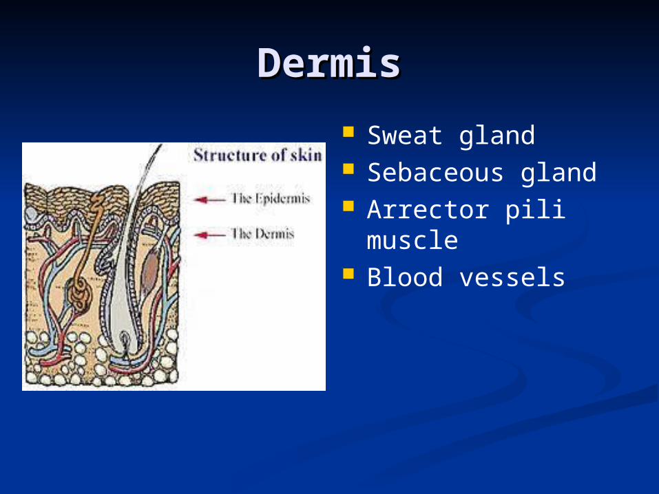
DermisDermis
Sweat gland Sebaceous gland Arrector pili muscle
Blood vessels

The SkinThe Skin
Appendages of the skinAppendages of the skin Receptors Receptors
Receptors are specialized nerve endings Receptors are specialized nerve endings that make it possible for the skin to that make it possible for the skin to act as a sense organact as a sense organ
Meissner’s corpuscle—capable of Meissner’s corpuscle—capable of detecting detecting
light touch;light touch; located close to the skin located close to the skin surfacesurface
Pacinian corpuscle—capable of detecting Pacinian corpuscle—capable of detecting
pressurepressure
Quick QuizQuick Quiz
Ok, quick quiz. Which layer of Ok, quick quiz. Which layer of the skin does not have a blood the skin does not have a blood supply?supply?

ANSWERANSWER
Correct! The epidermis doesn’t Correct! The epidermis doesn’t have a blood supply. The have a blood supply. The epidermis and subcutaneous epidermis and subcutaneous tissues do have a blood supply. tissues do have a blood supply.
Quick QuizQuick Quiz
Which layer has the pigment Which layer has the pigment cells?cells?

ANSWERANSWER
The epidermis again. Remember The epidermis again. Remember that it has many layers. The that it has many layers. The pigment cells begin development pigment cells begin development in the dermis and migrate to in the dermis and migrate to the epidermis as they mature. the epidermis as they mature.

Quick QuizQuick Quiz
What is that pigment called?What is that pigment called?

ANSWERANSWER
Melanin! It is produced by the Melanin! It is produced by the melanocytes.melanocytes.
This pigment is called melanin and This pigment is called melanin and is responsible for giving the skin is responsible for giving the skin some of its color. some of its color.
Dark skinned individuals can get melanoma despite the fact that the dark skinned individuals have more melanocytes than fair skinned individuals.
Quick QuizQuick Quiz
Which layer has the fatty Which layer has the fatty tissue?tissue?

ANSWERANSWER
The subcutaneous The subcutaneous tissue/hypodermis. tissue/hypodermis.

BurnsBurns The severity of a burn is determined by the depth of the The severity of a burn is determined by the depth of the injury as well as by the amount of surface area involvedinjury as well as by the amount of surface area involved. .
First-degreeFirst-degree (partial-thickness) burns - only the (partial-thickness) burns - only the surface layers of epidermis involved. surface layers of epidermis involved. Causes minor Causes minor discomfort and some skin reddening, but no blistering discomfort and some skin reddening, but no blistering and minimal damage.and minimal damage. Example: typical sunburn. Example: typical sunburn.
Second-degreeSecond-degree (partial-thickness) burns - (partial-thickness) burns - involve the involve the deep epidermal layers and always cause injury to the deep epidermal layers and always cause injury to the upper layers of the dermis. Causes blisters, reddening upper layers of the dermis. Causes blisters, reddening of the skin, severe pain, swelling and fluid loss.of the skin, severe pain, swelling and fluid loss. Scarring is also common. Scarring is also common.
Third-degreeThird-degree (full-thickness) burns — (full-thickness) burns — characterized by characterized by complete destruction of the epidermis and dermis and complete destruction of the epidermis and dermis and tissue death extends into subcutaneous tissues.tissue death extends into subcutaneous tissues. Often Often involve muscles and bone.involve muscles and bone. Not painful because nerve Not painful because nerve endings are destroyed. endings are destroyed.
This is the most serious type of burn. This is the most serious type of burn. The most serious complications of third degree burns is The most serious complications of third degree burns is fluid loss and risk of infection.fluid loss and risk of infection.


BurnsBurns
All of the following can cause All of the following can cause burns to the skin:burns to the skin: ElectricityElectricity Ultraviolet lightUltraviolet light Chemicals Chemicals

BurnsBurns
Determining the extent of burn Determining the extent of burn injury is possible by utilizing injury is possible by utilizing the “rule of nines”the “rule of nines” in adults; in adults; this estimates the body surface this estimates the body surface area effected by the burn. area effected by the burn. The body is divided into 11 areas The body is divided into 11 areas of 9% eachof 9% each
Additional 1% of body surface area Additional 1% of body surface area around genitalsaround genitals

The Rule of NinesThe Rule of Nines
**Be able to use the Rule of Nines to answer a question regarding %age of body burned based on a scenario provided on your exam**

Skin CancerSkin Cancer
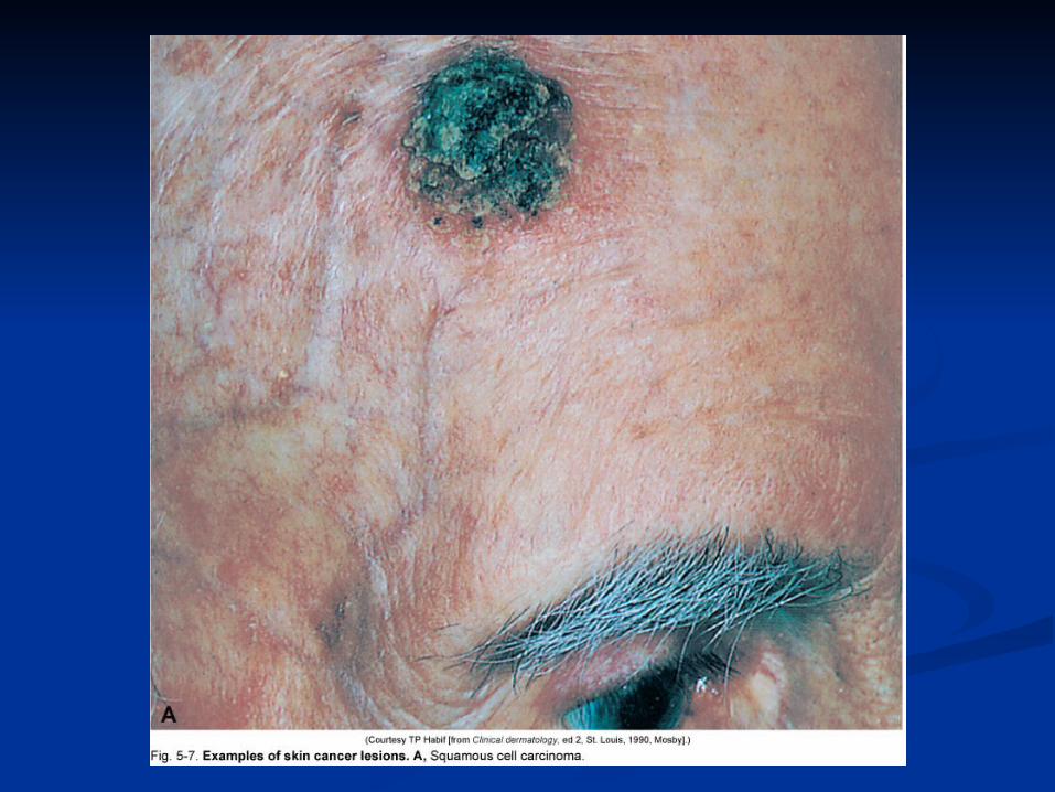
Three common typesThree common types Squamous cell carcinoma—is common; Squamous cell carcinoma—is common; lesions begin as hard, painless lesions begin as hard, painless nodules; it will metastasize.nodules; it will metastasize.
Metastasis is when cancer spreads to Metastasis is when cancer spreads to distant sites from where it originated. distant sites from where it originated.
Basal cell carcinoma— the most common Basal cell carcinoma— the most common type of skin cancer. type of skin cancer.
Melanoma— the most serious type of skinMelanoma— the most serious type of skin cancercancer. . Is indicated by a pigmented Is indicated by a pigmented spot that contains different colors. spot that contains different colors.


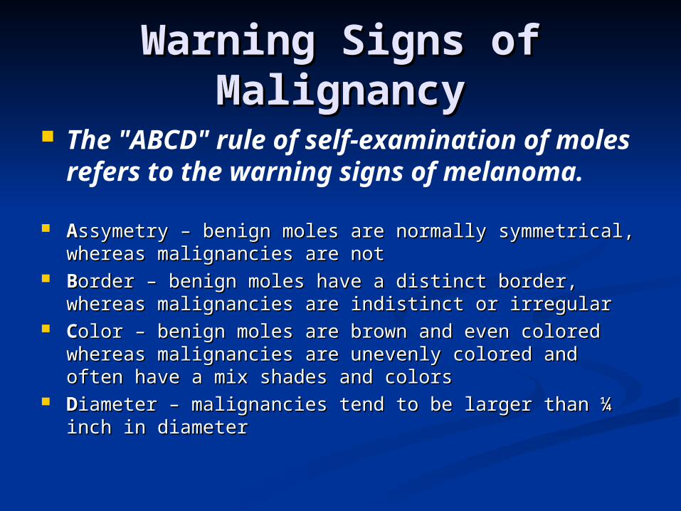
Warning Signs of Warning Signs of MalignancyMalignancy
The "ABCD" rule of self-examination of moles refers to the warning signs of melanoma.
AAssymetry – benign moles are normally symmetrical, ssymetry – benign moles are normally symmetrical, whereas malignancies are notwhereas malignancies are not
BBorder – benign moles have a distinct border, order – benign moles have a distinct border, whereas malignancies are indistinct or irregularwhereas malignancies are indistinct or irregular
CColor – benign moles are brown and even colored olor – benign moles are brown and even colored whereas malignancies are unevenly colored and whereas malignancies are unevenly colored and often have a mix shades and colorsoften have a mix shades and colors
DDiameter – malignancies tend to be larger than ¼ iameter – malignancies tend to be larger than ¼ inch in diameterinch in diameter

Skin CancerSkin Cancer
The most important causative The most important causative factor in common skin cancers is factor in common skin cancers is exposure to sunlight. exposure to sunlight. In other words, ultraviolet In other words, ultraviolet radiation is the most important radiation is the most important factor in causing skin cancer. factor in causing skin cancer.
UV radiation damages the DNA in UV radiation damages the DNA in skin cells which interferes with skin cells which interferes with normal mitosis and leads to cancer.normal mitosis and leads to cancer.

Decubitus UlcersDecubitus Ulcers
Decubitus ulcers (aka Decubitus ulcers (aka bedsores/pressure sores)bedsores/pressure sores) develop when excessive pressure slows develop when excessive pressure slows down blood flow to sensitive areasdown blood flow to sensitive areas
More common in bedridden or immobilized More common in bedridden or immobilized individualsindividuals
However, they are NOT always a sign of However, they are NOT always a sign of abuse. abuse.
MiscellaneousMiscellaneous
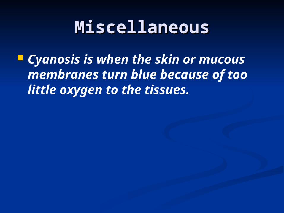
Cyanosis is when the skin or mucous membranes turn blue because of too little oxygen to the tissues.

Let’s Let’s Review!Review!

Which type of body Which type of body membrane lines the membrane lines the digestive tract?digestive tract?
A.A. Cutaneous membraneCutaneous membraneB.B. Tympanic membraneTympanic membraneC.C. Serous membraneSerous membraneD.D. Synovial membraneSynovial membraneE.E. Mucous membraneMucous membrane

ANSWERANSWER
Answer: EAnswer: E Mucous membrane lines cavities Mucous membrane lines cavities that are open to the exterior, that are open to the exterior, which includes the lumen of the which includes the lumen of the digestive tract. digestive tract.

The skin plays important The skin plays important roles in maintaining a roles in maintaining a stable body temperature.stable body temperature.
A.A. TrueTrueB.B. FalseFalse
ANSWERANSWER
Answer: AAnswer: A How? Via sweating and How? Via sweating and diversion of blood flow. diversion of blood flow.

What is the largest What is the largest sensory organ of the body?sensory organ of the body?
A.A.EyeEyeB.B.EarEarC.C.TongueTongueD.D.SkinSkinE.E.NoseNose

ANSWERANSWER
Answer: DAnswer: D All the sensory receptors in All the sensory receptors in the largest organ of the body the largest organ of the body make skin the clear winner. make skin the clear winner.

What characterizes second-What characterizes second-degree burns?degree burns?
A. blistersA. blistersB.B. swellingswellingC.C. severe painsevere painD.all of the aboveD.all of the above

ANSWERANSWER
Answer: DAnswer: D

That’s All, Folks!That’s All, Folks!