understanding, managing, and living with malignant · pdf filewhere do malignant wounds occur...
TRANSCRIPT
Understanding,Managing,
and Living withMalignant Wounds
Sherry A. Clark

Understanding, Managing, and Living with Malignant Wounds: A Resource Guide for Patients and Caregivers isprovided at no cost thanks to many in-kind donations. This valuable resource is available inhardcopy and electronic formats. To obtain a hardcopy, pdf, or iBook version choose from thefollowing:
Print version:Benton Hospice Service2350 NW Professional Dr. Corvallis, OR 97330
pdf version:www.bentonhospice.org/malignantwound.pdf
iBook version:Search the iBooks store for Understanding, Managing, and Living with Malignant Wounds
To submit feedback, comments, or arrange contributions send e-mail to: [email protected]
Medical Information DisclaimerThe medical information provided in Understanding, Managing, and Living with Malignant Wounds isobtained from a variety of public resources. Every effort has been made to ensure that links toexternal sites were current and correct. This information is intended as a patient educationresource only and should not be used for diagnosing or treating a health problem. It is not asubstitute for expert professional care. If you have or suspect you may have a health problem,please consult your health care provider.
Copyright © 2012 by Sherry A. Clark and Robert W. Durst
Front cover by Mark Allison
version 2.1

Understanding, Managing,and Living with Malignant Wounds
A Resource Guide for Patients and Caregivers
Sherry A. Clark

ContentsList of Illustrations ………………………………………………………………… iiiForeword …………………………………………………………………………… ivPreface …………………………………………………………………………… vDedication ………………………………………………………………………… viiAcknowledgments ……………………………………………………………… viiiIntroduction ……………………………………………………………………… ixUnderstanding Malignant Wounds ……………………………………………… 1
What is a malignant wound? ……………………………………………… 1What is the origin of a malignant wound? ………………………………… 1Do all malignant wounds look alike? ……………………………………… 1Where do malignant wounds occur? ……………………………………… 2Are malignant wounds easy to diagnose? ……………………………………2How does a malignant wound progress over time? ………………………2
Normal tissue ……………………………………………………………3Infiltration of cancer ………………………………………………………3Deeper invasion …………………………………………………………3Late stage …………………………………………………………………6
What is the incidence of malignant wounds? ………………………………6What can a patient and caregiver do? ………………………………………6
Managing Malignant Wounds ………………………………………………………8Moist wound beds ……………………………………………………………8Assessment ……………………………………………………………………8Moist wound care protocol……………………………………………………8Restoring optimal moisture ……………………………………………………9
Wound bed A ……………………………………………………………10Wound bed B ……………………………………………………………12
Optimal moisture dressing layers……………………………………………15Signs and symptoms ………………………………………………………… 15
Pain ……………………………………………………………………… 15Exudate ………………………………………………………………… 17Bleeding ………………………………………………………………… 18Odor …………………………………………………………………… 18Itching …………………………………………………………………… 20
Possible side effects and complications ……………………………………21Home and away preparedness ………………………………………………22
Living with Malignant Wounds ………………………………………………… 23Resources ………………………………………………………………………… 27
ii

List of Illustrations
Figure 1: Ulcerating and fungating lesions ……………………………………… 1Figure 2: Common sites for malignant wounds ………………………………… 2Figure 3: Malignant wound progressions (left to right) in the chest wall …… 4-5Figure 4: Example dressing layers: Wound bed A ……………………………… 10Figure 5: Elastic net tank top …………………………………………………… 12Figure 6: Example dressing layers: Wound bed B ……………………………… 13Figure 7: Storing dressings ……………………………………………………… 22
Book cover and illustrations by Mark Allison. Mark began his medical illustration career atRancho Los Amigos Hospital in Downey, CA, right after college. He has a degree in biologyfrom Santa Clara University. After several years working at the hospital, Mark continued hiseducation and expanded his skills into commercial art, illustration, design, and fine art. He isan award winning teacher of art and design for Linn Benton Community College. He alsoteaches for the community extension classes at Oregon State University, and continues hiscareer as a freelance illustrator and designer. He may be contacted through his website:www.MarkAllison.wordpress.com
Author photo by Robert Durst.
iii

ForewordCancer takes many faces, requires many responses, and challenges the patient and the caregiver in many ways. While malignant wounds are uncommon, our knowledge of how to carefor them is inadequate. At last, here is a handbook that not only helps the patient and the careprovider to understand the basic issues, but also provides practical approaches to the manyproblems of skin that has been invaded by disease.
Sherry A. Clark wrote this gift of a book after being diagnosed with breast cancer in 1993 andsuffering four separate recurrences. After surgery, radiation, and chemotherapy, sadly, Sherrywas left with malignant wounds. In our work as family physicians, we have encountered otherindividuals who have had similar skin problems. Managing their symptoms and problems hasbeen difficult, in part because we lacked clear and “common sense” educational materials.Now we have this excellent and helpful resource, a legacy from an amazing, eloquent author.
David H. Cutsforth, Jr. MDDavid R. Grube MD
iv

PrefaceFor a non-medically trained person, I thought that I knew a lot about cancer—breast cancer inparticular. During the nineteen years since my first cancer diagnosis, I’ve gradually accumulatedbits and pieces of information about the disease. I also have been very good at dodging thebullets of each recurrence (four), and took it for granted that I could do this forever. Imagine mysurprise when weeping sores appeared on my chest and I was told that nothing could be doneabout them. I asked my oncologist for a name for this ugly thing, a brochure about care of it, oranything from which to begin understanding what was happening to my body. He called itcutaneous metastasis. But no brochure, no understanding, no proper care. Would I have toaccept reduced quality of life, as well? No. Instead, I launched my own search for information.
Using lay and scientific search tools on the Internet, I began reading about breast cancer withcutaneous metastases. Published papers with that terminology mostly focused upon clinical trialsand specifics about rodent genetics. Nothing practical was published about how a patient shouldactually manage these wounds so that one could avoid soiled pajamas in the morning or reduceodor so that one could enjoy the company of friends. Instead, there was brief mention ofcommon materials that might bring relief: honey, yogurt, and dried plant material, for example,but not how they could be used in order to go about daily activities with confidence. I needed thehow, but it was not where I could find it in either print or electronic version.
Week after week, month after month, I searched trusted, scholarly sites on the Internet forsomething that would give me a practical solution. I was sent to a wound care nurse, whose onlysuggestions were to gently debride in the shower using a washcloth and soap, and to keep thewound dry during the day. After three months of this (later learned to be incorrect) treatment, Irequired hospitalization for severe cellulitis. A hospital wound nurse with actual experiencetreating malignant wounds reversed the previous protocol. She showed me the correct dressingsto use to keep the wound moist, what to expect, and a health products catalog from which topurchase suitable dressings as things progressed. Bless her! I also now had a new search term,malignant wound, for what was happening.
Armed with that catalog and medical insurance, I would be able to find dressings that wouldn’taggravate the situation or cause a new one. I continued my online search for almost two years.One day I came across the word fungating buried in the middle of a research paper aboutmalignant wounds. A medical dictionary definition for fungating described what I saw on my chest!
Using fungating in my search string called up a host of papers previously unknown to me.Fortuitously, the first of these was about how to manage fungating wounds. The paper was writtenby a nurse in Ohio and had been published only a few weeks earlier.
Other papers I found were about specific dressings, details about exudate, and discussion aboutbody image. I felt as though I’d won a big sweepstakes! Suddenly I had more to read than I hadtime.
My next prize discovery was written by a New Zealand nurse. His publication addressed an issuethat was problematic for me at the time. My wound and periwound area had progressed to such asize that a common, wide elastic bandage was too small and not shaped properly to cover andhold in place the increasingly larger dressings I needed. The publication included a slideshow
v
uploaded onto the Internet that described the use of tube-shaped, elastic netting cut to fitvarious body areas to hold dressings in place without slippage. This had the added benefit of notrequiring a second person to dress my wounds, improving my independence.
Thanks to nurses who published in their professional journals, I had resources from which Icould gather not only an understanding of what was going on but of how to care for mywounds. These were the tools I would use to improve my quality of life. Imagine how muchmore I could have accomplished, with more comfort and confidence, had these resources beenavailable at the onset of my wound development! I learned from the published literature that Iwas not alone with a cancer that creates malignant wounds. As a life-long educator, I had to sharewhat I’d discovered. There was only one thing to do—a last lesson to teach. My discoveries mustbecome a booklet, given to other malignant wound patients to guide them and restore quality totheir lives.
This booklet, with its collection of information and color illustrations, is intended to help readersbetter understand malignant wound care management. Never more should patients or theircaregivers lose precious time searching for what is going on or what they are supposed to do. Idon’t want patients, family, or friends to miss quality-of-life opportunities because of thesewounds.
Three years have passed since my finding of the first nodule under the skin of my chest. In spiteof time spent searching, I was determined not to let these wounds cause me to missopportunities. I traveled with my husband to Asia (I even took a Korean cooking class) and toEurope (where I rode atop a mechanical elephant in Nantes, France). We traveled the east coastof North America, where in one day we were in eight different states. We spent a month in theUK and Ireland visiting areas my ancestors emigrated from long ago.
I went through four different chemotherapy treatments, understanding that none would cure, buthoping that one might slow down the progress of the wounds. None worked. I tried a topicalcream (off-label use) in a clinical trial that didn’t work for me, either. I tried to get into otherclinical trials only to be turned away because I didn’t have “enough” bulk tumor.
I hired a new oncologist. I learned to ask medical practitioners before they began to offer advicewhether they knew the terms fungating, weeping malignant wound, etc. If they were unfamiliarwith these terms, I excused myself and left to find someone who did know. There was no time towaste on well-intentioned practitioners with out-of-date or incorrect information.
I retired early and focused upon home and hobby projects. I made my daughter’s wedding dressand enjoyed following her plans for a unique wedding ceremony. I focused upon bucket listprojects around the home, some last trips, and finally writing this booklet.
Even a determined me, with well-managed wounds, is no match for cancer. Eventually, the toll ofthe invading cancer upon internal structures became evident. It was not something to beimproved with different dressings or stronger medication. It was time to relinquish control of myhealth care needs to those far better equipped and trained. My husband and I phoned our localhospice service. This was one of the most difficult but the wisest decision we made as a couple.
My sincere hope is that Understanding, Managing, and Living with Malignant Wounds provides patientsand their caregivers with the tools and confidence that bring quality at the end of life.
vi

Dedication
To my caring, loving, soul-mate, Bob. I knew you were the one when you made me laugh.
vii

AcknowledgmentsTalented and caring people supported my efforts to create this booklet. What began as an idea for asmall brochure to fill a need for patients like me, evolved into multiple pages with color illustrationsdesigned for a larger audience. Self-publishing aside, I did not develop this informative, final productwithout the guidance and help of many others.
My physicians and many nurses in clinics, hospitals, and hospice, are overdue a giant thank you forthe treatment and care provided during my nineteen years experience with breast cancer. Arthur L.Clark put a check in my hand and told me to “Write the book!” I know patients, caregivers and theirhealth-care team will forever be thanking him for kick-starting the project. Mark Allison was theteacher of the first art class I ever took who spoke like a scientist. When this project began, a fewyears later, I knew who could create the illustrations I envisioned. Thank you, Mark.
Editing draft after draft of a document, written by a non-writer, must be tantamount to giving oneselfa root canal while going over a waterfall—or, something like that. For her outstanding, critical eyeand multitude of suggestions from start to finish, I am forever indebted to Ella May Wulff. Strugglingwith health issues of her own and that of her family, her perseverance got this project to the printshop. I am forever appreciative of Lucy Himelreich Noone for her last-minute proofreading andencouraging words when the project became overwhelming and my health was declining.
As the scope of this booklet grew, there became a need to ensure facts, availability of products, andmany other items I could not have imagined. Therefore, I cast my net to a broad range of reviewerswho responded with incredibly valuable input. These wonderful people include David Grube, MD;David Cutsforth, MD; Stephen Chui, MD; Shawn Foley, MD; Kelli Bergstrom, BSN, RN, CWOCN;Wayne Naylor, Senior Analyst at Cancer Control New Zealand, RN; Karen Daley, RN; ReenieSchwallie, RN, BSN, CHPIN; Robert Spiegel, RN, BSN, CHPN; Dawn Daniels, RN; and EmilyGilbert, Social Services, Benton Hospice Service.
Julie and Jessica, my wise, long-time friends whose varied support during this project is thanked fromthe bottom of my heart. I was blessed with encouraging words along the way from Carol, Gabriele,Jane, Kerry, Rick, Cliff, Diane, Ron and Kathy, Deron and Margo, members of Rickett’s circa ‘74, andmembers of the Order of the Holey Fungi Roundtable Group.
Yards of thanks to Jan and Lou at JanniLou Creations who allow Permission to Play instructors LuAnn,Nancy, Virginia, and Kathi, to teach cancer survivors a new craft, each month, in their quilt shop. ToChris, Anika and all my Silica Sisters who stimulated my creative juices while working in my glassstudio, I give you a perfectly fused thank you. To Chris, Eija, and others who offered transportationand humored me by fitting in a mocha or lunch in the process, I give you a delicious thank you.
For a fresh, clean house for four months I send a dust bunny-free thank you to Clean Endings, LLC.,a local housecleaning service. In 2011, the owner was introduced to Cleaning for a Reason through theowner of The Clean Team, a supplier of household cleaning products. The employees and staff atClean Endings quickly agreed that the gift of time for women battling cancer would be their “givingback to our community program”. See www.cleaningforareason.org for more information.
For assistance with future distribution of this booklet and its free, electronic availability I am gratefulto Micky Shields, ED and the nurses and staff at Benton Hospice Service. For availability in iBookformat, an iThanks goes to Kevin.
viii

IntroductionThe early sign of a malignant wound in the skin is frightening. The extent to which your wound willgrow and spread is unknown. The amount of time you have left to be fully mobile and free of painor other symptoms is unknown. Understanding, Managing, and Living with Malignant Woundsunfortunately cannot answer those unknowns. Instead, this booklet serves as a resource for thepatient and caregiver whose normal routines and quality of life have been challenged by thesewounds. The progress of a malignant wound from its earliest appearance to much later stages isassumed here to be managed by the patient and caregiver in the home.
This booklet begins with answers to questions asked by patients with malignant wounds. This isfollowed by a demonstration of how a malignant wound establishes itself in the skin, creating awound bed. For illustration purposes, the location of this wound is the chest wall of a patient with abreast cancer recurrence to that area. Following this wound through time, one gets a clear picture ofwhat is happening and learns the sensations the patient experiences as the wound develops. Byproviding an understanding of the process, the booklet lays the foundation for learning how tomanage malignant wounds as they continue to grow and spread. Throughout the booklet, terms thatmay not be familiar to everyone are italicized and defined in sidebars.
The management of malignant wounds in the chest wall with proper products and sound practicesmakes their care in the home feasible. This is not always true for wounds that appear in locations thepatient cannot see and reach. Some wounds may become more than the patient or caregiver is able orwilling to maintain without assistance.
This booklet describes a variety of symptoms and symptom management practices. Contrary to morecommon healing-wound-care, where a dry wound bed promotes healing, malignant wound carerequires a moist wound bed. Dressings needed to restore this optimal moisture environment aredemonstrated by two sample wound bed treatments requiring different approaches.
Proper care of the wound bed and symptom-appropriate medication satisfy the physical needs of thepatient, but the emotional and social impact of these wounds upon both patient and caregiver mustnot be underestimated. Wounds that are well managed provide quality time—free from discomfort orrisk of embarrassment. This booklet should serve to free up the patient and caregiver to maximizequality time with loved ones. The booklet concludes with a more personal conversation with theauthor about her experience living with malignant wounds.
I encourage patients and caregivers to seek further information and care only from medicalpractitioners who have previous experience supervising malignant wounds, their symptoms, andcomplications. Poor or wrong advice can have serious repercussions. Here, the author speaks fromexperience.
A list of resources at the end includes sources the author used but may not be available to everyone.The author was fortunate to have access to special or subscription-only research and medical data-bases. The patient and caregiver are cautioned against taking advice from a source that lacks validityor peer review. Before accepting any finding on the Internet or print form, discuss the matter withyour trusted, health care team.
ix

x

Understanding Malignant WoundsA small percentage of cancers will develop into malignant wounds in theskin. If they do, both the patient and caregiver need to know as muchas possible about these difficult wounds so that they can betterunderstand how to manage them and their complications.
What is a malignant wound?A malignant wound is visible evidence that cancer cells from a primarytumor have infiltrated the skin. Over time, it may grow deeper intohealthy underlying tissue layers. Unlike more common wounds, amalignant wound never heals. Despite its presence in skin, amalignant wound is not skin cancer per se but can arise from somekinds of melanoma as well as from other cancers.
What is the origin of a malignant wound?The origin of a malignant wound is usually a primary tumorsomewhere in the body. The route followed from a primary tumor toa malignant wound in the skin varies. Via one route, a primary tumorin or near the skin continues to grow and eventually becomes amalignant wound in the skin. A different route involves metastasis.Tumor cells detach from a primary tumor and travel to the skinthrough the blood, lymph vessels or between tissue planes. Onceestablished in the skin, these metastasized cancer cells multiply,producing early signs of the malignant wounds they will become.
Do all malignant wounds look alike?No. The early appearance of a malignant wound begins with one ormore raised nodules under the skin’s surface. These nodules eventuallydevelop into shapes that resemble a crater or a cauliflower (Figure 1).
1
malignant wound: an injury to(skin) tissue due to invasivecancerous growth
caregiver: in the context of thisbooklet, caregiver is the personthat provides the day-to-daycare of the patient. Caregiversmay be health professionals,family members, friends, socialworkers, or members of theclergy.
primary tumor: the originaltumor
infiltrating cancer: cancer thathas spread beyond the layer oftissue in which it developedand is growing intosurrounding, healthy tissues.Also called invasive cancer.
metastasis: the spread ofcancer from one part of thebody to another
lymph vessel: a thin tube thatcarries lymph (lymphatic fluid)and white blood cells throughthe lymphatic system
tissue plane: where twodifferent tissues meet and areusually joined by a thinconnective tissue layer, forexample where skin andmuscle meet
nodule: a growth or lump thatmay be malignant (cancer) orbenign (not cancer)
Figure 1. Ulcerating and fungating lesions Some wounds resemble a crater (left) with high edges and a sunken center, commonly called anulcerating lesion. Other wounds grow above the surface and resemble a cauliflower (right),commonly called a fungating lesion. Both types can occur in the same wound bed.

Crater-like wounds, commonly called ulcerating lesions, have high edgeswith a sunken center. Cauliflower-like wounds, referred to as fungatinglesions, look like clusters of raised nodules growing above the surfaceof the skin. Both ulcerating and fungating lesions can appear in thesame wound bed.
Where do malignantwounds occur?Malignant wounds occur in the skin.Because this is the largest organ ofthe body, there is potential for anypart of the skin to develop amalignant wound. In women, themost common primary tumor thatcan lead to a malignant wound is abreast cancer that has also spread tothe skin of the chest wall. In men,the most common primary tumor toinvade skin is lung cancer. In generalmalignant wounds can occur to theskin of the head and neck, back,abdomen, groin, armpit, anus, andgenitals from primary tumorsanywhere in the body (Figure 2).
Are malignant wounds easy to diagnose?Not always. Sometimes the appearance of a malignant woundprecedes symptoms of primary tumors. Seeing or feeling the raisednodule in the early stage of a malignant wound may be the first signof an underlying cancer somewhere else in the body. Unfortunately,these early signs may be mistaken for a variety of benign skinconditions. This can confuse diagnosis and delay important care ofthese wounds and of the primary tumor.
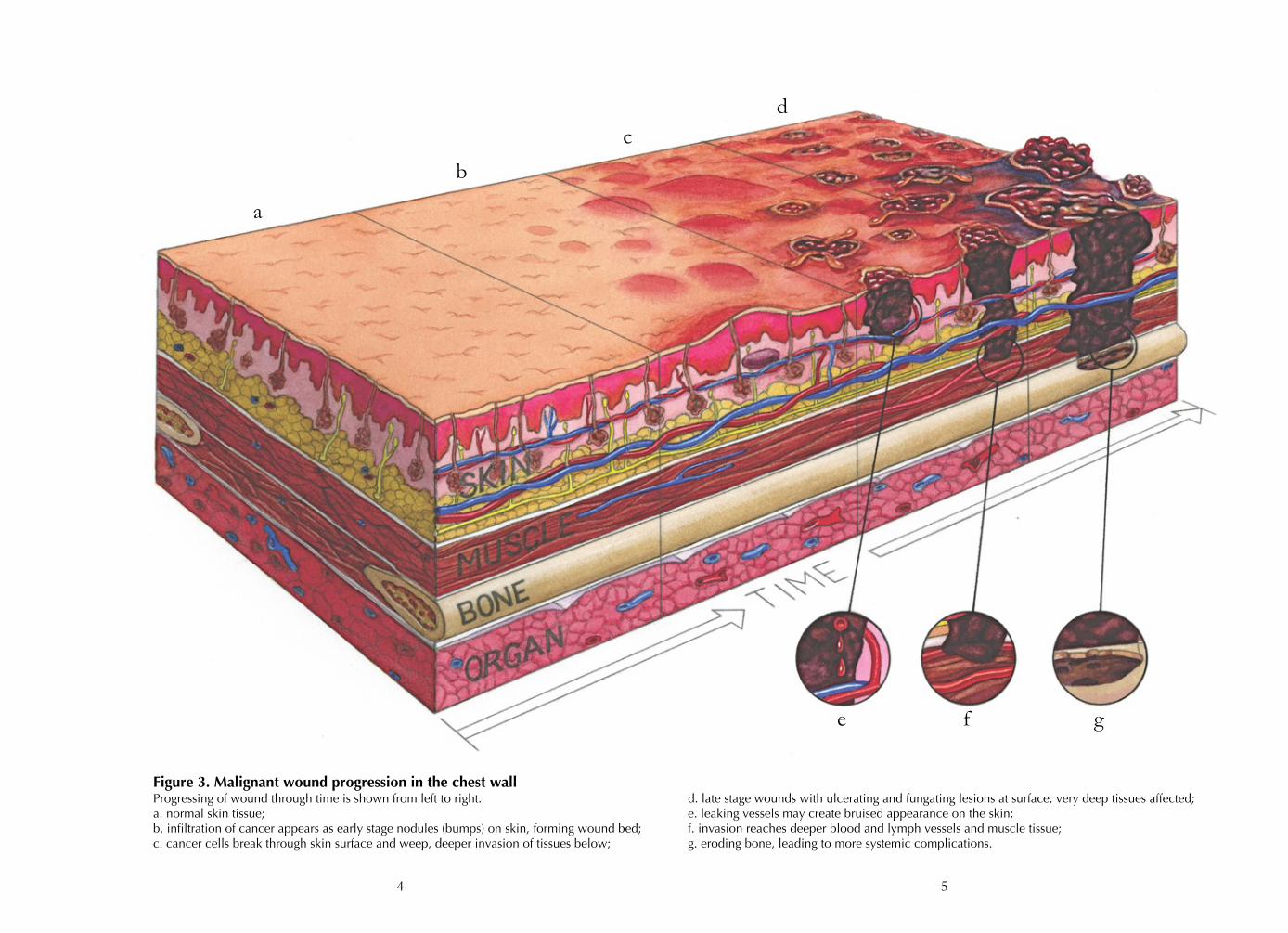
How does a malignant wound progress overtime?Figure 3 represents a block of malignant wound tissue as it wouldappear in the chest wall of a patient whose primary tumor was abreast cancer. Beginning with normal tissue (Figure 3a), we followthis same block through time (Figures 3b, c, d). The illustrationshows what can occur at the surface and beneath a malignant woundbed as the infiltrating cancer continues to grow. Early-stage nodules(Figure 3b) and ulcerating and fungating lesions (Figure 3c, d) areshown at the surface. As the nodules grow wider and deeper, thelikelihood of disruption and damage to the surface and underlying
2
ulcerating lesion: a type ofskin lesion where the surfacecells have died and been castoff
fungating lesion: a type of skinlesion that is marked by breaksin the skin, cancerous out-croppings, and necrosis (deathof living tissue)
wound bed: total area affectedby wounds in the skin
Figure 2. Common sitesfor malignant woundsPrimary tumors from any origincan lead to a malignant woundsomewhere on the skin. Thecircled areas are the morecommon sites malignantwounds tend to appear.

layers is demonstrated. The interval of time from Figure 3a to anysubsequent stage is unpredictable, ranging from weeks to years.
Normal TissueNormal skin tissue (Figure 3a) includes a healthy, intact surface.Beneath this unbroken surface is an intricate network of vessels. Inthe absence of cancer, these blood vessels bring nutrients and oxygento healthy cells while lymph vessels remove waste and excess fluid.Below the vessel networks is muscle tissue. Beneath this layer ofmuscle is bone tissue (a rib in our example). The organ tissue layershown here is the lung. As we follow this tissue block through time,we shall see how the infiltration of cancer cells affects each layer,beginning with skin tissue.
Infiltration of CancerA wound bed is being established (Figure 3b). Often this is when apatient first realizes there is a problem. As mentioned earlier, aprimary tumor may have gone undetected until it manifests itself inthe form of a raised nodule under the intact skin surface. Thesenodules may be flesh-colored or red, and may be visible to the eye, oronly detected with careful palpation. There is usually no pain at thisphase. Sometimes the nodules are associated with, or preceded by, arash. The periwound skin area may itch. Although it may appearnormal to the unaided eye, the periwound skin may actually havebeen damaged by previous oncology treatments (radiation, surgery,chemotherapy).
Deeper InvasionAs the nodules continue to grow (Figure 3c), the once intact skin maybe compromised. The surface area and depth of the wound bedincrease in size, affecting underlying vessel networks and possiblymuscle tissue. The continual breakdown of cells at the surfacereleases fluid and contributes to inflammation. If the skin is nolonger intact, the wound weeps exudate onto the wound bed. Exudatecontains water and toxic cellular materials that are irritating to thewound bed and periwound skin. This contributes to an environmentwhere opportunistic microbes (bacteria, yeast) can grow.
Early on, the surface of a weeping wound grows pale in color becausethe cancer is diverting blood vessels for its own growth deeper intothe wound. In time, wounds appear as a mixture of yellow, gray, orblack colors. This is a sign of necrosis. The combined effects ofexudate and necrotic tissue breakdown account for the foul odorassociated with weeping wounds. The invading cancer also affects theintegrity of vessel walls. This results in bleeding at or below the skin’ssurface. Leakage of blood from capillaries (Figure 3e) under the skingives rise to a bruised appearance at the surface. Fragile tissue just
3
palpation: examine by pressingon the surface of the body tofeel the organs or tissuesunderneath
periwound skin: the areasurrounding the wound bed
exudate: a mass of cells andfluid that has seeped out ofblood vessels or an organ, esp.in inflammation
weeping wound: a wound thatdischarges exudate
necrosis: the death of livingtissues
capillary: the smallest type ofblood vessel
4
Figure 3. Malignant wound progression in the chest wallProgressing of wound through time is shown from left to right.a. normal skin tissue; b. infiltration of cancer appears as early stage nodules (bumps) on skin, forming wound bed; c. cancer cells break through skin surface and weep, deeper invasion of tissues below;
a
b
cd
e f g
d. late stage wounds with ulcerating and fungating lesions at surface, very deep tissues affected; e. leaking vessels may create bruised appearance on the skin; f. invasion reaches deeper blood and lymph vessels and muscle tissue; g. eroding bone, leading to more systemic complications.
5

beneath a necrotic, malignant wound can bleed easily. The cancer notonly affects normal blood clotting factors but also weakens the wallsof larger blood vessels. This can lead to hemorrhaging.
The rapid growth of tumors at this stage can disrupt or overwhelmthe functioning of lymph vessels, too. The result is edema of the area,a source of pain and possibly reduced mobility. Deep invasion ofcancer can begin to encroach upon muscle tissue, also causing painand affecting mobility, among other discomforts.
Late StageFigure 3d illustrates continued growth into the chest wall. Thesurface of the malignant wound bed shows a variety of tumors atvarious stages of development. Beneath this visible surface chaos, thetumors create increasing risk to the patient. Not only is more muscletissue involved (Figure 3f), with more restricted movement, swelling,and pain, but the tumor has now begun the slow process of erodingbone (Figure 3g). With deeper tissue involvement come more systemiccomplications. Again, the time frame from early stage to late stagecan vary widely, depending upon the type of cancer and any primarytumor treatments.
What is the incidence of malignant wounds?Malignant wounds are uncommon. In fact, only 5–10% of patientswith internal, primary tumors develop malignant wounds. Dependingupon the status of the primary tumor, malignant wounds and theircare may not be the most debilitating consequence of the cancer.Only when the primary tumor is slow-growing or causes littlediscomfort does the care of the malignant wound become a primaryfactor in quality of life.
What can the patient and caregiver do?When both the patient and caregiver understand what a malignantwound is, they can better work with their trusted health care team tomanage these very complicated and devastating wounds. The nextsection of this booklet suggests how to manage these wounds in thehome. The patient may be receiving curative treatment for a primarytumor from an oncologist while independently managing thesewounds. When the decision has been made to end curative treatment,the patient transitions into palliative care. Palliative care is provided inprofessional facilities or can be set up in the home. The patient maybe in palliative care for a long period of time. When doctors, the patient, and family agree that the prognosis is
6
hemorrhage: in medicine, lossof blood from damaged bloodvessels. A hemorrhage may beinternal or external, andusually involves a lot ofbleeding in a short time.
edema: swelling caused byexcess fluid in body tissues
systemic: affecting the entirebody
curative treatment: treatmentand/or therapies designed toimprove a patient’s symptomsand cure the medical problem
palliative care: care given toimprove the quality of life ofpatients who have a serious orlife-threatening disease. Thegoal of palliative care is toprevent or treat as early aspossible the symptoms of adisease, side effects caused bytreatment of a disease, andpsychological, social, andspiritual problems related to adisease or its treatment. Alsocalled comfort care, supportivecare, and symptommanagement.

terminal, the patient may choose to enter into hospice care. Hospice isoften thought to be appropriate only during the last few weeks of aperson’s life. Current hospice practices, however, prefer to supportthe patient, family, and caregiver for approximately the last sixmonths of life. Although hospice services and supplies vary byregion, staffing, and funding, the aim is improving patient quality oflife by relieving pain and minimizing symptoms.
Regardless of patient prognosis or physical setting (home or carefacility), managing malignant wounds is possible. In the followingpages, wound symptoms, what to do about them, and what to watchfor, are described with an emphasis upon minimizing discomfort andmaximizing quality of life.
7
terminal: an illness that cannotbe cured and will cause death
hospice care: a program thatprovides special care forpeople who are near the endof life (~6 months) and for theirfamilies, either at home, infreestanding facilities, orwithin hospitals

Managing Malignant WoundsUnderstanding malignant wounds sets the foundation formanagement on a daily basis at home or in a care facility. Managingmalignant wounds is different from the type of wound care withwhich most people are familiar. These wounds will not heal, and theycontinue to deteriorate. Managing, in this context, refers tosuccessfully taking back a sense of control over the care of thepatient’s malignant wounds. It is not easy. It is not a quicklyperformed routine. It may require help from others. The reward,however, is more quality time. The pages that follow provide insightinto what the patient and caregiver may expect and how best to cope.
Moist Wound BedsWound care practices that maintain a moist wound bed are preferredto manage malignant wounds, because they minimize or preventcomplications (odor, exudate, bleeding). In the literature, moist woundhealing is a common term for treating chronic wounds. Becausemalignant wounds do not heal, we will use the term moist wound care.The management practices are the same—maintaining a moistwound bed.
AssessmentA medical practitioner (e.g. wound care or hospice nurse) withprevious malignant wound care experience determines the bestprotocol for the patient. They should first observe the wound bedand periwound skin and listen to the needs and concerns of thepatient and caregiver. Likewise, the patient should be prepared atvisits with questions to ask or concerns to resolve. Together theydetermine the optimal wear time for dressings according to thenature and volume of exudate, manufacturer’s instructions, andpatient’s activity level. A list of alternatives should be discussed aswell as complications to look for and how to correct themindependently.
Keep notes on what works or does not. Periodic reassessment isnecessary to accommodate the changing wound bed.
Moist Wound Care ProtocolMaintaining a balanced moisture environment is key to these types ofwounds. The opposite is true for most other types of wounds thepatient or caregiver may have encountered. Remember, malignantwounds will not heal, so drying out and scabbing over is not a goal.
8
moist wound healing: the goldstandard of chronic woundcare; involves the presence ofa balanced moistureenvironment within the woundbed. Malignant wounds,however, will not heal.

It is possible that the patient simultaneously develops wounds insome areas that have different needs (one bleeds, another does not).Having a variety of dressing types on hand is wise. Saturateddressings and blanched skin are early signs of a wound that is too wet.To correct for this, more frequent dressing changes or moreabsorbent dressing layers may be needed. Very dry dressings and hardscabs on wounds are signs of a too-dry wound bed. Know how torestore the proper moisture content to avoid discomfort. Both too-wet and too-dry wound beds require restoration to an optimalmoisture environment.
Dressing changes should be free of pain to the patient and free oftrauma to the wound bed. A medical practitioner determines if pre-medication is advised to alleviate anxiety or pain at dressing changes.Reading the instructions for each dressing type, and following therecommended procedures, also contributes to eliminating pain andreducing trauma to the wound bed at each dressing change.
Specialized dressings can be found in wound care supply catalogs. Onoccasion, and to keep costs down, common products can besubstituted for specialized wound dressings. Consult with yourmedical practitioner for cost-saving products that can be substitutedfor more expensive ones. To promote patient comfort, do notoverlook minimizing bulkiness, which affects body shape and maydiscourage social interaction.
Restoring Optimal Moisture Two different wound bed environments on the chest wall areillustrated in Figures 4 and 6. The management of each condition isdetermined by moisture status, signs of bleeding, the presence offoul odor, etc. Wound Bed A demonstrates wound dressings designedfor a problematic wound bed that is very odorous and has moderateto heavy exudate but is not bleeding. Wound Bed B is dressed forminor odor and some bleeding but does have balanced moisture. Byexploring combinations of dressing layers, the patient and caregivercan adapt to changes to the wound bed without sacrificing patientcomfort. For clarity, wound dressing layers are separated into threecategories: primary, secondary, and tertiary. Always apply freshdressings to a clean wound bed. If a shower is not possible atdressing change time, a rinse with water or saline solution will softendried exudate so that it can be removed with minimal damage. Note:gently cleaning with water or saline means no scrubbing or soap; justlet the moistened exudate wash away. Do not pick at stubbornsections.
9
blanched: whitened or madepale
saline solution: used here, thisis contact lens spray free ofocular lubricants orpreservatives

Wound Bed A Status: very odorous, moderate to heavy exudate, no bleeding. See Figure 4.
Primary The primary layer is in direct contact with the weeping wounds. Inthis example, to control odor, notice a sprinkling of powder(metronidazole). Apply powder more liberally to the weeping areas.This powder can be used daily to keep odor in check. Anti-microbiallotions or gels are not an option in this example, because oneshouldn’t add more moisture to an already wet (moderate or heavy)wound bed. Noticeable odor improvement should be detected after afew days.
10
metronidazole: a drug that isused to treat infection and isbeing studied in the treatmentof cancer.
Figure 4. Example dressing layers: Wound Bed ABottom to top shows layering required to restore optimal moisture of this wound bed.
Primary a. The wound bed is very odorous, with moderate to heavy exudate, and no bleeding. Notice sprinkling of metronidazole powder directly on surface for odor control.
b. Silicone mesh dressing with anti-bacterial cream smeared into the weave.Secondary c. Alginate dressing for moderate to heavy exudating wounds.
d. Activated carbon filter dressing to absorb odor.e. A highly absorptive dressing to capture excess exudate and prevent leakage.
Tertiary f. Elastic net, tubular bandage to keep dressings from slipping.
bcde
f
a

Directly on top of the powder, for odor control, is an open-weavemineral oil coated and non-adherent silicone mesh dressing. This issoft and conforms to body shape. The open weave allows exudate towick through to the absorbent secondary layer. The mineral oilcoating helps maintain a proper moisture environment and preventswound sticking, which could lead to unintentional debridement orbleeding. This silicone mesh is also beneficial because it encouragesgentle, autolytic debridement, which is desirable. These dressings areeasily cut to shape. Gauze is never advised as a primary layer becauseits fibers may adhere to the delicate wound bed.
Adding an additional prophylactic anti-microbial treatment to this meshcan be achieved by thinly smearing a silver-impregnated cream or gelinto the open weave of the silicone mesh before applying it directlyto the wound bed. Careful monitoring is required due to the additionof this moisture (anti-microbial cream) to an already moderate orheavy exudate wound bed. This is to prevent the wound bed frombecoming too wet.
If advised by a medical practitioner, a barrier film product on theperiwound skin can be applied at dressing changes to protect it fromexudate leakage or abrasion from dressing layers. It is possible thatthe patient simultaneously develops wounds in some areas that havedifferent needs (one bleeds, another does not). Having a variety ofdressing types on hand is wise.
SecondaryExudate moves away from the wound bed through the primary layerto these secondary layers. These absorptive layers collect the exudatefrom the weeping malignant wound. The dressing type, quantity, andfrequency of changes require careful monitoring to insure that themoisture content of the wound bed is optimized. In this example,exudate flow is moderate to heavy and requires two different dressingtypes to prevent strikethrough to garments while maintaining a propermoisture environment. However, two absorptive layers also addsignificant bulk.
The secondary dressing closest to the silicone mesh of the primarylayer is a dry, alginate dressing that can be cut to fit wound size andshape. An activated charcoal dressing, to trap odors, is added to this woundbed. These cannot be cut so size, so place them above the mostoffensive area but outside the alginate dressing. Activated charcoaldressings should not get saturated with exudate.
An additional, highly absorptive dressing completes the secondarylayering. Any excess moisture passing through the alginate and carbonfilter dressings is pulled away from the wound and is absorbed in this
11
debride: to remove of non-living tissue
autolytic debridement: usingthe body‘s own enzymes andmoisture to break down andgently slough off necrotictissue and exudate
prophylactic: in medicine,something that prevents orprotects
barrier film product: amoisture-vapor permeable thincoating applied onto thesurface of the skin
alginate dressing: made fromseaweed; one of the secondarydressings for wounds that havemoderate to heavy exudate
activated charcoal dressing: adressing containing highlyabsorbent carbon

outer dressing. This helps prevent leakage onto periwound skin orpassing through onto clothing or bedding. If these highly absorptivedressings prove too costly, other products (menstrual, incontinence, orgauze pads) can be substituted because these are not in direct contactwith the wounds. Products using gels to trap moisture should not becut because the gel can ooze onto the wound bed or out onto clothingor bedding.
TertiaryThe outer or tertiary layer should be non-adhesive and not composedof any material that would irritate periwound skin. Its purpose is tokeep all dressing layers comfortably in place while the patient carrieson with daily activities. The tertiary layer should not limit mobility orbe so tight as to affect blood or lymph flow.
A convenient and highly adaptable solution for holding dressingsagainst wounds on the chest wall consists of constructing a tank topfrom very stretchy elastic nettubular bandage (Figure 5). Soldin 25-yard units, one unit canmake several dozen tank topsfor the chest. Multiple sizesallow for covering everythingfrom a finger to a large torso.Found online (keywords:“elastic net tubular bandage”) or ordered from pharmacies, thetubular bandage can be washed and reused many times. Withpractice, the patient and caregiver will learn how long to cut a lengthof tubing and where to clip it for shoulder straps. This shouldprovide a comfortable fit and maximum control (to avoid slippage)of the layers beneath. Wide, elastic-wrap bandages or soft, cotton t-shirts are alternatives, or they can be used in combination with elasticnetting to securely keep dressings in place.
Wound Bed B Status: minor odor, balanced moisture, some bleeding. See Figure 6.
PrimaryThe primary layer is in direct contact with the weeping wounds. Inthis example, to control odor, notice a sprinkling of powder(metronidazole). Apply more liberally to the weeping areas. Can beused daily to keep odor in check. Noticeable odor improvementshould be detected after a few days.
12
Figure 5. Elastic net tank top

Directly on top of the powder, the primary layer consists of a dry,silver-impregnated, hydrofiber dressing that can be cut to fit the wound sizeand body contours. Moisture (water, saline, or exudate) converts thisdry, hydrofiber dressing into a wet, gel-like protective covering. Thecomposition of this specialized hydrofiber dressing promotes bloodclotting, which is advised for wounds with minor bleeding. The silvercontent also reduces odor-causing microbial growth.
Although a hydrofiber dressing is advised for bleeding wounds,caution should be exercised. Wound Bed B currently has balancedmoisture. Without a steady supply of moisture (water, saline, orexudate), this hydrofiber dressing can become dry and hard and mayadhere to the wound tissue. A hard dressing against a malignantwound is painful and can actually cause bleeding. Frequent
13
silver-impregnated, hydrofiberdressing: a primary dressingthat has silver ions added as ananti-microbial
Figure 6. Example dressing layers: Wound Bed BBottom to top shows layering required to maintain optimal moisture of this wound bed
and to stop minor bleeding.Primary a. The wound bed has minor odor, balanced moisture, and some bleeding.
Notice sprinkling of metronidazole powder directly on surface for odor control.b. A silver-impregnated, hydrofiber dressing converted to a protective gel-like covering
in the presence of moisture (exudate, water, or saline). Promotes clotting of minorbleeds.
Secondary c. A highly absorptive dressing to capture excess exudate and prevent leakage.Tertiary d. Elastic net, tubular bandage to keep dressings from slipping.
b
c
d
a

monitoring of moisture content may be necessary throughout theday. A spray of water or sterile saline solution can restore moisture toa dry hydrofiber dressing. At dressing time, a water or saline soakmakes it easy to remove a too-dry hydrofiber dressing with minimaldamage to tissue below. Do not attempt to remove an adheredhydrofiber dressing until it has been reconverted to a soft gel thateasily falls away from the wound bed.
It is possible that the patient simultaneously develops wounds insome areas that have different needs (one bleeds, another does not).Having a variety of dressing types on hand is wise. If, while restoringWound Bed B to optimal moisture environment, the wound bedbecomes too moist, use the silicone mesh and alginate as discussed inexample Wound Bed A, above. For isolated wounds that continue tobleed, small pieces of the silver impregnated hydrofiber can beapplied directly to those areas. Continue to monitor the entire woundbed for balanced moisture.
A medical practitioner will advise the best methods to follow, what towatch for, and when to change a silver impregnated hydrofiberdressing. If advised, a barrier film product on the periwound skin canbe applied at dressing changes to protect it from exudate leakage orabrasion from dressing layers.
Secondary In this scenario, the secondary layer of Wound Bed B consists of oneabsorptive dressing, because exudate production is minimal andmoisture of the wound bed is balanced. This single layer should beadequate to wick exudate from the wound bed and prevent leakage toclothing or bedding.
TertiaryThe outer or tertiary layer should be non-adhesive and not composedof any material that would irritate periwound skin. Its purpose is tokeep all dressing layers comfortably in place while the patient carrieson with daily activities. The tertiary layer should not limit mobility orbe so tight as to affect blood or lymph flow.
A convenient and highly adaptable solution for holding dressingsagainst wounds on the chest wall, consists of constructing a tank topfrom very stretchy elastic net tubular bandage (Figure 5). This tubularbandage comes in many widths for use all over the body. Theabdomen range comes in three sizes. With practice, the patient andcaregiver will learn how long to cut a length of netting and where toclip it for shoulder straps. This should provide a comfortable fit andmaximum control (to avoid slippage) of the layers beneath. The tubetank can be washed and reused many times. Wide elastic-wrap
14
bandages or soft, cotton t-shirts are alternatives, or they can be usedin combination with elastic netting to securely keep dressings inplace.
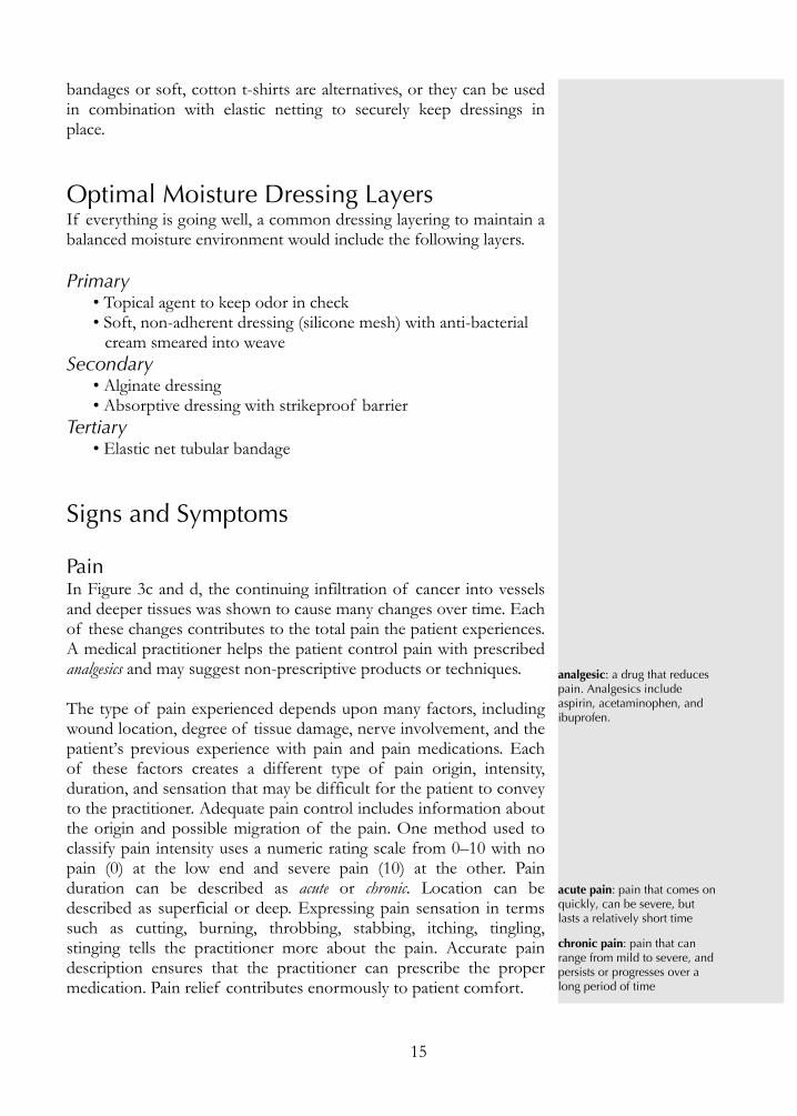
Optimal Moisture Dressing LayersIf everything is going well, a common dressing layering to maintain abalanced moisture environment would include the following layers.
Primary• Topical agent to keep odor in check• Soft, non-adherent dressing (silicone mesh) with anti-bacterial
cream smeared into weaveSecondary
• Alginate dressing • Absorptive dressing with strikeproof barrier
Tertiary• Elastic net tubular bandage
Signs and Symptoms
PainIn Figure 3c and d, the continuing infiltration of cancer into vesselsand deeper tissues was shown to cause many changes over time. Eachof these changes contributes to the total pain the patient experiences.A medical practitioner helps the patient control pain with prescribedanalgesics and may suggest non-prescriptive products or techniques.
The type of pain experienced depends upon many factors, includingwound location, degree of tissue damage, nerve involvement, and thepatient’s previous experience with pain and pain medications. Eachof these factors creates a different type of pain origin, intensity,duration, and sensation that may be difficult for the patient to conveyto the practitioner. Adequate pain control includes information aboutthe origin and possible migration of the pain. One method used toclassify pain intensity uses a numeric rating scale from 0–10 with nopain (0) at the low end and severe pain (10) at the other. Painduration can be described as acute or chronic. Location can bedescribed as superficial or deep. Expressing pain sensation in termssuch as cutting, burning, throbbing, stabbing, itching, tingling,stinging tells the practitioner more about the pain. Accurate paindescription ensures that the practitioner can prescribe the propermedication. Pain relief contributes enormously to patient comfort.
15
analgesic: a drug that reducespain. Analgesics includeaspirin, acetaminophen, andibuprofen.
acute pain: pain that comes onquickly, can be severe, butlasts a relatively short time
chronic pain: pain that canrange from mild to severe, andpersists or progresses over along period of time

Causes/Sources of Pain• Dressing changes• Inappropriate dressings• Exposure to air• Pressure on nerves• Inflammation• Infection
How to Control PainDisturbing the wound bed at dressing changes can create pain anddistress. A wound care practitioner with malignant wound careexperience knows how to minimize pain during dressing changes.Depending upon wound bed conditions, some dressings may be leftin place for long periods, thus minimizing wound disruption.Inappropriate dressings in contact with the wound bed (e.g. gauze;non-contouring) exacerbate the chaos at the surface. Dressings thatadhere, harden, or in any way harm the delicate surface tissue must becarefully removed and replaced with a type that does not aggravatethe situation. Pain caused by exposure to air can be minimized bytemporarily covering the wound with a moist dressing during dressingchanges. Water or a saline solution on a sterile, tight-weave padplaced on the wound while preparing new dressings may bring somerelief. The patient may also benefit from pre-medication with ananalgesic prior to removal of old dressings.
Pain arising from pressure upon nerves, inflammation, and/orinfection requires the skills of a medical practitioner, who works withthe patient to develop an analgesic protocol that provides steady, long-acting relief as well as short-acting medicine for those times when thereis breakthrough pain or anticipated pain (e.g. during dressing changes).
Effective control of pain from malignant wounds may involve acombination of appropriate analgesics, anti-inflammatories, and non-pharmacological products and techniques. Some patients findalternative pain relief therapies useful to reduce anxiety or thesensation of pain by creating a distraction. These therapies include:relaxation, music, massage, visualization, and aromatherapy.
Pain Warning SignsIf the pain changes, this may be a warning. Pain associated withredness, heat (including fever), or exudate could be sign of aninfection. Pain that ceases to be kept under control, by either long-acting or short-acting medication, should be brought to yourpractitioner’s immediate attention.
16
pre-medication: givingmedication in anticipation ofpain or anxiety of pain
long-acting: (a drug)maintaining its effects over along period of time, beingslowly absorbed and persistingin the tissues before beingexcreted
short-acting: (a drug) quicklyeffective, but requiringregularly repeated doses forlong-term treatment, beingrapidly absorbed, distributed inthe body, and excreted
breakthrough pain: intenseincreases in pain that occurwith rapid onset even whenpain-control medication isbeing used. Breakthrough paincan occur spontaneously or inrelation to a specific activity
Exudate Exudate is the fluid that weeps from a malignant wound that hasprogressed to the stage represented in Figure 3c. Management ofmalignant wounds requires maintaining a balanced, moistenvironment. Too much or too little moisture can lead to increasedpain and complications. The release of exudate can make it difficultto maintain this balance. Exudate from malignant wounds containssubstances that can irritate the wound bed and periwound skin.
Outer dressings that have become saturated may strikethrough ontoclothing, or, at night, bedding. This is a sign that dressing type is notadequate for current wound bed status. Proper management ofexudate from the malignant wound contributes to patient comfortand well-being.
Causes/Sources of Exudate• Fluid released from or around malignant cells
How to Control ExudateExudate is managed with dressings designed to match the volume offluid released (light to heavy). Containing exudate requires advicefrom a medical practitioner familiar with weeping malignant wounds.There is a wide range of absorbent dressings available from medicalsuppliers. However, depending upon the status of the malignantwound, it may be difficult to find the perfect dressing to controlexudate. A clever solution may involve off-label use of absorbentproducts (menstrual or incontinence pads) to achieve an appropriatelevel of absorbency. Off-label usage keeps costs down. To addressstrikethrough, a different dressing type or an additional absorbentlayer, may help match the interval of time between dressing changes.
A wound bed that is too wet or too dry may contribute to pain orlead to complications. If the tissue looks blanched, it is too wet. Ifsurface exudate has dried or hardened above a weeping wound (like ascab), then the moisture content of that wound bed is too dry.Sometimes changing to a slightly different dressing for a few daysrestores proper moisture balance. It is important to recognize theappearance of best moisture environment, how to maintain it, andwhat to do if its status changes. It is also important to pay attentionto periwound skin that may be irritated by exudate. Somepractitioners recommend the use of barrier film products forperiwound skin protection. Keeping absorbent dressings off ofperiwound skin is advised.
Exudate Warning signsIf there is a sudden change in volume or color of the exudate,consult your medical practitioner.
17
strikethrough: the point atwhich absorbed fluid reachesthe outer surface or edge of adressing and leaks ontoclothing
off-label use: use of a productfor something other than itsintended use
BleedingIn addition to creating pain, dressing changes may also inducebleeding. The fragile tissue making up the wound bed can easily bedisturbed, resulting in bleeding. Dressings that rub against, or adhereto, this tissue may cause bleeding either while being worn or whenbeing removed. The infiltration of cancer may lead to erosion ofblood vessels (Figure 3e), abnormally thin-walled blood vessels, orfailure to coagulate. Sometimes the patient doesn’t know until the nextdressing change that there was a minor bleeding episode. The sightof blood on the dressing can be alarming to the patient and caregiver.
Causes/Sources of Bleeding• Dressing changes• Inappropriate dressings• Vessel damage (erosion)• Vessel abnormality (thin walls)• Coagulation defect
How to Control BleedingThere are several categories of bleeding. Their control variesdepending upon the extent and cause of the bleeding. Minor bleedingmay remedy itself or can be controlled by local pressure, ice packs,and/or alginate dressings placed directly against the bleeding wound.
Discuss preparation for moderate bleeding with your medicalpractitioner. It may be necessary to have appropriate dressings onhand. Depending upon the location and status of the malignantwound, trained medical staff may be required to control bleeding.
Concern about major bleeding should be discussed with your trustedhealth-care team.
OdorFor some patients, odor is more troubling than pain. It can also bethe cause of depression and social isolation for the patient. Withtime, dressings become saturated with exudate and collect pieces ofnecrotic flesh and microbial waste products. Both exudate andnecrotic tissue support the growth of microorganisms that produceodors that may be offensive to the patient and to anyone near them.Odor can also be a sign of a serious infection in the wound bed.With proper dressings and other products, odor can be minimized oreven eliminated.
18
coagulate: to congeal; to clot
Causes/Sources of Odor• Infrequent dressing changes• Exudate (skin or saturated dressing)• Necrotic tissue• Infection
How to Control OdorDetermining the source of the odor is key to controlling it. Theadvice of the experienced medical practitioner includes which wounddressing type best suits the needs of the wound bed and periwoundskin, as well as how often that dressing needs to be replaced. Asaturated dressing may be a sign that more frequent dressing changesare needed. This is a good time to review the types of dressings beingused.
Depending upon location and status of the malignant wound, gentlerinsing with water or saline solution removes most exudate and anyloose necrotic tissue on the skin surface. Not all necrotic tissue in themalignant wound can or should be removed because the blood andlymph vessels below the wound are fragile and easily ruptured. If ashower is not possible at this time, a water or saline pre-soak softensdried exudate so that it can be removed with minimal damage. Note:gently cleaning with water or saline means no scrubbing or soap, juststand or sit and let the moistened exudate wash away. Do not pick atstubborn sections.
Cleaning the wound is the first step in controlling odor. Topicalagents such as metronidazole 5% (available in powder or gel form)can reduce wound odor, but may take a few days to show results. Ifthe wound is optimally moist, or too wet, the powder form isappropriate. If the wound is too dry, the gel form may be useful.Alternative topical agents include sugar paste, medical honey, andyogurt. Their use can be explained by an experienced medicalpractitioner familiar with applying these to the wound.
There are also dressings impregnated with anti-bacterial material (e.g.silver, iodine, medical honey). A medical practitioner can bestdetermine if additional products (e.g. activated charcoal filter) orprocedures will increase patient comfort and relief. As malignantwounds progress, different dressings for odor control may be addedto, or taken away from, the various layers.
If preventing odor in the wound is insufficient, then absorbing ormasking odor in the surroundings (home) may be necessary. In thehome, charcoal, kitty litter, or coffee filters placed near the patientcan help to mask or trap odors. Some patients also mask the odor bydabbing perfume or an essential oil onto the outermost dressing.
19

Odor Warning SignsSome patients may develop reactions to anti-microbial topical agentsor to silver-impregnated dressings. Alternatives (silver versus iodine)should be discussed with the medical practitioner, with a samplealternative readily available should a problem arise.
A sudden change in odor should be brought to the attention of themedical practitioner.
If the source of bad odor is an infection, only a medical practitionercan determine if and which antibiotics may be required for control.
ItchingThe sensation of itching in the periwound area can be a significantsource of irritation and may interfere with social activities. Themedical term for distressing itching is pruritis. For the patient with amalignant wound, this sensation may be localized or widespread. Itmay be so severe that the patient scratches uncontrollably, damagingthe wound bed or periwound skin, even to the point of bleeding.Scratching that results in broken skin puts the patient at risk ofinfection.
Causes/Sources of Itching• Excoriated (epidermis removed) skin due to contact with exudate• Dehydrated or stretched skin• Infection• A sign of a more serious blood or organ disorder
How to Control ItchingPeriwound area pruritis does not respond to antihistamines. If thecause of itching is irritation from exudate in contact with the skin,especially in the periwound area, then gentle washing with water or asaline spray helps stop the itching sensation. Barrier film productsmay prevent irritation from exudate leakage. Gentle patting or softmassage above the troublesome area can help stop the itch-scratchcycle. If the cause is dehydrated skin, first consult with a medicalpractitioner before using any product on periwound skin. Othermethods that may be useful to reduce itching include distraction,music therapy, relaxation, and imagery techniques.
Only a medical practitioner can determine the cause of itching andoffer alternatives when simple remedies fail. Infections and seriousdisorders may require prescribed medicine. If the cause is severedamage to the peripheral nerve supply, then alternative options mayinclude cancer specific hormone therapy, chemotherapy, or TENS(Transcutaneous Electrical Nerve Stimulation).
20
pruritis: itching. Severe itchingmay be a side effect of somecancer treatments and asymptom of some types ofcancers.
Possible Side Effects and ComplicationsCancer, malignant wounds, and their treatments assault on manyfronts. Prescribed and non-prescribed medication to control some ofthe perviously listed symptoms bring with them a variety of sideeffects. As demonstrated in the previous pages, the malignantwounds affect many tissue structures and functions. The sum canlead to a host of side effects and complications that also requiremanagement. The following list demonstrates the breadth ofcomplications that malignant wounds can inflict upon a patient:
• Swelling and inflammation• Weakness and fatigue• Interrupted sleep patterns• Nausea• Impaired mobility• Shortness of breath• Diarrhea or constipation• Incontinence or urinary retention• Impaired cognitive skills• Lack of appetite• Tumor fever• Cellulitis• Malignant-related ascites
These complications develop slowly over time despite a well managedwound bed. Managing these complications or their combinationsrequires the skills of the entire health care team, because treatmentsmay be counterproductive. There is no simple fix-all as the woundsprogress. Instead, be aware and watch for signs. Keep notes. Informyour health care team.
21
tumor fever: a fever associatedwith a tumor and believed tobe associated with the releaseof pyrogens
cellulitis: a common,potentially serious bacterialskin infection
ascites: abnormal buildup offluid in the abdomen that maycause swelling. In late-stagecancer, tumor cells may befound in the fluid in theabdomen
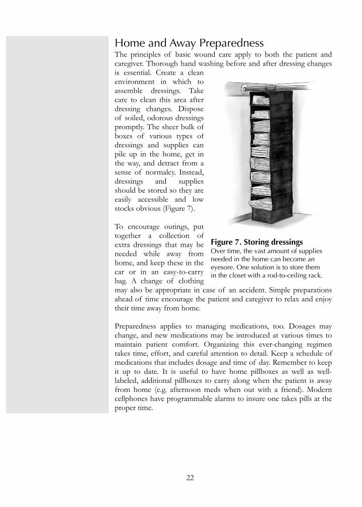
Home and Away PreparednessThe principles of basic wound care apply to both the patient andcaregiver. Thorough hand washing before and after dressing changesis essential. Create a cleanenvironment in which toassemble dressings. Takecare to clean this area afterdressing changes. Disposeof soiled, odorous dressingspromptly. The sheer bulk ofboxes of various types ofdressings and supplies canpile up in the home, get inthe way, and detract from asense of normalcy. Instead,dressings and suppliesshould be stored so they areeasily accessible and lowstocks obvious (Figure 7).
To encourage outings, puttogether a collection ofextra dressings that may beneeded while away fromhome, and keep these in thecar or in an easy-to-carrybag. A change of clothingmay also be appropriate in case of an accident. Simple preparationsahead of time encourage the patient and caregiver to relax and enjoytheir time away from home.
Preparedness applies to managing medications, too. Dosages maychange, and new medications may be introduced at various times tomaintain patient comfort. Organizing this ever-changing regimentakes time, effort, and careful attention to detail. Keep a schedule ofmedications that includes dosage and time of day. Remember to keepit up to date. It is useful to have home pillboxes as well as well-labeled, additional pillboxes to carry along when the patient is awayfrom home (e.g. afternoon meds when out with a friend). Moderncellphones have programmable alarms to insure one takes pills at theproper time.
22
Figure 7. Storing dressings Over time, the vast amount of suppliesneeded in the home can become aneyesore. One solution is to store themin the closet with a rod-to-ceiling rack.

Living with Malignant WoundsAs painful, odorous and messy as malignant wounds can be, it is possible to live with them. I havedone so for over three years. It wasn’t straightforward. I had no guide or manual to follow. In myhome, I have shelves of country tour guides and stacks of operator manuals for everything froma desk fan to my overly complicated cell phone. But for wounds that were visible signs of myimminent death, there was nothing available. Surely I’m not the only one who’s ever beendiagnosed with this aspect of an advanced cancer. I’m special, but not that special.
I might not be able to stop my cancer’s progression by learning about it in published sources, butI was certainly going to find out what these wounds were. I researched and took notes. Lots ofnotes. I found a common theme among articles in research and medical, online databases towhich I was fortunately allowed access. Patients with poorly or incorrectly treated malignantwounds were showing up in hospitals. At the front-line, hospital nurses were tending to thesepatients. Many nurses were not prepared for what they encountered. Using their professionaljournals as the classroom, nurses began teaching their colleagues. But, who was going to teachthe patients? Like a tourist in a strange land, patients needed a guidebook. A manual withillustrations and troubleshooting tips is valuable to the seamstress when the sewing machine fails.How is this any different for a patient who is isolated because of wound odor? With a goodmanual, one might be able to fix things and carry on. If anyone needs to fix something and carryon, it is another cancer patient like me, with malignant wounds.
That’s when I decided to formally organize my reprints and various handwritten notes and setout to fill that gap. Thanks to a jump-start with in-kind donations and firm support of familyand friends, I opened my laptop and began writing this booklet, for you.
Researching and writing the technical parts was an on-again, off-again three-years’ journey. Alongthe way, I met many caring people. I wanted to share the technical details of exudate control, butthey wanted to learn how I ordered supplies with my medical insurance. I spoke about movingfrom short-acting to long-acting analgesics, but they insisted I tell how I coped with odor on along flight to Europe. Even as publication of this booklet drew near, I continued to be remindedto include a part about how I lived these past three years with devastating wounds. Thoserequests were not only from patients with malignant wounds. Some were from physicians treatingcancer patients. Others were from cancer survivors or ones whose lives have been touched bycancer. They may be future malignant wound patients themselves. They may be future caregivers.They needed reassurance in the form of technical details alongside my story to believe one can livewith malignant wounds.
After almost six years cancer-free, I found a small bump and, like all my previous findings overthe past nineteen years, this was biopsied and found to be positive for cancer. I’ve always foundthat a poor use of the word positive. Now I was back in curative care, with two possible regimensto follow, should one not work. I would need a port installed for the delivery of nasty chemicals,and I would again lose my hair. Days after surgery, I flew to the Midwest to attend the babyshower of my expected twin nephews. I would be too sick to visit them after their birth, so thiswas as close as I could get. After the baby shower and while staying at another brother’s home, Iwas busy mopping up damage caused by a severe water leak. The words from the discharging
23

nurse echoed in my ears, “Don’t do anything strenuous for several weeks.” She doesn’t know theClarks. I just hoped the tube in my jugular was placed very securely.
I brought my laptop to the infusion center and either researched online or watched old KatharineHepburn movies. Nodules continued to appear under the skin. There were so few, I could countthem. I was gaining two new ones a week. It was time to try another chemo cocktail and resumecounting. The nodules were winning, I was bald, and one of the older nodules above my sternumwas behaving particularly oddly. It was time to let my body rest and rethink the battle plan.Meantime, we had an autumn trip to Europe for business and pleasure to organize. I needed torecover and figure out how to pack for a cold, wet Europe while bald and having issues withbumps on my chest. The one over the sternum now wept a yellow, thick fluid. I ordered extracamisoles to hold my foam prostheses and packed laundry soap so I could wash fouled camisoleseach day. I filled a zippered pouch with all the supplies I had gathered at home or found inpharmacy sections of grocery stores to cope with that oozing liquid. I made sure I had one boxof anything sterile and absorbent. But the scissors had to go in my TSA-approved pouch.
Off we went to the Netherlands, Belgium, and France. I borrowed a bike and retraced a fewmiles of the path to Delft I had trailblazed at age seventeen. I explored the former playing cardcapital of the world and lost all sense of time in a cooking store. Having taught Big Puppetworkshops in my community, I couldn’t miss the amazing talents on display at Les Machines inNantes, France. As a young reader, I had loved Jules Verne. Nantes was his hometown. Despitean icy wind and a soiled camisole, I climbed the iron, circular stairs to ride atop their life-sizedmechanical elephant. Naturally, Les Machines’ mechanical animals are built in the style of Verne’simagination. More nodules were breaking through the skin. Things were picking up speed. Westopped at a pharmacy (chemist) and looked at choices for absorbent dressings not found in theUS. Nothing new there except the print on the box—we don’t read French.
After returning home, I learned about a clinical trial using chemotherapy and the off-label use ofa topical cream. My previous chemotherapy disqualified me from the trial, but I was veryinterested in the cream part. I telephoned the researcher back East. Why not? What have I got tolose? Over the next few weeks, we developed a rapport. I would conduct the cream part of thetrial and send her my data (which was to become anecdotal to her, not part of the trial). Abaseline image of the nodules and unusual skin coloration was marked with a black, permanentink pen on a large, clear plastic sheet placed above my chest. Each week, with the clean baselineimage place upon my chest, we marked changes, took a digital photo, and sent that to theresearcher. The nodules were merging so quickly that it was next to impossible to draw anythinguseful. We stopped trying and gave up on the cream.
Odor reared its ugly head, and I visited a wound care nurse. I was told to use a soft washcloth inthe shower and scrub away that pesky, hardened scab. This became a daily visit to hell. I was anemotional wreck after showering. Sometimes the wound bled. Each day, however, there was anew scab to scrub. I started using thin menstrual pads, stuffed down my camisole, to catch thesmelly stuff and prevent my clothes from collecting it. A new business and pleasure trip wascoming up. Now I had to pack for a longer time in stranger (to me) lands, because these woundswere coming along, too. It was going to look odd to TSA, but we were going to be traveling withsix cans of contact lens spray just in case clean water for rinsing my wounds was not available.Six cans in two weeks was about right!
24
I don’t know how many advanced cancer patients with malignant wounds have been to the DMZin Korea, and safely (i.e. with armed guards) stepped about twenty feet into North Korea, butcount me in that number. I am probably the only one in this wound group who managed to getan impromptu, one-on-one lesson at a Korean cooking school. To get to that lesson, I had tonavigate without understanding any Korean. I had to take mass transportation, alone, and get offas close to the school as possible. Weeping wounds or not, I wanted to learn how to makeBimimbap. Make sure you count me in with the weeping wound crowd who ventured intoenormous lava tubes at Jeju Island. It was peak cherry blossom season in Japan, so include me inthe tally for those who climbed the stairs of Himeji Castle and stood in awe of the temples andshrines in Nara. However, it was time to really focus on the odorous and heavier flow from mywounds. They were a nuisance and starting to interfere with my foreign adventures. We returnedhome determined to find a better way to cope with the wounds.
We learned, however, that day-to-day management of these wounds is not a skill set of mostoncologists. My wounds would not heal. They were getting worse. I had tried unsuccessfully toget through the day without smelling awful or constantly changing my clothes. I found a differentoncologist who only saw advanced breast cancer patients. But, as luck would have it, a few weeksbefore I was to begin a new chemotherapy protocol, I developed a severe case of cellulitis andwas hospitalized. In the hospital, a team of surgeons tried to get me to consent to a skin graftthat would “remove all the cancer” and I’d be fine. That simply wasn’t true, and the attendingoncologist, ironically my former one, told them so in front of me. Just before I was discharged, awound care nurse paid me a visit. From her, I learned I should not have been scrubbing the openwounds all those months. I also learned about better products for absorption of the fluid andprevention of infection. Between the surgeons and the wound care nurse, I had an oddcombination of mistrust and trust of the medical community. I had to pay more attention anddefinitely do more research.
I took every possible opportunity to search for information about wounds associated withadvanced cancer. The articles published by medical researchers, naturally, did not have helpfulinformation about absorbent dressings or controlling odor. I was looking for any clue that mightlead me to what I needed for daily care. While the new chemotherapy regimen was underway, Icame across the term fungating. Like all other unknown words I was finding, I looked up thedefinition. Fungating described me. This was what I had on the skin of my chest. A new set ofsearches with that term uncovered brand new articles by different authors (mostly nurses). Abrilliant and useful guide was Kelli Bergstrom’s article (see Resources). I now had a guidebook. Ialso saw pictures of others with these wounds. Emotionally, I was not sure I could face anyfurther advancement. My husband and I talked a lot about what was to come. We talked withfamily and friends. My future was horrific and cruel.
I continued researching. The wounds had progressed to such a size that the wide sports bandagewe used to keep dressings in place was no longer working. That’s when I came across WayneNaylor’s teaching video (for nurses) using elastic net tubing to secure dressings. Another brilliantdiscovery, and just in time for my needs. The new chemotherapy was not slowing down theprogression of the wounds in my skin. It was time to transition from curative to palliative care. IfI was going to face the future that I’d seen in Bergstrom’s article, I needed to take charge, form aplan, and prioritize my life. My oncologist supported my decision to stop curative care, butcontinued to guide pain management. For wound care management (odor, bleeding, leakage), Iwas on my own.
25
Helping plan our daughter’s wedding took my mind off the chaos taking place on my chest.Personally, I think battling 30 yards of tulle is more difficult than managing weeping wounds!Our son has worked at dozens of weddings and took on tasks that were beyond my skills andenergy level. Each time a problem with planning came up, he assured me that regardless of anyoops on wedding day, everyone would have a great time. We had an oops when $100 of heliumballoons escaped from an open van during setup. Watching the limo drive away with the happycouple was something I never thought I’d witness when first diagnosed with cancer nineteenyears ago. My son was right, everyone had lots of fun.
The complications of, and medications for, advanced cancer were slowing me down. Before itwas no longer an option, I had one more trip to take. Would it be Africa or UK/Ireland? I hadbeen to six of seven continents. Should I get that seventh one and have a proud claim? Should Iuse the information I’d been gathering about my ancestral roots and travel around UK/Ireland?Given the condition of my wounds and now knowing more about serious complications, I chosethe latter. Packing for a month away from home with a very large mass of weeping wounds onmy chest required much planning and anticipation of problems. I humored myself by saying thatall the space I was taking up with dressings and related supplies, I would fill with mementoes. SoI packed a lot!
There seemed to be a lot of mold in those castles and cottages of my ancient land. I thought I’dpicked up an infection in my lungs, because breathing became difficult. True, I was a bit out ofshape, but it was so difficult to climb stairs that I frequently let others in our group go while Istayed on one level—often complete with gift shop. It was not on my travel itinerary, but I had aride in an ambulance to a London ER because breathing had become seriously difficult.American and British English sound different inside an ambulance. I was about to be prickedwith a little lancet by an EMT when I told him my blood type, “B positive.” To this he asked,“When was that diagnosed?” Diagnosed? That’s an odd way to ask when I learned my bloodtype. Did he really need to know when I learned my blood type? We both stared at each othertrying to decipher what the other had said. Finally, I replied, “I learned it in high school biologyclass.” This really surprised him. His surprise just confused me. My finger was pricked regardless.A few minutes later, the other EMT solved the riddle we had created. The first EMT thought Iwas referring to positive for hepatitis B. Silly English language. On we went to the ER, but noscan was done. I left to go pack for the next day’s flight.
Back home, a scan revealed that my breathing problem was not from mold infection from aquaint cottage in the Cotswolds. Instead, I had pleural effusion and the beginnings of boneerosion. Not so glamorous. Time to reassess, again. This time, the decision was made totransition from palliative to hospice care. We were going to need the support of a knowledgeableteam. We phoned our local hospice service, and almost overnight, the burden of managing allthat these wounds required was lessened. True, it meant relinquishing some of the control I hadso carefully organized. But it gave me back time to focus on the quality of my final months ofliving. That was more than a fair trade. It allowed me to write this resource guide.
This is the background story of how I have lived with my malignant wounds. Cancer survivors,caregivers, and others want assurance that managed malignant wounds won’t rob them of living.Perhaps I have demonstrated that it is possible to squeeze as much quality out of a life aspossible. That squeezing will be accomplished in different ways by different people. You nowknow my story. Go out there and live yours!
26

ResourcesAlexander, S. Malignant Fungating Wounds: Epidemiology, Aetiology, Presentation and
Assessment. Journal of Wound Care 18. no. 7 (2009): 273–4, 276–8, 280.
Alexander, S. Malignant Fungating Wounds: Key Symptoms and Psychosocial Issues. Journal ofWound Care 18. no. 8 (2009): 325–9.
Alexander, S. Malignant Fungating Wounds: Managing Malodour and Exudate. Journal ofWound Care 18. no. 9 (2009): 374–82.
Alexander, S. Malignant Fungating Wounds: Managing Pain, Bleeding, and Psychosocial Issues.Journal of Wound Care 18. no. 10 (2009): 418–425.
American Cancer Society http://www.cancer.org/ December 3, 2011.
Bergstrom, Kelli. Assessment and Management of Fungating Wounds. Journal of Wound,Ostomy, and Continence Nurses Society 38. no.1 (2011): 31–7.
Dubenske, Lori L. and others. Caregivers’ Differing Needs Across Key Experiences of theAdvanced Cancer Disease Trajectory. Palliative and Supportive Care 6 (2008): 265–272.
Fineberg, Cohen and others. Communication with Families Facing Life-Threatening Illness: AResearch-Based Model for Family Conferences Journal of Palliative Medicine 14. no. 4(2011): 421–7.
Fowler, Evonne. Plain Talk About Wound Pain. Ostomy Wound Management 51. no. 11a (2005):Supplement 4–6.
Hudson, P. and others. Meeting the Supportive Needs of Family Caregivers in Palliative Care:Challenges for Health Professionals. Journal of Palliative Medicine 7. no. 1 (2004): 19–25.
Ivetic, Olga, and Patricia A. Lyne. Fungating and Ulcerating Malignant Lesions: A Review ofthe Literature. Journal of Advanced Nursing 15 (1990): 83–8.
Kifer, Zelia Ann. Fast Facts for Wound Care Nursing: Practical Wound Management in a Nutshell. NewYork, NY: Springer 2012.
King, Michael J., and Elisa G. DiFalco. Lymphedema: Skin and Wound Care in an AgingPopulation. Ostomy Wound Management 51. no. 11a (2005): Supplement 14-16.
Kirsner, Robert. New Approaches to a Timeless Dilemma. Ostomy Wound Management 51. no.11a (2005): Supplement 2–3.
Lancaster, Chris, Melissa O’Brien, Joanna Lawton. Wound Management of Cancerous
Wounds–NSCCAHS. Northern Sydney Central Coast Hospital Guide #039 (2009):1–32.
27
LeBlanc, Karen and Robert M. Arnold. Evaluation of Malignant Ascites #176–177. Journal ofPalliative Medicine Fast Facts and Concepts 13. no. 8 (2010): 1027–9.
Lo, Shu-Fen, Wen–Yu Hu, Mark Hayter and others. Experiences of Living with a MalignantFungating Wound: A Qualitative Study. Journal of Clinical Nursing 17 (2008): 2699–7.
McMurray, Vivien. Managing Patients with Fungating Malignant Wounds. Nursing Times 99. no.13 (2003): 55–7.
National Cancer Institute http://www.cancer.gov/ (January 2012).
Naylor, Wayne. Assessment and Management of Pain in Fungating Wounds. British Journal ofNursing 10. no. 22 (2001): Supplement s33–s52.
Naylor, Wayne. Dressing Difficult Wounds. http://www.cancernursing.org/forums/topic.asp?TopicID=131 presented 11 November 2008.
Queen, Douglas and others. Chronic Wound Pain and Palliative Cancer Care. Ostomy WoundManagement 51. no. 11a (2005): Supplement 9–11.
Piggin, C. Malignant Fungating Wounds: An Analysis of the Lived Experience. Journal ofWound Care 18. no. 2 (2009): 57–63.
Proot, Ireen, and others. Vulnerability of Family Caregivers in Terminal Palliative Care AtHome: Balancing Between Burden and Capacity. Scandinavian Caring Sciences 17. no. 2(2003): 113–21.
Seaman, Susie. Management of Malignant Fungating Wounds in Advanced Cancer. Seminars inOncology Nursing 22. no. 3 (2006): 185–93.
Selby, T. Managing Exudate in Malignant Fungating Wounds and Solving Problems forPatients. Nursing Times 105. no. 18 (2009): early online publication.
Saunders, Dame Cicely, http://www.cicelysaundersfoundation.org (March 2012). Dame Cicelyis widely acknowledged as the founder of the modern hospice movement.
UptoDate part of Wolters Kluwer Health http://www.uptodate.com/patients/index.html(February 2012)
World Health Organization http://www.who.int/cancer/en/ (February 2012)
World Wide Wounds http://worldwidewounds.com/ (February 2012)
28

Notes
29

Notes
30


Sherry A. Clark, a retired educator, lives in Oregon with her family.During her twenty years of teaching, she combined her love for thesciences with the introduction of computers in schools and taughtdozens of classes at middle school, high school, and university levels.This included a year as an exchange teacher in Auckland, NewZealand, where her family learned all things Kiwi. Beginning at age six-months, she is a long-time traveler who managed to set foot on six ofseven continents. Quilting and glass fusing are favorite hobbies. She isalso a breast cancer survivor with four recurrences. Her recent
recurrence, untreatable, developed into malignant wounds. She came out of retirement tooffer this booklet as her last lesson: to help and encourage others with malignant wounds toimprove the quality at their end of life.P.S. Shortly after the completion of this booklet, the cancer that Sherry had fought for somany years caught up with her. Thanks to proper care as outlined in this booklet andprovided by Hospice at her end of life, she was active until the very end. She will be missedby one and all.