unc’s biomedical imaging research center (bric)
TRANSCRIPT

Roger Sit, Ph.D., CHP
NCHPS Spring 2013 Meeting
February 28, 2013
UNC’s Biomedical Imaging
Research Center (BRIC)

Outline
Introduction
History
Describe capabilities of the Center developed
over last few years
Describe the evolution taking place presently
Describe the capabilities that are planned
Tie in to Health Physics or Radiation safety
Describe some of the challenges
Future – new imaging modalities

The BRIC
Biomedical Research Imaging Center
Established in 2005 to serve the imaging needs
of UNC-Chapel Hill biomedical researchers and to
advance the rapidly developing science of
biomedical imaging. The Center enables a better
understanding of disease, including cancer and
neurologic diseases and studies the effects of
genetic changes on disease development and
progression.

Example: Therapeutic agent for
Ovarian Cancer
Ho-165 (n,g) Ho-166
Used mesopourous silica nanoparticles as carrier
Study done in ovarian tumor-bearing mice
Ho-166 emits low energy gamma for imaging as well
as high energy betas for therapy
SPECT/CT used for tissue biodistribution studies
PET/CT used for disease progression
Conclusion: The retention of holmium in nanoparticles
during irradiation and in vivo after intraperitoneal
administration as well as their efficacy in extending survival
in tumor-bearing mice underscores their potential as a
radiotherapeutic agent for ovarian cancer metastasis.

Truly Interdisciplinary
Psychiatry
Neurology
Psychology
Pathology
Oncology
Physics
Biology
Rheumatology
Cardiology
Gastroenterolo
gy
Public health
Genetics
Dentistry
Pharmacology
Biomedical
engineering
Chemistry
Bioinformatics
Pharmacy
Pharmacology
Nursing
Radiology
And
others…….
hopefully
Radiation
Safety

Humble beginnings

Early Years
Half focused on fMRI imaging of humans
3-T Siemens Trio (whole body)
3-T Siemens Allegra dedicated to neurological
exams of the brain
Half focused on research imaging of animals
SPECT/CT
PET/CT
CT
Ultrasound
MRI
Optical

Siemens Trio Siemens Allegra

Animal Imaging: MRI
Bruker
9.4 T
MRI
Scanner

Animal Imaging: CT
SCANCO mCT
40
GE eXplore
CT120
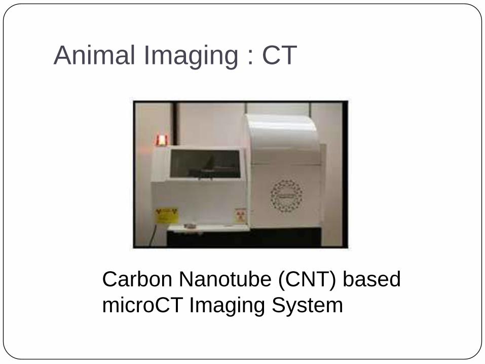
Animal Imaging : CT
Carbon Nanotube (CNT) based
microCT Imaging System

Animal Imaging: SPECT/CT
GE eXplore speCZT
System

Animal Imaging: PET/CT
GE eXplore Vista

Animal Imaging: Ultrasound
Visualsonics Vevo2100

Animal Imaging: Optical
IVIS Kinetic Optical
System

Animal Imaging: Optical
VisEn
FMT-2500
In-vivo
Optical
Imaging
System

NC Cancer Research Fund
In 2007, the North Carolina Legislature
established the NCCRF and began a long term
financial commitment to research and treatment
of cancer to improve the health and well being of
the citizens of North Carolina.
The fund enabled:
highly successful recruitment of new investigators
major expansion of research capabilities
new buildings

BRIC Growth
Addition of a 500 sq. feet enclosure

New Imaging Research Building

2012 A Banner year
Installed the fourth PET/MR fusion imaging
device in the US. This is the Siemens Biograph
mMR combining the functional imaging of PET
with the superior soft tissue contrast of MRI
imaging.
Big advantage in having simultaneous imaging.
PET/CT had been the best imaging technique but
requires imaging with a PET scanner, then CT,
then overlaying images.
PET/MR provides better image, lower patient
dose, and quicker scans

Siemens Biograph mMR


2012 continued
Installed the second dose-on-demand mini-
cyclotron in the US. This is the ABT Biomarker
Generator

ABT Biomarker Generator -
Cyclotron

AND
BRIC Building

New Capabilities for New
Building
GE PETtrace 17-MeV cyclotron
Irradiator
Siemens 7T Full Body Scanner
850 MHz NMR
PET/CT Full body scanner

Vault for Two Cyclotrons

Vault Door: Plug on Rails

GE PETtrace Cyclotron

Irradiator

Siemens 7T Full Body Scanner

Bruker Ascend 850 MHz NMR


New Building Sub-basement
Cyclotron, radiochemistry, QC
NMR Suite
7 T MRI
Animal facilities: 9.4 T, optical, ultrasound, PET/CT, SPECT/CT, microCT
Basement
Shell space for commercial partner
Irradiator
1st Floor
3T MRI
PET/MR
PET/CT

IRB Sub-basement

IRB Basement

IRB First floor

HP Challenges Plethora of machines and materials concerns
Work with multiple shielding contractors
Work with multiple equipment contractors
Work with both branches of NC-RPS
Bridging the knowledge gap for funding organization, architects, and /or construction management
Integrate safety issues that may not have always been a consideration (magnetic fields, cryogenics, high rf, etc)
Trying to convince line management that additional resources are necessary
Working out case-by-case procedures for external research collaborators

Future
X-ray Luminescence CT
X-ray Fluorescence CT
Cerenkov imaging

The End
Thank you