ultrastructural analysis of hepatitis c virus particles · ultrastructural analysis of hepatitis c...
TRANSCRIPT
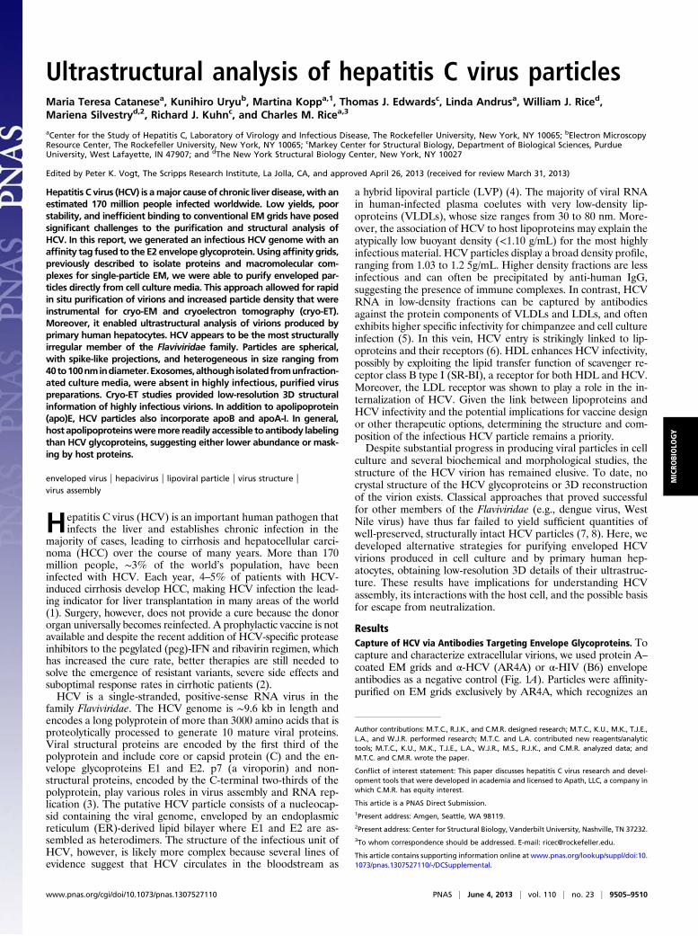
Ultrastructural analysis of hepatitis C virus particlesMaria Teresa Catanesea, Kunihiro Uryub, Martina Koppa,1, Thomas J. Edwardsc, Linda Andrusa, William J. Riced,Mariena Silvestryd,2, Richard J. Kuhnc, and Charles M. Ricea,3
aCenter for the Study of Hepatitis C, Laboratory of Virology and Infectious Disease, The Rockefeller University, New York, NY 10065; bElectron MicroscopyResource Center, The Rockefeller University, New York, NY 10065; cMarkey Center for Structural Biology, Department of Biological Sciences, PurdueUniversity, West Lafayette, IN 47907; and dThe New York Structural Biology Center, New York, NY 10027
Edited by Peter K. Vogt, The Scripps Research Institute, La Jolla, CA, and approved April 26, 2013 (received for review March 31, 2013)
Hepatitis C virus (HCV) is amajor cause of chronic liver disease,with anestimated 170 million people infected worldwide. Low yields, poorstability, and inefficient binding to conventional EM grids have posedsignificant challenges to the purification and structural analysis ofHCV. In this report, we generated an infectious HCV genome with anaffinity tag fused to the E2 envelope glycoprotein. Using affinity grids,previously described to isolate proteins and macromolecular com-plexes for single-particle EM, we were able to purify enveloped par-ticles directly from cell culture media. This approach allowed for rapidin situ purification of virions and increased particle density that wereinstrumental for cryo-EM and cryoelectron tomography (cryo-ET).Moreover, it enabled ultrastructural analysis of virions produced byprimary human hepatocytes. HCV appears to be the most structurallyirregular member of the Flaviviridae family. Particles are spherical,with spike-like projections, and heterogeneous in size ranging from40to100nmindiameter.Exosomes,althoughisolatedfromunfraction-ated culture media, were absent in highly infectious, purified viruspreparations. Cryo-ET studies provided low-resolution 3D structuralinformation of highly infectious virions. In addition to apolipoprotein(apo)E, HCV particles also incorporate apoB and apoA-I. In general,host apolipoproteinsweremore readily accessible to antibody labelingthan HCV glycoproteins, suggesting either lower abundance or mask-ing by host proteins.
enveloped virus | hepacivirus | lipoviral particle | virus structure |virus assembly
Hepatitis C virus (HCV) is an important human pathogen thatinfects the liver and establishes chronic infection in the
majority of cases, leading to cirrhosis and hepatocellular carci-noma (HCC) over the course of many years. More than 170million people, ∼3% of the world’s population, have beeninfected with HCV. Each year, 4–5% of patients with HCV-induced cirrhosis develop HCC, making HCV infection the lead-ing indicator for liver transplantation in many areas of the world(1). Surgery, however, does not provide a cure because the donororgan universally becomes reinfected. A prophylactic vaccine is notavailable and despite the recent addition of HCV-specific proteaseinhibitors to the pegylated (peg)-IFN and ribavirin regimen, whichhas increased the cure rate, better therapies are still needed tosolve the emergence of resistant variants, severe side effects andsuboptimal response rates in cirrhotic patients (2).HCV is a single-stranded, positive-sense RNA virus in the
family Flaviviridae. The HCV genome is ∼9.6 kb in length andencodes a long polyprotein of more than 3000 amino acids that isproteolytically processed to generate 10 mature viral proteins.Viral structural proteins are encoded by the first third of thepolyprotein and include core or capsid protein (C) and the en-velope glycoproteins E1 and E2. p7 (a viroporin) and non-structural proteins, encoded by the C-terminal two-thirds of thepolyprotein, play various roles in virus assembly and RNA rep-lication (3). The putative HCV particle consists of a nucleocap-sid containing the viral genome, enveloped by an endoplasmicreticulum (ER)-derived lipid bilayer where E1 and E2 are as-sembled as heterodimers. The structure of the infectious unit ofHCV, however, is likely more complex because several lines ofevidence suggest that HCV circulates in the bloodstream as
a hybrid lipoviral particle (LVP) (4). The majority of viral RNAin human-infected plasma coelutes with very low-density lip-oproteins (VLDLs), whose size ranges from 30 to 80 nm. More-over, the association of HCV to host lipoproteins may explain theatypically low buoyant density (<1.10 g/mL) for the most highlyinfectious material. HCV particles display a broad density profile,ranging from 1.03 to 1.2 5g/mL. Higher density fractions are lessinfectious and can often be precipitated by anti-human IgG,suggesting the presence of immune complexes. In contrast, HCVRNA in low-density fractions can be captured by antibodiesagainst the protein components of VLDLs and LDLs, and oftenexhibits higher specific infectivity for chimpanzee and cell cultureinfection (5). In this vein, HCV entry is strikingly linked to lip-oproteins and their receptors (6). HDL enhances HCV infectivity,possibly by exploiting the lipid transfer function of scavenger re-ceptor class B type I (SR-BI), a receptor for both HDL and HCV.Moreover, the LDL receptor was shown to play a role in the in-ternalization of HCV. Given the link between lipoproteins andHCV infectivity and the potential implications for vaccine designor other therapeutic options, determining the structure and com-position of the infectious HCV particle remains a priority.Despite substantial progress in producing viral particles in cell
culture and several biochemical and morphological studies, thestructure of the HCV virion has remained elusive. To date, nocrystal structure of the HCV glycoproteins or 3D reconstructionof the virion exists. Classical approaches that proved successfulfor other members of the Flaviviridae (e.g., dengue virus, WestNile virus) have thus far failed to yield sufficient quantities ofwell-preserved, structurally intact HCV particles (7, 8). Here, wedeveloped alternative strategies for purifying enveloped HCVvirions produced in cell culture and by primary human hep-atocytes, obtaining low-resolution 3D details of their ultrastruc-ture. These results have implications for understanding HCVassembly, its interactions with the host cell, and the possible basisfor escape from neutralization.
ResultsCapture of HCV via Antibodies Targeting Envelope Glycoproteins. Tocapture and characterize extracellular virions, we used protein A–coated EM grids and α-HCV (AR4A) or α-HIV (B6) envelopeantibodies as a negative control (Fig. 1A). Particles were affinity-purified on EM grids exclusively by AR4A, which recognizes an
Author contributions: M.T.C., R.J.K., and C.M.R. designed research; M.T.C., K.U., M.K., T.J.E.,L.A., and W.J.R. performed research; M.T.C. and L.A. contributed new reagents/analytictools; M.T.C., K.U., M.K., T.J.E., L.A., W.J.R., M.S., R.J.K., and C.M.R. analyzed data; andM.T.C. and C.M.R. wrote the paper.
Conflict of interest statement: This paper discusses hepatitis C virus research and devel-opment tools that were developed in academia and licensed to Apath, LLC, a company inwhich C.M.R. has equity interest.
This article is a PNAS Direct Submission.1Present address: Amgen, Seattle, WA 98119.2Present address: Center for Structural Biology, Vanderbilt University, Nashville, TN 37232.3To whom correspondence should be addressed. E-mail: [email protected].
This article contains supporting information online at www.pnas.org/lookup/suppl/doi:10.1073/pnas.1307527110/-/DCSupplemental.
www.pnas.org/cgi/doi/10.1073/pnas.1307527110 PNAS | June 4, 2013 | vol. 110 | no. 23 | 9505–9510
MICRO
BIOLO
GY

E1/E2 conformational epitope, but not by the B6 monoclonal an-tibody. This indicates that both HCV glycoproteins are displayedon the surface of released HCV particles. Particles were roughlyspherical and well preserved, but with heterogeneous and not al-ways symmetrical structures (Fig. 1B) and diameters ranging from40 to 100 nm (Fig. 1C). Their distribution across the grid was nothomogeneous and overall particle density was low. This observa-tion was confirmed in a quantitative virus capture assay in whichprotein G–coated magnetic beads were conjugated to AR4A, B6,or an apolipoprotein (apo)E-reactive antibody and the amount ofprecipitated HCVRNAmeasured by RT-quantitative PCR. ApoEwas chosen for HCV RNA capture because it is both requiredfor the production of cell culture–derived HCV (HCVcc) and isa component of HCVcc particles (9, 10). Although AR4A onlycaptured about 2% of the input viral genomes, 10-fold more viralRNA was precipitated using an apoE-targeting antibody (Fig. 1D).Similarly, a small fraction of the beads coupled to AR4A, but notB6, displayed bound particles by transmission EM (TEM), in-dicating that, albeit with low efficiency, intact virions were specifi-cally captured using envelope-reactive antibodies (Fig. 1E).
Generation of E2-Tagged HCV Particles. The number of virionscaptured using envelope-specific antibodies was suboptimal for
further ultrastructural analyses. We hypothesized that the lowyield might be due to poor accessibility of the viral glycoproteins.To increase particle density on the EM grid, we generated aJ6/JFH-1 derivative with a 39-aa-long, tandem affinity tag fused tothe N terminus of E2 (tag-HCV) immediately upstream of thehypervariable region 1. This region was shown to tolerate insertions,is a target of neutralizing antibodies and therefore exposed on in-fectious virus (6). The first two residues of E2 were duplicated toensure proper cleavage at the E1/tagE2 junction followed by sixhistidine residues (6xHis) and two copies of a streptavidin tag II(StrepII) tag (Fig. S1A). Immunoblot analysis of lysates preparedfrom cells electroporated with tag-HCV demonstrated correctprocessing, with both tags detectable (Fig. S1B). Importantly, theinsertion did not markedly impair infectious virus production(Fig. S1C). Incubation of virus-containing media with beadsrecognizing either tag captured both E2 and capsid protein, asexpected for isolation of assembled particles (Fig. S1D).
Ultrastructural Characterization of tag-HCV with Affinity Grids. Topurify tag-HCV, we developed custom-made EM affinity gridscoated with a monolayer of lipids functionalized with a nickel-nitrilotriacetic acid group (Ni-NTA) to interact with the 6xHis-tag (Fig. 2A). High specificity of capture was achieved during thebinding step by using low concentrations of imidazole (20 mM),as demonstrated by the lack of WT-HCV particles on the grids(Fig. 2B). The captured tag-HCV particles had a broad sizerange (n = 317; mean = 64 nm, SD = 11 nm) similar to thatobserved with glycoprotein-specific monoclonal antibodies andWT-HCV (Fig. 2C) but particle density on the grid improvedsignificantly (compare Fig. 2B with Fig. 1B). Similarly, beadscoupled to α-His antibody precipitated about eightfold moreHCV RNA than AR4A (Fig. 2D vs. Fig. 1D) and bead-boundvirions were easily visualized by TEM (Fig. S2).
C
D
B
α-HCV
E
α-HIV41
-45
46-5
051
-55
56-6
061
-65
66-7
071
-75
76-8
081
-85
86-9
091
-95
96-1
00
0
10
20
30
Diameter (nm)
AEM gridProt AAntibody
α-HCV α-HIV-HCV
-HIV-ap
oE
1.6%
0.3%
11.9%
Num
bero
fPar
ticle
s
5
6
7
HC
VR
NA
copi
es(L
og10
)
Fig. 1. Characterization of HCV virions captured via glycoproteins-specificantibody. (A) Schematic of protein (prot) A EM grids. (B) Representativeimages of negatively stained HCV virions captured using protein A EM gridscoated with α-HCV or α-HIV antibodies. (Scale bar: Upper, 100 nm; Lower, 20nm.) (C) Size histogram of HCV particles (n = 111; mean = 62 nm, SD = 11nm). (D) HCV RNA capture assay using protein G beads coupled to α-HCV,α-HIV, or α-apoE antibody and incubated with equal amounts of virus-containing media (2 × 10⁷ viral genome copies). RNA was extracted fromeach pull-down and HCV genome copy numbers were determined by RT-quantitative PCR. Means and SD from three independent experiments areshown. The percent of input HCV RNA captured by each antibody is in-dicated. (E) Images of negatively stained HCV virions adsorbed to protein Gbeads coupled to α-HCV or α-HIV antibody (Scale bars: Upper, 100 nm;Lower, 20 nm.) A bead-bound HCV particle is indicated (arrow).
CA
tag-HCV wt-HCV
D
Ni2+
LipidsBait
41-4
546
-50
51-5
556
-60
61-6
566
-70
71-7
576
-80
81-8
586
-90
91-9
596
-100
0
10
20
30wt-HCV tag-HCV
Diameter (nm)
%To
talC
aptu
red
Part
icle
s
-His
-HIV-a
poE
5
6
7
8.2%
0.3%
12.3%
HC
VR
NA
copi
es(L
og10
)
BEM grid
Fig. 2. Ultrastructural analysis of tag-HCV virions with affinity grids. (A)Schematic of the affinity grid, an EM grid coated with a Ni-NTA lipidmonolayer. (B) Representative images of negatively stained HCV virionscaptured using 2% (vol/vol) Ni-NTA affinity grids. (Scale bars: Upper, 100 nm;Lower, 20 nm.) (C) Comparative analysis of HCV size distribution with dif-ferent capture methods. The diameter of WT-HCV particles captured onprotein A grids via α-E1/E2 antibody (black bars) and tag-HCV bound to af-finity grids through the His-Ni interaction (gray bars) was measured and thenumber of virions in each size group is expressed as percent of total capturedparticles. (D) The copy number of tag-HCV genomes precipitated by His-,HIV-, or apoE-specific antibodies coupled to protein G beads was measuredby RT-quantitative PCR. Means and SDs from three independent experi-ments are shown and the percent of input HCV RNA captured by eachantibody is indicated.
9506 | www.pnas.org/cgi/doi/10.1073/pnas.1307527110 Catanese et al.

HCV Virions Are Assembled as Lipoviral Particles. ApoE-specificantibodies were superior at capturing viral RNA than α-E1/E2 orHis- and Strep-tag antibodies (Figs. 1D and 2D). Immunolabel-ing paralleled these results with particles staining more efficientlyfor apoE than E2 (Fig. 3A). Immunogold labeling revealed ad-ditional apolipoproteins associated with cell culture–producedHCVcc: apoA-I and apoB. Double-immunolabeling experimentsfor apoE and E2 showed that more than 95% of the stained par-ticles were apoE+, whereas a low percentage (∼3%) stained only forE2 (Fig. 3A). Single- and double-stained virus particles had similarsize ranges (Fig. 3B). However, significantly more apoE- thanE2-reacting gold particles was observed per virion in the apoE+/E2+ population (Fig. 3C), suggesting that this viral envelope epitopemay be less accessible and possibly masked. Taken together, ourresults are consistent with the LVP model for extracellular HCV.
HCV Produced by Primary Human Hepatocytes. Given that hepatomacells are unable to produce authentic VLDLs (4), we were in-terested in characterizing HCV particles grown inmore physiologiccultures, human fetal liver cells (HFLCs), which are polarized andmay better recapitulate in vivo lipoprotein and virus assembly.These cells are permissive for HCV but infection is short-lived withlow virus yields and little evidence of spread. However, inhibition ofinnate immune responses enhances permissiveness, spread, andvirus yield (11). To reduce possible background resulting from in-put cell culture–produced virus, HCV infection was initiated byRNA transfection in the presence of a tank binding kinase 1(TBK1) inhibitor, BX795 (Fig. S3A). BothWT- and tag-HCV titersin HFLC approached those of HCVcc in Huh-7.5.1 cells by thethird week posttransfection (Fig. S3B). Nuclear translocation ofan HCV protease-targeted tagRFP-nls-IPS reporter was used tovisualize the number of cells successfully infected over time (12).
At peak virus titers (day 26), the majority of HFLC transfectedwith WT- and tag-RNA was HCV+ (Fig. S3C). Despite lim-ited starting material, well-preserved, HFLC-produced virions(HFLC-HCV) could be captured using both protein A–coatedEM grids with AR4A (Fig. S3D) or affinity grids (Fig. 4A), withthe latter method being more efficient, as previously observedfor HCVcc. When the same supernatant was applied to regularEM grids, no particles were observed, suggesting that the affinitypurification in situ was crucial. As for HCVcc, no particles weredetected with α-HIV antibody or when WT-HCV was applied toaffinity grids. The size range of apoE+ particles captured fromHuh-7.5.1 andHFLC culture media was similar (60.17± 21.03 nmand 65.13 ± 16.56 nm respectively; P = 0.193) (Fig. 4B). Particlesimmunoreactive for apoA-I and apoB were detected, albeit toa lesser extent than apoE (Fig. 4C). These results suggest thatvirions derived from HFLC, like HCVcc, are assembled as LVPsincorporating host-derived apolipoproteins.
Cryo-EM of HCV.To attempt 3D reconstruction of the HCV virion,structurally preserved enveloped HCVcc particles were purifieddirectly from cell culture media using affinity grids with 20%(vol/vol) Ni-NTA and frozen in their native state (Fig. 5 A–C andFig. S4). A total of 318 particle images were isolated and pro-cessed with RobEM software. Particle sizes ranged from 45 to86 nm in diameter, with a mean diameter of 68 nm (Fig. 5C).A total of 150 particles were randomly selected to generate arandom model using Auto3DEM. Random model generationyielded a model of 58 Å resolution as determined by 0.5 Fouriershell correlation (FSC). The resulting model appeared as a smoothsphere, as expected for a heterogeneous set of particles, where anyidentifying features would be averaged out. This model was used foriterative orientation search on the entire dataset using Auto3DEM.No result better than 58 Å was achieved. Because of the disparateparticle size range, the dataset was sorted into three size classes(50–60, 61–70, and >71 nm) and the procedure was repeated oneach size class with similar results. Efforts to use reconstructionsof the alphavirus Ross River virus (22 Å) or the flavivirus denguevirus (30 Å) as initial models also yielded featureless spheres.Finally, the two smaller size classes were put through a commonlines reconstruction method using the EMICOFV program.Orientation for particles was hand selected and the resultingreconstruction was yet another smooth sphere. Single-particlereconstruction methods failed because of extreme pleomorphismof the particles. Exosome-like structures 50–100 nm in diameterwere occasionally visible, some of which were multivesicularwith two or more distinguishable bilayers (Fig. 5B and Movie S1).Exosome-like particles were observed on affinity grids only whentag-HCV samples were applied, suggesting that they containaccessible HCV E2.
Ultrastructural Characterization of Purified HCV. With the goal ofgaining insights into the ultrastructure of the infectious HCVparticle, we generated highly purified HCVcc preparations. In-fectious particles were concentrated by binding and elution froma heparin column followed by buoyant density fractionation ona 10–40% (wt/vol) iodixanol gradient. Particles with a densityof ∼1.13 g/mL contained more than 40% total infectivity, 20% ofthe HCV RNA, and thus a higher specific infectivity comparedwith the input (range 1:10–1:50 vs. 1:1,000–1:5,000; Fig. 6A).TEM analysis of peak infectious fractions captured on affinitygrids revealed well-preserved, spherical particles with a broadsize range (Fig. 6 B and C). The average diameter and overallappearance of purified particles was similar to unpurified HCVcc(67 ± 12 nm vs. 64 ± 11 nm). However, virions 81–85 nm in sizewere enriched in purified samples (12.1% vs. 2.8% of total boundparticles; Fig. 6B) and different structures were occasionallyobserved (Fig. 6C, Right). Given the 2D nature of TEM images,it was difficult to determine whether these structures represented
B C
A
E2+
apoE
+
apoE
+/E2+
0
25
50
75
100
125
Dia
met
er(n
m)
E2 apoE0
5
10
15
Num
berG
old/
Virio
n ***
a b
fe
dc
g h
Fig. 3. Viral and host proteins exposed on the envelope of HCV virions. (A)Representative images of tag-HCV particles purified on 2% (vol/vol) Ni-NTAaffinity grids and immunolabeled for the (a) viral glycoprotein E2, (b) apoE,(c) apoA-I, (d) apoB, and then (e) double-stained for E2 (arrows, 8 nm gold)and apoE (arrowheads, 18 nm gold), (f) StrepII tag, (g) 6xHis tag, (h) or in-cubatedwith secondary antibodyonly. (Scale bar: 100 nm.) (B) Size distributionof tag-HCV particles immunoreactive for E2 (n = 2, mean= 60 nm, SD = 11 nm),apoE (n = 20, mean = 53 nm, SD = 15 nm), or double-positive for E2 and apoE(n = 38, mean = 61 nm, SD = 22 nm). (C) Number of E2- and apoE-reactivegold particles per virion in apoE+/E2+ group (***P = 0.0003).
Catanese et al. PNAS | June 4, 2013 | vol. 110 | no. 23 | 9507
MICRO
BIOLO
GY

discrete particles with internal features or separate particles thatoverlapped. Interestingly, despite being present in cell culturesupernatant, exosomes were not found in purified samples of highspecific infectivity. Immunoblot analyses of purified virions (tag-HCV) captured with His-tag Dynabeads revealed apoE, apoA-I,and apoB100, confirming the immunolabeling results (Fig. 6D).
Cryo-ET of Purified HCV. In further attempts to obtain additional 3Dstructural information of purified HCV, we collected tomographictilt series of vitrified, unfixed particles with high specific infectivitypurified from iodixanol gradients. The quality of the tomogramswas affected by the limited tilt range we were able to collect (∼−60°to +30°). The missing wedge made the tomograms streaky andlimited the z-resolution of the reconstructions. Unfortunately,attempts at generating affinity grids using widermesh grids resultedin insufficient binding of virus particles to enable cryoelectron to-mography (cryo-ET) studies. Two reconstructed tomograms illus-trating the general morphology of HCV particles are available inMovies S2 and S3. The majority of virions were round but notsymmetrical with the outer rim heavily covered by globular proteinsdistributed unevenly around the viral envelope. Examples of par-ticles with apparent patches of double-layer membrane, spikes, andinternal structures are shown in Fig. 7 A–D. Striations in the viralenvelope, indicated by transversal densities are visible in Fig. 7Eand might represent transmembrane proteins (Movie S4).
DiscussionTEM has been instrumental to the discovery and characterizationofmany clinically relevant viruses, some from infected cell cultures,as in the case of adeno-, entero-, and reo-viruses, and others fromunpurified clinical samples (i.e., hepatitis A and B) (13). HCVrepresents an unusual case in that it was first identified by molec-ular cloning and has proven very difficult to visualize from infectedsera and tissues (14). Despite the development of systems topropagate infectious virus in cell culture (15), low titers and poorstability remained important challenges to the structural analysisof HCV. Attempts, by us and others, to scale up the number ofpurified particles applied to EM grids by concentration or pre-cipitation were not successful because of aggregation and serumcontaminants, whereas virus stocks produced in serum-free me-dium yielded fewer infectious particles.
To gain insights into the ultrastructure of fully assembledvirions, we first used EM grids coated with AR4A, but only a verysmall percentage of HCV RNA was precipitated and very fewparticles were visualized by TEM. AR4A potently neutralizes thevirus used in this study and displays one of the highest inhibitoryeffects among a panel of α-HCV antibodies screened (16, 17),arguing against insufficient binding properties (Fig. 1).To increase the efficiency of particle capture, as an alternative
approach we constructed a genome with a long, hydrophilictandem affinity tag on E2 that was not impaired in infectiousvirus production (Fig. S1) and purified it directly on custom-madeEM affinity grids interacting with the tags. The affinity grid, firstdescribed by Kelly et al. (18), enabled fast isolation, preservation,and enrichment of HCV virions that were instrumental to ultra-structural studies without the need for prior biochemical purifica-tion. Indeed, no particles were visualized if the same cell culture–derived media were applied to regular EM grids, indicating that theaffinity purification in situ was a key step. Provided that tags canbe inserted on accessible regions of the viral surface, the methoddescribed here can be applied to ultrastructural studies of othervirions that are unstable, produced in low amounts, or not easilybound to conventional grids. The 6xHis tag allowed for betterpurification of HCV particles than the StrepII tag, possibly becauseof its higher accessibility (Fig. S1D); nevertheless, the presence of
BA
CHCVcc HFLC-HCV
E2 ApoE
ApoB ApoA-I
25
50
75
100
125
Dia
met
er(n
m)
Fig. 4. Ultrastructure of HCV purified from primary human hepatocytescultures. TEM images of virions from HFLC supernatant harvested at day 26posttransfection and concentrated 10-fold. (A) Tag-HCV particles purified on2% (vol/vol) Ni-NTA affinity grids. (Scale bar: 100 nm.) (B) Size distribution ofapoE+ particles produced in Huh-7.5.1 (HCVcc) or HFLC (HFLC-HCV), capturedon affinity grids. (C) TEM images of HFLC-tag-HCV showing immunolabelingwith the indicated antibodies. (Scale bar: 20 nm.)
D
45-4
647
-48
49-5
051
-52
53-5
455
-56
57-5
859
-60
61-6
263
-64
65-6
667
-68
69-7
071
-72
73-7
475
-76
77-7
879
-80
81-8
283
-84
85-8
687
-88
0
10
20
30
40
Diameter (nm)
Num
bero
fPar
ticle
s
*
BA
C
Fig. 5. Cryo-EM analysis of HCVcc virions. (A) Cluster of HCV virions fromHuh-7.5.1 cultures purified on 20% (vol/vol) Ni-NTA affinity grids. (B) Areaof the grid showing HCV virions on the carbon film (arrows), exosomes(arrowheads), and multivesicular structures (*) inside the holes. (Scale bar:100 nm.) A reconstructed tomogram of this field is available in Movie S1. (C)Low-dose images of HCV particles at 78,000× magnification, representativeof three size classes (50–60, 61–70, and >71 nm). (D) Size distribution of HCVparticles (n = 318; mean = 64 nm, SD = 11 nm).
9508 | www.pnas.org/cgi/doi/10.1073/pnas.1307527110 Catanese et al.

the StrepII tag was confirmed by immunolabeling with peptide-specific antibody (Fig. 3A, f).We were interested in characterizing HCV particles grown in
primary hepatocytes (HFLCs) that represent more physiologiccultures. We previously showed that paramyxovirus V protein ex-pression promotes productive HCV infection in HFLCs by antag-onizing IFN induction and signaling following infection withHCVcc (11). Here, we report a further improvement of virusproduction in HFLCs using HCV RNA transfection rather thanHCVcc infection. Productive infection was facilitated by a chemicalinhibitor of TBK-1, a downstream signaling molecule of severalpathogen-associated pattern recognition receptors including reti-noic acid-inducible gene-I (RIG-I), a primary sensor for HCV.Increasing HCV titers were observed over time, indicative of virusspread, and high numbers of infectious particles [up to 105 50%tissue culture infectious dose per milliliter (TCID50/mL) of uncon-centrated supernatant] were obtained without the potential forcontamination by hepatoma-derived HCVcc inocula. The com-bination of optimized titers and affinity grid enrichment enabledthe characterization of primary human hepatocyte-derived HCVvirions (HFLC-HCV; Fig. 4).Both HCVcc and HFLC-HCV purified with affinity grids
appeared well preserved (as suggested by the lack of positivestaining), spherical, and very heterogeneous in size (40–100 nmin diameter), with spike projections but no distinguishable arrays.Thus, HCV does not seem to have structural similarities withother members of the Flaviviridae, such as dengue or West Nilevirus, which display a relatively smooth surface in their maturestage, an icosahedral symmetry and a diameter of 50 nm (7, 8).Not only the size, but also the density of HCV particles was
reported to be very broad both in vivo and in cell culture systemsand to vary depending on the host cell in which the virus is as-sembled. This suggests that the host contributes to define themakeupof the virus inways thatmight affect its structure and composition (5).Initial studies with human and chimpanzee plasma favored the hy-pothesis of association of HCV virions with the VLDL fractions (19,20). Since then, a growing body of literature has emphasized how thematuration and release of HCV particles are tightly linked to theVLDL biosynthetic pathway, with the strongest evidence being thatapoE is essential for virus assembly and found to be a component ofHCVcc particles (9, 10).In this study, we confirmed that apoE is incorporated in the
majority of captured HCVcc and demonstrated that this is true forprimary-derived HCV virions as well. In agreement with viralRNA immunocapture data (Fig. 1), double-immunolabelingexperiments showed that significantly more copies of apoE thanE2 were observed per particle (Fig. 3). These data indicate thata host-derived protein is better exposed on the outer rim of theviral envelope than the viral glycoproteins, which would be con-sistent with apoE playing a role in the initial attachment of HCVto the host cell. Interestingly, larger particles did not displaystronger staining for apoE or E2. This may suggest that the sitesaccessible to antibody binding on the virion were saturated be-cause of steric hindrance. Alternatively, a larger size may reflecthigher lipid content or the presence of additional host proteins.Other apolipoproteins were suggested to associate with serum-
or cell culture-derived HCV (4). In this study, we show in-corporation of apoA-I and apoB-100 into both HCVcc- andprimary hepatocyte–derived virions. ApoA-I, the major struc-tural protein of HDL, interacts with SR-BI, which is the en-dogenous receptor for HDLs in the liver. Interestingly, SR-BI isalso an HCV receptor, and HDLs were shown to facilitate HCVentry via SR-BI and to attenuate virus neutralization, suggestingthat HCV has developed an advantageous strategy to hijack thephysiological interaction of HDL with SR-BI (6). Our findingthat apoA-I is exposed on the surface of virions may providea link to explaining the complex interplay between HCV, HDL,and SR-BI. ApoB-100 is the longer isoform of the apoB protein,
B
C
A
41-4
546
-50
51-5
556
-60
61-6
566
-70
71-7
576
-80
81-8
586
-90
91-9
596
-100
0
5
10
15
20
25HCV-containing supernatantpurified HCV
Diameter (nm)
%of
Tota
lCap
ture
dPa
rticl
es
D
kDa
70
550
28
20
E2
apoE
core
apoA-I
β-actin
apoB
34
43
lysate virion
mockHCV
wttag
0 2 4 6 8 10 12 14 160
10
20
30
40
50
1.0
1.1
1.2
1.3infectivity HCV RNA density
Fractions
%of
Inpu
t Density
(g/ml)
Fig. 6. Biophysical and ultrastructural characterization of purified HCV. (A)High-titer, heparin-purified tag-HCV virions were fractionated accordingto density on a 10–40% (wt/vol) iodixanol gradient. For each fraction, viralRNA, infectivity, and density were determined and expressed as percent ofinput. (B) Size distribution of highly infectious tag-HCV particles in fraction 9(1.13 g/mL) of the buoyant density gradient (black bars; n = 430; mean =67 nm, SD = 12 nm) is compared with unpurified HCV-containing supernatant(gray bars; n = 317; mean = 64 nm, SD = 11 nm). Data are expressed as percentof total captured particles. (C) Left: Low-magnification TEM image of tag-HCV virions from fraction 9 captured on 2% (vol/vol) Ni-NTA affinity grids.(Scale bar: 100 nm.) Right: Close up views of structures observed in purifiedpreparations with high specific infectivity (Scale bar: 20 nm.) (D) Immunoblotfor the indicated proteins in mock- or HCV RNA–electroporated Huh-7.5.1cells, tag-, andWT-HCV extracellular virions that were heparin- and iodixanol-purified, precipitated with Dynabeads and eluted with 0.9 M imidazole.
A
B
C
D
*
*
*
E
F
Fig. 7. Cryo-ET of purified HCVcc virions. (A–D) Two virtual slices throughsome of the reconstructed purified HCV particles with spikes (arrows), viralenvelope (arrowheads), and internal structures (*) indicated. (E and F) Twosections through the 3D tomogram of an HCV particle showing striations inthe envelope (arrows) and viral membrane (arrowheads).
Catanese et al. PNAS | June 4, 2013 | vol. 110 | no. 23 | 9509
MICRO
BIOLO
GY

specifically produced in the liver, required for VLDL biogenesisand found on VLDL, intermediate density lipoprotein (IDL),and LDL. Its role in the assembly and infectivity of HCV pro-duced in cell culture systems is still being debated (4). Never-theless, HCV RNA–containing particles purified from low-density fractions (i.e., VLDLs, LDLs) of chronically infectedpatients were found to be positive for apoB, supporting the in-corporation of apoB in LVPs (4). Huh-7.5 cells were thought tobe impaired in the formation of apoB-containing VLDLs andtherefore unable to produce apoB-associated HCV virions (4).Notably, here we show that extracellular virions purified fromHuh-7.5 cultures expose apoB on their envelope. ApoB is con-sidered a nonexchangeable apolipoprotein; therefore, we can inferthat viral particles acquire it in the liver cell and not from serumlipoproteins. In line with this hypothesis, HCV produced in pri-mary human hepatocytes, unlike HCVcc, are secreted in serum-free media and yet contains apoB. Interestingly, most HCVcc- andHFLC-derived virions reacted with only one apoB immunogoldparticle, which might indicate a similar stoichiometry to VLDL,which assembles around a single molecule of apoB.In comparing the ultrastructure of HCV from cell culture
media with that of purified samples with narrower density andhigher specific infectivity, two differences were noticed. First,exosome-like structures were observed in HCV-containing su-pernatant but not in preparations enriched in infectious par-ticles. These vesicles were captured via tag-specific methods,indicating that they exposed at least the E2 envelope glycopro-tein on their surface. They were characterized by a relativelysmooth surface compared with the particles found in highly in-fectious samples that were heavily covered by electron-denseglobular material, suggesting a different composition and likelyseparate biogenesis. Second, when we looked at highly in-fectious, purified HCV, we observed an enrichment in particleswith a diameter of 81–85 nm displaying internal structures (Fig. 6).However, it was not possible to determine whether these particlesaccounted for the increased specific infectivity.Interestingly, as one would expect for an enveloped virus, the
cryo-ET images did not show evidence of a continuous bilayer. Thiscould be due to the low resolution of the images ormay indicate thatthe particle is embedded in a nonunique amount of lipoprotein,therefore delimited it by a lipid monolayer. In this context, theHCVenvelope glycoproteins may position their transmembrane domainsparallel to the lipid monolayer, as suggested by Bartenschlager et al.(4), unless the capsid is in close proximity of the lipoprotein-delimiting monolayer. In that case, a partial bilayer might formand E1/E2 would be able to acquire a transmembrane topology,thus anchoring the lipoprotein to the capsid. This hypothesiswould be consistent with some of our images (Figs. 6C and 7).
In summary, our results reveal the hybrid nature of HCV,assembled as a mixed particle with a thick shell of host-derivedapolipoproteins coating the viral envelope that presumably aidboth release and entry of the virus as well as escape from cir-culating neutralizing antibodies, ultimately allowing this veiledpathogen to fly under the radar.
Materials and MethodsVirus Purification.Virus-containingmedia was harvested every 4 h for 4 d afterswitching electroporated cells to low-serum media [1.5% (vol/vol) FBS]. High-titer HCVcc stocks were obtained by concentration of the infectious super-natant in a stirred ultrafiltration cell (Model 8400 with 100-kDa MWCOmembranes; Millipore). Concentrated samples were purified over heparincolumn (GE Hitrap Heparin) according to the manufacturer’s instructions.Heparin-eluted virus was fractionated over a 10–40% (wt/vol) iodixanolbuoyant density gradient (Optiprep; Sigma) to isolate fractions with thehighest infectivity (range 1.12–1.16 g/mL).
Cryo-EM and Cryo-ET: Sample Preparation and Data Collection. Holey carbongrids (400 mesh, Ted Pella) were coated with 20% (vol/vol) Ni-NTA lipid meshto generate affinity grids suitable for cryo-EM. Ten-nanometer gold particles(Aurion Gold Sol, EMS) were added to the virus suspension to serve as fiduciarymarkers for tomography. Grids were floated carbon side down on a 50-μL dropof virus solution containing 20 mM imidazole for 30 min, blotted for 2.0 s ina Gatan Cryoplunge Cp3 with 70–80% chamber humidity, and plunged intoliquid ethane. Cryo-EM images were collected using a Titan Krios electronmicroscope (FEI) at 300 keV under low-dose conditions, using an Ultrascan 9504k CCD (Gatan). For cryo-ET, imaging was done on a JEOL 3200FSC electronmicroscope (JEOL USA) operating at 300 KeV under control of SerialEM soft-ware using low-dose conditions. Images were collected on a Gatan Ultrascan4k camera at 50,000× nominal magnification and 2× binning, with a final pixelsize of 4.40A and dose per frame of 2.4e−/A2. An energy filter was inserted forall recorded images with slit width set to 20 eV. Tilt series were collected in 2°increments at the maximum range allowed by the grid: from −62° to +30° inthe best case and −64° to +20° in the worst. The tilt range was limited by themesh size of the grids. Tilt series were aligned and reconstructed using Pro-tomo software. Back-projected reconstructions were viewed using Imod.
Detailed methods and the associated references can be found in SIMaterials and Methods.
ACKNOWLEDGMENTS. The authors thank Dr. Thomas Walz and Daniel Zachs(Harvard Medical School), and Dr. Zheng Liu and Guimei Yu (Purdue University)for assistance with the preparation of affinity grids, Dr. Mansun Law (TheScripps Research Institute) for providing the AR4A and B6 antibodies, andDr. Cynthia de la Fuente for editing the manuscript. This work was supported byNational Institutes of Health (NIH) Grants AI072613 and AI075099 (to C.M.R.), theGreenbergMedical Research Institute, The Starr Foundation, and The RockefellerUniversity Women and Science Fellowship (to M.T.C.). The cryo-ET facilityat New York Structural Biology Center is supported by Research FacilitiesImprovement Program Grant C06 RR017528-01-CEM from the National Centerfor Research Resources and NIH and the JEOL 3200FSC electron microscopewas purchased with funds from NIH Grant S10 RR17291.
1. Lavanchy D (2011) Evolving epidemiology of hepatitis C virus. Clin Microbiol Infect
17(2):107–115.2. Delang L, et al. (2013) Hepatitis C virus-specific directly acting antiviral drugs. Curr Top
Microbiol Immunol 369:289–320.3. Moradpour D, Penin F (2013) Hepatitis C virus proteins: From structure to function.
Curr Top Microbiol Immunol 369:113–142.4. Bartenschlager R, Penin F, Lohmann V, André P (2011) Assembly of infectious hepatitis
C virus particles. Trends Microbiol 19(2):95–103.5. Lindenbach BD (2013) Virion assembly and release. Curr Top Microbiol Immunol 369:
199–218.6. Zeisel MB, Felmlee DJ, Baumert TF (2013) Hepatitis C virus entry. Curr Top Microbiol
Immunol 369:87–112.7. Kuhn RJ, et al. (2002) Structure of dengue virus: Implications for flavivirus organiza-
tion, maturation, and fusion. Cell 108(5):717–725.8. Mukhopadhyay S, Kim BS, Chipman PR, Rossmann MG, Kuhn RJ (2003) Structure of
West Nile virus. Science 302(5643):248.9. Gastaminza P, et al. (2010) Ultrastructural and biophysical characterization of hepa-
titis C virus particles produced in cell culture. J Virol 84(21):10999–11009.10. Merz A, et al. (2011) Biochemical and morphological properties of hepatitis C
virus particles and determination of their lipidome. J Biol Chem 286(4):3018–
3032.
11. Andrus L, et al. (2011) Expression of paramyxovirus V proteins promotes replicationand spread of hepatitis C virus in cultures of primary human fetal liver cells. Hep-atology 54(6):1901–1912.
12. Jones CT, et al. (2010) Real-time imaging of hepatitis C virus infection using a fluo-rescent cell-based reporter system. Nat Biotechnol 28(2):167–171.
13. Roingeard P (2008) Viral detection by electron microscopy: Past, present and future.Biol Cell 100(8):491–501.
14. Choo QL, et al. (1989) Isolation of a cDNA clone derived from a blood-borne non-A,non-B viral hepatitis genome. Science 244(4902):359–362.
15. Steinmann E, Pietschmann T (2013) Cell culture systems for hepatitis C virus. Curr TopMicrobiol Immunol 369:17–48.
16. Giang E, et al. (2012) Human broadly neutralizing antibodies to the envelope gly-coprotein complex of hepatitis C virus. Proc Natl Acad Sci USA 109(16):6205–6210.
17. Catanese MT, et al. (2013) Different requirements for SR-BI in hepatitis C virus cell-free versus cell-to-cell transmission. J Virol, in press.
18. Kelly DF, Abeyrathne PD, Dukovski D, Walz T (2008) The Affinity Grid: A pre-fabri-cated EM grid for monolayer purification. J Mol Biol 382(2):423–433.
19. Thomssen R, et al. (1992) Association of hepatitis C virus in human sera with beta-lipoprotein. Med Microbiol Immunol (Berl) 181(5):293–300.
20. Prince AM, Huima-Byron T, Parker TS, Levine DM (1996) Visualization of hepatitis Cvirions and putative defective interfering particles isolated from low-density lip-oproteins. J Viral Hepat 3(1):11–17.
9510 | www.pnas.org/cgi/doi/10.1073/pnas.1307527110 Catanese et al.