ujpb.orgujpb.org/vol9/sp16revised.pdfautism. one of the major factors that characterize autism is a...
TRANSCRIPT


i
TABLE of CONTENTS
iii Journal Staff
v Editor’s note
Sharon YuHusan Chuang
vii Preface
Ann M. Kring
01 EXPLORING THE HEMISPHERIC LATERALIZATION OF THEORY OF MIND
Ashley-Nicole Harrison—University of Western Ontario
16 ADHD SEVERITY, PEER VICTIMIZATION, AND INTIMATE PARTNER VIOLENCE IN
YOUNG ADULT WOMEN
Cherry Youn—University of California, Berkeley
30 MUSIC-SHAPE ASSOCIATIONS AND THE EMOTIONAL MEDIATION HYPOTHESIS
Sheila Rajagopalan—University of California, Berkeley
51 DEFICITS IN ANXIETY AND SOCIAL BEHAVIORS INDUCED BY EARLY-LIFE STRESS
CAN BE ANNUATED BY CANNABINOID TYPE 1 RECEPTOR ANTAGONISM
Patrick M. Einhorn—Boston College; McLean Hospital and Harvard
Medical School
81 EXAMINING LANGUAGE PROCESSING UNDER AN EMBODIED COGNITION
FRAMEWORK
Sumer Vaid—University of Chicago

ii

iii
JOURNAL STAFF
♦EDITOR-IN-CHIEF♦
Sharon YuHsuan Chuang
♦EXECUTIVE DIRECTOR♦
Vanita Borwankar
Fall 2015
Jigyasa Sharma
Spring 2016
♦MARKETING DIRECTOR♦ Jigyasa Sharma • Juwon Kim
Fall 2015
Juwon Kim
Spring 2016
♦ASSOCIATE EDITORS♦
Sophia Brink*
Vanita Borwankar*
Olivia Cavagnaro*
Amanda Huang*
Juwon Kim*
Kevin Kim*
Roya Massoudi
Kimberly Policarpio
Jigyasa Sharma*
Anne Tambe
♦SELECTION COMMITTEE♦
Carly Gibbs
Chaeyoon Kim
Esmond Kim
Juliana Nicolas
Jack Serna
Nana Smith
Stephani Toussaint
*Editors also on the selection committee
♦SPECIAL THANKS TO♦
Chief Technology Officer: Michelle Koo • Design Manager: Kimberly Policarpio
Alumni Sponsor: Grayson Chao

iv
v
EDITOR’S NOTE elcome to the 9th edition of the Undergraduate Journal of Psychology at Berkeley.
In this edition, you will find articles and literature reviews on topics from
cognitive psychology to biological psychology to clinical psychology.
The academic community at Berkeley celebrates the dedication and passion of
researchers around the world. Excellence in not only one’s ability to conduct sound
research, but also the ability to critically analyze and communicate the results is what
drives the academic community. In this journal, we give you the first tastes of some of
the brightest minds in the future of psychological research.
This year, the Undergraduate Journal of Psychology at Berkeley successfully faced
challenges in restructuring the organization with a tighter knit group of hardworking
editors, had talented new leaders who stepped up and took responsibility, and
established new directions in expanding the journal. In the upcoming year, our goal
will be to increase global visibility and recognition of the journal, and as follows, inspire
and encourage undergraduate researchers to take up the challenge to pursue original
research and publish their hard work.
I would like to thank Vanita Borwankar for her excellent counsel as executive director
providing me with a great second opinion and backup. I also want to thank Jigyasa
Sharma for her leadership and taking up responsibility in time of great need. Thank you
both for making this year as successful as it is. I’m grateful to Katherine Wood, former
Editor-in-Chief, who provided me with exceptional guidance and support, and finally, I
want to thank the editing team for their dedication and effort.
With great honor and pride,
SHARON YUHSUAN
CHUANG
Editor-in-Chief
W

vi

vii
PREFACE Welcome to the 2016 edition of the Undergraduate Journal of Psychology!
his is my first year as Chair in the Department of Psychology at Berkeley, and one of the pleasures of this job has been to work with the student editors who have put
together this journal. They have worked tirelessly to select and edit articles that reflect the very best of psychological science.
We at Berkeley have been committed to fostering research of the highest quality, in order to understand the brain and mind, individual personalities and social interactions, lifespan development, cognition, and mental illness. What makes psychology so exciting is that is a “hub” for the social and life sciences. The papers contained in this volume reflect this breadth, with contributions spanning different domains of psychology.
Our faculty have the great good fortune to teach and collaborate with a very talented group of undergraduates at Berkeley. Our students not only engage in the intensive study of a problem that reflects their personal interests, but, as important, gain skills in the scientific method. An important part of this skill set is clearly writing about complicated laboratory observations. Written communication about our research is still a hallmark of our science the articles you will read in this volume each represent the terrific exemplars of psychological science.
Congratulations to all of the participants – contributors and editors alike - who have created another amazing edition of the Undergraduate Journal of Psychology.
ANN M. KRING Professor and Chair
Department of Psychology
University of California, Berkeley
T

viii
1
Ashley-Nicole Harrison
University of Western Ontario
This study was conducted with 149 university students to evaluate how theory of mind
mechanisms are lateralized in the brain. Three experiments were conducted in which reaction
times were measured in response to the final frame of a false belief animation. In the first
experiment, the image was flashed in participants right or left visual field, and the participant
responded with the corresponding hand (i.e., the hand on the same side as the visual field
presentation). In the second experiment, participants responded with their dominant hand while
images alternated between the visual fields. In the third experiment, participants responded to an
image presented in the center of the screen (to both visual fields) with alternating hand
responses. Significant reaction time differences between the right and left hand or right and left
visual field were not found in any of the experiments. This provided support for the weak
hypothesis, indicating that theory of mind may be right lateralized. I would like to thank several people for the incredible amount of support and encouragement that
I have received while completing my thesis, and throughout my University career. First, I must
thank my supervisor Dr. Adam Cohen, for his guidance, patience, and assistance. I would also
like to thank my research partner Danna for making this process much more enjoyable, as well as
the other members of our research lab, and the students who participated in my study. Finally, I
would like to thank my parents, brother, friends, and the rest of my family for their unconditional
love and support.
ne of the key factors that distinguish
humans from other animals is our
uniquely social disposition
(Fletcher, Simpson, Campbell, & Overall,
2013). Every individual participates in
countless number of social groups, including
their community, family, sports teams, peer
group, etc. Human beings must constantly
navigate the social world. Consequently, a
great deal of research has been conducted to
identify the mechanisms that allow humans
to do so. Some of this research has focused
on neurotransmitters (Heinrichs & Gaab,
2007), joint attention (Seemann, 2011),
infant-caregiver attachment (Smith, 2013),
and cultural influence (Hannover & Kuhnan,
2009), among other factors. Another process that is critical for
social function is theory of mind. Theory of
mind refers to the ability of an individual to
infer the mental states of others, including
their beliefs, knowledge, thoughts,
intentions, and desires (Baron-Cohen, 1997).
Consider a scenario in which a person
observed an individual take a Snickers bar
from a pile of assorted candies and then
Exploring the Hemispheric Lateralization of
Theory of Mind
O

LATERALIZATION OF THEORY OF MIND | ASHLEY-NICOLE HARRISON
2
questioned the individual’s reasoning for
choosing the Snickers bar. For individuals
who possess theory of mind, answering this
question requires simple thought. For
example, the individual may like Snickers
more than other types of candy, has allergies
to other types of candy, or may not be in the
“mood” for a Snickers bar.. However, if an
individual does not possess theory of mind,
and therefore cannot reason another person's
thought processes, he or she will find this
event confusing. Without theory of mind,
it’s impossible to understand other people’s
motivations for executing behaviours,
predict future behaviours, and make
judgments about other people’s personality
characteristics. Baron-Cohen (1997) refers
to this as a state of “mind-blindness”, which
appears to be the reality for individuals with
autism. One of the major factors that
characterize autism is a social deficit. This
social deficit compromises autistic
individuals’ abilities to answer questions
appropriately, uphold regular conversation,
and connect with other human beings. A
lack of theory of mind is the likely role in
this deficit. Theory of mind is most often
evaluated using a task known as a false-
belief paradigm. A false belief paradigm is a
story, presented as a video or series of
pictures, which requires the viewer to make
an inference about a character’s thoughts. In
a basic false belief paradigm, an agent
places an object in one location (location A).
The agent then exits the room, and while the
agent is gone, another actor moves that same
object to a different location (location B).
Finally, the agent comes back into the room,
and the participant is asked where the agent
will look for the object. A person who
possesses theory of mind will respond by
saying that the agent will look for the object
in the location that they originally placed it
(location A) in. In return, this demonstrates
an ability to reason the agent’s thoughts (i.e.,
the agent did not see the actor move the
object, so he/she will think it is still where
he/she left it). Conversely, a person who
does not possess theory of mind will expect
the agent to look for the object in the
location to which it was moved (location B)
to. The individual is unable to understand
the agent’s thoughts, so they assume that the
agent knows what they know (i.e., that the
object is in location B). The false belief paradigm has been
used in previous research to evaluate the
way how theory of mind develops during
childhood. For example, research by Scott
and Baillargeon (2009) indicated that babies
could successfully complete false belief
tasks as early as 18-months-old. In their
study, infants watched a series of false belief
sequences, which involved an agent
assembling and disassembling two penguins.
The penguins looked identical, except for
the fact that one penguin was composed of
two separate pieces, while the other penguin
was one completed piece. In the
familiarization trials (conducted
immediately before the critical trials),
infants were shown that the two-piece
penguin could be disassembled, and that the
agent consistently sought the two-piece
penguin so that she could hide her key inside
of it (she could not place her key inside of
the one-piece penguin). In one condition, the
agent saw the two-piece penguin being
placed under a transparent cover, but
reached (while holding the key) for the one-
piece penguin under an opaque cover. This
was an example of an unexpected event,
because the key leads the viewer to assume
that the agent will reach for the two-piece
penguin (to hide her key inside of it). Infants
looked reliably longer at unexpected events
in comparison to expected events. Longer
gaze times indicated that the infants were
surprised by the agent’s actions, suggesting
that they were able to reason logically about
the agent’s desire to hide her key. In every

LATERALIZATION OF THEORY OF MIND | ASHLEY-NICOLE HARRISON
3
condition, the infants looked reliably longer
at the unexpected events, indicating that
they did possess theory of mind. While research reliably indicates that
infants succeed on false belief tasks,
findings become confusing with children in
early to middle childhood. It should be noted
that false belief tasks given to children are
different than false belief tasks given to
infants. An infant’s success on a false belief
task is gauged by measuring their gaze
times, whereas children are often required to
make verbal responses. Surprisingly,
children ages 3-5 are unable to provide
accurate verbal responses to tasks requiring
theory of mind reasoning. Apperly and
Butterfill (2009) reason that this is due to
cognitive systems being activated at
different points in childhood. The previous literature has identified
two major perspectives regarding theory of
mind processing. One perspective argues
that theory of mind abilities must be efficient
enough to respond to constant changes in the
environment. This perspective proposes that
one or more modules specialized for theory
of mind reasoning develop in the brain
before or during infancy (Apperly &
Butterfil, 2009). Alternatively, another
group of researchers argue that theory of
mind must be flexible, to allow for reasoning
in a variety of situations. However, this type
of flexible reasoning is effortful and
cognitively demanding, and is based on
information learned in early childhood. Both
views have garnered research support, and
Apperly and Butterfill argue that neither is
likely to be entirely correct or incorrect.
Instead, they argue for a two-systems theory
of belief reasoning that comprises both
perspectives, and may explain young
children’s inability to succeed on false belief
tasks. The researchers argue that the
efficient, inflexible, cognitively
undemanding system is present in infancy.
This system, they reason, should allow
infants to make very basic belief inferences;
thus allowing them to succeed on basic false
belief tasks. In later childhood and
adulthood, humans become capable of
making more complex mental inferences.
This is achieved when the flexible cognitive
processes that guide belief reasoning come
online. However, these flexible cognitive
processes are less efficient and more
cognitively demanding than the other
system. Considering this theory, it might be
the case that children start to succeed on
more complex false belief tasks only when
the flexible and demanding cognitive
processes come into play. Another possible explanation for
children’s inability to succeed on false belief
tasks might be the structure of their brain,
particularly the corpus callosum. The corpus
callosum is a bundle of neural fibers that
connect the left and right hemispheres of the
brain, and is required for the transmission of
information between the hemispheres.
Research has indicated that corpus callosum
density might impact information transfer in
the brains of young children. In 2011,
Westerhausen et al. conducted a study with
20 children, in which the researchers
examined the structural and functional
changes that occur in the corpus callosum.
The researchers used fMRI to examine the
structural changes, and a speech
discrimination task to examine the
functional changes. The same children were
tested using the same measures at 6 and 8
years old. Their findings indicated that
during this time period, a child’s corpus
callosum goes through a refinement process
by which it becomes thinner. This process
allows for information to be transferred
more quickly between the hemispheres,
likely aiding in theory of mind reasoning. In order to gain a clearer
understanding of why some individuals fail
the false belief task (i.e., young children and
autistic individuals) it is first necessary to
LATERALIZATION OF THEORY OF MIND | ASHLEY-NICOLE HARRISON
4
understand the neural mechanisms that
underlie theory of mind reasoning. A
possible first step toward identifying critical
brain regions is to understand the nature by
which theory of mind is lateralized in the
brain. Brain lateralization refers to the
concept that the mechanisms required for
certain functions exist (in part or total) in
one hemisphere of the brain (Saxe &
Wexler, 2005). Lateralization can indicate
that a function is localized to one
hemisphere, or that both hemispheres play
an asymmetrical role in implementing the
function. Researchers have not yet determined
how theory of mind is lateralized, but
previous studies have indicated that it may
be specialized to the right hemisphere.
Evidence for this was provided by Saxe and
Wexler (2005), who used fMRI methods to
analyze four brain areas implicated in
previous theory of mind research. These
areas are the right temporo-parietal junction
(RTPJ), the left temporo-parietal junction
(LTPJ), the posterior cingulate (PC), and the
medial prefrontal cortex (MPFC). In this
study, participants read stories in which the
protagonist was of either a ‘familiar’ or
‘foreign’ background, and had ‘normal’ or
‘norm-violating’ desires. The
familiar/foreign background represented
social information, while the normal/norm-
violating desires represented mental state
information. The researchers reasoned that
any brain areas that were activated by
mental state information, but not social
information, likely played a role in theory of
mind reasoning. Only the RTPJ exhibited
this pattern, providing support for the right
lateralization of theory of mind. Similarly, Young, Camprodon,
Hauser, Pascual-Leone, and Saxe (2010)
also collected evidence for the important
role of the RTPJ in theory of mind
reasoning. In their experiment, they used
transcranial magnetic stimulation (TMS) to
interfere with neural activity in the RTJP,
and asked participants to make moral
judgments. The experiment included a
condition where the participants viewed an
agent trying, but failing, to inflict harm on
another individual. When participants RTJPs
were disrupted, they tended not to consider
this action immoral. The researchers
reasoned that because participants RTPJs
were not functioning, they were not able to
make inferences about what the agent was
thinking (i.e., that the agent wanted to harm
the other person). Therefore, participants
had to rely on external cues to make
judgments about the morality of the action.
The external cues indicated no wrongdoing
(i.e., the person was not harmed), so
participants concluded that nothing immoral
had occurred. This further supports the
notion that mechanisms required for
inferring others’ mental states exist in the
right hemisphere. While several studies have provided
support for the right lateralization of theory
of mind, conflicting findings have also been
collected (Saxe & Wexler, 2005). The
present study will investigate this topic
further, to gain more insight as to whether
theory of mind is right lateralized. To do so,
the present study will evaluate time delays
in information being transferred between the
hemispheres, known as interhemispheric
transfer time. Previous research (Weber et
al., 2005) indicates that when information is
received in one hemisphere of the brain, and
can be responded to using mechanisms in
the same hemisphere, reaction times are
faster than when both hemispheres are
involved. This is likely because the
information does not have to cross over the
corpus callosum, which slows response
times. Evaluating interhemispheric transfer
time is very simple: researchers present
information to the participants right or left
visual field, and ask them to respond with
the hand ipsilateral or contralateral to the
LATERALIZATION OF THEORY OF MIND | ASHLEY-NICOLE HARRISON
5
visual field. Vision is organized
contralaterally, meaning that information
viewed in the right visual field is processed
in the left hemisphere of the brain, and vice
versa. Hand control is also organized
contralaterally; meaning that the right hand
is controlled by the left hemisphere of the
brain, and the left hand is controlled by the
right hemisphere. Therefore, when
information is received in one hemisphere
(e.g., an image is shown in the right visual
field, thereby processed in the left
hemisphere), but the other hemisphere is
required for a response (e.g., the participant
must press a button with their left hand,
which is controlled by the right hemisphere),
information has to cross the corpus
callosum, slowing response times. When these types of tasks are
conducted (i.e., tasks that require hand
responses) a predictable response time
pattern is typically observed. In most cases,
participants respond more quickly when
using their dominant or preferred hand. This
advantage has been demonstrated in a
number of studies, most notably a series of
studies conducted by Annett and colleagues
in the early 1970s. In one study, Annett,
Hudson, and Turner (1974) measured the
time taken by participants to arrange 10 pegs
in a row using only one hand, alternating
hands between trials. Their results indicated
that participants responded more slowly, as
well as less consistently, with their non-
preferred hand. Furthermore, the time
difference between preferred and
nonpreferred hands persisted even after
participants received training for the task.
Based on their 1979 study, Annett, Annett,
Hudson, and Turner argued that the
preferred hand motor advantage exists
because the individual is better able to
initiate movements with that hand. This
study also involved a peg-moving task, in
which participants moved 10 pegs on a
board to a parallel row using one hand.
Annett et al. observed that participants had
to make more corrective movements, and
that corrective movements were slower,
when using their nonpreferred hand.
Because a preference for the right hand is far
more common than a preference for the left
hand, right hand responses are consistently
faster on motor tasks.
The Present Study The present study aims to evaluate
the notion that theory of mind is right
lateralized. Three experiments were
conducted with varying visual field
presentations and hand responses. In
Experiment 1, the final frame of a false
belief animation (i.e., an image of the agent
looking in location A or location B for an
object) was presented as a still image in
either the participant’s right or left visual
field. On trials when the image was
presented in the right visual field,
participants responded with their right hand.
Similarly, participants responded with their
left hand when the image was flashed in
their left visual field. The visual field and
response hand was restricted to the same
side (i.e., both left or both right) so that the
same cerebral hemisphere was required to
interpret the image, as well as respond to the
image (and no information crossed the
corpus callosum). In Experiment 2,
participants responded with only their
dominant hand, while visual field
presentation alternated between blocks of
trials. This was done to diminish the right
hand motor advantage previously discussed.
In the final experiment, participants
alternated response hand between trials,
while images were flashed in the center of
the screen (visible to both visual fields).
This was done to isolate the right hand
motor advantage. The present study proposes a strong
and a weak hypothesis. The strong
hypothesis predicts that participants will

LATERALIZATION OF THEORY OF MIND | ASHLEY-NICOLE HARRISON
6
respond more quickly to images flashed in
the left rather than right visual field. If this
time delay is observed, it will provide
support for the right lateralization of theory
of mind. The reason this would indicate
right lateralization is as follows: when the
right hemisphere of the brain receives the
image, is used to make a mental inference
about the false belief task, and controls the
hand producing a response, no information
will cross over the corpus callosum.
Therefore, a faster response time will
indicate that processing is occurring
exclusively in the right hemisphere,
indicating that theory of mind mechanisms
exist there. The weak hypothesis predicts that
participants will respond equally as quickly
to left and right visual field information. As
noted, a right hand advantage is consistently
observed on motor tasks. If reaction times
are not faster using the right hand, it will
indicate that some right hemisphere
cognitive process is offsetting the right hand
motor advantage. It could then be reasoned
that the process offsetting a motor advantage
is theory of mind reasoning, thus providing
mild support for the right lateralization of
theory of mind. Several secondary predictions were
also made. First, it was expected that
participants would respond more quickly to
trials in the true belief condition (i.e., when
the agent in the animation had a true belief
about the location of his/her object) rather
than the false belief condition. It was also
predicted that participants would respond
more quickly to trials in the expected
condition (i.e., when the agent looks in the
expected location for his/her object) rather
than the unexpected condition. Furthermore,
it was predicted that the results would
indicate a significant interaction between
expectedness (expected vs. unexpected) and
side (left vs. right visual field
presentation/hand response). Previous
research has indicated that unexpected
events require more theory of mind
processing in order to produce an accurate
response (Saxe & Wexler, 2005). Therefore,
the left hand response time advantage should
be more pronounced on these trials. The independent variables of the
current study include expectedness (whether
the sequence of events in the false belief
animation are expected or unexpected),
belief (whether the agent has a true or false
belief about the object’s location), visual
field (left or right), and ( Experiment 3)
response hand (left or right). The dependent
variable is the participants’ response times.
Experiment 1 Methods
Participants. Experiment 1 was
conducted with 78 undergraduate students
age 17.3 – 23.1 (M = 18.61, SD = 0.73), 45
of which were female and 33 of which were
male. The participants were students at a
Canadian university recruited through a
participant pool. Participants were awarded
research credit for their participation in the
study, which counted toward their mark in
an introductory psychology course. Materials. False belief animation.
This was an animated version of a basic
false belief paradigm. In the animation, an
agent entered a room while holding a ball
(refer to Appendix A). He or she placed the
ball in a box (box A) and either left the
frame (in the false belief condition) or
remained in the frame (in the true belief
condition). Next, an animal came into the
frame and moved the ball from the box (box
A), to another box beside it (box B). The
animal then left the frame, and the agent
approached the boxes. This animation
played for nine seconds, and disappeared.
Once the animation sequence disappeared, a
“+” appeared in the center of the screen for
one second, until a final image was flashed
for 200ms on either the right (right visual

LATERALIZATION OF THEORY OF MIND | ASHLEY-NICOLE HARRISON
7
field condition) or left (left visual field
condition) side of the computer screen. In
the final image, the agent was looking for
his or her ball in either box A or box B. In
the expected condition, the agent looked for
his/her ball in a logical place. For example,
if the agent watched the animal move his or
her ball to box B, and looked for the ball in
box B, this would be logical and therefore
expected. Conversely, if the agent saw the
animal move the ball to box B, but looked
for the object in box A, this would be
unexpected. The same materials were used
in Experiments 1, 2, and 3. Design. The present study included a
2 (Hand and Visual Field: left vs. right) x 2
(Expectedness: expected vs. unexpected) x 2
(Belief: true belief vs. false belief) design
with 3 within-subjects factors. Because the
animation varied on three major factors
(expected/unexpected, right visual field/left
visual field, and true belief/false belief), this
created eight critical conditions (i.e., eight
combinations of the three factors).
Altogether, the task involved 40 critical
trials, five per condition. For example, there
were five trials in which the agent did
something expected, had a true belief, where
the image was flashed in the right visual
field. Similarly, there were five trials in
which the agent did something expected,
had a true belief, where the image was
flashed in the left visual field, and so on. The false belief animation trials also
varied depending on four non-major
variables. These included the colour of the
room, the type of animal, the gender of the
agent, and the side of the screen from which
the agent entered. These variables were
termed non-major because they were not
tested for significance; they were introduced
to deter participants from creating low-level
strategies for their responses. Procedure. Trials were conducted in
a study room on the university’s campus.
Participants were tested two at a time, and
each pair began testing at the same time.
Participants were seated at desks on opposite
sides of the testing room, with their backs
facing one another. They were instructed to
mute any cell phones, and given the letter of
information and the consent form.
Participants were seated 57cm away from
the computer screen, to ensure that the
image would fall onto the intended visual
field. The computers used were Dell
XPS15s, with a screen resolution of 1920 x
1080. The animation was presented at a
visual angle of 10.5 degrees, and in the first
two experiments, had an eccentricity of 7.25
degrees to the left or right of the center of
the screen. Once properly aligned, participants
began the false belief task, which took
approximately 20 minutes to complete.
Participants were instructed to keep their
index finger on the “H” key until prompted
otherwise, and to respond to the prompts
using only that finger. After seeing the final
image in each false belief trial, participants
were instructed to press the “Y” key if they
believed the agent was looking in the
expected location, and the “N” key if the
location was unexpected. Participants first
completed practice trials, for which they
received immediate feedback (i.e., the words
“correct” or “incorrect” appeared on the
screen immediately after they responded).
Once the participants correctly completed
three practice trials, they began the critical
trials. If the participant failed to successfully
complete three out of eight practice trials,
they would be taken back to the original
instructions to begin again. After completing
the practice trials, participants began the
critical trials. The critical trials were
identical to the practice trials, except that
participants did not receive feedback.
Stimuli were presented and response times
recorded using PsychoPy version 1.81.01. After completing the false belief
task, participants were asked to briefly

LATERALIZATION OF THEORY OF MIND | ASHLEY-NICOLE HARRISON
8
summarize the instructions they received
during the task in a word document on the
computer. This was done to verify that they
understood and were engaged in the task.
Results Several sets of participant data were
excluded for various reasons. First, trials
with incorrect responses were removed prior
to the analyses. Second, an outlier analysis
was run to remove reaction times not within
three standard deviations (plus or minus) of
the participants mean. Reaction times not
within three standard deviations of the mean
were likely the result of a mistake (e.g., the
participant responded without processing the
stimuli, waited for further instruction
without realizing they were expected to
make a response, etc.). Finally, subjects
within an overall accuracy of less than
62.5% (less than chance) were excluded
from the analysis. Eight participants were
excluded for failing to meet the accuracy
cutoff. Mean reaction times for all
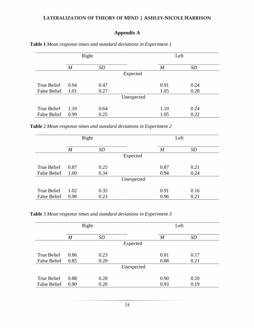
conditions are summarized in Appendix A
Table 1. Data was analyzed using a 3-way
Repeated Measure Analysis of Variance
(ANOVA). There was no main effect of
visual field or handedness observed, as
illustrated in Appendix B Figure 2. Secondary predictions included a
main effect of belief and expectedness. No
significant effect of belief was observed, but
the analysis did indicate a significant effect
of expectedness, F(1, 67) = 5.87, p = .018,
= .081, indicating that participants
responded more quickly when the agents
behaviour was congruent with the
participants expectation (refer to Appendix
B Figure 3). Furthermore, an interaction
between belief and expectedness was
observed, F(1, 67) = 5.22, p = .025, =
.072. A series of post hoc t-tests were
conducted to identify which means were
driving the significant interaction, and a
significant difference was observed between
the false-expected and true-expected
conditions, t(67) = 2.67, p = .10, d = .324,
and the true-expected and true-unexpected
conditions, t(67) = - 2.55, p = .013, d = .309.
This indicates that when the agent had a true
belief and searched for his or her object in
the expected location, participants
responded more quickly than when the agent
had a false belief or searched in an
unexpected location. Based on previous
research by Saxe and Wexler (2005), an
exploratory analysis was conducted to
determine whether a greater reaction time
difference between right hand and left hand
responses existed on unexpected trials as
compared to expected trials. This analysis
did not produce a significant result. Analyses of variance are somewhat
limited in terms of providing support for
hypothesized effects. Therefore, a Bayesian
analysis was conducted to identify the odds
that reaction times did not differ
significantly between right hand/visual field
conditions versus left hand/visual field
conditions. The Bayes factors did not find an
effect, and produced an odds factor of
6.62:1.
Experiment 2 Methods
Participants. Experiment 2 was
conducted with 40 undergraduate students
age 18.2 – 20.3 (M = 19.02, SD = 0.57), 22
of which were female and 18 of which were
male. The participants were recruited and
compensated in the same way as Experiment
1. Procedure. The procedure in
Experiment 2 was the same as Experiment 1,
except that participants were instructed to
only respond with their dominant hand.
Results Data was analyzed using a 3-way
Repeated Measure Analysis of Variance
LATERALIZATION OF THEORY OF MIND | ASHLEY-NICOLE HARRISON
9
(ANOVA). No significant main effect of
visual field (refer to Appendix B Figure 4),
expectedness, or belief was observed, and no
interactions were observed. However, a
marginal effect of belief was observed, F(1,
39) = 3.21, p = .081, = .076, indicating
that participants responded marginally faster
when the agent had a true belief about his or
her object’s location. The same exclusion
criteria were applied as in Experiment 1, and
four participants were excluded for
insufficient accuracy. Mean response times
for all conditions are summarized in
Appendix C. The Bayesian analysis did not
indicate a difference in response times to left
versus right visual field information, with an
odds factor of 3.44:1.
Experiment 3 Methods Participants. Experiment 3 was
conducted with 31 undergraduate students
age 18.1 – 22.7 (M = 19.13, SD = 1.17), 16
of which were female and 15 of which were
male. The participants were recruited and
compensated in the same way as Experiment
1. Procedure. Images were presented
in the center of the computer screen. The
procedure was otherwise identical to
Experiment 1.
Results Data was analyzed using a 3-way
Repeated Measure Analysis of Variance
(ANOVA). No significant main effect of
response hand (refer to Appendix A Figure
5) or belief was observed. However, the
analysis did indicate a main effect of
expectedness (refer to Appendix A figure 6),
F(1, 29) = 5.29, p = .029, = .154,
indicating that participants responded more
quickly when the agent looked for his or her
object in the expected location. Mean
response times for all conditions are
summarized in Appendix A Table 3. The
same exclusion criteria were applied as in
Experiment 1, and five participants were
excluded for insufficient accuracy. The
Bayesian analysis did not find a difference
in response times with left versus right hand
responses, with an odds factor of 6.85:1.
Discussion The present study was conducted to
gain more insight as to the way that theory
of mind mechanisms are lateralized in the
brain. Statistical analyses indicated that
participants did not respond more quickly to
images flashed in the left visual field,
discrediting the strong hypothesis. However,
the results did provide support for the weak
hypothesis. As previously discussed, a
distinct right hand motor advantage typically
leads to faster right hand response times on
motor tasks. Because the right hand motor
advantage was not observed in the present
study, it can be inferred that it was offset by a right
hemisphere cognitive advantage. This
provides mild support for the right
lateralization of theory of mind. The secondary predictions were also
somewhat supported. Participants did
respond more quickly in the expected
conditions in Experiments 1 and 3, and
responded marginally more quickly in the
true belief condition in Experiment 2.
Furthermore, an interaction between
expectedness and belief was observed,
meaning that participants responded more
quickly to trials in which the agent had a
true belief and behaved expectedly, than
when the agent had a false belief and when
the agent behaved unexpectedly. This is
likely because the false belief condition
requires participants to inhibit their own
knowledge. That is, the participant knows
that the object has been moved, but her or
she must inhibit this knowledge to reason
about the agent’s beliefs regarding the
object's location. Similarly, the unexpected

LATERALIZATION OF THEORY OF MIND | ASHLEY-NICOLE HARRISON
10
condition likely requires more theory of
mind reasoning to rationalize the agents
unexpected behaviour. These processes
probably slowed participants’ responses. These findings are somewhat
consistent with previous research concerning
theory of mind lateralization. As previously
discussed, Saxe and Wexler (2005) and
Young et al. (2010) each found support for
the right lateralization of theory of mind.
However, findings in these studies were
likely more pronounced than in the present
study due to the different methodologies
used. The Saxe and Wexler (2005) study
used neuroimaging methods, and the Young
et al. (2010) study inhibited function in the
RTPJ. In contrast, the present study
measured behavioural responses. Because so
many processes interact to produce a
behavioural response (i.e., language centers
process the instructions, facial recognition
centers respond to the animated character,
attention centers respond to the task, etc.) it
becomes more difficult to isolate a specific
brain region. Many processes interact to
produce a neural output as well, but when
using a neuroimaging method such as fMRI
or MRI, researchers can select a particular
brain region and measure its activation in
response to an input (i.e., a task or stimulus).
Similarly, TMS allows researchers to
manipulate a brain region of interest by
inhibiting its function. Conversely, a
behavioural task does not allow researchers
to isolate specific brain regions in a similar
way. This may explain the more robust
findings observed in previous studies.
Implications There are several practical
implications that can be drawn from the
present study. First, the observed findings
add to the literature supporting the right
lateralization of theory of mind. This
contributes to the breadth of research
devoted to understanding the functions of
different brain regions. This information is
also useful for understanding the neural
components of autism. As previously noted,
autistic individuals lack theory of mind
capabilities, which hinders their social
function. The first step toward
understanding the nature of theory of mind
deficits is to understand how theory of mind
processing occurs in normal functioning
individuals. Once the foundational
components of social function are better
understood, it will likely become possible to
develop interventions and treatments to
improve social functioning in individuals
with autism. Furthermore, the right lateralization
of theory of mind might provide further
insight as to why young children fail false
belief tasks. If theory of mind is organized
bilaterally (i.e., can be reasoned about using
mechanisms in both hemispheres), then
information should cross the corpus
callosum few times when making a verbal
response to a theory of mind task.
Conversely, if theory of mind mechanisms
are right lateralized, this should increase the
number of times that information must cross
the corpus callosum to generate a verbal
response. For example, consider a scenario
in which an image is flashed in the right
visual field, and thereby received in the left
hemisphere. Note that verbal responses are
generated in the left hemisphere (because
language is left lateralized; Frost, Binder,
Springer, Hammeke, Bellgowan, & Patrick,
1999). If theory of mind reasoning occurs in
both hemispheres, then information does not
need to be transferred over the corpus
callosum for any reason (i.e., information is
received in the left hemisphere, reasoned
about in the left hemisphere, and the
response is generated in the left
hemisphere). However, if theory of mind
reasoning is right lateralized, then
information must cross over the child’s
corpus callosum (which, as previously

LATERALIZATION OF THEORY OF MIND | ASHLEY-NICOLE HARRISON
11
noted, is dense prior to the age of 5 before it
undergoes a refinement process;
Westerhausen et al., 2011) to produce an
accurate response. It’s then possible that a
child’s dense corpus callosum could hinder
information transfer to the extent that he or
she fails the task.
Limitations The major limitation of the present
study is the inclusion of left-handed
participants, which could have muddled the
data in several ways. First, left-handed
individuals might have a left hand motor
advantage, which could potentially skew
reaction times in favor of the hypothesis
(faster reaction times to left visual field
information). Second, left-handed
individuals are more likely to have an
atypical neural makeup, meaning that certain
functions might not be lateralized in a
typical way (for example, their language
functions might not be left lateralized to the
same extent as many other people; Pujol,
Deus, Losilla, & Capdevila, 1999).
However, there’s a low possibility that
results were significantly impacted by the
inclusion of left-handed participants. Only
13 of the 149 participants were left-handed
(8.7%), and only a minority of left-handed
individuals are likely to be neurologically
atypical. Another potential limitation relates
to the image being lateralized in the correct
visual field. In order for the image to have
fallen onto the intended visual field, it was
necessary for the participant to focus on the
cross hair (i.e., “+”) in the center of the
screen during the false belief task. If
participants looked away or lost focus, the
image would not have fallen squarely in one
visual field, possibly skewing the results.
However, this is also unlikely to have
significantly impacted the results. This is
because the sequence of images occurred
very quickly (i.e., the cross hair appeared for
one second and the image appeared for
200ms), so participants would have had very
little time to shift their gaze. Furthermore,
images were not lateralized in Experiment 3
(they were flashed in the center of the
screen), but results were similar to the
previous experiments. A final limitation relates to possible
confusion among participants regarding the
false belief animation. In Experiment 3,
participants were instructed to switch
response hands while the image was
presented in the center of the screen. During
this experiment, it was observed that one
participant did not switch their hands
between blocks of trials when instructed to
do so. In Experiment 1, it was clear which
hand the participants were expected to use,
because the response hand corresponded to
the visual field presentation. However, there
is a possibility that participants may have
been confused about which hand to use in
Experiment 3 - since the image was
presented in the center of the screen - which
could have affected the results. Following
the observation of the student who failed to
switch hands, researchers monitored
participants more closely to ensure that they
responded with the correct hand.
Future Research and Conclusion The present study provides many
opportunities for further study, incorporating
different methods and sample groups.
Researchers could conduct similar studies,
but with more precise methods. For
example, a future study could use
neuroimaging methods to scan participant’s
brains while responding to the false belief
animation - to examine which areas become
activated while making theory of mind
judgments. Furthermore, neuroimaging
methods would also be useful for
determining whether participant’s brains are
organized in a typical way, to filter out
participants who might skew the results.
LATERALIZATION OF THEORY OF MIND | ASHLEY-NICOLE HARRISON
12
Another avenue for future research
could examine children’s difficulties on
false belief tasks. This could involve
conducting the present study with young
children, and comparing their response times
to those observed among adults. Research
could investigate whether the difference in
the amount of time taken to respond to right
visual field versus left visual field
information is greater in children than
adults. If so, this would indicate that
children’s corpus callosums are slowing
information transfer between the
hemispheres, and hindering theory of mind
processing. Finally, future studies could also
examine theory of mind using verbal
responses rather than button press responses.
Generating a verbal response requires
different brain mechanisms than a button
press response (i.e., brain centers involved
in speech rather than motor control).
Therefore, it would be interesting to see
whether the activation of different brain
mechanisms affects the results in any way. While many strides have been made
toward understanding theory of mind
reasoning, much remains unclear.
Uncovering the mechanisms underlying
theory of mind not only advances our
understanding of social processing, but also
has vast implications for research and
intervention regarding mental disorders,
such as autism. The present study supported
the right lateralization of theory of mind, but
additional research is necessary to confirm
the role of the RTPJ in theory of mind
reasoning. Theory of mind remains a rich
area of study, and as research progresses, a
clearer understanding of its mechanisms will
be obtained.
References Annett, M., Annett, J., Hudson, P.T.W., &
Turner, A. (1979). The control of
movement in the preferred and non-
preferred hands. The Quarterly journal
of experimental psychology,31(4),
641-652. doi:
10.1080/14640747908400755. Annett, M., Hudson, P.T.W., & Turner, A.
(1974). The reliability of differences
between the hands in motor
skill. Neuropsychologia, 12(4), 527-
531. doi: 10.1016/0028-
3932(74)90083-9. Apperly, I.A., & Butterfill, S.A. (2009). Do
humans have two systems to track
beliefs and belief-like states?
Psychological Review, 116(4), 953-
970. doi: 10.1037/a0016923. Baron-Cohen, S. (1997). Mindblindeness:
An Essay on Autism and Theory of
Mind. US: MIT Press. Fletcher, G., Simpson, J.A., Campbell, L., &
Overall, N.C. (2013). The Science of
Intimate Relationships. UK: Wiley
Blackwell Publications. Frost, J.A., Binder, J.R., Springer, J.A.,
Hammeke, T.A., & Bellgown, P.S.F.
(1999). Language processing is
strongly left lateralized in both sexes:
Evidence from functional MRI. Brain:
A Journal of Neurology, 122(2), 199-
208. doi: 10.1093/brain/122.2.199. Hannover, B., & Kuhnen, U. (2009). Culture
and social cognition in human
interaction. New York, NY, US:
Psychology Press. Heinrichs, M. & Gaab, J. (2007).
Neuroendocrine mechanisms of stress
and social interaction: Implications for
mental disorders. Current Opinion in
Psychiatry, 20(2), 158-162. doi:
10.1097/YCO.0b013e3280146a13. Pujol, J., Deus, J., Losilla, J.M., &
Capdevilla, A. (1999) Cerebral
lateralization of language in normal
left-handed people studies by
functional MRI. Neurology, 52(5),
1038-1043. doi:
10.1212/WNL.52.5.1038.

LATERALIZATION OF THEORY OF MIND | ASHLEY-NICOLE HARRISON
13
Saxe, R., & Wexler, A. (2005). Making
sense of another mind: The role of the
right temporo-parietal junction.
Neuropsychologia, 43(10), 1391-1399.
doi:
10.1016/j.neuropsychologia.2005.02.0
13. Scott, R. M., & Baillargeon, R. (2009).
Which penguin is this? Attributing
false beliefs about object identity at 18
months. Child development, 80(4),
1172-1196. doi: 10.1111/j.1467-
8624.2009.01324.x. Seemann, A (2011). Joint attention: New
development in psychology,
philosophy of mind, and social
neuroscience. Cambridge, MA, US:
MIT Press. Smith, T.S. (2013). Attachment, interaction,
and synchronization: How innate
mechanisms in attachment give rise to
emergent structure in networks and
communities. Handbook of
neurosociology. New York, NY, US:
Spriner Science + Business Media.
Weber, B., Treyer, V., Oberholzer, N.,
Jaermann, T., Boesiger, P., Brugger,
P., Regard, M., Buck, A., Savazzi, S.,
& Marzi, C. A. (2005). Attention and
interhemispheric transfer: a behavioral
and fMRI study. Journal of Cognitive
Neuroscience, 17(1), 113-123. doi:
10.1162/0898929052880002. Westerhausen, R., Luders, E., Specht, K.,
Ofte, S.H., Toga, A.W., Thompson,
P.M., Helland, T., & Hugdahl, K.
(2011). Structural and functional
reorganization of the corpus callosum
between the age of 6 and 8 years.
Cerebral Cortex, 21(5), 1012-1017.
doi: 10.1093/cercor/bhq165. Young, L., Camprodon, J., Hauser, M.,
Pascual-Leone, A., & Saxe, R. (2010).
Disruption of the right temporo-
parietal junction with transcranial
magnetic stimulation reduces the role
of beliefs in moral judgment. PNAS,
107(15), 6753-6758. doi:
10.1073/pnas.0914826107.
LATERALIZATION OF THEORY OF MIND | ASHLEY-NICOLE HARRISON
14
Appendix A Table 1 Mean response times and standard deviations in Experiment 1
Right Left M SD M SD
Expected
True Belief 0.94 0.47 0.91 0.24 False Belief 1.01 0.27 1.05 0.28
Unexpected
True Belief 1.10 0.64 1.10 0.24 False Belief 0.99 0.25 1.05 0.22
Table 2 Mean response times and standard deviations in Experiment 2
Right Left M SD M SD
Expected
True Belief 0.87 0.25 0.87 0.21 False Belief 1.00 0.34 0.94 0.24
Unexpected
True Belief 1.02 0.35 0.91 0.16 False Belief 0.98 0.23 0.96 0.21
Table 3 Mean response times and standard deviations in Experiment 3
Right Left M SD M SD
Expected
True Belief 0.86 0.23 0.81 0.17 False Belief 0.85 0.20 0.88 0.21
Unexpected
True Belief 0.88 0.28 0.90 0.20 False Belief 0.90 0.20 0.93 0.19

LATERALIZATION OF THEORY OF MIND | ASHLEY-NICOLE HARRISON
15
Appendix B
Figure 1. A series of still images illustrating
the sequence of events in the false belief
task.
Figure 3. Mean response times in the
expected and unexpected conditions in
Experiment 1.
Figure 2. Mean response times for images
flashed in the right and left visual field in
Experiment 1.
Figure 4. Mean response times for images
flashed in the right and left visual field in
Experiment 2.
Figure 5. Mean response times for right and
left hand responses in Experiment 3.
Figure 6. Mean response times in the
expected and unexpected conditions in
Experiment 3.
16
Cherry Youn
University of California, Berkeley
Peer victimization (PV) and intimate partner violence (IPV) are ubiquitous public health
concerns across ages and cultures, but previous studies indicate that they are more salient in
females than males, especially for individuals with psychiatric illnesses, such as Attention-
Deficit/Hyperactivity Disorder (ADHD; Blachman & Hinshaw, 2002; Cardoos & Hinshaw,
2001; Guendelman, Ahmad, Meza, Owens, & Hinshaw, 2015). PV describes physical
aggression, verbal threats, and relational harassment by other peers (Crick & Grotpeter, 1995;
Hawker & Boulton, 2000), and IPV describes physical, verbal, sexual, and/or psychological
abuse by a current or former partner or spouse (Centers for Disease Control and Prevention
[CDC], 2014). To better understand the risk factors associated with IPV and PV during
adulthood, this study will examine (1) the association between childhood ADHD severity and
adulthood IPV and PV, (2) the association between adolescent PV and adulthood PV, and (3)
adolescent PV as a predictor of IPV. The longitudinal study included three waves of data,
gathered every five years, with 228 female participants: Wave 1 consisted of parent reports on
ADHD symptoms; Wave 2 included self-reports on PV; and Wave 3 comprised of self-reports on
IPV and PV. Linear regressions showed significant associations between ADHD severity and
both adolescent IPV(β > .009, p < 0.001) and PV (β > 0.004, p <.001) . There was also a
significant association between ADHD severity and adult PV when co-varying
sociodemographic and cognitive variables (child IQ, mother’s education, household income, and
age). We found significant associations between adolescent PV and adulthood PV (β = 0.059, p
< 0.05). However, adolescent PV was not a significant predictor of adulthood IPV (β = 0.070, p
> 0.05). Clinical and research implications of the study are discussed.
eer victimization (PV) and intimate
partner violence (IPV) are serious
public health concerns that affect 50%
and 15% of women, respectively (e.g.,
Thompson et al., 2006); rates of each
increase for individuals with psychiatric
illnesses, such as Attention-
Deficit/Hyperactivity Disorder (ADHD).
Furthermore, the rate of PV is significantly
higher in children with ADHD than the
comparison groups (Cardoos & Hinshaw,
2011; Wiener, 2009; Humphrey, Storch, &
Geffken, 2007). These findings are not
surprising, as findings show that peer
relations is one of the significant
impairments in childhood ADHD (Hinshaw
& Melnick, 1995), and that girls have a
higher prevalence of conduct and
ADHD Severity, Peer Victimization, and
Intimate Partner Violence in Young Adult
Women
P

ADHD, PV, & IV | CHERRY YOUN
17
internalizing problems associated with
ADHD (Pajer, 1998; Lee & Hinshaw, 2006).
Previous research suggests that IPV is
significantly more prevalent at young
adulthood in women with mental illnesses
(Capaldi, Knoble, Shortt, & Kim, 2012;
Moffitt & Caspi, 1999; Trevillion, Oram,
Feder, & Howard, 2012). Less explored,
however, are associations between ADHD
and IPV. Moreover, previous studies show
that ADHD is a potential risk factor for
subsequent IPV (Fang, Massetti, Ouyang,
Grosse, & Mercy, 2010), specifically when
considering the social dysfunction exhibited
in adolescents with childhood ADHD
(Guendelman et al., 2015). Yet, there is
limited research on the peer relationships of
young adults with ADHD and even fewer
studies that examine romantic relationships
of adults with ADHD. The majority of
research on ADHD has focused on children
and adolescents, resulting in a gap in the
literature regarding interpersonal
relationships in adulthood. In an attempt to
address the gap in the literature, the current
study examined (1) the association between
childhood ADHD severity and IPV/PV, (2)
the persistence of adolescent PV into
adulthood PV, and (3) adolescent PV as a
predictor of adulthood IPV. The overall aim
of this study is to better understand the risk
factor(s) that result in the presence of IPV
and persistence of PV in females with
ADHD.
PV in Female Adolescents with ADHD
Children with ADHD experience a
high rate of peer rejection even after brief
interactions (Erhardt & Hinshaw, 1994).
They have fewer friends than their peers
without ADHD and are often ostracized by
other peer groups (Hinshaw & Melnick,
1995). The associated peer rejection and
isolation are explained, in part, by their
insensitive reciprocal interpersonal
behaviors and discordant interactions
(Hubbard & Newcomb, 1991). Such social
maladjustment in children with ADHD is
highly associated with (1) externalizing
behaviors such as disruptive behavior
(Hodges, Boivin, Vitaro, & Bukowski,
1999), lack of cooperation (Perren &
Alsaker, 2006), and conflict with peers
(Perren & Alsaker, 2006); (2) internalizing
behaviors, which include difficulties in
emotion regulation (Hodges, Boivin, Vitaro,
& Bukowski, 1999; Hodges & Perry, 1999);
and (3) social skills problems (Fox &
Boulton, 2006). A key result is rejection
from their peer groups (Hodges & Perry,
1999).
ADHD is one of the most common
childhood psychiatric diagnoses that often
persists well into adolescence and adulthood
(Glass, Flory, & Hankin, 2012; Goldman,
Genel, Bezman, & Slanetz, 1998). Hence,
social dysfunction often continues into
adolescence and young adulthood, as ADHD
persists in adolescence with 50% to 80% of
children continuing to meet criteria for
ADHD (Barkley, Fischer, Edelbrock, &
Smallish, 1990). This persistence accounts
for the underdevelopment of skills
fundamental to maintaining functional
relationships, as well as the stability of
disharmony in peer relations over time (Coie
& Dodge, 1983). Furthermore, clinical
research highlights that social difficulties are
often treatment-refractory for children with
ADHD, especially evident in current
psychosocial and pharmacological treatment
(Bagwell, Molina, Pelham, & Hoza, 2001).
In other words, even though medications
and behavioral treatments lead to behavioral
improvements in a majority of cases, peer
reputation is harder to improve.
IPV in Young Adult Women
IPV is a serious public health
concern that affects up to 54% of the women
ADHD, PV, & IV | CHERRY YOUN
18
in the United States (CDC 2003; Thompson
et al., 2006). Max and colleagues
highlighted that the United States spent 5.8
billion dollars for women who have
experienced IPV (2004). The data continue
to reflect the pervasiveness and costly
outcomes of the epidemic of IPV. Research
reveals that the risk factors for IPV include
young age, lower income and education
level of women, previous experience and/or
witness of violence in family, and
developmental psychopathology (e.g.,
conduct problems or antisocial behaviors;
Stith et al, 2004; Thoennes & Tjaden, 1990).
Despite findings that reflect symptoms of
ADHD are significant risk factors for IPV
(Guendelman et al., 2015), there is very little
research on how ADHD severity plays a role
in predicting IPV.
The Relation between PV and IPV
Furthermore, the extent to which PV
contributes to increased IPV risk among
young women with childhood ADHD has
not yet been examined. Much of what is
known about PV and IPV includes only the
development of violent behaviors and its
perpetration (United States Department of
Health and Human Services, 2001; Williams
et al., 2008). Considering that there is a
strong association between ADHD and the
continued prevalence of negative peer status
as well as the externalizing problems shown
to be risk factors of IPV (Bagwell et al.,
2001), this study aims to investigate the
associations between childhood ADHD
severity and IPV/PV in adolescence and
early adulthood. I hypothesize a strong
association between adolescent PV and
adulthood PV. Additionally, I also predict
that adolescent PV is a predictor of IPV in
young adulthood.
Method Participants
This study included 228 female
participants from the Berkeley Girls with
ADHD Longitudinal Study (BGALS). They
initially participated at research summer
programs conducted by the Principal
Investigator, Stephen P. Hinshaw, Ph.D.,
and staff. The programs ran from 1997 to
1999, when the participants were 6 to 12
years old (M = 9.1 years). The population
sample includes 140 participants diagnosed
with ADHD and 88 in a matched
comparison group. Comparison girls were
recruited from fliers that advertised summer
programs for girls, and did not meet the
diagnostic criteria for ADHD. Participants
with (a) Full Scale IQ lower than 70, (b)
neurological disorders or psychosis, (c)
autism or other developmental disorders,
and (d) other medical or physical conditions
that prevented them from participating in the
study were excluded from the population
sample. Both groups were recruited from
various sites that range from schools,
pediatric practices, and clinics in the San
Francisco Bay Area. The participants of this
study were diverse both ethnically (53%
Caucasian, 27% African American, 11%
Hispanic, 9% Asian American) and in terms
of socioeconomic status (M = $55,000,
ranging from $10,000 or below to $75,000
or over). Participants in the ADHD group
were required to meet the Diagnostic and
statistical manual of mental disorders, 4th
edition (DSM-IV) diagnostic criteria for
ADHD via the Diagnostic Interview
Schedule for Children (4th ed, DISC-IV;
Shaffer, Fisher, Lucas, Dulcan, & Schwab-
Stone, 2000). To promote generalizability of
the ADHD sample, participants with
common comorbidities of ADHD, such as
oppositional defiant disorder (ODD) and
conduct disorder (CD), were included in the
ADHD group. Out of the girls with ADHD
ADHD, PV, & IV | CHERRY YOUN
19
(n = 140), 63% (n = 88) and 20% (n = 29)
met criteria for ODD and CD, respectively.
Girls with mental disorders, medical issues,
or conditions, such as psychosis, overt
neurological disorder, mental retardation,
pervasive developmental disorder, non-
ability to speak English, that hindered them
from participating in the research summer
programs were excluded from the study.
Procedure Follow-up assessments have been
completed and analyzed throughout the
ongoing longitudinal investigation.
Participants were asked to participate in
follow-up assessments every five years, and
are currently participating in the fourth wave
of follow-up assessments. This study will
only examine the first three completed
follow-up studies. Wave 1 consists of
participants in their childhood whose ages
range from 6 to 12 (M = 9.1, SD = 1.7),
Wave 2 consists of participants in
adolescence whose ages range from 11.3 to
18.2 years (M = 14.2, SD = 1.7); these data
include 209 of the original 228 participants
(92%). Wave 3 consists of participants in
young adulthood whose ages range from 17
to 24 years (M = 19.6), including 216 of the
original 228 participants (95%). Participants
who were not included in this study were
due to attrition or lack of experience in
romantic relationship.
Measures ADHD Severity. Along with the
DISC structured interview, the Swanson,
Nolan, and Pelham IV rating scale (SNAP-
IV; Swanson et al., 2001) was administered
on both ADHD and control groups to obtain
an ADHD diagnosis. This parent scale
includes a total of 26-items according to the
symptom criteria in the DSM-IV for ADHD
and ODD: nine ADHD inattentive, nine
ADHD hyperactive/impulsive, and eight
ODD symptoms. The SNAP-IV is scored
based on a four-point Likert scale which
ranged from 0=not at all, 1=just a little,
2=quite a bit, to 3=very much. Previous
studies have shown the SNAP-IV to be a
reliable questionnaire and valid source for
gathering information on ADHD severity in
participants (Hinshaw, 2002). For this study,
an ADHD severity index was created by
summing the 18 ADHD symptoms,
weighted by their severity scores. PV and IPV. The Social
Relationships Interview (SRI; Brugha et al.,
1987) was assessed in both Wave 2 and
Wave 3 to measure PV and IPV. This
project-derived interview includes topics
related to peers, friendships, and romantic
relationships. Relevant questions were based
on conceptual models of friendship
attainment and social/dating relationships.
PV, in Wave 2 and Wave 3, was calculated
using a variable that averaged three
questions rated on a Likert scale (1 = never,
2 = less than once per month, 3 = once or
twice per month, 4 = once a week, 5 = a few
times a week, and 6 = everyday): (1) "have
you ever been hit?", (2) "have you ever been
teased to your face?", and (3) "have you
ever been teased behind your back?". Across
these three items, Cronbach's alpha in our
sample = .65, revealing adequate internal
consistency. The composite score for IPV at
Wave 3 combined the presence of physical
abuse and verbal abuse by the romantic
partner. The participant asked was to report
one of the four conditions: (1)
pushing/shoving, (2) slapping, (3) hitting
other parts of the body, and (4) pulling hair,
in order to meet the criteria for physical
abuse. In addition, indicating either (1)
swearing or name calling, or (2) threatening
physical violence met the criteria for verbal
abuse. This study applied a stringent
method through which a participant was
considered to be abused by a romantic
ADHD, PV, & IV | CHERRY YOUN
20
partner only if both physical and verbal
abuse criteria were met.
Covariates Four sociodemographic and
cognitive variables –child IQ, mother’s
education, household income, and age– were
used in this study as covariates. The
Wechsler Intelligence Scale for Children-
Third Edition (WISC-III; Wechsler, 1991)
was administered at Wave 1 to determine
the full-scale IQ scores for all participants.
The full scale IQ score was calculated to
assess if its association with poor social life
outcome was, in turn, related to PV, because
previous studies indicate that low IQ has
been linked to poor social functioning
(Dunlop & Savulescu, 2015).
Socioeconomic status was calculated using
the level of education for the parent in the
home measured at Wave 1, along with total
gross household income (Guendelman et al.,
2015), as previous studies have found that
low income is a significant risk factor for
IPV (Tjaden & Thoennes, 2009). Mother’s
education level was measured on a 6-point
scale (1 = less than 8th grade; 6 = advanced
or professional degree). Finally, age in
months at Wave 3 was collected from the
Wechsler Individual Achievement Test,
Second Edition (WIAT-II; Wechsler, 2001),
administered during in-person Wave 3
assessments.
Data Analytic Plan All statistical analyses were
performed with SPSS, Version 22 (IBM
Corp 2014). First, bivariate correlations
among ADHD severity at Wave 1, PV at
Wave 2, and IPV/ PV at Wave 3 were
calculated. A total of six correlations were
analyzed in this study: (1) correlation
between ADHD severity at Wave 1 and PV
at Wave 2, (2) ADHD severity at Wave 1
and IPV at Wave 3, (3) PV at Wave 2 and
IPV, (4) ADHD severity at Wave 1 and PV
at Wave 3, (5) PV at Wave 2 and Wave 3,
and (6) IPV at Wave 3 and PV at Wave 3. Second, linear regressions among the
four aforementioned variables were
conducted. Four regressions are featured: (1)
ADHD severity at Wave 1 predicting PV at
Wave 2, (2) ADHD severity at Wave 1
predicting PV at Wave 3, (3) ADHD
severity predicting IPV at Wave 3, and (4)
PV at Wave 2 predicting PV at Wave 3. In
these regressions, the covariates were placed
on step 1 of the equation and ADHD
severity (or, for the fourth regression, PV)
was placed on step 2.
Results Intercorrelations and Descriptive
Analyses A correlation matrix among the four
primary variables, Wave 1 ADHD severity,
Wave 2 PV, Wave 3 IPV, and Wave 3 PV,
examined the relations among predictor and
outcome variables investigated in the current
study (see Table 1). Several significant
associations were found. As expected, Wave
1 ADHD severity, Wave 2 PV, and Wave 3
PV and IPV were significantly associated
with one another. Wave 1 ADHD severity
was positively associated with Wave 2 PV (r
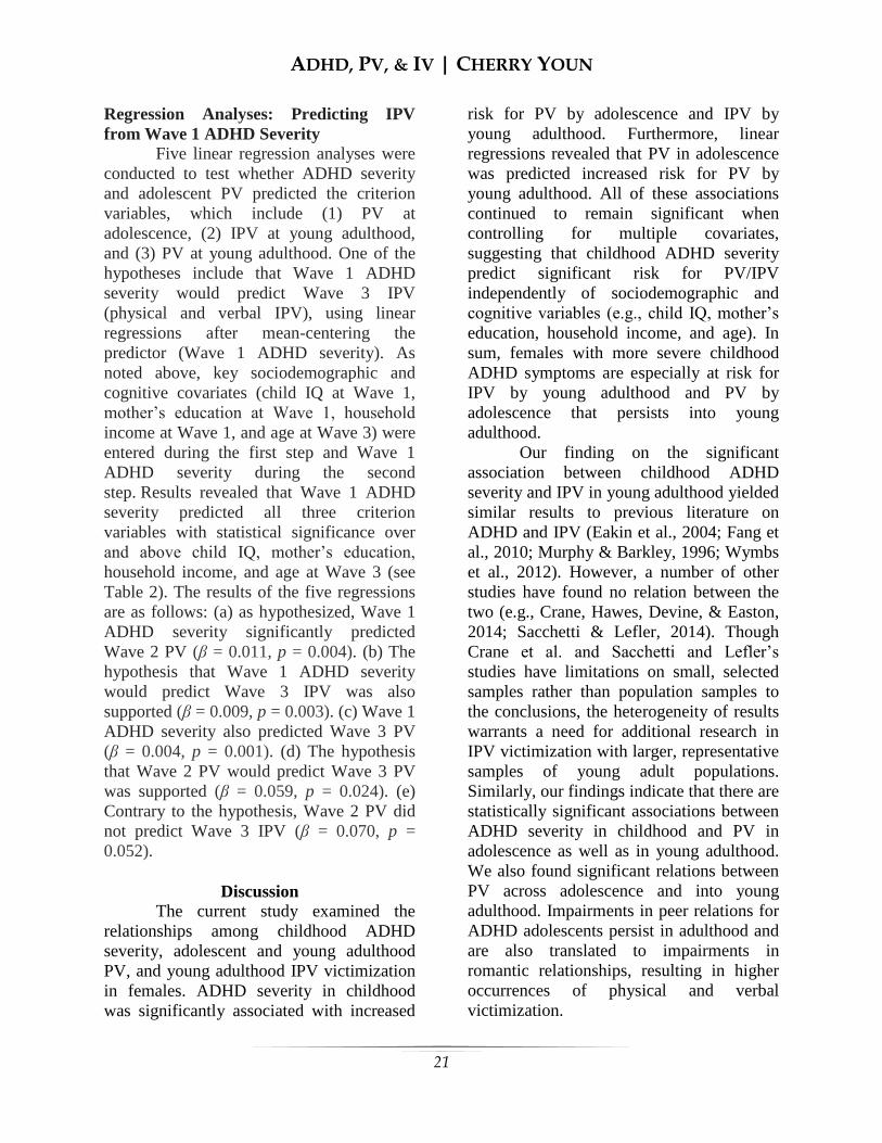
= 0.212, p = 0.003; see Figure 1). Similarly,
Wave 1 ADHD severity was positively
associated with Wave 3 PV (r = 0.198, p =
0.005; see Figure 2), and IPV (r = 0.217, p =
0.001; see Figure 3). Wave 2 PV was
significantly correlated with Wave 3 PV in
the expected direction (r = 0.193, p = 0.009;
see Figure 4). Wave 3 PV was positively
associated with IPV (r = 0.396, p = 0.000;
see Figure 5). The association between
Wave 2 PV and Wave 3 IPV was the sole
intercorrelation that showed a lack of
statistical significance (r = 0.029, p = 0.683;
see Figure 6).
ADHD, PV, & IV | CHERRY YOUN
21
Regression Analyses: Predicting IPV
from Wave 1 ADHD Severity Five linear regression analyses were
conducted to test whether ADHD severity
and adolescent PV predicted the criterion
variables, which include (1) PV at
adolescence, (2) IPV at young adulthood,
and (3) PV at young adulthood. One of the
hypotheses include that Wave 1 ADHD
severity would predict Wave 3 IPV
(physical and verbal IPV), using linear
regressions after mean-centering the
predictor (Wave 1 ADHD severity). As
noted above, key sociodemographic and
cognitive covariates (child IQ at Wave 1,
mother’s education at Wave 1, household
income at Wave 1, and age at Wave 3) were
entered during the first step and Wave 1
ADHD severity during the second
step. Results revealed that Wave 1 ADHD
severity predicted all three criterion
variables with statistical significance over
and above child IQ, mother’s education,
household income, and age at Wave 3 (see
Table 2). The results of the five regressions
are as follows: (a) as hypothesized, Wave 1
ADHD severity significantly predicted
Wave 2 PV (β = 0.011, p = 0.004). (b) The
hypothesis that Wave 1 ADHD severity
would predict Wave 3 IPV was also
supported (β = 0.009, p = 0.003). (c) Wave 1
ADHD severity also predicted Wave 3 PV
(β = 0.004, p = 0.001). (d) The hypothesis
that Wave 2 PV would predict Wave 3 PV
was supported (β = 0.059, p = 0.024). (e)
Contrary to the hypothesis, Wave 2 PV did
not predict Wave 3 IPV (β = 0.070, p =
0.052).
Discussion The current study examined the
relationships among childhood ADHD
severity, adolescent and young adulthood
PV, and young adulthood IPV victimization
in females. ADHD severity in childhood
was significantly associated with increased
risk for PV by adolescence and IPV by
young adulthood. Furthermore, linear
regressions revealed that PV in adolescence
was predicted increased risk for PV by
young adulthood. All of these associations
continued to remain significant when
controlling for multiple covariates,
suggesting that childhood ADHD severity
predict significant risk for PV/IPV
independently of sociodemographic and
cognitive variables (e.g., child IQ, mother’s
education, household income, and age). In
sum, females with more severe childhood
ADHD symptoms are especially at risk for
IPV by young adulthood and PV by
adolescence that persists into young
adulthood. Our finding on the significant
association between childhood ADHD
severity and IPV in young adulthood yielded
similar results to previous literature on
ADHD and IPV (Eakin et al., 2004; Fang et
al., 2010; Murphy & Barkley, 1996; Wymbs
et al., 2012). However, a number of other
studies have found no relation between the
two (e.g., Crane, Hawes, Devine, & Easton,
2014; Sacchetti & Lefler, 2014). Though
Crane et al. and Sacchetti and Lefler’s
studies have limitations on small, selected
samples rather than population samples to
the conclusions, the heterogeneity of results
warrants a need for additional research in
IPV victimization with larger, representative
samples of young adult populations.
Similarly, our findings indicate that there are
statistically significant associations between
ADHD severity in childhood and PV in
adolescence as well as in young adulthood.
We also found significant relations between
PV across adolescence and into young
adulthood. Impairments in peer relations for
ADHD adolescents persist in adulthood and
are also translated to impairments in
romantic relationships, resulting in higher
occurrences of physical and verbal
victimization.

ADHD, PV, & IV | CHERRY YOUN
22
Contrary to our hypothesis, there was
no significant association between Wave 2
PV and Wave 3 IPV. Girls who were
victimized in adolescence by their peers
were more likely to be involved in romantic
relationships where they are physically
and/or verbally victimized, but those
victimized in romantic relationships were
not necessarily victimized in adolescence by
peers. This result could be interpreted by the
environment that the adolescent females
with ADHD are in: significant rates of
females with ADHD are single parents and
young mothers (Russell, Ford, Rosenberg, &
Kelly, 2014), excluding them from social
environments such as school and work
where most social interactions occur.
Another interpretation could be made from
the durations of relationships formed by
girls with ADHD. Previous literature shows
that women with ADHD have serious
problems in interpersonal relationships, and
are involved in significantly less number of
intimate relationships due to poor social
functioning abilities (Babinski et al., 2011),
and exhibit less satisfying romantic
relationships (Biederman et al., 2006;
Kessler et al., 2006; Murphy & Barkley,
2006). In other words, girls with higher
ADHD severity are unable to commit to
long-term relationships, if any at all,
resulting in the lack of data in our study to
support the presence of IPV. However, out
of those who engage in romantic
relationships, many are victimized and
become involved in IPV.
Limitations and Conclusions A few study limitations should be
considered. First, PV and IPV victimization
were assessed from a small subsection of the
self-report questionnaire. Not using
standardized PV and IPV instruments may
have led to unreliability of measurement as
well as false negatives, where participants
underestimate the rate and frequency of the
PV/IPV due to a number of factors including
recall bias and psychological trauma.
Moreover, the self-report questionnaire did
not assess for exact time, severity, or
frequency of PV/IPV, hindering the study
from further analyses on the severity of
PV/IPV in girls with more severe symptoms
of ADHD. Lastly, the study only focused on
the participants’ exposure to both physical
and verbal IPV, excluding sexual and
psychological victimization. The presence of
child abuse and neglect, a significant risk
factor for future IPV (Bensley, Van Eenwyk,
& Wynkoop Simmons, 2003; Ehrensaft et
al., 2003), was also not assessed for this
study. Future research on the association
between the presence and severity of
different victimization and ADHD severity
over several developmental periods is
recommended. Taking these limitations into account, this
study introduces a conceptually plausible
pathway between peer victimization and
physical and verbal IPV in females with
ADHD. In other words, prior experience of
peer victimization has shown to function as
a gateway for subsequent victimization of
other kinds, including peer victimization in
adulthood and IPV. In conclusion, the
current study found that childhood ADHD
severity is linked to adolescent and young
adult PV and young adult IPV. Given the
developmental significance of peer
relationships, additional research on the
causes and treatment of poor social
functioning in ADHD severity in
adolescence and adulthood is warranted.
References Babinski, D. E., Pelham, W. E., Jr., Molina,
B. S., Gnagy, E. M., Waschbusch, D.
A., Yu, J., … Karch, K. M. (2011).
Late adolescent and young adult
outcomes of girls diagnosed with
ADHD in childhood: an exploratory

ADHD, PV, & IV | CHERRY YOUN
23
investigation. Journal of Attention
Disorders, 15(3), 204-214. Bagwell, C. L., Molina, B. S., Pelham, W.
E., Jr., & Hoza, B. (2001). Attention-
deficit hyperactivity disorder and
problems in peer relations:
predictions from childhood to
adolescence. Journal of the
American Academy of Child and
Adolescent Psychiatry, 40(11), 1285-
1292. Barkley, R. A., Fischer, M., Edelbrock, C.
S., & Smallish, L. (1990). The
adolescent outcome of hyperactive
children diagnosed by research
criteria: I. An 8-year prospective
follow-up study. Journal of the
American Academy of Child and
Adolescent Psychiatry, 29(4), 546-
557. Bensley, L., Van Eenwyk, J., & Wynkoop
Simmons, K. (2003). Childhood
family violence history and women’s
risk for intimate partner violence and
poor health. American Journal of
Preventive Medicine, 25(1), 38–44. Biederman, J., Faraone, S. V., Spencer, T. J.,
Mick, E., Monuteaux, M. C., &
Aleardi, M. (2006). Functional
impairments in adults with self-
reports of diagnosed ADHD: A
controlled study of 1001 adults in the
community. Journal of Clinical
Psychiatry, 67(4), 524-540. Blachman, D. R., & Hinshaw, S. P. (2002).
Patterns of friendship among girls
with and without attention-
deficit/hyperactivity disorder.
Journal of Abnormal Child
Psychology, 30(6), 625-640. Brugha, T. S., Sturt, E., MacCarthy, B.,
Potter, J., Wykes, T., & Bebbington,
P. E. (1987). The Interview Measure
of Social Relationships: The
description and evaluation of a
survey instrument for assessing
personal social resources. Social
Psychiatry, 22(2), 123-128. Capaldi, D. M., Knoble, N. B., Shortt, J. W.,
& Kim, H. K. (2012). A systematic
review of risk factors for intimate
partner violence. Partner Abuse, 3,
231–280. Cardoos, S. L., & Hinshaw, S. P. (2011).
Friendship as protection from peer
victimization for girls with and
without ADHD. Journal of
Abnormal Child Psychology, 39(7),
1035-1045. Centers for Disease Control and Prevention.
(2003). Costs of intimate partner
violence against women in the
United States. Atlanta: National
Center for Injury Prevention and
Control. Centers for Disease Control and Prevention.
(2014). Intimate partner violence.
Retrieved from
http://www.cdc.gov/ViolencePrevent
ion/intimatepartnerviolence/index.ht
ml.
Coie J. D., & Dodge K. A. (1983).
Continuities and changes in
children’s social status: a five-year
longitudinal study. Merrill-Palmer
Quarterly, 29(3), 261-282.
Crane, C. A., Hawes, S. W., Devine, S., &
Easton, C. J. (2014). Axis I
psychopathology and the
perpetration of intimate partner
violence. Journal of Clinical
Psychology, 70(3), 238-247.
Crick, N. R., & Grotpeter, J. K. (1995).
Relational aggression, gender, and
social-psychological adjustment.
Child Development, 66(3), 710–712.
Dunlop, M., & Savulescu, J. (2015).
Distributive justice and cognitive
enhancement in lower, normal
intelligence. Monash Bioethics
Review, 32(3), 189-204.
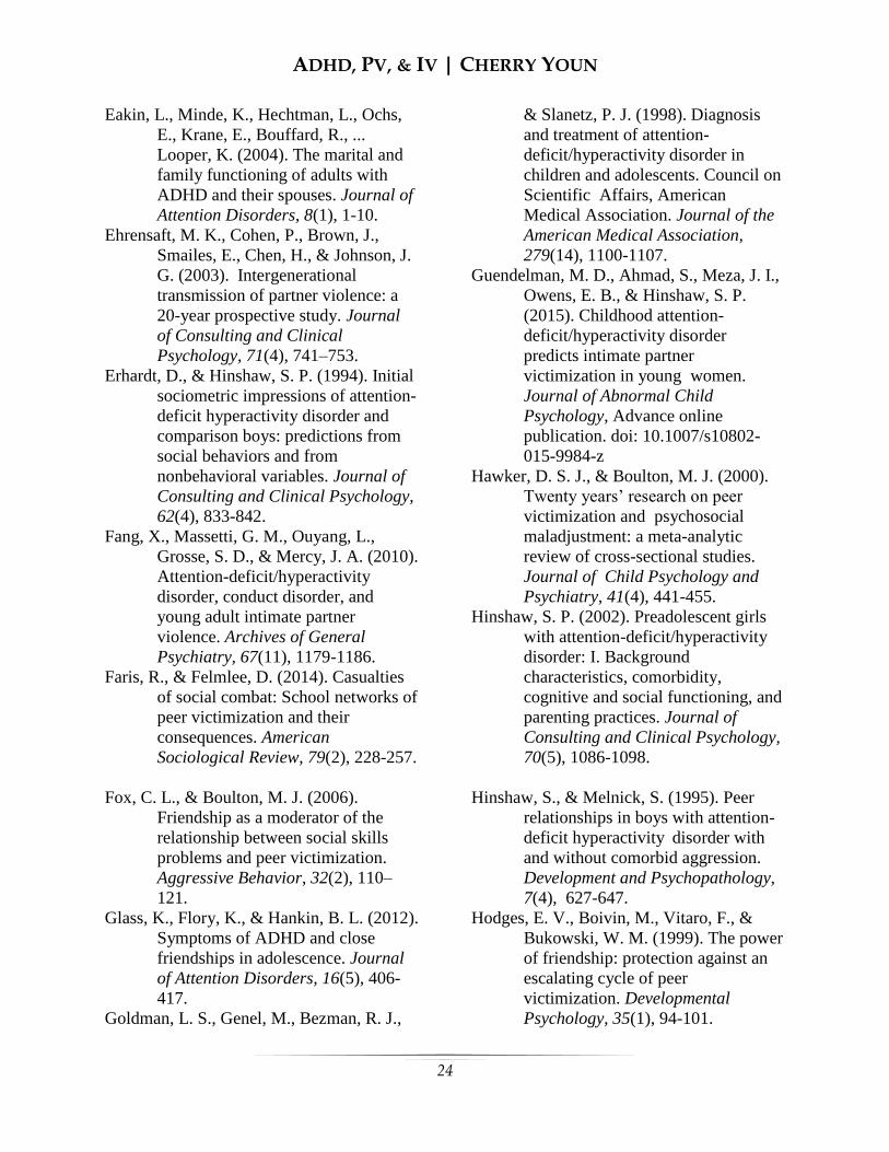
ADHD, PV, & IV | CHERRY YOUN
24
Eakin, L., Minde, K., Hechtman, L., Ochs,
E., Krane, E., Bouffard, R., ...
Looper, K. (2004). The marital and
family functioning of adults with
ADHD and their spouses. Journal of
Attention Disorders, 8(1), 1-10. Ehrensaft, M. K., Cohen, P., Brown, J.,
Smailes, E., Chen, H., & Johnson, J.
G. (2003). Intergenerational
transmission of partner violence: a
20-year prospective study. Journal of Consulting and Clinical
Psychology, 71(4), 741–753.
Erhardt, D., & Hinshaw, S. P. (1994). Initial
sociometric impressions of attention-
deficit hyperactivity disorder and
comparison boys: predictions from
social behaviors and from
nonbehavioral variables. Journal of
Consulting and Clinical Psychology,
62(4), 833-842.
Fang, X., Massetti, G. M., Ouyang, L.,
Grosse, S. D., & Mercy, J. A. (2010).
Attention-deficit/hyperactivity
disorder, conduct disorder, and
young adult intimate partner
violence. Archives of General
Psychiatry, 67(11), 1179-1186.
Faris, R., & Felmlee, D. (2014). Casualties
of social combat: School networks of
peer victimization and their
consequences. American
Sociological Review, 79(2), 228-257.
Fox, C. L., & Boulton, M. J. (2006).
Friendship as a moderator of the
relationship between social skills
problems and peer victimization.
Aggressive Behavior, 32(2), 110–
121. Glass, K., Flory, K., & Hankin, B. L. (2012).
Symptoms of ADHD and close
friendships in adolescence. Journal
of Attention Disorders, 16(5), 406-
417.
Goldman, L. S., Genel, M., Bezman, R. J.,
& Slanetz, P. J. (1998). Diagnosis
and treatment of attention-
deficit/hyperactivity disorder in
children and adolescents. Council on
Scientific Affairs, American
Medical Association. Journal of the
American Medical Association,
279(14), 1100-1107.
Guendelman, M. D., Ahmad, S., Meza, J. I.,
Owens, E. B., & Hinshaw, S. P.
(2015). Childhood attention-
deficit/hyperactivity disorder
predicts intimate partner
victimization in young women.
Journal of Abnormal Child
Psychology, Advance online
publication. doi: 10.1007/s10802-
015-9984-z
Hawker, D. S. J., & Boulton, M. J. (2000).
Twenty years’ research on peer
victimization and psychosocial
maladjustment: a meta-analytic
review of cross-sectional studies.
Journal of Child Psychology and
Psychiatry, 41(4), 441-455.
Hinshaw, S. P. (2002). Preadolescent girls
with attention-deficit/hyperactivity
disorder: I. Background
characteristics, comorbidity,
cognitive and social functioning, and
parenting practices. Journal of
Consulting and Clinical Psychology,
70(5), 1086-1098.
Hinshaw, S., & Melnick, S. (1995). Peer
relationships in boys with attention-
deficit hyperactivity disorder with
and without comorbid aggression.
Development and Psychopathology,
7(4), 627-647.
Hodges, E. V., Boivin, M., Vitaro, F., &
Bukowski, W. M. (1999). The power
of friendship: protection against an
escalating cycle of peer
victimization. Developmental
Psychology, 35(1), 94-101.
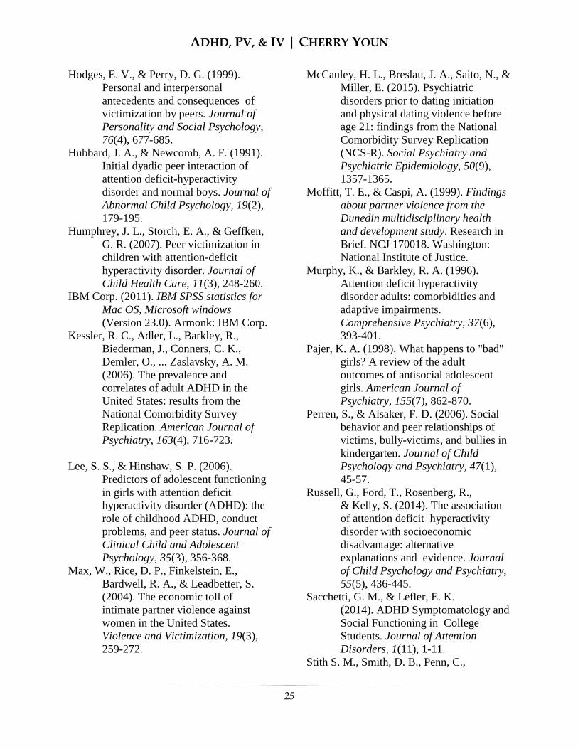
ADHD, PV, & IV | CHERRY YOUN
25
Hodges, E. V., & Perry, D. G. (1999).
Personal and interpersonal
antecedents and consequences of
victimization by peers. Journal of
Personality and Social Psychology,
76(4), 677-685.
Hubbard, J. A., & Newcomb, A. F. (1991).
Initial dyadic peer interaction of
attention deficit-hyperactivity
disorder and normal boys. Journal of
Abnormal Child Psychology, 19(2),
179-195.
Humphrey, J. L., Storch, E. A., & Geffken,
G. R. (2007). Peer victimization in
children with attention-deficit
hyperactivity disorder. Journal of
Child Health Care, 11(3), 248-260. IBM Corp. (2011). IBM SPSS statistics for
Mac OS, Microsoft windows
(Version 23.0). Armonk: IBM Corp. Kessler, R. C., Adler, L., Barkley, R.,
Biederman, J., Conners, C. K.,
Demler, O., ... Zaslavsky, A. M.
(2006). The prevalence and
correlates of adult ADHD in the
United States: results from the
National Comorbidity Survey
Replication. American Journal of
Psychiatry, 163(4), 716-723.
Lee, S. S., & Hinshaw, S. P. (2006).
Predictors of adolescent functioning
in girls with attention deficit
hyperactivity disorder (ADHD): the
role of childhood ADHD, conduct
problems, and peer status. Journal of
Clinical Child and Adolescent
Psychology, 35(3), 356-368. Max, W., Rice, D. P., Finkelstein, E.,
Bardwell, R. A., & Leadbetter, S.
(2004). The economic toll of
intimate partner violence against
women in the United States.
Violence and Victimization, 19(3),
259-272.
McCauley, H. L., Breslau, J. A., Saito, N., &
Miller, E. (2015). Psychiatric
disorders prior to dating initiation
and physical dating violence before
age 21: findings from the National
Comorbidity Survey Replication
(NCS-R). Social Psychiatry and
Psychiatric Epidemiology, 50(9),
1357-1365. Moffitt, T. E., & Caspi, A. (1999). Findings
about partner violence from the
Dunedin multidisciplinary health
and development study. Research in
Brief. NCJ 170018. Washington:
National Institute of Justice.
Murphy, K., & Barkley, R. A. (1996).
Attention deficit hyperactivity
disorder adults: comorbidities and
adaptive impairments.
Comprehensive Psychiatry, 37(6),
393-401.
Pajer, K. A. (1998). What happens to "bad"
girls? A review of the adult
outcomes of antisocial adolescent
girls. American Journal of
Psychiatry, 155(7), 862-870.
Perren, S., & Alsaker, F. D. (2006). Social
behavior and peer relationships of
victims, bully-victims, and bullies in
kindergarten. Journal of Child
Psychology and Psychiatry, 47(1),
45-57.
Russell, G., Ford, T., Rosenberg, R.,
& Kelly, S. (2014). The association
of attention deficit hyperactivity
disorder with socioeconomic
disadvantage: alternative
explanations and evidence. Journal
of Child Psychology and Psychiatry,
55(5), 436-445.
Sacchetti, G. M., & Lefler, E. K.
(2014). ADHD Symptomatology and
Social Functioning in College
Students. Journal of Attention
Disorders, 1(11), 1-11.
Stith S. M., Smith, D. B., Penn, C.,

ADHD, PV, & IV | CHERRY YOUN
26
Ward, D., & Tritt, D. (2004). Risk
factor analysis for spouse physical
maltreatment: a meta-analytic
review. Journal of Aggression and
Violent
Behavior, 10(1), 65-98.
Swanson, J. M., Kraemer, H. C., Hinshaw,
S. P., Arnold, L. E., Conners, C. K.,
Abikoff, H. B., ... Wu, M. (2001).
Clinical relevance of the primary
findings of the MTA: Success rates
based on severity of ADHD and
ODD symptoms at the end of
treatment. Journal of the American
Academy of Child and Adolescent
Psychiatry, 40(2), 168-179.
Thoennes, N., & Tjaden, P. G. (1990). The
extent, nature, and validity of sexual
abuse allegations in
custody/visitation disputes. Child
Abuse and Neglect, 14(2), 151-163.
Thompson, R. S., Bonomi, A. E., Anderson,
M., Reid, R. J., Dimer, J. A., Carrell,
D., & Rivara, F. P. (2006). Intimate
partner violence: prevalence, types,
and chronicity in adult women.
American Journal of Preventive
Medicine, 30(6), 447-457.
Trevillion, K., Oram, S., Feder, G.,
& Howard, L. M. (2012).
Experiences of domestic violence
and mental disorders: A systematic
review and meta-analysis. PloS One,
7. Advance online publication.
doi:10. 1371/journal.pone.0051740.
Wechsler, D. (1991). Wechsler Intelligence
Scales for Children —Third Edition
[Measurement Instrument]. San
Antonio, TX: The Psychological
Corporation.
Wechsler, D. (2001). Wechsler Individual
Achievement Test (2nd ed.)
[Measurement Instrument]. New
York, NY: Psychological
Corporation. Wiener, J., & Mak, M. (2009). Peer
victimization in children with
Attention-Deficit/Hyperactivity
Disorder. Psychology in the Schools,
11(3), 116-131.
Wymbs, B., Molina, B., Pelham, W.,
Cheong, J., Gnagy, E., Belendiuk,
K., ... Waschbusch, D. (2012). Risk
of intimate partner violence among
young adult males with childhood
ADHD. Journal of Attention
Disorders, 16(5), 373-383.

ADHD, PV, & IV | CHERRY YOUN
27
Appendix
Table 1Intercorrelations among the primary variables 1. 2. 3. 1. Wave 1 ADHD Severity 2. Wave 2 PV .212** 3. Wave 3 IPV .217** .029 4. Wave 3 PV .198** .193** .396***
Note: PV = peer victimization, IPV = intimate partner violence
* Correlation is significant at the .05 level (2-tailed).
** Correlation is significant at the .01 level (2-tailed).
*** Correlation is significant at the .001 level (2-tailed). A correlation matrix among the four primary variables, which include Wave 1 ADHD severity,
Wave 2 PV, Wave 3 IPV, and Wave 3 PV, examined the relations among predictor and outcome
variables investigated in this study. All but the correlation between Wave 2 PV and Wave 3 IPV
showed statistical significance at the .05 level or higher. This data was retrieved from the
Berkeley Girls with ADHD Longitudinal Study (BGALS).
Table 2 Linear regressions among primary variables while accounting for covariates β SE p-value Wave 1 ADHD Severity Wave 2 PV .011 .004 .001 Wave 3 IPV .009 .003 .001 Wave 3 PV .004 .001 .001 Wave 2 PV Wave 3 IPV .070 .052 .180 Wave 3 PV .059 .024 .014
Note: PV = peer victimization, IPV = intimate partner violence
Results indicate that Wave 1 ADHD severity predicted all three criterion variables with
statistical significance over and above four key socioeconomic and cognitive covariates (child IQ
at Wave 1, mother’s education at Wave 1, household income, and age) at Wave 3. All
hypotheses other than the one that Wave 2 PV would predict Wave 3 PV were supported with
statistical significance. This data was retrieved from the Berkeley Girls with ADHD
Longitudinal Study (BGALS).
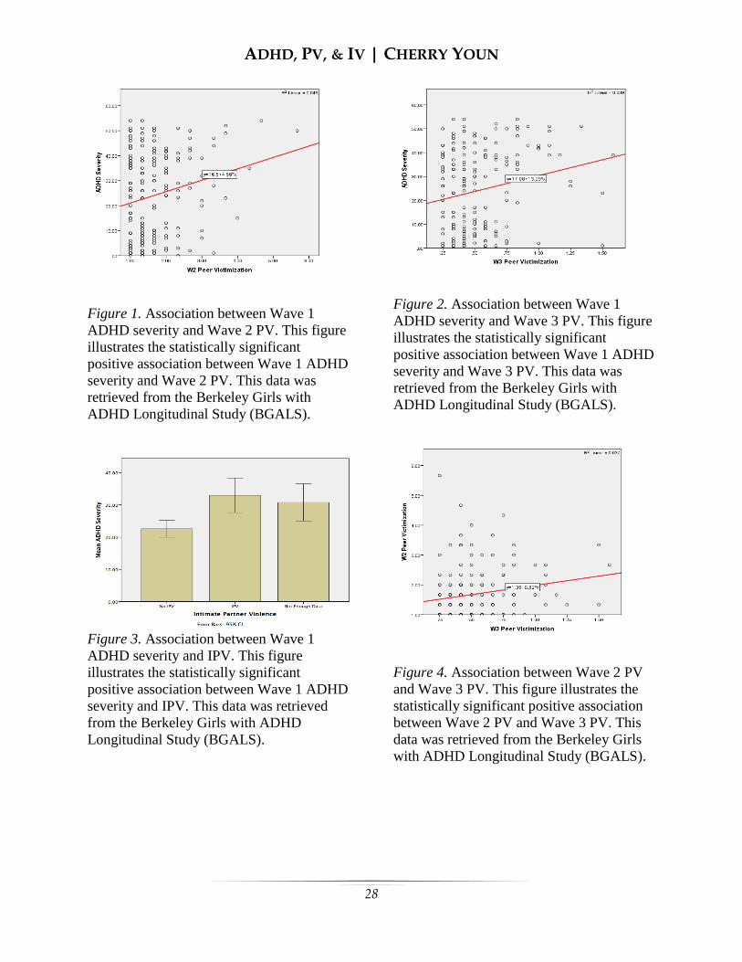
ADHD, PV, & IV | CHERRY YOUN
28
Figure 1. Association between Wave 1
ADHD severity and Wave 2 PV. This figure
illustrates the statistically significant
positive association between Wave 1 ADHD
severity and Wave 2 PV. This data was
retrieved from the Berkeley Girls with
ADHD Longitudinal Study (BGALS).
Figure 3. Association between Wave 1
ADHD severity and IPV. This figure
illustrates the statistically significant
positive association between Wave 1 ADHD
severity and IPV. This data was retrieved
from the Berkeley Girls with ADHD
Longitudinal Study (BGALS).
Figure 2. Association between Wave 1
ADHD severity and Wave 3 PV. This figure
illustrates the statistically significant
positive association between Wave 1 ADHD
severity and Wave 3 PV. This data was
retrieved from the Berkeley Girls with
ADHD Longitudinal Study (BGALS).
Figure 4. Association between Wave 2 PV
and Wave 3 PV. This figure illustrates the
statistically significant positive association
between Wave 2 PV and Wave 3 PV. This
data was retrieved from the Berkeley Girls
with ADHD Longitudinal Study (BGALS).
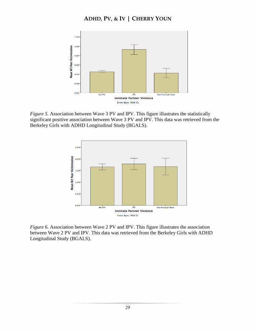
ADHD, PV, & IV | CHERRY YOUN
29
Figure 5. Association between Wave 3 PV and IPV. This figure illustrates the statistically
significant positive association between Wave 3 PV and IPV. This data was retrieved from the
Berkeley Girls with ADHD Longitudinal Study (BGALS).
Figure 6. Association between Wave 2 PV and IPV. This figure illustrates the association
between Wave 2 PV and IPV. This data was retrieved from the Berkeley Girls with ADHD
Longitudinal Study (BGALS).

30
Sheila Rajagopalan
University of California, Berkeley
Previous research has shown that non-synesthetes make systematic associations across a variety
of sensory combinations (Spence, 2011). Studies conducted in the Palmer Visual Aesthetics and
Perception Lab at UC Berkeley have found that for a variety of visual and musical stimuli, a
shared affective response mediates cross-modal associations. This study aims to test whether
people make systematic associations between diverse musical genres and line-shapes and
additionally whether these associations could be mediated by the perceived emotional,
perceptual, and/or musical qualities that are shared by the respective stimuli. Participants listened
to music samples from 33 different genres while 48 line-shapes were displayed on a computer
screen. They were then asked to select the three most consistent and the three least consistent
shapes for each musical selection. Finally, they rated each of the musical and shape stimuli along
a series of 13 bipolar dimensions from three categories: emotional (Calm/Agitated,
Disharmonious/Harmonious, Dislike/Like, Not Angry/Angry, and Sad/Happy), perceptual
(Light/Heavy, Open/Closed, Simple/Complex, and Smooth/Sharp), and musical
(Monotonous/Interesting, Slow/Fast, Soft/Loud, and Sparse/Dense). Results support previous
research in that participants typically matched line-shapes and music samples that shared
emotional, perceptual, and musical features. The dimensions which demonstrated the strongest
correlations for musical selections and line-shapes were Calm/Agitated (r=0.95, p<0.001),
Light/Heavy (r=0.95, p<0.001), and Soft/Loud (r=0.95, p<0.001). This suggests that emotional,
perceptual, and musical features could indeed all be mediating cross-modal associations.
The author would like to thank Dr. Palmer, Dr. Malfatti, and the rest of the team in the UC
Berkeley Visual Aesthetics and Perception Lab for their immense help with this project.
e experience the world as an
integrated combination of
information presented through
different sensory modalities. As we
encounter particular combinations of
sensory stimuli more frequently than others,
it should follow that we eventually learn to
associate certain stimuli of one sensory
modality with those of another. Which
sensory stimuli do we pair together? What is
the nature of the method in which we make
these associations?
One phenomenon related to these
sentiments is the neurological phenomenon
of synesthesia, which affects up to 5% of the
population (Simner et al., 2006). Synesthetes
experience unusual mixing of the senses,
such that stimulation in one sense also
Music-Shape Associations and the Emotional
Mediation Hypothesis
W

MUSIC SHAPE & EMOTIONAL MEDITATION | SHEILA RAJAGOPALAN
31
triggers perception in a different sense. For
synesthetes, everyday activities like reading
or listening to music arouse extraordinary
sensations of color, smells, tastes, shapes,
and other perceptions that non-synesthetes
simply do not experience (Simner &
Hubbard, 2013). While synesthetes tend to
make absolute correspondences with definite
cross-modal experiences, non-synesthetes
tend to make only relative correspondences
without cross-modal experiences (Gallace &
Spence, 2006). Non-synesthetic cross-modal
matching effects are nevertheless robust in
the general population, particularly for
children (Bond & Stevens, 1970; Braaten,
1993; Cohen, Henik, & Walsh 2009; Imai,
Kita, Nagumo, & Okada, 2008; Lewkowicz
& Turkewitz, 1980; Mondloch & Maurer,
2004; Morgan, Goodson, & Jones, 1975).
Of the various sensory combinations
that have been studied for non-synesthetes,
the most well documented is visual-auditory
associations. Research has shown that
people use certain low-level visual
metaphors to represent low-level auditory
features. A study conducted by Walker
(1987) found that across cultures, people
match high vertical placement with high
pitch, large objects with high volume, and
long duration with long horizontal length.
Additional work done previously in
the Palmer Visual Aesthetics and Perception
Lab at UC Berkeley has shown that non-
synesthetes also make systematic auditory-
visual cross-modal associations for a variety
of higher-level musical stimuli (such as
musical passages) as well. The results of this
research suggest that these associations are
mediated by shared emotional content across
sensory stimuli, a theory coined the
“emotional mediation hypothesis” (Palmer,
Schloss, Xu, & Prado-Leon, 2013).
Evidence for the emotional mediation
hypothesis stems from studies showing that
participants match cross-modal stimuli that
are rated similarly along emotional
dimensions. This effect was first
demonstrated for music-to-color
associations (Schloss, Lawler, & Palmer,
VSS-2008; Palmer, Langlois, Tsang,
Schloss, & Levitin, VSS-2011; Griscom &
Palmer, VSS-2012; Griscom & Palmer,
VSS-2013), but later extended to music-to-
texture associations (Langlois, Peterson, &
Palmer, VSS-2014; Peterson, Langlois, &
Palmer, VSS-2014). Further research done
by Malfatti (2014) has shown that emotion
also mediates associations between line-
shapes and color, while research by
Whiteford has shown that emotion mediates
associations between color and musical
genres (Whiteford, Schloss, & Palmer, VSS-
2013). The present study aims to supplement
previous research in an attempt to discover
whether emotional, perceptual, and/or
musical characteristics mediate the
formation of associations between musical
genres and line-shapes.
Previous research into the correlation
between shape and music has found that
people match high pitches with sharp shapes
and low pitches with round shapes (Kohler,
1929; Marks, 1987). The connection
between shape and music is somewhat
intuitive for those with musical training
(Kussner, 2013). In fact, musicians often
make reference to the “shape” of a musical
passage, oftentimes making gestures as if
shaped movement can communicate some
aspect of the music (Kussner, 2013). In an

MUSIC SHAPE & EMOTIONAL MEDITATION | SHEILA RAJAGOPALAN
32
attempt to examine what the “shape” of
music refers to, which acoustical parameters
relate to shape, and whether musical shapes
can be made more accessible by visualizing
them, Kussner had musicians and non-
musicians alike listen to passages of music
and draw a visual representation of that
music. He found that musicians made more
consistent drawings in line with the overall
expressive quality of the music, while non-
musicians used more diverse representation
strategies and neglected temporal aspects of
pitch (Kussner, 2013).
This correlation works inversely
from shape to music as well. Graphic design
of a composition is a common aid in the
creation of an envisioned musical work
(Goldberg & Schrak, 1986). The creation of
the Unité Polyagogique Informatique
CEMAMu (UPIC) system by the composer
Iannis Xenakis is perhaps the most famous
example. The UPIC system allows
composers to use graphic notation to
represent musical effects that are too
complicated to be specified with the
traditional staff notation. For instance, the
system represents convoluted orchestral
musical features such as glissandi for many
instruments with lines in a pitch-versus-time
domain. The lines and various other
graphical objects have specific functions
that a computer recognizes to synthesize
sound (Marino, Serra, & Raczinski, 1993).
In this way, the composer can draw music
and is freed from the limitations of
traditional notation which cannot represent a
great variety of sound phenomena (Marino,
Serra, & Raczinski, 1993).
The relationship between shape and
music is also of artistic interest. There have
been multiple artistic attempts to create
multimedia works in both the visual and
sonic domains. The artistic technique of
“graphical sound” involves photographing
lines and shapes, then arranging those
images on an analog optical sound track to
produce notes and chords. This technique
has been used in art films for decades
(Goldberg & Schrak, 1986). Various scenes
in Disney’s 1940 film Fantasia show lines
and shapes that transform to reflect the
sound and rhythm of the soundtrack. The
connection between music and form is an
important aspect of artistic works by Kupka,
Kandinsky, and Klee, who use music as a
primary source of inspiration and whose
work is often discussed using the lexicon for
musical composition (Goldberg & Schrak,
1986).
In studying associations made
between shape and music, as well as the
mechanisms of those associations, we hope
to enrich the fields of visual art, music, and
psychology by offering a greater
understanding of cross-modal correlations in
the general population.
Methods
Participants
For this study, 23 participants were
recruited through the UC Berkeley Research
Participation Program and 9 participants
were recruited through the UC Berkeley
Research Subject Volunteer Pool, for a total
of 32 participants (8 male, 24 female).
Participants ranged from 18 to 34 years of
age (mean= 22.6), were fluent in English,
and self-identified as having either normal
or corrected-to-normal vision. Participants
included in analysis were non-synesthetic, as

MUSIC SHAPE & EMOTIONAL MEDITATION | SHEILA RAJAGOPALAN
33
determined by a pre-screening questionnaire
adapted from the Eagleman Battery. Further
online assessments were carried out as
needed through the synesthete.org website
(see Appendix A) (Eagleman, Kagan,
Nelson, Sagaram, & Sarma, 2007). An
additional three participants were excluded
from the data analysis because their data
were incomplete (two participants) or they
were identified as shape-sound synesthetic
(one participant).
Materials
The line-shapes used in the
experiment varied along four dimensions:
number of line segments (4 vs. 8), angularity
(angular vs. curved), line thickness (thin vs.
thick), and number of intersections (0% vs.
50% of the number of line segments). Lines
were produced in Adobe Illustrator using a
script for random line generation. Once
shapes of 4 and 8 line segments were
randomly produced, subsets of the shapes
were selected for having no intersections (0
%) or 50% intersections (e.g., 2 intersections
for 4-line-segments shapes and 4
intersections for 8-line-segment shapes).
These selected shapes were then edited in
Adobe Illustrator to create variants with
different curvatures (angular vs. curved) and
line thickness (thick vs. thin). Three shape
variants of each four-factor combination
were produced, for a total of 48 lines shapes
(see Figure 1). These 48 shapes were then
arranged systematically by factors and
spatially permuted in eight different sets to
counter any bias that may have arisen from
specific placements of each shape within the
grid. The first four sets were designed to
balance placement of the four shape
dimensions and variants, so that each row
corresponded to a different variant and each
column, quadrant, and semi-quadrant
corresponded to one of the four shape
dimensions. These four sets were then
rotated 180° to produce the second four sets
(see Appendix B for all sets). The shapes
were presented on a neutral gray background
(CIE x= 0.312, y=0.318, Y=19.26), at the
center of a 21.5-inch Mac desktop monitor
that was calibrated using a Minolta CS100
Chroma Meter. Participants were seated
approximately 70cm from the monitor.
The 33 musical selections were a
subset of the selections used in a previous
experiment carried out in the Palmer Visual
Perception and Aesthetics Lab at UC
Berkeley (Whiteford, Schloss, & Palmer,
VSS-2013) (see Figure 2). The stimuli were
instrumental selections from 33 different
musical genres that were edited using
Audacity software to have an appropriate
peak amplitude. Each was a sample of 15
seconds and was edited to include a 2-
second fade-in and fade-out. The music was
played through closed-ear headphones for
the experiment.
Participants rated each of the 33
musical selections and 48 line-shapes along
a series of 13 bipolar dimensions. Five of
these dimensions were chosen as being
primarily emotional (Calm/Agitated,
Disharmonious/Harmonious, Dislike/Like,
Not Angry/ Angry, and Sad/Happy), four as
primarily perceptual or geometric
(Light/Heavy, Open/Closed,
Simple/Complex, and Smooth/Sharp), and
four as primarily musical
(Monotonous/Interesting, Slow/Fast,
Soft/Loud, and Sparse/Dense).
MUSIC SHAPE & EMOTIONAL MEDITATION | SHEILA RAJAGOPALAN
34
Figure 1: The 48 line-shapes used in the study. Line-shapes varied along four dimensions, which
are indicated by the labels beneath: number of line segments (“4” vs. “8”), angularity (“a” for
angular vs. “c” for curved), line thickness (presence of “t” for thick), and number of intersections
(presence of “i” if shape has intersections).
Table 1: The titles, of the 33 musical selections used in the study, as well as the performing
artists and genres.
Procedure
Participants initially signed a consent
form and completed a demographic
questionnaire. The first task involved
making associations between the 33 musical
stimuli and the 48 shape stimuli. Participants
then rated each of the musical selections and
each of the line stimuli along a series of 13
bipolar dimensions concerning their
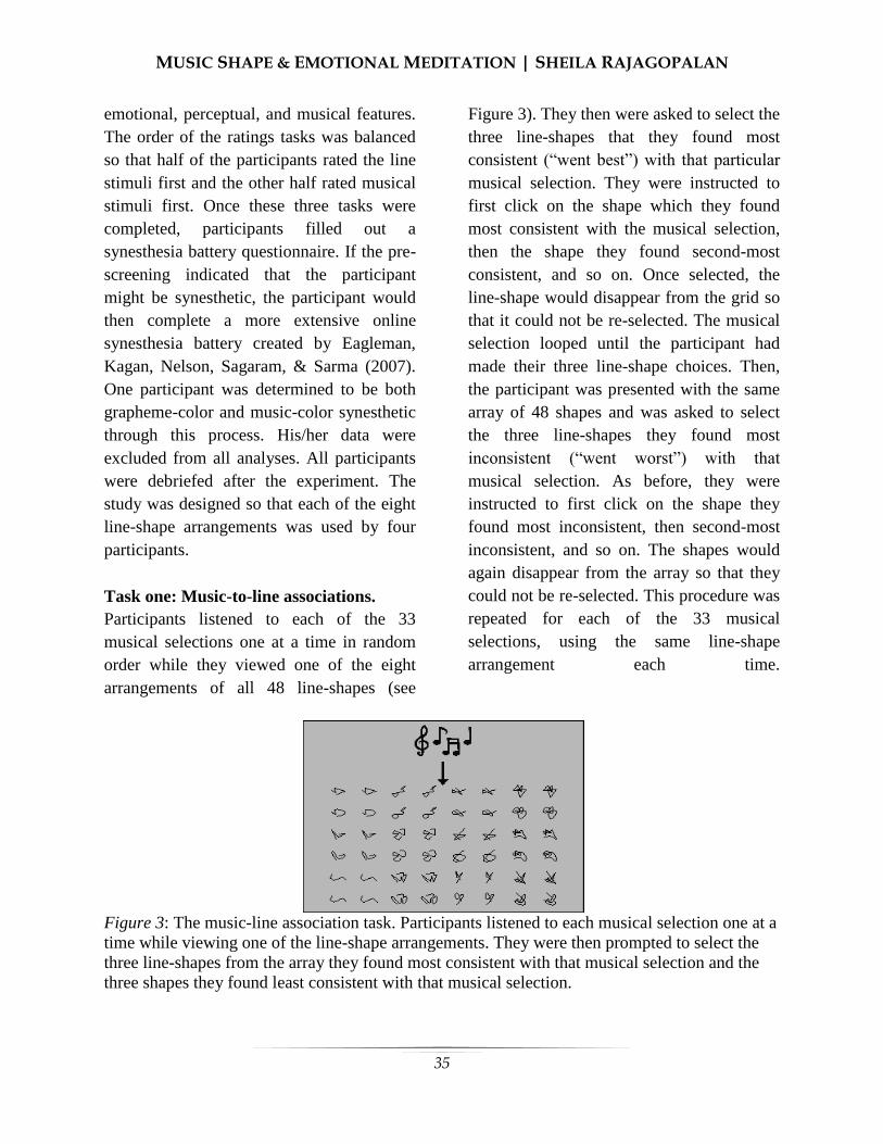
MUSIC SHAPE & EMOTIONAL MEDITATION | SHEILA RAJAGOPALAN
35
emotional, perceptual, and musical features.
The order of the ratings tasks was balanced
so that half of the participants rated the line
stimuli first and the other half rated musical
stimuli first. Once these three tasks were
completed, participants filled out a
synesthesia battery questionnaire. If the pre-
screening indicated that the participant
might be synesthetic, the participant would
then complete a more extensive online
synesthesia battery created by Eagleman,
Kagan, Nelson, Sagaram, & Sarma (2007).
One participant was determined to be both
grapheme-color and music-color synesthetic
through this process. His/her data were
excluded from all analyses. All participants
were debriefed after the experiment. The
study was designed so that each of the eight
line-shape arrangements was used by four
participants.
Task one: Music-to-line associations.
Participants listened to each of the 33
musical selections one at a time in random
order while they viewed one of the eight
arrangements of all 48 line-shapes (see
Figure 3). They then were asked to select the
three line-shapes that they found most
consistent (“went best”) with that particular
musical selection. They were instructed to
first click on the shape which they found
most consistent with the musical selection,
then the shape they found second-most
consistent, and so on. Once selected, the
line-shape would disappear from the grid so
that it could not be re-selected. The musical
selection looped until the participant had
made their three line-shape choices. Then,
the participant was presented with the same
array of 48 shapes and was asked to select
the three line-shapes they found most
inconsistent (“went worst”) with that
musical selection. As before, they were
instructed to first click on the shape they
found most inconsistent, then second-most
inconsistent, and so on. The shapes would
again disappear from the array so that they
could not be re-selected. This procedure was
repeated for each of the 33 musical
selections, using the same line-shape
arrangement each time.
Figure 3: The music-line association task. Participants listened to each musical selection one at a
time while viewing one of the line-shape arrangements. They were then prompted to select the
three line-shapes from the array they found most consistent with that musical selection and the
three shapes they found least consistent with that musical selection.

MUSIC SHAPE & EMOTIONAL MEDITATION | SHEILA RAJAGOPALAN
36
Tasks 2 and 3: Stimulus ratings. The order
of the second and third tasks was varied so
that half of the participants performed the
line-dimension rating task first and the other
half performed the music-dimension rating
task first.
Line-dimension rating task. The participant
was presented with each of the 48 line-
shapes one at a time in random order and
was asked to rate each shape along a series
of 13 bipolar dimensions (Calm/Agitated,
Disharmonious/Harmonious, Dislike/Like,
Light/Heavy, Monotonous/Interesting, Not
Angry/Angry, Open/Closed, Sad/Happy,
Simple/Complex, Slow/Fast, Smooth/Sharp,
Soft/Loud, and Sparse/Dense). Each line-
shape appeared at the center of the computer
screen, and a slider scale with an adjustable
bar was placed below it. Each end of the
scale was labeled with a word indicating one
extreme pole of the dimension (See Figure
4). The participant was instructed to rate the
shape along that bipolar dimension by
sliding the bar’s position along the
continuous scale and clicking to record their
response. The slider bar was one pixel in
width, and the scale itself was 400 pixels in
width, so that their response correlated to a
rating between -200 and 200 for that
dimension. The center of the scale indicated
neutrality and was marked with a tick for the
participant’s reference. The slider was
placed in the middle of the scale at the start
of each iteration.
Task 4: Music-dimension rating task. In
this task, participants were presented with
each musical selection one at a time in
random order and asked to rate each along
the same 13 bipolar dimensions using the
same procedure as for the line-dimension
task described above.
Figure 4: The line-dimension and music-dimension rating tasks. Participants were presented
with each line-shape and musical stimulus one at a time in random order, then were instructed to
move a bar along a continuous scale to rate each stimulus along a series of 13 bipolar dimensions
(e.g., Disharmonious/Harmonious). Note that arrows were not displayed in the experiment, but
are included here to signify the possible movements of the slider bar. Similarly, the participant
did not see musical notes during the music-dimension rating task (just the dimensional scale), but
it is included here to represent the fact that the participants heard music during that task.

MUSIC SHAPE & EMOTIONAL MEDITATION | SHEILA RAJAGOPALAN
37
Results
Music-Shape Association scores
(MSAs) were computed to examine the
correlation between each musical selection
and line-shapes chosen as consistent with
that selection along each of the 13 bipolar
dimensions. The calculation is modeled after
the Music-Color Association scores used by
Palmer et al. (2013) and is computed as
follows:
(1) Cd,m = (3c1,d,m + 2c2,d,m + c3,d,m)/3
(2) Id,m = (3i1,d,m + 2i2,d,m + i3,d,m)/3
(3) MSAd,m= Cd,m – Id,m
where cj,d,m is the average rating for the jth
line chosen as consistent (c) with musical
selection m along dimension d, and ij,d,m is
the average rating for the jth line chosen as
inconsistent (i) with musical selection m
along dimension d. Each shape’s
dimensional rating is weighted by the order
in which it was picked, so that the values for
the most consistent and inconsistent shapes
are multiplied by three, second-most
consistent and inconsistent multiplied by 2,
and so on. The average values for the most
inconsistent shapes (Id,m ) are subtracted
from the average values for the most
consistent shapes (Cd,m) to compute a single
Music-Shape Association score for each
musical selection along each dimension.
Figures 5,6, and 7 show the MSA values (y-
axis) plotted against the average ratings for
each musical selection (x-axis) in order to
observe music-line shape correlation along
the emotional, perceptual, and musical
dimensions, respectively.
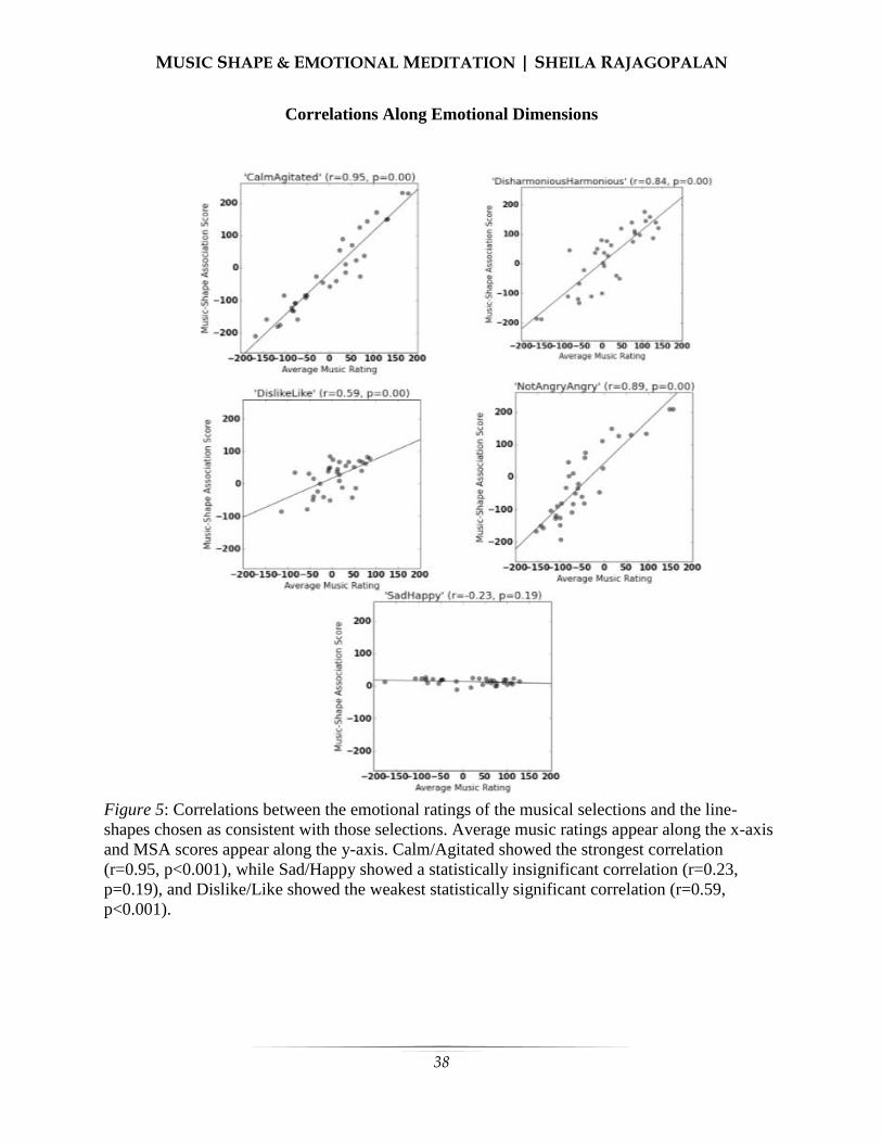
MUSIC SHAPE & EMOTIONAL MEDITATION | SHEILA RAJAGOPALAN
38
Correlations Along Emotional Dimensions
Figure 5: Correlations between the emotional ratings of the musical selections and the line-
shapes chosen as consistent with those selections. Average music ratings appear along the x-axis
and MSA scores appear along the y-axis. Calm/Agitated showed the strongest correlation
(r=0.95, p<0.001), while Sad/Happy showed a statistically insignificant correlation (r=0.23,
p=0.19), and Dislike/Like showed the weakest statistically significant correlation (r=0.59,
p<0.001).
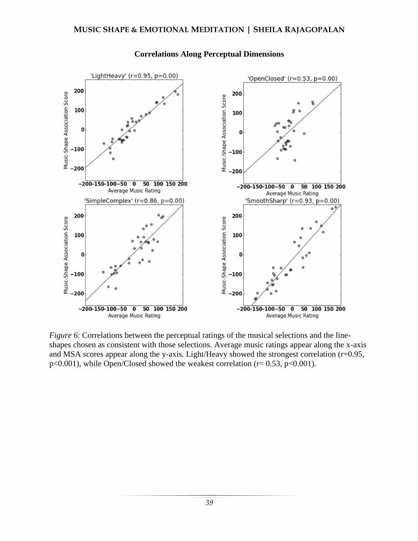
MUSIC SHAPE & EMOTIONAL MEDITATION | SHEILA RAJAGOPALAN
39
Correlations Along Perceptual Dimensions
Figure 6: Correlations between the perceptual ratings of the musical selections and the line-
shapes chosen as consistent with those selections. Average music ratings appear along the x-axis
and MSA scores appear along the y-axis. Light/Heavy showed the strongest correlation (r=0.95,
p<0.001), while Open/Closed showed the weakest correlation (r= 0.53, p<0.001).

MUSIC SHAPE & EMOTIONAL MEDITATION | SHEILA RAJAGOPALAN
40
Correlations Along Musical Dimensions
Figure 5: Correlations between the musical ratings of the musical selections and the line-shapes
chosen as consistent with those selections. Average music ratings appear along the x-axis and
MSA scores appear along the y-axis. Soft/Loud showed the highest correlation (r=0.95,
p<0.001), while Monotonous/Interesting showed the weakest correlation (r=0.59, p<0.001).
All of the 13 dimensions showed
statistically significant correlations (p <
0.001) between the ratings for the musical
selections and the corresponding ratings of
the line-shapes that were chosen as going
best/worst with those selections (see Figures
5-7). The only exception was for
Sad/Happy (r = -0.23, p = 0.19).
Among the five emotional
dimensions, three showed very strong
correlations between the emotional ratings
of the music samples and line-shapes paired
with each music sample (Calm/Agitated r =
0.95, Disharmonious/Harmonious r = 0.84,
Not Angry/Angry r = 0.89). On the other
hand, the Dislike/Like domain (r = 0.59)
demonstrated the weakest statistically
significant correlation. Among the four
perceptual dimensions, three showed very
strong positive correlations (Light/Heavy r =
0.95, Smooth/Sharp r = 0.93,
Simple/Complex r = 0.86), whereas
Open/Closed showed a weaker correlation (r
= 0.53). Among the four musical
dimensions, three showed very strong
positive correlations (Soft/Loud r = 0.95,

MUSIC SHAPE & EMOTIONAL MEDITATION | SHEILA RAJAGOPALAN
41
Sparse/Dense r = 0.91, Slow/Fast r = 0.85),
while Monotonous/Interesting showed a
weaker correlation (r = 0.59).
A principal components analysis on
the emotional ratings of the musical genres
showed that the five dimensions
(Calm/Agitated,
Disharmonious/Harmonious, Disklike/Like,
Not Angry/Angry, Sad/Happy) could be
reduced to two principal components that
explained 91.1% of variance in the data. The
first principal component (PC1) explained
73.3% of the data, and the second (PC2)
explained 17.8% of the data. PC1
corresponded roughly to the agitation level
of the music (PC1 loadings: Calm/Agitated
= 0.963, Disharmonious/Harmonious = -
0.958, Not Angry/Angry = 0.91,
Dislike/Like = -0.822, Sad/Happy = 0.561).
PC2 roughly represented the happiness of
the music (PC2 loadings: Sad/Happy=
0.814, Dislike/Like= 0.346,
Disharmonious/Harmonious= 0.196,
Calm/Agitated= 0.188, Not Angry/Angry= -
0.183).
We performed an analogous
principal components analysis for the
emotional ratings of the line-shapes. The
solution was very similar to that of the
emotional ratings of the musical genres. The
five emotional dimensions could be reduced
to two principal components that explained
79.4% and 17.5% of variance in the data,
respectively, for a total of 96.9% of the data
explained by the two components. The first
principal component corresponded roughly
to the harmony of the lines (PC1 loadings:
Disharmonious/Harmonious = 0.987, Not
Angry/Angry = -0.974, Calm/Agitated = -
0.956, Dislike/Like = 0.941, Sad/Happy =
0.498), whereas the second principal
component corresponded roughly to the
happiness of the lines (PC2 loadings:
Sad/Happy = 0.860, Calm/Agitated = 0.251,
Not Angry/Angry = 0.188, Dislike/Like =
0.134, Disharmonious/Harmonious = -
0.133).
Discussion
The current study reveals that
participants consistently matched musical
selections with line-shapes that affected a
shared emotional response. The shared
Agitation and Angriness levels of the stimuli
were the most significant mediating factors,
similar to findings in the Whiteford et al.
study on musical genres and color (2013)
and the Malfatti study on color and line
shape (2014). These results support the
emotional mediation hypothesis. The
perceptual and musical dimensions tended to
have very high correlations as well,
indicating that any of these three
dimensional categories could play an
important role in mediating systematic
music-to-line associations.
When analyzing musical stimuli
across dimensional categories, it was
apparent that certain musical dimensions
correlated highly with certain emotional
dimensions (p < 0.01 for all correlations;
See Appendix C for full correlation table).
Most notably, the Calm/Agitated dimension
correlated strongly with several musical
dimensions (Soft/Loud r = 0.958, Slow/Fast
r = 0.909, Sparse/Dense r = 0.908,
Monotonous/Interesting r = 0.566),
indicating that the agitation of a musical
selection corresponds strongly to its
loudness, quickness, and density, but not
MUSIC SHAPE & EMOTIONAL MEDITATION | SHEILA RAJAGOPALAN
42
necessarily to its level of interest for the
subject. It is unclear whether we have a
representative enough sample of musical
genres to generalize across all music, but
this is certainly an intriguing relation that
deserves further exploration.
Similar effects occurred across
dimensional categories for the line ratings
(See Appendix C for full correlation table).
In particular, the Smooth/Sharp dimension
correlated highly with various emotional
dimensions (Not Angry/Angry r = 0.949,
Calm/Agitated r = 0.940, Dislike/Like r = -
0.847, Sad/Happy r = -0.357, p < 0.05 in all
cases). This result indicates that the sharp
lines in our sample are viewed as angry and
agitated, and are generally disliked.
The relative Calmness and Harmony
of the line-shapes and music tended to be the
most important associative factors. The line-
shapes that were rated as most calm were
curved, non-intersecting, lines with few
segments; the most harmonious lines were
the lines that shared those same
characteristics. This implies that
harmonious, calm lines are those that are
simple, non-intersecting, and curved. The
most agitated lines were the angular,
intersecting, thick lines with the most line
segments; the most disharmonious were the
angular, intersecting lines with the most line
segments. Agitated, disharmonious lines can
then be interpreted as possessing those
qualities. Interestingly, line thickness did not
seem to play a vital role in determining the
perceived Calmness or Harmony of the line-
shapes. For music, the Calmest genres were
Piano, Indie, and Soundtrack, whereas the
most Agitated were Heavy Metal, Dubstep,
and Ska. The most Harmonious musical
genres were Mozart, Smooth Jazz, and
Indie, whereas the most Disharmonious
were Dubstep, Heavy Metal, and Gamelan.
It is important to note that the
musical selections used in this study were
chosen by Whiteford et al. to be
representative of the genre, but the
measurements we report here should be
interpreted in terms of each particular
selection, rather than representative of the
entire genre. Furthermore, the evidence
presented here is purely correlational, and
therefore does not establish a causal relation
between emotional, perceptual, or musical
feature mediation in shape-music
associations. Future research could examine
correlations between line-shapes and
musical genres on a broader scale and
clarify the respective roles of emotional,
perceptual, and musical mediating factors in
producing the observed pattern of
associations. It would also be interesting to
study how these associations are affected by
various levels of perceptual organization.
The results presented here could be
useful in creating multimedia art works that
strongly evoke certain emotions by utilizing
emotionally complementary shapes and
music. Commercially, the associations made
between music and form could be utilized to
create album covers that accurately
represent the emotions of the music. Using
an understanding of correlated shapes and
music, music educators can reinforce their
pupil’s learning by incorporating stimuli for
multiple senses, which could help those
students who differentially prefer visual or
auditory learning. Although it is not clear
that the stimuli used in the study actually
made the participants feel the emotions they
MUSIC SHAPE & EMOTIONAL MEDITATION | SHEILA RAJAGOPALAN
43
assigned to the stimuli, further research in
this area could help predict emotional
responses to music and/or shapes, as well as
the emotions felt during painting or music
composition. Art and music therapists could
use that information to develop a more
enriched understanding of the emotions
experienced by their patients by viewing the
musical and perceptual features of that
patient’s creations. Furthermore, they could
soothe agitated patients with fittingly calm
artwork and music as identified through
research supplemental to this study. In a
psychiatric setting, therapists may be able to
better communicate with patients who have
trouble interpreting emotions in other people
(such as those with Asperger’s Syndrome)
by using visual or musical stimuli as
identified through this study in conjunction
with past and future studies.
References
Bond, B., & Stevens, S. S. (1969). Cross-
modality matching of brightness to
loudness by 5-year-olds. Perception
& Psychophysics, 6, 337–339. Braaten, R. (1993). Synesthetic
correspondence between visual
location and auditory pitch in infants.
Paper presented at the 34th Annual
Meeting of the Psychonomic
Society. Cohen Kadosh, R., Henik, A., & Walsh, V.
(2009). Synaesthesia: Learned or
lost? Developmental Science, 12,
484–491. Eagleman, D.M., Kagan, A.D., Nelson, S.S.,
Sagaram, D., & Sarma, A.K. (2007).
A standardized test battery for the
study of synesthesia. Journal of
Neuroscience Methods, 159(1), 139-
145. Gallace, A., & Spence, C. (2006).
Multisensory synesthetic interactions in the speeded classification of visual
size. Perception & Psychophysics,
68, 1191–1203. Goldberg, T., & Schrack, G. (1986).
Computer-Aided Correlation of
Musical and Visual Structures.
Leonardo, 19 (1), 11-17. Retrieved
2015, from JSTOR. Griscom, W., & Palmer, S. (2012, May).
The Color of Musical Sounds: Color
Associates of Harmony and Timbre
in Non-synesthetes, Poster Presented
at the 12th
annual meeting of the
Vision Sciences Society, FL. Griscom, W., & Palmer, S. (2013, May).
Cross-modal Sound to Sight
Associations with Musical Timbre in
Non-synesthetes, Poster Presented at
the 13th
annual meeting of the Vision
Sciences Society, FL. Imai, M., Kita, S., Nagumo, M., & Okada,
H. (2008). Sound symbolism
facilitates early verb learning.
Cognition, 109, 54–65. Köhler, W. (1929). Gestalt psychology. New
York: Liveright. Kussner, M. (2013). Music and Shape.
Literary and Linguistic Computing,
28(3), 472-479. Retrieved 2015,
from Oxford Journals. Langlois, T., Peterson, J., Palmer, S. (2014,
May). Visual Texture, Music, and
Emotion, Poster presented at the 14th
annual meeting of the Vision
Sciences Society, FL. Lewkowicz, D. J., & Turkewitz, G. (1980).
Cross-modal equivalence in early
infancy: Auditory–visual intensity
matching. Developmental Psychology, 16, 597–607.
Malfatti, Michela (2014) Shape-to-color

MUSIC SHAPE & EMOTIONAL MEDITATION | SHEILA RAJAGOPALAN
44
associations in non-synesthetes:
perceptual, emotional, and cognitive
aspects. PhD thesis, University of
Trento. Marino, G., Serra, M., & Raczinski, J.
(1993). The UPIC System: Origins
and Innovations. Perspectives of
New Music, 31(1), 258-269.
doi:10.2307/833053 Marks, L. E. (1987). On cross-modal
similarity: Auditory–visual interactions in speeded
discrimination. Journal of
Experimental Psychology: Human
Perception and Performance, 13,
384–394. Mondloch, C. J., & Maurer, D. (2004). Do
small white balls squeak? Pitch–
object correspondences in your
children. Cognitive, Affective &
Behavioral Neuroscience, 4, 133–
136. Morgan, G. A., Goodson, F. E., & Jones, T.
(1975). Age differences in the
associations between felt
temperatures and color choices. The American Journal of Psychology, 88,
125–130. Palmer, S. E., Schloss K. B., Xu, Z., Prado-
León, L. R. (2013). Music-colour
associations are mediated by
emotion. Proceedings of the National
Academy of Sciences, USA, 110, 8836–8841.
Palmer, S., Langlois, T., Tsang, T., Schloss,
K., & Levitin, D. (2011, May).
Color, Music, and Emotion, Poster
presented at the 11th
annual meeting
of the Vision Sciences Society, FL. Peterson, J., Langlois, T., & Palmer, S.
(2014, May). Cross-modal
associations from musical timbres
and intervals to visual textures,
Poster presented at the 14th
annual
meeting of the Vision Sciences
Society, FL. Schloss, K., Lawler, P., & Palmer, S. (2008,
May). The Color of Music, Poster
presented at the 8th
annual meeting
of the Vision Sciences Society, FL. Simner, J., Mulvenna, C., Sagiv, N.,
Tsakanikos, E., Witherby, S. A.,
Fraser, C., et al. (2006). Synaesthesia: The prevalence of
atypical cross-modal experiences.
Perception, 35, 1024–1033. Simner, J., & Hubbard, E. (Eds.). (2013).
The Oxford handbook of synesthesia.
New York, New York: Oxford
University Press. Spence, C. (2011). Crossmodal
correspondences: A tutorial review.
Attention, Perception, &
Psychophysics, 73 (4). 971-995.
Retrieved 2015, from JSTOR. Walker, R. (1987). The effects of culture,
environment, age, and musical
training on choices of visual
metaphors for sound. Perception &
Psychophysics, 42 (5), 491-502.
Retrieved 2015, from Google
Scholar. Whiteford, K., Schloss, K. B., & Palmer, S.
E. (2013). Music-Color Associations
from Bach to the Blues: Emotional
Mediation in Synesthetes and Non-
synesthetes. Poster presented at the
13th Annual Meeting of the Vision
Science Society, Naples, Florida,
May 2013.

MUSIC SHAPE & EMOTIONAL MEDITATION | SHEILA RAJAGOPALAN
45
Appendix A
Synesthesia Battery Pre-screening Questionnaire
Subject #: _________
1. Does seeing, thinking of, or hearing a number (e.g. Arabic or Chinese) cause a perception
of color?
☐ Never ☐ Rarely ☐ Sometimes ☐ Often ☐ All the time
2. Does seeing, thinking of, or hearing a letter cause a perception of color?
☐ Never ☐ Rarely ☐ Sometimes ☐ Often ☐ All the time
3. Does the concept of dates such as days (e.g. Monday, Tuesday), or months (e.g. January,
February) trigger a color?
☐ Never ☐ Rarely ☐ Sometimes ☐ Often ☐ All the time
4. Do you visualize numbers, letters or time units like weekdays or months as being spread out
in a 3D space around you?
☐ Never ☐ Rarely ☐ Sometimes ☐ Often ☐ All the time
5. Do individual keys on a piano or other instrument cause the perception of different colors?
☐ Never ☐ Rarely ☐ Sometimes ☐ Often ☐ All the time
6. Do different musical chords cause perception of different colors?
☐ Never ☐ Rarely ☐ Sometimes ☐ Often ☐ All the time
7. Do different musical instruments cause perception of different colors?
☐ Never ☐ Rarely ☐ Sometimes ☐ Often ☐ All the time
8. Do individual keys on a piano or other instrument cause the perception of different shapes?
☐ Never ☐ Rarely ☐ Sometimes ☐ Often ☐ All the time
9. Do different musical chords cause perception of different shapes?

MUSIC SHAPE & EMOTIONAL MEDITATION | SHEILA RAJAGOPALAN
46
☐ Never ☐ Rarely ☐ Sometimes ☐ Often ☐ All the time
10. Do different musical instruments cause perception of different shapes?
☐ Never ☐ Rarely ☐ Sometimes ☐ Often ☐ All the time
11. Do tastes trigger a color for you? For example, does the taste of chocolate, citrus, or banana
trigger a color?
☐ Never ☐ Rarely ☐ Sometimes ☐ Often ☐ All the time
12. Do smells trigger a color for you? For example, does the odor of steak or fries cause the
perception of a color in you?
☐ Never ☐ Rarely ☐ Sometimes ☐ Often ☐ All the time
13. Does pain trigger a color for you? For example, do different levels of pain you experience
at different times, say while having a headache, cause you to perceive color?
☐ Never ☐ Rarely ☐ Sometimes ☐ Often ☐ All the time
14. Does seeing or thinking of a person make you perceive a color?
☐ Never ☐ Rarely ☐ Sometimes ☐ Often ☐ All the time
15. Do tactile sensations trigger a color for you? For example, when you experience touch
sensations of different kinds, on different parts of your body, do you also perceive color?
☐ Never ☐ Rarely ☐ Sometimes ☐ Often ☐ All the time
16.Do heat and cold trigger colors in you? For example, does touching cold water or feeling
warm water in a shower cause you to perceive different colors?
☐ Never ☐ Rarely ☐ Sometimes ☐ Often ☐ All the time
17. Do you perceive different colors while experiencing a sexual orgasm?
☐ Never ☐ Rarely ☐ Sometimes ☐ Often ☐ All the time
18. Do different emotions like joy, gloom cause perception of color?

MUSIC SHAPE & EMOTIONAL MEDITATION | SHEILA RAJAGOPALAN
47
☐ Never ☐ Rarely ☐ Sometimes ☐ Often ☐ All the time
19. Does seeing a picture or a scene also cause you to hear a sound?
☐ Never ☐ Rarely ☐ Sometimes ☐ Often ☐ All the time
20. Does hearing a sound cause a distinct odor, like the noise of water gushing arouses the
smell of a rose?
☐ Never ☐ Rarely ☐ Sometimes ☐ Often ☐ All the time
21. Does seeing a picture, object or a scene cause you to perceive a distinct smell?
☐ Never ☐ Rarely ☐ Sometimes ☐ Often ☐ All the time
22. Does hearing a sound cause a sensation of touch for you? For example, does hearing an
airplane fly past cause a distinct sensation of touch?
☐ Never ☐ Rarely ☐ Sometimes ☐ Often ☐ All the time
23. Does hearing a sound cause a sensation of taste for you? For example, does the ticking of a
clock cause a sour taste in your mouth?
☐ Never ☐ Rarely ☐ Sometimes ☐ Often ☐ All the time
24. Does seeing a picture, object, or a scene cause the sensation of a taste for you?
☐ Never ☐ Rarely ☐ Sometimes ☐ Often ☐ All the time
25. Does sign language (e.g. American Sign Language, British Sign Language) cause a
perception of color for you?
☐ Never ☐ Rarely ☐ Sometimes ☐ Often ☐ All the time
26. Does the alphabet (e.g. Cyrillic, Greek, Hebrew) cause experiences of color for you?
☐ Never ☐ Rarely ☐ Sometimes ☐ Often ☐ All the time
27. We have described a few types of synesthesia. Many other unusual blendings of the senses
have been reported. Do you suspect that you experience an unusual blending that other people

MUSIC SHAPE & EMOTIONAL MEDITATION | SHEILA RAJAGOPALAN
48
do not have (other than the ones listed above)? Those could include automatically hearing a
sound when you see movement, or the sense of a shape being triggered by a taste, or
experiencing a color when feeling pain.
☐ Yes, I believe I may have other forms of unusual sensory experiences
☐ Not that I know of
28. Please describe any other unusual blending of the senses that you think you might have
and that other people do not have. If you would like to add any comments about the
experiences that are mentioned in this survey as well, feel free to do so.
29. Do you have any training in music? If so, please describe the extent of your training and
address your knowledge of music theory, the instruments you play, and the length of time
you have spent studying or playing.
30. Do you have any training in visual art, especially drawing, graphics programming or
painting? If so, please describe the extent of your training and address your technical
knowledge, the art forms you have studied or practiced, and the length of time you have spent
studying or practicing.
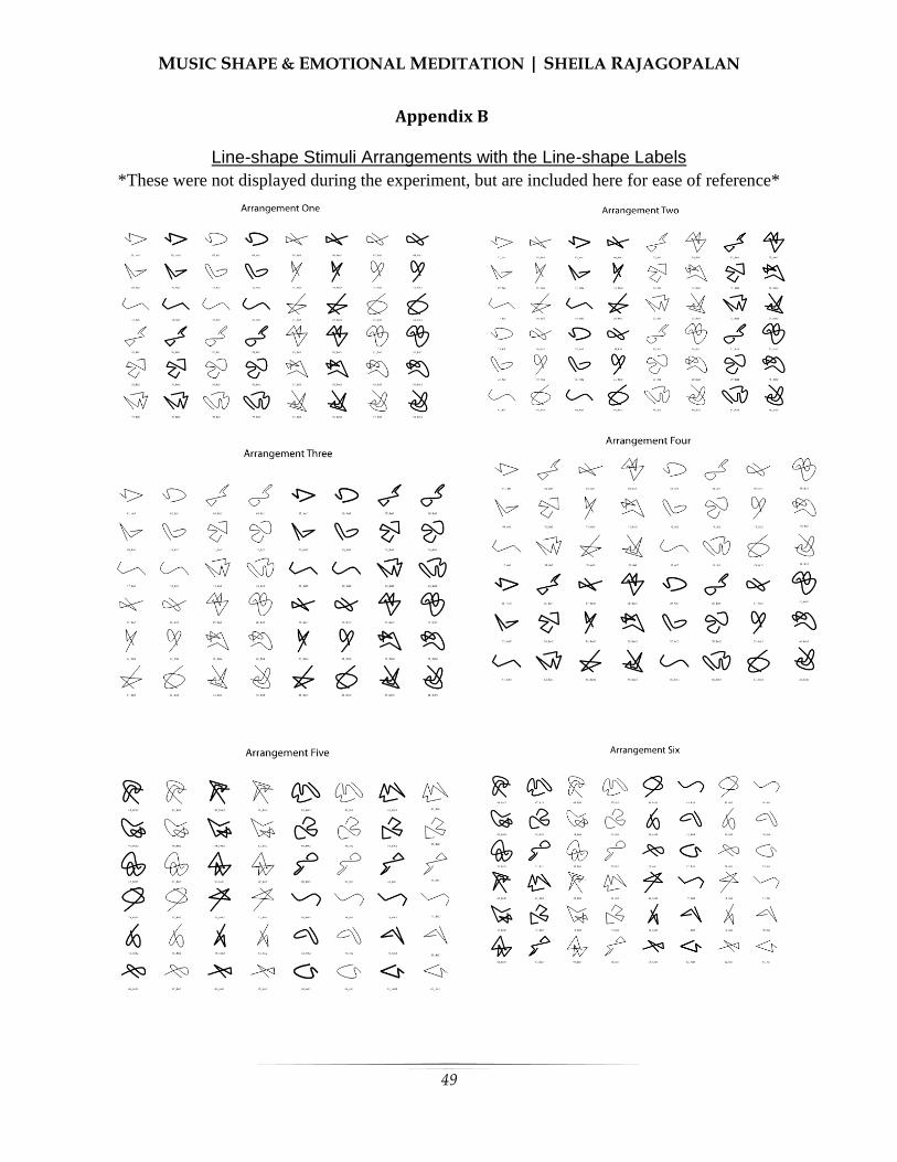
MUSIC SHAPE & EMOTIONAL MEDITATION | SHEILA RAJAGOPALAN
49
Appendix B
Line-shape Stimuli Arrangements with the Line-shape Labels
*These were not displayed during the experiment, but are included here for ease of reference*

MUSIC SHAPE & EMOTIONAL MEDITATION | SHEILA RAJAGOPALAN
50
Appendix C
Line-Shape Dimensional Correlation and Musical Genre Dimensional Correlation Tables

51
Patrick M. Einhorn
Boston College
McLean Hospital and Harvard Medical School
Early life stress (ELS) has been implicated in increasing anxiety and aggression in rats, possibly
by means of modulating inhibitory GABA interneurons colocalized with cannabinoid receptors
in the prefrontal cortex. We sought to investigate the developmental effects of maternal
separation (MS) and cannabinoid type 1 (CB1) receptor antagonism on anxiety and social
behaviors in male and female Sprague-Dawley rats. With the use of retrospective analysis, we
wished to discover if juvenile social play behaviors could be used to predict those exhibited in
adulthood. As adults, rats were exposed to the CB1 antagonist, rimonabant (Rim), or vehicle
(Veh) prior to the elevated plus maze, marble burying, and social interaction. With
immunohistochemistry, we investigated whether changes in calbindin and calretinin-expressing
GABAergic interneurons in the medial prefrontal cortex may mechanistically underlie behavioral
deficits. Sex differences in adult social behavior resulted from MS, most notably an increase in
aggression and evasion in male adults. Rim treatment uncovered a trend of decreased anxiety in
MS, but increased anxiety in controls (CON). Rim reduced social and explorative behaviors in
males, but increased the crossovers and stretch-attend postures in females in the elevated plus
maze. Juvenile social play behaviors were predictive of some behaviors in adulthood. A
moderate trend was observed for the interaction of condition, cell type, region, and cortical layer.
This trend, along with prior research, suggests that differences in anxiety and social behavior
seen in adults are differentially affected by Rim treatment dependent on changes in GABAergic
interneuron subpopulations resulting from early life stress.
Early Life Stress (ELS)
arly life stress (ELS) and other
developmental insults significantly
increase one’s susceptibility to long-
lasting psychiatric pathologies and
substance-abuse disorders later in adulthood
(Cadet, 2014). Although exposure to stress,
both prior to and during adolescence, can
promote the onset of behavioral and
cognitive disturbances, early childhood
appears to be the most significant sensitive
period of development (Leussis, Freund,
Brenhouse, Thompson, & Andersen, 2012).
Models of ELS, particularly maternal
separation (MS), are quite effective in
promoting the emergence of these
Deficits in anxiety and social behaviors
induced by early-life stress can be attenuated
by Cannabinoid Type 1 Receptor Antagonism
E
MATERNAL SEPARATION & CANNABINOID ANTAGONISM | PATRICK EINHORN
52
pathologies during late adolescence and
early adulthood, just as in the human
population (Huot, Lenox, & McNamara,
2002; Andersen & Teicher, 2008).
Medial Prefrontal Cortex and Behavioral
Development
Leading up to adolescence, many
developmental processes, such as cell
migration and differentiation, take place
along with the development and maturation
of the dopaminergic, GABAergic, and
endocannabinoid systems, which continue to
mature during childhood (Viveros et al.,
2011). As a result, childhood stress can
induce long-standing behavioral deficits,
such as heightened risk of developing
problems with drug abuse and
psychopathology (Lupien et al., 2009). Of
particular interest is the medial prefrontal
cortex (mPFC). The mPFC mediates the
stress response of the hypothalamic-
pituitary-adrenal axis (Seidel, Poeggel,
Holetschka, Helmeke, & Braun, 2011;
Groenewegen & Uylings, 2000). Recent
evidence also indicates that the mPFC is
integral to the reinforced learning and
acquisition of drug-seeking behaviors and
the mediation of its rewarding effects
(Tzschentke, 2000). The mPFC acts as an
inhibitory brake on emotional neural activity
through its projections to the amygdala, and
also on reward-driven behaviors through
projections to the nucleus accumbens
(Seidel et al., 2011; Elzinga & Bremner,
2002). Therefore, the mPFC is crucial to
higher-order functions such as planning,
decision-making, personality expression,
motivation, and social behavior. Lesions to
the mPFC disrupt executive function and
decrease a subject’s behavioral inhibition,
leading to the facilitation of drug-seeking
behaviors (Weissenborn et al., 1997), as
well as relapse (Kalivas et al., 2005; Kalivas
& Volkow, 2005).
ELS and Drug Use
Prior preclinical research indicates
that ELS, such as MS, is also predictive of
later life illicit drug-seeking behaviors and
drug usage (Gordon, 2002; Lynch, Mangini,
& Taylor, 2005; Lehmann, Stöhr, & Feldon,
2000). Some of these behaviors may be due
in part to repeated stress, which has been
found in adult rats to increase neuronal
atrophy (Liston et al., 2006), as well as
decrease both dendritic density and dendritic
branching within layers 2 and 3 of the
mPFC’s anterior cingulate cortex (ACC) and
prelimbic (PL) areas (Radley et al., 2004;
Radley et al., 2006). Accordingly, there is a
need for further investigation into MS’s role
in increasing susceptibility to drug abuse
later in development using mPFC
modulation.
ELS and Social Behaviors
ELS has also been implicated in
increasing anxiety and reducing sociality in
both rats and humans. Animals exposed to
ELS spent more time in the closed arms of
the elevated plus maze (EPM), indicating
the presence of anxiety (Girardi, Zanta, &
Suchecki, 2014). MS also promotes
depressive-like behavior (immobility),
increases stress hormone response in the
forced swim test, and increases aggression
between males when compared to controls
(Veenema et al., 2006). To date, there has
been no investigation into the effects of

MATERNAL SEPARATION & CANNABINOID ANTAGONISM | PATRICK EINHORN
53
marble burying (MB) in measuring anxiety.
Rather than measuring anxiogenic avoidance
behavior, like in the EPM, MB uncovers
perseverative anxious behavior; when the
animal subject is placed into a novel setting
with glass marbles evenly spaced
throughout, anxious subjects will bury more
marbles when compared to controls,
providing an effective means of measuring
anxiety-like behavior (Handley, 1991).
Resultantly, we predict that MS rats will
likely exhibit more anxiety than controls by
exhibiting increased MB and decreased time
in the open arms of the EPM. Juvenile social
play will be used as a predictive model of
adult anxiety following ELS.
Juvenile social play behavior is
rewarding and offers a multitude of
developmental benefits. For example, rough-
and-tumble play, consisting of pouncing and
pinning play partners, allows juvenile rats to
gain motor training (Pellis & Pellis, 2007),
as well as experience with unexpected
situations and stimuli (Pellis & Pellis, 2005;
Baldwin and Baldwin, 1977). As a result,
early life socialization may function to foster
a rat subject’s ability to interact with one
another as they age, thereby establishing
stable adult social relations (Panksepp,
1981).
MS increases aggressive play
fighting (biting and tail-pulling) in juvenile
male rats (Veenema et al., 2009); behaviors
which remain into adulthood (Veenema et
al., 2006). Consequently, it may be possible
to use juvenile social play as a means of
predicting adult social behaviors. We expect
an increase in aggressive behaviors in MS
rats during juvenile social play and adult
social interaction (Veenema et al., 2009;
Veenema et al., 2006). Little previous
research has investigated the effects of MS
on anxiety and social behaviors in female
rats. This experiment hopes to bridge the
gap in the previous literature by
investigating juvenile and adult gender
differences in these behaviors, as well as the
role of the mPFC, particularly GABAergic
interneurons, implicated in regulating top-
down inhibition on anxiety and social
behavior. Given the mPFC’s role in
coordinating social and anxious behavior, as
well as MS’s ability to disrupt mesocortical
development, adult social deficits are
expected. As such, the MS subject should
continue to exhibit aggressive behaviors.
GABA
Recent evidence has shown that
many behavioral deficits promoted by MS
are highly correlated with GABAergic
dysfunction within the PFC (Le Magueresse
& Monyer, 2013). GABA neurons can be
found throughout the brain’s cortex, with the
majority of GABA neurons synapsing
locally while functioning as interneurons
(Inan, Petros, & Anderson, 2013). GABA
neurons and the circuitry they regulate are
particularly immature in early life stages.
These GABA neurons require a protracted
period of development in order to carefully
refine cortical circuitry during development
(Reynolds, Zhang, & Beasley, 2001; Cotter
et al., 2002). Manipulation of infant-mother
relationships during neonatal development
produces both short and long-lasting effects
in the GABAergic system (Giachino et al.,
2007; Pryce & Feldon, 2003). Additionally,
early GABAergic signaling can affect many
aspects of cellular maturation and synaptic

MATERNAL SEPARATION & CANNABINOID ANTAGONISM | PATRICK EINHORN
54
plasticity (Heck et al., 2007) that extend into
early adulthood (Hashimoto, 2009; Lewis,
Hashimoto, & Volk, 2005).
Maintaining healthy cortical function
requires a carefully calibrated balance
between inhibition and excitation (Okun &
Lampl, 2009; Haider et al., 2006). By
shaping neural responsiveness, preventing
excess excitation, refining cortical receptive
fields, and maintaining the synchronization
of cortical activity, GABA interneurons
allow their circuits to remain malleable in
order to react to stressful stimuli in a healthy
manner (Rossignol, 2011). Thus, the proper
regulation and recalibration of these circuits
are vital to their maintenance and continued
development in an effort to achieve normal
cognitive and behavioral functioning.
GABA Dysfunction
Any reduction in the density of
GABAergic neurons can create pervasive
disturbances within cortical circuits due to
their control over the modulation and
calibration of activation and inhibition
(Rossignol, 2011). Within the PFC, the role
of GABAergic interneurons appears to be at
least partly to blame for the emergence of
psychopathological and drug-related
behaviors (Keverne, 1999; Miller &
Marshall, 2004;). Through the recruitment
of the hypothalamic-pituitary-adrenal (HPA)
axis, along with the adrenal-medullary
system’s sympathomimetic response, stress
rapidly induces central and peripheral
nervous system effects (Skilbeck, Johnston,
& Hinton, 2010). It has been hypothesized
that GABAergic interneuron dysfunction
could be due in part to either excitotoxicity
induced by excess excitatory transmission
(Yizhar et al., 2011; Marin, 2012), or the
early maturation of GABAergic interneurons
as a result of a sustained increase in
transmission within the neural circuitry
(Yizhar et al., 2011; Marin, 2012).
With reduced GABAergic
interneuron signaling, one could predict
excessive excitation within the circuit to be
responsible for the neuronal damage seen
within the population (Olney & Farber,
1995). In rats, acute and recurrent stress can
reduce GABA binding affinity in the PFC
(Biggio et al., 1981; Caldji, Diorio, &
Meaney, 2000). Likewise, ELS reduces the
number of high-affinity GABA binding sites
in the mPFC (Caldji et al., 2000; Plotsky et
al., 2005), as well as increases anxiety and
HPA axis hyperresponsiveness to stress
(Giachino et al., 2007). Though the
mechanism for GABAergic neuron loss is
not yet fully understood, it is evident that
ELS does disrupt mesocortical inhibition via
changes in GABAergic interneurons
(Nemeroff, 2003). Though many MS models
have focused on behavioral deficits
exhibited in adulthood (Réus et al., 2011;
Holland, Ganguly, Potter, Chartoff, &
Brenhouse, 2014), there is significant
evidence to indicate the emergence of
psychopathological and drug-related
symptoms as early as adolescence
(Andersen & Teicher, 2009).
Calcium-Binding Protein (CBP) and
GABA Interneurons
CBP-expressing interneurons are of
particular interest because these cell types
are widely distributed across the nervous
system and express proteins that are
considered modulators of intracellular
MATERNAL SEPARATION & CANNABINOID ANTAGONISM | PATRICK EINHORN
55
calcium (Schwaller, Meyer, & Schiffman,
2002; Mueller et al., 2005; Giachino et al.,
2007). Research has shown that these
neurons help control synaptic plasticity
(Gurden et al., 1998; Schwaller et al., 2002)
and the cell’s ability to maintain sustained
firing by regulating intracellular calcium
(Lin, Arai, Lynch, & Gall, 2003). The CBPs
expressed in these neuronal populations
reversibly bind calcium, thereby influencing
the cells’ likelihood of propagating an action
potential (Hendry et al., 1989; Kawaguchi &
Kubota, 1997). These interneurons balance
GABAergic interneuron excitability,
neurotransmitter release, and its resulting
GABA-mediated inhibition on its synaptic
projections. MS studies investigating the
relative density and distribution of these
populations have yielded conflicting results,
though it appears that overall, MS may
reduce CBP-type protein expression in
GABAergic interneurons in the mPFC
(Helmeke, Ovtscharoff, Poeggel, and Braun,
2008).
Changes in populations of
GABAergic interneurons that are
immunopositive for CBPs increase an
individual’s vulnerability to
psychopathological behavior later in life
(Lewis et al., 2005). There are far fewer
studies documenting the histology of
Calbindin (CB) and Calretinin (CR) within
the mPFC of those at risk for psychological
illness and substance abuse than other
classes of CBP-expressing GABA
interneurons, such as Parvalbumin (PV). As
such, this investigation has been restricted to
Calbindin (CB) and Calretinin (CR)
expressing GABA interneurons in the
mPFC.
Most studies investigating CB
regarding anxiety, depression, or maternal
stress have found a net reduction in cortical
populations as compared to controls
(Pascual et al., 2007; Lephart & Watson,
1999; Xu et al., 2011). However, one study
has documented no changes in CB (Cotter et
al., 2000), while another found a net
increase in CB expressing GABAergic
interneurons (Daviss & Lewis, 1993).
Unfortunately, in the only previous study
that has investigated changes in CR
following MS, no change in CR expression
was found in male Octodon Degus when
compared to controls (Helmeke et al., 2008).
These conflicting results warrant further
investigation into medial prefrontal changes
in CR and CB interneuron populations. By
representing ELS through repeated neonatal
MS for four hours daily from P2-P20, in
conjunction with fluorescent
immunohistochemistry, we were able to
compare the relative distribution and the
frequency of CB and CR GABAergic
interneurons within the mPFC of male
adolescent (P40) Sprague-Dawley rats. This
method allowed for the analysis of co-
localization of both CB and CR
subpopulations throughout layers 2/3 and
5/6 across the ACC, PL, and IL regions of
the mPFC. Given previous findings, we
expect to see a reduction in CB and CR
interneurons in MS subjects during
adolescence.
A recent study by Wedzony and
Chocyk (2009) reported that CB-expressing
GABAergic interneurons were colocalized
with cannabinoid type (CB1) receptors
within the mPFC of male Wistar rats. Given
that adolescent marijuana usage increases
MATERNAL SEPARATION & CANNABINOID ANTAGONISM | PATRICK EINHORN
56
the proclivity of users to develop substance
abuse and various other psychoses, along
with the colocalization of CB1 receptors on
GABAergic interneurons immunopositive
for CB (but not PV or CR), it is possible that
marijuana usage may exacerbate the
disturbances already incurred in the
prefrontal GABA system as a result of ELS
(Wedzony & Chocyk, 2009).
Endocannabinoids
The endocannabinoid system
modulates many different neural activities in
various regions of the brain, particularly the
regulation of stress and emotional behavior
in the PFC and HPA axis (Herkenham et al.,
1990; Katona, 2001; Mackie, 2005).
Cannabis and its primary psychoactive
component, Δ9-tetrahydrocannabinol
(THC), exhibit the ability to alter a subject’s
reaction to stimuli, which has implicated the
endocannabinoid system’s role in stress
management (Patel et al., 2004; Rodriguez
et al., 2013). In healthy individuals,
endogenous endocannabinoids are
synthesized as needed and use retrograde
transmission to reduce further presynaptic
transmission (Herkenham et al., 1990;
Katona 2001). In the brain, the CB1 receptor
has been isolated as the most abundant
endocannabinoid receptor and is likely
responsible for much the endocannabinoid
activity in the central nervous system
(Mackie, 2005). CB1 and cannabinoid type
2 (CB2) receptors are Gi/G-protein coupled.
When activated by an agonist, cyclic
adenosine monophosphate, and thus ATP,
are reduced by CB1’s G-protein activity
inhibiting the secondary messenger,
adenylyl cyclase, while also enhancing the
activity of mitogen-activated protein kinase
(Svíženská, Dubovy, & Sulcova, 2008).
Similarly, the activity of cAMP-dependent
protein kinase is inhibited by reduced cAMP
production (Svíženská et al., 2008). Through
G-protein coupling to ion channels, CB1
activation increases the activity of inward
rectifying and A-type potassium channels,
reduces the activity of outward D-type
potassium channels, and inhibits activity at
P/Q-type and N-type calcium channels
(Howlett & Mukhopadhyay, 2000; Pertwee,
1997). When CB1 is not activated by
cannabinoids, PKA phosphorylates protein
in potassium channels to reduce the outward
movement of potassium, thereby increasing
membrane potential. Similarly, cannabinoid
activity at CB1 inhibits neurotransmitter
release by reducing presynaptic calcium
channel activity (Svíženská et al., 2008).
Unfortunately, there is little research
documenting the effects of neonatal stress
on an endocannabinoid system that has been
taken “offline” by a direct CB1 antagonist.
Given MS’s ability to induce changes in
CBP-expressing GABAergic interneurons,
CB1’s role in calcium-driven synaptic
reactivity, and the colocalization of CB1
receptors on CBP-expressing GABAergic
interneurons, manipulation using MS and
CB1 antagonism should uncover how these
systems interact to contribute to anxiety and
social behavioral deficits.
Cannabinoid Antagonism and
Rimonabant
Rimonabant (SR141716), is a
selective synthetic CB1 antagonist (Padwal
& Majumdar, 2007; Zador et al. 2015). As a
direct CB1 antagonist, rimonabant binds
MATERNAL SEPARATION & CANNABINOID ANTAGONISM | PATRICK EINHORN
57
CB1 and exerts an effect opposite to that of
THC. Rimonabant blocks CB1 with high
selectivity and efficacy in lower
concentrations, while acting on both CB1
and CB2 at increasing levels of
concentration (Svíženská et al., 2008; Jagger
et al., 1998). Rimonabant may, in fact, exert
behavioral effects that reflect receptor
activity inhibited below baseline since CB1
receptors are tonically active in the absence
of cannabinoid signaling (Ward & Raffa,
2012; Fong & Heymsfield, 2009). Not only
does rimonabant mitigate the inhibitory
effect of CB1 agonists on Ca2+
channels, but
it also increases Ca2+
channel currents when
acting in the absence of an agonist (Pan,
Ikeda, & Lewis, 1998). Given that MS
increases susceptibility to adult
psychopathology and drug use, the
inhibition of the endocannabinoid system by
rimonabant should allow for further analysis
of the system’s role in modulating risk
trajectory by measuring its effects on
anxiety and social behaviors. The effect of
rimonabant on CB1 receptors should
increase presynaptic calcium influx, which
increases neuronal excitability, synaptic
activity, and neurotransmitter release. With
inhibitory dysfunction following MS,
antagonistic activity at CB1 receptors may
mitigate the behavioral deficits (increased
anxiety and increased aggressive social
behavior) by stimulating activity at
dysfunctional GABAergic interneurons
previously inhibited by MS. However,
recent research findings have proven
inconsistent with this prediction.
Measures of anxiety induced by
cannabinoid agonism and antagonism in
mouse and rat models have yielded
conflicting results, indicating that there may
be a species difference to account for the
incongruity. As for measures of anxiety,
lower doses of rimonabant (1-3 mg/kg)
increased the exploration of the open arms
in the EPM in mice, indicating an anxiolytic
effect. Also, the use of another CB1
antagonist, AM-251, was shown to dose-
dependently increase anxiety-like EPM
behaviors in wild-type mice (Haller, Varga,
Ledent, & Fruend, 2004). Conversely, a
study by Gomes, Cassarotto, Resstel, and
Guimaraes (2011) found that the CB1
agonist WIN, along with endocannabinoid
synthesizing protein inhibitors, AM404 and
URB597, all reduced marble burying
behavior in mice. In male rats, MS and
exposure to the cannabinoid agonist
CP55,940, increases time in the closed
portion of the EPM and perimeter areas in
open-field testing, indicating a significant
increase in anxiety-like behaviors, which
were much more prominent when rats were
exposed to both MS and CP55,940 (Klug &
van den Buuse, 2012). Furthermore, indirect
inhibition of the cannabinoid system of mice
through enzyme inhibition reduced MB,
which could be prevented by rimonabant
(Kinsey, O’Neal. Long, Cravatt, &
Lichtman, 2011).
Unfortunately, no studies have
investigated the relationship of MB and the
endocannabinoid system in rats. It is
apparent that the endocannabinoid system
plays a sizable role in the maintenance of
many social, aggressive, anxious, and
depressive behaviors; therefore, when
coupled with early life insults, exposure to
cannabinoid antagonist/inverse agonist
rimonabant may have the capacity for

MATERNAL SEPARATION & CANNABINOID ANTAGONISM | PATRICK EINHORN
58
inducing robust changes in behavior.
Despite conflicting research, rat subjects
treated with rimonabant are expected to
exhibit increased anxiety and decreased
open arm exploration in the EPM. However,
it is important to concede that this prediction
is not inconsistent with the hypothesized
activity of rimonabant at the receptor and
synaptic levels, especially in regard to MS
animals.
CB1 and Social Behavior
CB1-related changes in social
behavior have conflicted across animal
models. A preclinical study by Rodriguez-
Arias et al. (2013) concluded that the CB1
receptor is critical in mediating the display
of aggressive behaviors, due in part to its
role in stress regulation. Accordingly,
knock-out mice lacking the CB1 receptor
were quicker to engage in aggressive
behaviors while also exhibiting more
aggressive behaviors than wild-type controls
(Rodrigues-Arias et al., 2013). The
administration of a CB1 agonist to
aggressive wild-type/control mice was also
effective in mitigating aggressive behavior.
As a result, one would likely hypothesize
that an antagonist would propagate
aggression, given that CB1 knockout mice
displayed significantly increased aggression
and also that aggressive behavior in wild-
type mice was reduced by administration of
a CB1 agonist. However, not all studies
have found similar results.
Navarro et al. (1997) found that
acute intraperitoneal (i.p.) administration of
rimonabant (3 mg/kg) increased anxiety-like
behavior in rats in the EPM. A similar study
by Schneider, Schomig, and Leweke (2008)
concluded that chronic and acute treatment
with the synthetic CB1 agonist, WIN
55,212-2 (WIN), during puberty (P40-65)
produced significant changes in male rats’
social behavior and play behavior during
both puberty and adulthood. Rats
chronically exposed to WIN during puberty
exhibited significantly less exploration of
novel conspecifics in social recognition
testing, reduced total social interaction with
conspecifics, and an increase in social
evasion behaviors. However, acute pubertal
exposure to WIN induced a net reduction in
social behavior, but a significant increase in
spontaneous social play behavior.
Furthermore, acute WIN administration to
adult rats promoted evasive behaviors and
playful activities, as well as, significantly
reduced social recognition, investigation of
unknown social partners, anogenital
exploration, and overall social behavior
(Schneider et al., 2008). Notably, injection
of WIN reduced social interaction in rats,
whereas agonists that indirectly raise the
endocannabinoid levels by reducing their
metabolizing enzymes, such as the fatty acid
amide hydrolase inhibitor, URB597, foster
social interaction (Trezza & Vanderschuren,
2008). Currently, it is unclear if a CB1
antagonist, such as rimonabant, would
propagate aggressive behavior or reduce
social interactions in adults, though we
expect to uncover these effects. However,
such disparate findings raise the possibility
that the effects of rimonabant are biphasic
by dose, or that such conflicting findings
may be related to maturational, species, or
gender differences.

MATERNAL SEPARATION & CANNABINOID ANTAGONISM | PATRICK EINHORN
59
Predictive Social Behaviors
Social play behavior was assessed
after MS during the juvenile stage (P25) to
establish a behavioral baseline in MS versus
CON subjects. Subjects were retested again
in adulthood (social investigation) following
exposure to rimonabant or vehicle to
evaluate its effects on exhibited social
behavior. Particularly, given that marijuana
usage induces such robust behavioral and
social effects in its users, the use of
rimonabant should be effective in detecting
inducible behavioral disturbances in social
and anxiety-like behavior. In addition to
increasing susceptibility to psychological
illness, ELS and MS have been known to
alter aggression and social interaction.
Therefore, the use of anxiety paradigms and
social play interaction both with and without
rimonabant exposure should allow for
analysis of the endocannabinoid system’s
control of social and anxious behaviors.
The EPM and marble burying
paradigm testing was counterbalanced and
always preceded social investigation such
that social interaction did not confound
innate measures of anxiety. Given that
recent findings have implicated both MS and
rimonabant as contributing to decreased
social behavior, increased anxiety and
aggression, as well as, CB1 knock-out mice
displaying increased aggression, we expect
the administration of rimonabant will
enhance these behaviors in control subjects.
However, given the reduction of
GABAergic inhibition seen in MS subjects,
which should be activated by rimonabant,
we expect MS subjects to exhibit behaviors
more like that of controls receiving vehicle.
Moreover, by pairing predictive and
retrospective analysis of social behavior
with immunohistochemistry, we sought to
further investigate whether CB and CR
populations in the mPFC may contribute
mechanistically to the behavioral changes
exhibited in subjects following MS.
Methods
Animal Subjects
On gestation day 14, pregnant female
Sprague-Dawley Rats were obtained from
Charles River Laboratories (Wilmington,
Mass., USA). Postnatal day 0 (P0) was
designated as the pup’s date of birth. Food
and water were made available ad libitum to
all rats, which were housed constant
temperature and humidity on a 12-hr
light/dark cycle (light period 0700h-1900h).
For Experiment 2, new male subjects (5 MS
and 5 controls), were used to control for the
effects of behavioral paradigms and RIM
treatment. All experiments conducted herein
were in accordance with the 1996 guide for
the Care and Use of Laboratory Animals
(NIH) and approved by the Institutional
Animal Care and Use Committee at Mclean
Hospital. Experimental apparati were
thoroughly cleaned with 30% ethanol
solution between all trials; bedding was
changed between all trials, when
appropriate.
Maternal Separation
The MS paradigm used is identical to
procedures previously conducted by this
laboratory (Leussis et al., 2012), and similar
to those used by others (Veenema &
Neumann, 2008; Holland et al., 2014). MS
was initiated on P2 when all litters were
culled to 10 pups: 5 males and 5 females. By

MATERNAL SEPARATION & CANNABINOID ANTAGONISM | PATRICK EINHORN
60
means of random assignment, these litters
were designated as an MS or an animal
facility reared control group (CON).
Additionally, pups in Experiment 1 were
randomly assigned to treatment with
rimonabant or vehicle within a litter.
As seen in Figure 1, a total of 36 rats
(18 male and 18 female, N=36) were used in
Experiment 1. Of which, 6 males and 5
females were assigned to the MS condition,
while 3 males and 3 females were randomly
assigned to treatment with rimonabant
(MS+Rim). The remaining MS subjects
were assigned to treatment with Veh
(MS+Veh). Of the 22 remaining controls, 3
males and 4 females were designated to
treatment with rimonabant (CON+Rim)
while the rest received vehicle (CON+Veh).
Within Experiment 2, 10 male rats (N=10)
were used: 5 MS and 5 CON.
MS animals were isolated at 22
degrees Celsius for 4 hours daily from P2 to
P20. The CON rats were not disturbed after
P2 except for weekly cage changing and
daily weighing. On P21, rats were weaned
and assigned to same-sex group housing
with 2-4 rats per cage. All subjects remained
in group housing until the end of
experimentation, (see Figure 2 for the course
of subjects and experiments).
MATERNAL SEPARATION & CANNABINOID ANTAGONISM | PATRICK EINHORN
61
Experiment 1
Drug. Rimonabant (SR141716A,5-
(4-Chlorophenyl)-1-(2,4-dichloro-phenyl)-
4-methyl-N - (piperidin-1-yl) - 1H-pyrazole-
3-carboxamide) HCl was obtained from
Sigma-Aldrich and suspended in dimethyl
sulfoxide (DMSO). DMSO was used as the
vehicle (Veh). Rimonabant (Rim, 3 mg/kg)
or Veh was administered systemically by
means of intraperitoneal (i.p) injection with
a 15m period of pretreatment before testing.
Injection and pretreatment time period is
similar to methods used by based on
methods of similar previous literature
(Ward, Rosenberg, Dykstra, & Walker,
2010; Blasio et al., 2013; Järbe et al., 2011).
Measures of Anxiety-like
Behaviors.
Elevated Plus Maze. The EPM
paradigm used is identical to previous
procedures previously by this laboratory
(Andersen & Teicher, 1999) and similar to
those used by others (Bredeworld, Smith,
Dumais, & Veenema, 2014; Lukkes,
Engelman, Zelin, Hale, & Lowry, 2012).
Rats were placed into the EPM located 50
cm above the ground with two 50 cm long
closed arms and two 50 cm long open arms
for 5 min. During this testing period, the
time spent by the rat in the closed and open
arms was recorded. Additionally, the total
number of crossovers between open and
closed arms, as well as, stretch-attend (SA)
postures was recorded.
Marble Burying. To investigate an
additional measure of anxiety-like behavior,
the marble-burying paradigm used was
similar to that used by Skelton et al. (2010)
and Kinsey et al. (2011). Rats were placed
for 15 min into an opaque cage (41 cm x36
cm x 10 cm) with 5cm of bedding with 24
evenly spaced glass marbles placed on top
of the bedding. At the end of the testing
period, the number of buried marbles was
measured and recorded. A marble is
classified as having been buried if 67.7% or
more of its surface was covered with
bedding.
Measures for Social Behaviors.
Juvenile Social Play. All rats were
tested for social behaviors at P25, and again
in adulthood after treatment with Rim or
vehicle. Testing was conducted in a room
separate from the housing. Within this
protocol, an MS animal was paired with a
novel same-sex CON conspecific in a glass-
enclosed open field chamber (50 cm x26 cm
x32 cm) for 15 min. All animals were
isolated for 24 hours on the day prior to
testing to promote willingness to socialize.
All interactions were videotaped in 720p
(HD) using Kodak PIXPRO SPZ1 on a
tripod under normal lighting to score the
frequency of each exhibited behaviors. As
described in Pellis, Pellis, and Bell (2010)
and in Veenema et al. (2013), the following
behaviors were scored based on their distinct
characteristics: pounces, pins, chases, bites,
boxing, social sniffing, and nape attacks.
Scoring was conducted individually for both
the MS and CON subjects within each
testing dyad.
Adult Social Interaction. Similar to
aforementioned social play testing, social
interaction was conducted during adulthood
between postnatal days 104 through 114
(P104+). Interactions were assessed in an

MATERNAL SEPARATION & CANNABINOID ANTAGONISM | PATRICK EINHORN
62
opaque ovular open field (97 cm x 69 cm x
33 cm) to mitigate the subjects’ propensities
to remain in corners. On the day before
testing, rats were placed in the novel open
field for 5 min of habituation time. On
testing day, the rats were isolated for 3.5
hours prior to testing to promote willingness
to interact. All interactions were videotaped
in 720p (HD) using Kodak PIXPRO SPZ1
on a tripod under normal lighting to score
the frequency of each exhibited behaviors.
In addition to the behaviors measured in
social play testing, additional social
behaviors, as well as, evasive and self-
grooming behaviors were also assessed.
These behaviors include: (1) self-grooming,
licking or biting own fur and rubbing
forepaws overhead; (2) evasive behaviors,
including running, leaping, or swerving
away from social partner; (3) additional
social behaviors, including (a) contact
behaviors, such as grooming (chewing or
biting partner’s fur), crawling over/under
social partner; (b) social exploration
behaviors, including anogenital exploration
(sniffing or licking the anogenital area of the
social partner), non-anogenital investigation
(licking or sniffing any other area of the
social partner); and (4) tail manipulations,
including grabbing, biting, and pulling the
partner’s tail; and (d) approaching and
following behaviors, including following,
approaching the social partner, or both.
(Schneider et al., 2008).
Euthanasia. Two days after
experimental testing, rats were anesthetized
with pentobarbital (60mg/kg, i.p.) 15 min
prior to decapitation in accordance with
IACUC guidelines.
Statistical Analysis. Data were
analyzed using a three-way analysis of
variance (ANOVA) with repeated measures
where appropriate, with factors being sex,
MS, and Rim treatment as main factors. In
cases where a main effect or interaction was
found, separate two-way ANOVAs split by
one of the independent factors or pairwise
comparisons of appropriate group
combinations were used to clarify results. If
p<0.05, differences were considered
statistically significant.
Experiment 2
Euthanasia, Perfusion, and Slicing.
Prior to perfusion, subjects at age P40 were
administered 0.5 ml of pentobarbital by IP
injection. Approximately 5 min later, when
the animals were fully anesthetized, the
subjects were transcardially perfused with
cold (4°C) phosphate buffered saline (0.1 M
PBS, pH 7.4), followed by cold (4°C) 4%
paraformaldehyde in 0.1 M sodium
phosphate buffer. The animals’ brains were
then removed from the skull and post-fixed
overnight in 4% paraformaldehyde in 0.1 M
sodium phosphate buffer. Afterward, brains
were rinsed with 0.1 M PBS, immersed in a
solution of 30% sucrose in 0.1 M PBS, and
stored at 4°C for two days. The fixed brains
were then removed from the sucrose
solution and sliced to the level of the mPFC
in 40-µm coronal sections using a sliding
microtome. Sliced sections were collected in
0.1 M PBS as serial free-floating sections
placed into wells containing freezing-
solution (10% 0.2 M PBS, 30% dH20, 30%
glycerol, 30% ethylene glycol, by volume),

MATERNAL SEPARATION & CANNABINOID ANTAGONISM | PATRICK EINHORN
63
such that each well contained every sixth
serial section.
Fluorescent
Immunohistochemistry. Sliced serial
sections were washed three times with 0.1 M
PBS for 5 m. Following washing, the tissue
was transferred to a well contain 10%
normal donkey serum (NDS) in PBS with
0.1% Triton X and allowed to block for 1
hour. The blocked tissue was then allowed
to incubate overnight at 4°C in primary
antibodies: anti-CB (Swant; Mouse; 1:500)
and anti-CR (Merck Millipore; Goat;
1:10,000). Upon completion of incubation,
the sections were washed with 0.2% PBS
with 0.1% Triton X three times for 5m.
Washed tissue sections were then incubated
for 1 hour at room temperature in secondary
antibodies in PBS. The secondary antibodies
used to fluorescently label for CB and CR
are as follows: Alexa Fluor 488 (1:300;
donkey anti-goat) and Alexa Fluor 568
(1:400; goat anti-mouse). Tissue sections
were mounted on microscope slides with a
fine-tipped brush and cover-slipped with
VECTASHIELD (Vector Labs) mounting
medium.
Stereology and Microscopy.
Microscope slides containing
immunoreactive, fluorescent-labeled tissue
were placed under a high-powered
microscope to evaluate the frequency and
distribution of neuronal cells in the mPFC
(3.20-2.70 Bregma) labeled for CB, CR, or
double labeled (DBL) for both markers (see
Appendix). Two complete coronal sections
containing both left and right hemispheres,
one rostral (3.20 Bregma) and one caudal
(2.70 Bregma), were analyzed per subject.
The ACC, PL, and infralimbic (IL) cortical
regions of the mPFC were examined and
measured for relative areas through tissue
layers 2/3 and 5/6. Approximately 50-70
randomized stereological sections were
selected and sequentially examined; single
and dual-labeled immunopositive neurons
were labeled and recorded.
Statistical Analysis. All
stereological data was computed using the
density (immunoreactive cells/area) of CBP-
expressing GABAergic interneurons with
the ACC, PL, and IL of the mPFC across
layers 2/3 and 5/6. Data were analyzed using
a three-way analysis of variance (ANOVA)
with repeated measures where appropriate
and with factors being MS, type of CBP-
expressing GABAergic interneuron, and
also an investigation of effects across mPFC
regions and cell layers. In cases of where
main effects or interactions were found,
separate two-way ANOVAs split by one of
the independent factors or pairwise
comparisons of appropriate group
combinations were used to clarify results. If
p<0.05, differences were considered
statistically significant.
Results
Elevated Plus Maze
Data analysis for time spent in the
open arms revealed that females, regardless
of condition, spent more time in the open
arms of the EPM than males (F(1,28)=5.36,
p=0.028). No other significant effects of
time spent in the open arms of the EPM
were found. Though nonsignificant, male
CON+Veh subjects, on average, spent more
MATERNAL SEPARATION & CANNABINOID ANTAGONISM | PATRICK EINHORN
64
time in the open arms of the EPM than
MS+Veh subjects (Figure 3). Likewise,
male MS+Rim subjects spent more time in
the open arms than MS+Veh and
CON+RIM, while female MS+Rim subjects
spent less time in the open arms when
compared to females MS+ VEH. The EPM
arm crossovers were significantly affected
by sex (F(1,28)=5.433, p=0.027) and also
treatment (F(1,28)= 4.827, p=0.036).
Similarly, in females, a significant
interaction was isolated for the condition in
regard to crossovers (F(1,14)=6.516,
p=0.023). Additionally, a significant
interaction between treatment and
crossovers was documented in females
(F(1,14)=4.843, p=0.045) (Figure 4).
Treatment with Rim resulted in a trend of
reduced frequency of SA postures
(F(1,14)=3.278, p=0.081). In females, Rim
treatment significantly decreased SA
postures (F(1,14)= 6.728, p=0.021). There
appeared to be a trend of condition on SA
postures (F(1,14)=2.912, p=0.11) in
females; on average, MS subjects displayed
fewer SA postures than controls. No
significant effects of condition or treatment
were observed in SA postures in male.
Marble Burying
Data analysis of MB revealed no
significant interaction of ELS condition or
treatment. In males, a trend of condition and
treatment was determined to be responsible
for exhibited changes in MB behavior
(F(1,14)=3.513, p=0.082); in general,
treatment with Rim reduced MB in MS
animals but increased the number of buried
marbles in controls. No interactions were
discovered in females.
Juvenile Social Play
The number social sniffs
(F(1,18)=9.447, p=0.007) were affected by
sex (Figure 5 a&b). Specifically, females
engaged in fewer social sniffs than males
(p=0.002). A trend was found for sex and
pouncing (F(1,18)=3.066, p=0.097);
juvenile females, on average, pounced more
than males. Similarly, a trend of condition
was also found for pouncing behaviors
(F(1,18)=2.798, p=0.112), that is, MS
subjects engaged in more pounces than
controls. A trend of biting behavior
appeared to be affected by sex and
condition: Sex X Condition (F(1,18)=4.189,
p=0.056), MS males engaged in more bites
than controls, but female controls engaged
in more bites than MS subjects (Figure 5
a&b).
Adult Social Interaction
A Treatment X Condition X Sex
interaction was observed in chasing
behavior (F(1,28)= 6.072, p=0.02). A
Treatment X Sex interaction indicated
significant changes in anogenital
explorations (F(1,28)= 4.157, p=0.051) and
social grooming (F(1,28)= 6.97, p=0.013);
post-hoc analysis revealed that males
receiving Rim engaged in fewer anogenital
explorations and social grooms than Veh
males (p=0.01 and p=0.004, respectively).
Acute Rim treatment in adult rats
significantly decreased chases
(F(1,28)=6.072, p=0.02), approaches
(F(1,28)= 8.58, p=0.007), and social
grooming (F(1,28)=5.706, p=0.024);
Furthermore, MS produced significant
changes in the frequency of pounces

MATERNAL SEPARATION & CANNABINOID ANTAGONISM | PATRICK EINHORN
65
(F(1,28)= 14.209, p=0.001), chases
(F(1,28)= 6.072, p=0.02), genital
explorations (F(1,28)= 4.517, p=0.043),
evasive behaviors (F(1,28)= 9.713,
p=0.004), and tail manipulations (F(1,28)=
20.324, p<0.001). Sex differences existed in
chasing (F(1,28)= 6.072, p=0.02),
approaching (F(1,28)= 7.409, p=0. 011),
self-grooming (F(1,28)= 3.035, p=0.092)
and social grooming (F(1,28)= 4.303,
p=0.047) behaviors. A significant
interaction was found between treatment and
condition for chases (F(1,28)= 6.072,
p=0.02). Please refer to Appendix A for
graphs of all adult social behaviors.
Predictability of Juvenile Social Play on
Adult Anxiety and Social Measures
In regard to anxiety behaviors
displayed during adulthood, a significant
and positive correlational relationship
(r=0.534, p=0.04) was found between
juvenile chases and adult MB. Significant,
positive correlations were found between the
frequency of arm crossovers with time spent
in the open arms of the EPM (r=0.598,
p=0.003) and also with the frequency of SA
(r=0.745, p<0.001).

MATERNAL SEPARATION & CANNABINOID ANTAGONISM | PATRICK EINHORN
66
Correlational analysis also revealed
that juvenile pinning was predictive of adult
approaches (r=0.545, p=0.029). Juveniles
who engaged in more chases exhibited fewer
self-grooms during adulthood (r=-0.500,
p=0.049). A significant, negative correlation
was found between juvenile social sniffing
and adult approaches (r=-0.623, p=0.01). In
males only, juvenile pinning was predictive
of increased approaches as adults (r=0.598,
p=0.003). Additionally, juvenile males who
engaged in more social sniffing engaged in
fewer adult approaches (r=-0.536, p=0.01)
and boxes (r=-0.444, p=0.039). Similarly,
juvenile males who engaged in more boxing
also crawled over/under their partner more
in adult social interaction. In females,
juveniles boxing behavior was indicative of
increased crawling (r=0.772, p=0.042) and
anogenital exploration as adults (r=0.849,
p=0016).
Experiment 2.
Stereology/Histology.
Condition X (Marker X Region X
Layer) ANOVA revealed a moderate trend
of interaction (F(1,8)= 22.865, p=0.128) in
Condition X (Region X Marker X Layer)
(Appendix B). As seen in Figures 7, 8, and
9, significant interactions were also found in
Layer (F(1,8)= 6.918, p=0.03), Marker
(F(1,8)=38.413, p=0.001), Region X Layer
(F(1,8)=21.024, p=0.002), Marker X Layer
(F(1,8)=35.391, p=0.001), and also Region
X Layer X Marker (F(1,8)=22.865,
p=0.001). Condition X (Marker X Region)
ANOVA, revealed a significant difference
of immunoreactive cells per region
(F(1,8)=6.435, p=0.035) and
(F(1,8)=16.055, p=0.004), respectively.
More specifically, less immunoreactive cells
were isolated in the ACC and IL regions
compared to PL. Additionally, Condition X
(Marker X Region X Layer) revealed a
significant interaction of marker (F(1,8)=
38.413, p=0.001), that is, there were far
fewer cells double labeled for CB and CR
than those expressing only one marker
(Figures 7-9,Appendix B).
MATERNAL SEPARATION & CANNABINOID ANTAGONISM | PATRICK EINHORN
67
Discussion
Experiment 1
Neonatal maternal separation (MS)
and Rimonabant (Rim) treatment in rats
produced robust differences in measures of
anxiety and social behavior. These results
are summarized as follows: (1) females
exhibited increased time in the open arms of
the elevated plus maze (EPM), (2) a trend of
reduced time spent in closed arms of the
EPM in males treated with rimonabant
(Rim), when compared to MS+Veh and
CON+Rim, (3) Rim treatment increased
crossovers and reduced stretch-attend (SA)
postures in females in the EPM, (4)
treatment with Rim may have reduced
marble burying (MB) in MS, but increased
MB in controls, (5) a trend of MS increasing
aggressive behaviors in juvenile males, but
decreasing aggression in juvenile females,
(6) treatment with Rim reduced social and
explorative behaviors in adult males, (7) MS
subjects exhibited increased aggressive and
evasive behaviors in adult social interaction,
(8) juvenile behaviors were predictive of
some adult behaviors.
Anxiety
The revealed trend of increased
anxiety in MS subjects parallels both
significant findings (Huot, Thrivikraman,
Meaney, & Plotsky, 2001; Kalinichev,
Easterling, Plotsky, & Holtzman, 2002), as
well as trends in previous literature (Mathieu
et al., 2011; Muhammad & Kolb, 2011). As
a result, MS appears to be contributing to
increased adult anxiety. This effect may be
due to changes in CBP-expressing
GABAergic interneuron subpopulations. As
seen in previous research, reductions in
GABAergic interneurons have been found
within the mPFC and also other limbic areas
involved in stress response (Pascual et al.,
2007; Lephart & Watson, 1999; Leussis et
al., 2012; Zadrozna et al., 2011), which may
underlie observed behavioral deficits.
(1) Overall, females exhibited
increased time spent in the open arms of the
EPM when compared to males, indicating a
significant sex-difference in adult anxiety
and confirming results from a previous study
using Long-Evans rats by Kalinichev et al.,
(2002). Furthermore, when isolated to male
subjects, a nonsignificant trend in males
suggests that MS condition could have
contributed to increased anxiety in the EPM
and corresponds to significant findings in
previous research (Daniels, Pietersen,
Carstens, & Stein, 2004; Huot et al., 2001;
Kalinichev et al., 2002). Previous research
has noted that maternal deprivation (24-hour
single isolation) induces sex-dependent and
region-dependent changes in
endocannabinoid gene expression (Marco et
al., 2014) in response to maternal
deprivation, genetic expression of proteins
responsible for synthesizing and
metabolizing endocannabinoids increased in
the prefrontal cortex of males, while these
genes only increased expression in the
hippocampus of female subjects. (2)
Correspondingly, Rim treatment may have
had an anxiolytic effect in open arm
exploration in MS males, but an anxiogenic
effect in MS females, although this failed to
reach significance. As such, it appears that
anxiety differences exist between males and
females, which are differentially affected by
the neonatal environment. Resultantly,
gender-specific genetic changes in the

MATERNAL SEPARATION & CANNABINOID ANTAGONISM | PATRICK EINHORN
68
endocannabinoid system in response to
stress could help clarify why males in the
human population appear to be at an
increased risk of developing adult-onset
psychosis or drug abuse following the use of
marijuana (Arendt, Rosenberg, Foldager,
Perto, & Munk-Jørgensen, 2005). It is also
possible that not only do pre-existing/innate
sex differences exist, but also that MS may
contribute to long-standing changes in both
GABAergic interneurons and colocalized
CB1 receptors. Though our study did not
yield a significant effect of MS or Rim on
time spent in the open arms of the EPM, we
must concede that this finding may be
statistically driven due to a low sample size.
Moreover, the disparity in baseline anxiety
behaviors between male and female subjects
further highlights the need for an
investigation into sex differences in all
facets of future research.
Hesitancy and Risk-Assessment
(3) As a measure of risk-assessment
or hesitancy, a strong trend of reduced SA
and increased crossovers was observed in
subjects treated with Rim. When the
analysis was restricted to female subjects,
this result was significant, indicating that
females may have been less hesitant than
males. This finding implicates sex
differences as differentially contributing to
the effects of Rim. Again, this may be due to
sex differences contributing to the
differential distribution of CB1 receptors
across the brain and to changes in the
colocalization of CB1 receptors on
GABAergic interneurons. As a result,
further investigation into female
immunohistochemistry is necessary. (4) No
significant findings were revealed in MB;
however, there is a strong trend of increased
MB in MS subjects, but a decrease in MB in
CON. This finding indicates that, on
average, MS subjects displayed increased
perseverative anxiety than controls, despite
this effect being nonsignificant.
Furthermore, this result appears to be in line
with earlier predictions based on
GABAergic interneuron activity, which will
be discussed later.
Social Behaviors
Juvenile social play revealed a
number of differences in behaviors as a
result of sex and also MS condition. At P25,
all females exhibited fewer social sniffs than
males, indicating that males were more
social and explorative at this time. However,
juvenile females engaged in more pounces
than males, which is surprising and
conflicting with previous findings (Parent &
Meaney, 2008). However, our difference
was small and may be due to the study’s
less-than-ideal sample size. (5) A trend in
biting behavior suggests that MS increased
aggressive biting behavior in males, which
parallels significant previous findings
(Veenema et al., 2006; Veenema et al.,
2009). Interestingly, there was also a trend
of reduced biting behavior in juvenile
females, revealing the possibility that sex
differences due to MS may have reduced
aggression in females, likely due to cortical
changes in GABAergic interneurons.
Therefore, the use of females in future
immunohistochemical studies is needed.
(6) In adult social behavior,
treatment with Rim significantly reduced
social grooming in comparison to those
MATERNAL SEPARATION & CANNABINOID ANTAGONISM | PATRICK EINHORN
69
receiving vehicle. Also, treatment with Rim
reduced anogenital exploration in males.
Thus, it appears that treatment with Rim
reduced rats’ willingness to engage in
nonaggressive social behaviors, as well as,
increasing the reluctance of males to explore
and interact with conspecifics. (7) As
expected, MS was responsible for inducing a
significant increase in aggressive and
evasive behaviors as seen by an increase in
adult pouncing, evading, and tail
manipulating. This finding indicates that
although MS increased male and decreased
female aggression in juveniles, both sexes
went on to exhibit heightened aggression
and social reclusion as adults relative to
controls. Our finding of increased male
aggression in juveniles and adults supports
previous research (Veenema et al., 2009;
Veenema et al., 2006). Clarity of this effect
in females remains opaque due to the lack of
previous research in females. However,
Taylor et al. (2000) have argued that stress
fosters a “tend-and-befriend,” rather than
“fight-or-flight” response in females, likely
due to female reproductive hormones
promoting attachment and caregiving.
Similarly, adult females engaged in
significantly more self- and social-
grooming behaviors. Though previous
findings confirm that adult females engage
in an increased number of self-grooming
behaviors due to hormonal differences
(Moore, 1986), the exhibited increase in
social grooming indicates that adult females
were more willing to socially engage its
conspecific in a nonaggressive manner. This
finding appears to be in line with the
hierarchal social system seen in rats; it is
more advantageous for males to be
aggressive to gain social standing just as
increased sociality in females would
increase reproductive success.
Predictive Behavioral Measures
(8) Juvenile social play did predict
some adult social behaviors. Juvenile
pinning behavior was indicative of
approaches in adults, that is, juveniles who
engaged in more pinning behavior also made
more approaches to their social conspecific
as adults. This finding indicates that subjects
which were more willing to engage in social
interaction as juveniles continued to be more
social as adults. Juveniles who engaged in
more chasing were less likely to exhibit self-
grooming and more likely to engage in adult
chasing. Taken together, this finding
suggests that rats that were actively engaged
in socialization as juveniles continued to
remain relatively more active as an adult at
the expensive of self-grooming.
Additionally, juvenile boxing behavior was
indicative of increased crawling over and
under social partners as adults. In females,
juveniles who engage in boxing behavior
were more likely to engage in anogenital
exploration and crawling over and under as
adults. This indicates that juveniles who
engaged in co-defensive behaviors were
more willing to socialize with adult partners
in a nonaggressive manner. To our
knowledge, this is one of the first behaviors
to proactively predict adult social
interactions based on juvenile play behavior.
It is our hope that continued emphasis is
placed on measures of juvenile behaviors as
predictive markers for those exhibited later
in adulthood. Such predictive measures,
should they prove reliable, could unlock an
entirely new way to quantify and understand
developmental neurological changes and
corresponding behaviors. When applied to
translational research, such predictive
paradigms would have the potential to

MATERNAL SEPARATION & CANNABINOID ANTAGONISM | PATRICK EINHORN
70
expedite both the development of new
pharmaceuticals, as well as more quickly
uncover long-term detriments resulting from
new and experimental treatments.
Experiment 2 The results of the present study
demonstrate important differences in the
effects of neonatal MS on prefrontal
GABAergic interneuron subpopulations,
which are summarized as follows: (1) a
trend of difference in immunoreactive cell
density by Condition X (Marker X Region X
Layer) and (2) significant differences of
immunoreactive CBP-expressing cells
across mPFC regions and layers. (1) The uncovering of a moderate
trend in the interaction of Condition X
(Marker X Region X Layer) indicates that
condition may have contributed to changes
in the density and distribution of CBP-
expressing GABAergic interneurons in the
mPFC, despite being nonsignificant.
However, this nonsignificant finding is
consistent with previous studies in humans
and degus (Cotter et al., 2000; Helmeke et
al., 2008, respectively), thereby suggesting
that species differences are minimal. (2) Given prior research indicating a
decrease in CBP-expressing GABA
interneurons, it was expected that deficits
resulting from GABAergic hypoinhibition
would be mitigated by Rim treatment, given
its ability to increase transmission and
activity at the presynaptic terminal.
Resultantly, if there were no difference in
CB and CR in the mPFC following MS, it
would appear that conditional changes in
parvalbumin only are responsible for ELS-
induced hypoinhibition in GABAergic
interneurons (Seidel et al., 2011; Holland et
al., 2014; Leussis et al., 2012). This result is
interesting given that CB, and not CR or PV,
is colocalized with CB1 (Wedzony &
Chocyk, 2009), and that a trend of decreased
anxiety was found in MS subjects treated
with Rim. Furthermore, the observed trend
of increased anxiety in CON subjects treated
with Rim continues to support this idea.
Therefore, increasing our subject samples
size could potentially reduce margins of
error and reveal a significant interaction of
the condition in CB, thereby explaining the
resulting trend of decreased MS anxiety.
Conversely, it is possible that CB and CR, as
a result of their relatively low frequency in
comparison to PV, may contribute to these
conditional effects, albeit being
nonsignificant. Future investigation into ELS-induced
changes in GABA interneurons is still
necessary. Significant differences were
found in CBP-marked cells across mPFC
regions and layers, which may or may not be
statistically driven due to the low frequency
of DBL cells relative to those labeled for CB
or CR. It appears that the limited population
sizes of these neuron subtypes could be
driving conflicting or obscured
measurements in this experiment and those
prior. Particularly large sample sizes may
help isolate significant neuronal change;
however, differences in staining and
stereological techniques may also be
responsible for such varied results,
especially if these populations are
congregated in clusters, or “hotspots,”
throughout particular regions of the mPFC.
This result seems likely as the evaluation of
CON subjects uncovered significantly varied
distributions of CB and CR across layers 2/3
and 5/6 of all regions of interest in the
mPFC. By acknowledging the possibility of
both layer and regional hotspots, subsequent
investigations should be better able to
elucidate if such a phenomenon is
responsible for so many inconsistent
findings.
References
MATERNAL SEPARATION & CANNABINOID ANTAGONISM | PATRICK EINHORN
71
Andersen, S. L., & Teicher, M. H. (1999).
Serotonin laterality in amygdala
predicts performance in the elevated
plus maze in rats. Neuroreport, 10(17),
3497-3500.
Andersen, S. L., & Teicher, M. H. (2009).
Desperately driven and no brakes:
Developmental stress exposure and
subsequent risk for substance abuse.
Neuroscience & Biobehavioral
Reviews, 33(4), 516-524.
Arendt, M., Rosenberg, R., Foldager, L.,
Perto, G., & Munk-Jorgensen, P.
(2005). Cannabis-induced psychosis
and subsequent schizophrenia-spectrum
disorders: Follow-up study of 535
incident cases. The British Journal of
Psychiatry : The Journal of Mental
Science, 187, 510-515.
Baldwin, J. D., & Baldwin, J. I. (1977). The
role of learning phenomena in the
ontogeny of exploration and play.
Primate Bio-Social Development, , 343-
406.
Beasley, C. L., Zhang, Z. J., Patten, I., &
Reynolds, G. P. (2002). Selective
deficits in prefrontal cortical
GABAergic neurons in schizophrenia
defined by the presence of calcium-
binding proteins. Biological Psychiatry,
52(7), 708-715.
Bell, H. C., Pellis, S. M., & Kolb, B. (2010).
Juvenile peer play experience and the
development of the orbitofrontal and
medial prefrontal cortices. Behavioural
Brain Research, 207(1), 7-13.
Biggio, G., Corda, M., Concas, A.,
Demontis, G., Rossetti, Z., & Gessa, G.
(1981). Rapid changes in GABA
binding induced by stress in different
areas of the rat brain. Brain Research,
229(2), 363-369.
Blasio, A., Iemolo, A., Sabino, V.,
Petrosino, S., Steardo, L., Rice, K. C.,
& Zorrilla, E. P. (2013). Rimonabant
precipitates anxiety in rats withdrawn
from palatable food: Role of the central
amygdala. Neuropsychopharmacology,
38(12), 2498-2507.
Caldji, C., Diorio, J., & Meaney, M. J.
(2000). Variations in maternal care in
infancy regulate the development of
stress reactivity. Biological Psychiatry,
48(12), 1164-1174.
Cotter, D., Stevenson, R., Kerwin, R.,
Beasley, C., & Everall, I. (2000). No
alterations in the density of
parvalbumin or calretinin
immunoreactive neurons in the anterior
cingulate cortex (BA 24b/c) in
schizophrenia or bipolar disorder.
Schizophrenia Research, 41(1), 107-
108.
Cotter, D., Landau, S., Beasley, C.,
Stevenson, R., Chana, G., MacMillan,
L., & Everall, I. (2002). The density
and spatial distribution of GABAergic
neurons, labelled using calcium binding
proteins, in the anterior cingulate cortex
in major depressive disorder, bipolar
disorder, and schizophrenia. Biological
Psychiatry, 51(5), 377-386.
Daniels, W., Pietersen, C., Carstens, M., &
Stein, D. (2004). Maternal separation in
rats leads to anxiety-like behavior and a
blunted ACTH response and altered
neurotransmitter levels in response to a
subsequent stressor. Metabolic Brain
Disease, 19(1-2), 3-14.
Daviss, S., & Lewis, D. (1993). Calbindin-
and calretinin-immunoreactive local
circuit neurons are increased in density
in the prefrontal cortex of schizophrenic
subjects. Soc Neurosci Abstr, , 19 201.
Elzinga, B. M., & Bremner, J. D. (2002).
Are the neural substrates of memory the
final common pathway in posttraumatic

MATERNAL SEPARATION & CANNABINOID ANTAGONISM | PATRICK EINHORN
72
stress disorder (PTSD)? Journal of
Affective Disorders, 70(1), 1-17.
Enoch, M. (2011). The role of early life
stress as a predictor for alcohol and
drug dependence.
Psychopharmacology, 214(1), 17-31
Fong, T., & Heymsfield, S. (2009).
Cannabinoid-1 receptor inverse
agonists: Current understanding of
mechanism of action and unanswered
questions. International Journal of
Obesity, 33(9), 947-955.
Giachino, C., Canalia, N., Capone, F.,
Fasolo, A., Alleva, E., Riva, M., &
Peretto, P. (2007). Maternal deprivation
and early handling affect density of
calcium binding protein-containing
neurons in selected brain regions and
emotional behavior in periadolescent
rats. Neuroscience, 145(2), 568-578.
Girardi, C. E. N., Zanta, N. C., & Suchecki,
D. (2014). Neonatal stress-induced
affective changes in adolescent wistar
rats: Early signs of schizophrenia-like
behavior. Frontiers in Behavioral
Neuroscience, 8
Gomes, F. V., Casarotto, P. C., Resstel, L.
B., & Guimarães, F. S. (2011).
Facilitation of CB1 receptor-mediated
neurotransmission decreases marble
burying behavior in mice. Progress in
Neuro-Psychopharmacology and
Biological Psychiatry, 35(2), 434-438.
Gordon, H. W. (2002). Early environmental
stress and biological vulnerability to
drug abuse. Psychoneuroendocrinology,
27(1), 115-126.
Groenewegen, H. J., & Uylings, H. B.
(2000). The prefrontal cortex and the
integration of sensory, limbic and
autonomic information. Progress in
Brain Research, 126, 3-28.
Gurden, H., Schiffmann, S. N., Lemaire, M.,
Böhme, G. A., Parmentier, M., &
Schurmans, S. (1998). Calretinin
expression as a critical component in
the control of dentate gyrus long‐term
potentiation induction in mice.
European Journal of Neuroscience,
10(9), 3029-3033.
Haider, B., Duque, A., Hasenstaub, A. R., &
McCormick, D. A. (2006). Neocortical
network activity in vivo is generated
through a dynamic balance of excitation
and inhibition. The Journal of
Neuroscience : The Official Journal of
the Society for Neuroscience, 26(17),
4535-4545.
Haller, J., Varga, B., Ledent, C., Barna, I., &
Freund, T. F. (2004).
Context‐dependent effects of CB1
cannabinoid gene disruption on
anxiety‐like and social behaviour in
mice. European Journal of
Neuroscience, 19(7), 1906-1912.
Handley, S. L. (1991). Evaluation of marble-
burying behavior as a model of anxiety.
Pharmacology Biochemistry and
Behavior, 38(1), 63-67.
Heck, N., Kilb, W., Reiprich, P., Kubota, H.,
Furukawa, T., Fukuda, A., & Luhmann,
H. J. (2007). GABA-A receptors
regulate neocortical neuronal migration
in vitro and in vivo. Cerebral Cortex
(New York, N.Y.: 1991), 17(1), 138-
148. doi:bhj135 [pii]
Heim, C., & Nemeroff, C. B. (2001). The
role of childhood trauma in the
neurobiology of mood and anxiety
disorders: Preclinical and clinical
studies. Biological Psychiatry, 49(12),
1023-1039.
Helmeke, C., Ovtscharoff, W., Poeggel, G.,
& Braun, K. (2008). Imbalance of
immunohistochemically characterized
interneuron populations in the

MATERNAL SEPARATION & CANNABINOID ANTAGONISM | PATRICK EINHORN
73
adolescent and adult rodent medial
prefrontal cortex after repeated
exposure to neonatal separation stress.
Neuroscience, 152(1), 18-28.
Hendry, S., Jones, E., Emson, P., Lawson,
D., Heizmann, C., & Streit, P. (1989).
Two classes of cortical GABA neurons
defined by differential calcium binding
protein immunoreactivities.
Experimental Brain Research, 76(2),
467-472.
Herkenham, M., Lynn, A. B., Little, M. D.,
Johnson, M. R., Melvin, L. S., de Costa,
B. R., & Rice, K. C. (1990).
Cannabinoid receptor localization in
brain. Proceedings of the National
Academy of Sciences of the United
States of America, 87(5), 1932-1936.
Holland, F. H., Ganguly, P., Potter, D. N.,
Chartoff, E. H., & Brenhouse, H. C.
(2014). Early life stress disrupts social
behavior and prefrontal cortex
parvalbumin interneurons at an earlier
time-point in females than in males.
Neuroscience Letters, 566, 131-136.
Howlett, A. C., & Mukhopadhyay, S.
(2000). Cellular signal transduction by
anandamide and 2-
arachidonoylglycerol. Chemistry and
Physics of Lipids, 108(1), 53-70.
Huot, R. L., Plotsky, P. M., Lenox, R. H., &
McNamara, R. K. (2002). Neonatal
maternal separation reduces
hippocampal mossy fiber density in
adult long evans rats. Brain Research,
950(1), 52-63.
Huot, R. L., Thrivikraman, K., Meaney, M.
J., & Plotsky, P. M. (2001).
Development of adult ethanol
preference and anxiety as a
consequence of neonatal maternal
separation in long evans rats and
reversal with antidepressant treatment.
Psychopharmacology, 158(4), 366-373.
Inan, M., Petros, T. J., & Anderson, S. A.
(2013). Losing your inhibition: Linking
cortical GABAergic interneurons to
schizophrenia. Neurobiology of
Disease, 53, 36-48.
Järbe, T. U., LeMay, B. J., Vemuri, V. K.,
Vadivel, S. K., Zvonok, A., &
Makriyannis, A. (2011). Central
mediation and differential blockade by
cannabinergics of the discriminative
stimulus effects of the cannabinoid CB1
receptor antagonist rimonabant in rats.
Psychopharmacology, 216(3), 355-365.
Kalivas, P. W., & Volkow, N. D. (2005).
The neural basis of addiction: A
pathology of motivation and choice.
American Journal of Psychiatry,
162(8), 1403-1413.
Kalivas, P., Volkow, N., & Seamans, J.
(2005). Unmanageable motivation in
addiction: A pathology in prefrontal-
accumbens glutamate transmission.
Neuron, 45(5), 647-650.
Katona, I., Rancz, E. A., Acsady, L., Ledent,
C., Mackie, K., Hajos, N., & Freund, T.
F. (2001). Distribution of CB1
cannabinoid receptors in the amygdala
and their role in the control of
GABAergic transmission. The Journal
of Neuroscience : The Official Journal
of the Society for Neuroscience, 21(23),
9506-9518. doi:21/23/9506 [pii]
Kawaguchi, Y., & Kubota, Y. (1997).
GABAergic cell subtypes and their
synaptic connections in rat frontal
cortex. Cerebral Cortex (New York,
N.Y.: 1991), 7(6), 476-486.
Keverne, E. B. (1999). GABA-ergic neurons
and the neurobiology of schizophrenia
and other psychoses. Brain Research
Bulletin, 48(5), 467-473.
Kinsey, S. G., O'Neal, S. T., Long, J. Z.,
Cravatt, B. F., & Lichtman, A. H.
(2011). Inhibition of endocannabinoid
MATERNAL SEPARATION & CANNABINOID ANTAGONISM | PATRICK EINHORN
74
catabolic enzymes elicits anxiolytic-like
effects in the marble burying assay.
Pharmacology Biochemistry and
Behavior, 98(1), 21-27.
Klug, M., & van den Buuse, M. (2012).
Chronic cannabinoid treatment during
young adulthood induces sex-specific
behavioural deficits in maternally
separated rats. Behavioural Brain
Research, 233(2), 305-313.
Le Magueresse, C., & Monyer, H. (2013).
GABAergic interneurons shape the
functional maturation of the cortex.
Neuron, 77(3), 388-405.
Lehmann, J., Stöhr, T., & Feldon, J. (2000).
Long-term effects of prenatal stress
experience and postnatal maternal
separation on emotionality and
attentional processes. Behavioural
Brain Research, 107(1), 133-144.
Lephart, E. D., & Watson, M. A. (1999).
Maternal separation: Hypothalamic-
preoptic area and hippocampal
calbindin-D 28K and calretinin in male
and female infantile rats. Neuroscience
Letters, 267(1), 41-44.
Leussis, M. P., Freund, N., Brenhouse, H.
C., Thompson, B. S., & Andersen, S. L.
(2012). Depressive-like behavior in
adolescents after maternal separation:
Sex differences, controllability, and
GABA. Developmental Neuroscience,
34(2), 210.
Lewis, D. A., Hashimoto, T., & Volk, D. W.
(2005). Cortical inhibitory neurons and
schizophrenia. Nature Reviews
Neuroscience, 6(4), 312-324.
Lin, B., Arai, A. C., Lynch, G., & Gall, C.
M. (2003). Integrins regulate NMDA
receptor-mediated synaptic currents.
Journal of Neurophysiology, 89(5),
2874-2878.
Liston, C., Miller, M. M., Goldwater, D. S.,
Radley, J. J., Rocher, A. B., Hof, P. R.,
& McEwen, B. S. (2006). Stress-
induced alterations in prefrontal cortical
dendritic morphology predict selective
impairments in perceptual attentional
set-shifting. The Journal of
Neuroscience : The Official Journal of
the Society for Neuroscience, 26(30),
7870-7874.
Lukkes, J. L., Engelman, G. H., Zelin, N. S.,
Hale, M. W., & Lowry, C. A. (2012).
Post-weaning social isolation of female
rats, anxiety-related behavior, and
serotonergic systems. Brain Research,
1443, 1-17.
Lupien, S. J., McEwen, B. S., Gunnar, M.
R., & Heim, C. (2009). Effects of stress
throughout the lifespan on the brain,
behaviour and cognition. Nature
Reviews Neuroscience, 10(6), 434-445.
Lynch, W. J., Mangini, L. D., & Taylor, J.
R. (2005). Neonatal isolation stress
potentiates cocaine seeking behavior in
adult male and female rats.
Neuropsychopharmacology, 30(2), 322-
329.
Mackie, K. (2005). Distribution of
cannabinoid receptors in the central and
peripheral nervous system.
Cannabinoids (pp. 299-325) Springer.
Marco, E. M., Echeverry-Alzate, V., Lopez-
Moreno, J. A., Gine, E., Penasco, S., &
Viveros, M. P. (2014). Consequences of
early life stress on the expression of
endocannabinoid-related genes in the
rat brain. Behavioural Pharmacology,
25(5-6), 547-556.
Marín, O. (2012). Interneuron dysfunction in
psychiatric disorders. Nature Reviews
Neuroscience, 13(2), 107-120.
Mathieu, G., Oualian, C., Denis, I., Lavialle,
M., Gisquet-Verrier, P., & Vancassel,
S. (2011). Dietary n-3 polyunsaturated

MATERNAL SEPARATION & CANNABINOID ANTAGONISM | PATRICK EINHORN
75
fatty acid deprivation together with
early maternal separation increases
anxiety and vulnerability to stress in
adult rats. Prostaglandins, Leukotrienes
and Essential Fatty Acids, 85(3–4),
129-136.
Micheva, K. D., & Beaulieu, C. (1996).
Quantitative aspects of synaptogenesis
in the rat barrel field cortex with special
reference to GABA circuitry. Journal of
Comparative Neurology, 373(3), 340-
354.
Miller, C. A., & Marshall, J. F. (2004).
Altered prelimbic cortex output during
cue-elicited drug seeking. The Journal
of Neuroscience : The Official Journal
of the Society for Neuroscience, 24(31),
6889-6897.
Moore, C. L. (1986). A hormonal basis for
sex differences in the self-grooming of
rats. Hormones and Behavior, 20(2),
155-165.
Muhammad, A., & Kolb, B. (2011).
Maternal separation altered behavior
and neuronal spine density without
influencing amphetamine sensitization.
Behavioural Brain Research, 223(1), 7-
16.
Navarro, M., Hernández, E., Muñoz, R. M.,
del Arco, I., Villanúa, M. A., Carrera,
M. R. A., & de Fonseca, F. R. (1997).
Acute administration of the CB1
cannabinoid receptor antagonist SR
141716A induces anxiety‐like
responses in the rat. Neuroreport, 8(2),
491-496.
Nemeroff, C.B. (2003). The role of GABA
in the pathophysiology and treatment of
anxiety disorders. Psychopharmacology
Bulletin, 37(4), 133-146.
Okun, M., & Lampl, I. (2009). Balance of
excitation and inhibition. Scholarpedia,
4(8), 7467.
Olney, J. W., & Farber, N. B. (1995).
Glutamate receptor dysfunction and
schizophrenia. Archives of General
Psychiatry, 52(12), 998-1007.
Panksepp, J. (1981). The ontogeny of play in
rats. Developmental Psychobiology,
14(4), 327-332.
Parent, C. I., & Meaney, M. J. (2008). The
influence of natural variations in
maternal care on play fighting in the rat.
Developmental Psychobiology, 50(8),
767-776.
Pascual, R., Zamora-León, P., Catalán-
Ahumada, M., & Valero-Cabre, A.
(2007). Early social isolation decreases
the expression of calbindin D-28k and
dendritic branching in the medial
prefrontal cortex of the rat.
International Journal of Neuroscience,
117(4), 465-476.
Patel, S., Roelke, C. T., Rademacher, D. J.,
Cullinan, W. E., & Hillard, C. J. (2004).
Endocannabinoid signaling negatively
modulates stress-induced activation of
the hypothalamic-pituitary-adrenal axis.
Endocrinology, 145(12), 5431-5438.
Pellis, S. M., & Pellis, V. C. (2007). Rough-
and-tumble play and the development
of the social brain. Current Directions
in Psychological Science, 16(2), 95-98.
Pertwee, R. G. (1997). Pharmacology of
cannabinoid CB 1 and CB 2 receptors.
Pharmacology & Therapeutics, 74(2),
129-180.
Plotsky, P. M., Thrivikraman, K., Nemeroff,
C. B., Caldji, C., Sharma, S., &
Meaney, M. J. (2005). Long-term
consequences of neonatal rearing on
central corticotropin-releasing factor
systems in adult male rat offspring.

MATERNAL SEPARATION & CANNABINOID ANTAGONISM | PATRICK EINHORN
76
Neuropsychopharmacology, 30(12),
2192-2204.
Pryce, C. R., & Feldon, J. (2003). Long-term
neurobehavioural impact of the
postnatal environment in rats:
Manipulations, effects and mediating
mechanisms. Neuroscience &
Biobehavioral Reviews, 27(1), 57-71.
Radley, J., Sisti, H., Hao, J., Rocher, A.,
McCall, T., Hof, P., & Morrison, J.
(2004). Chronic behavioral stress
induces apical dendritic reorganization
in pyramidal neurons of the medial
prefrontal cortex. Neuroscience, 125(1),
1-6.
Radley, J. J., Rocher, A. B., Miller, M.,
Janssen, W. G., Liston, C., Hof, P. R.,
& Morrison, J. H. (2006). Repeated
stress induces dendritic spine loss in the
rat medial prefrontal cortex. Cerebral
Cortex (New York, N.Y.: 1991), 16(3),
313-320.
Raffa, R., & Ward, S. (2012).
CB1‐independent mechanisms of
Δ9‐THCV, AM251 and SR141716
(rimonabant). Journal of Clinical
Pharmacy and Therapeutics, 37(3),
260-265.
Réus, G. Z., Stringari, R. B., Ribeiro, K. F.,
Cipriano, A. L., Panizzutti, B. S.,
Stertz, L., & Quevedo, J. (2011).
Maternal deprivation induces
depressive-like behaviour and alters
neurotrophin levels in the rat brain.
Neurochemical Research, 36(3), 460-
466.
Reynolds, G. P., Zhang, Z. J., & Beasley, C.
L. (2001). Neurochemical correlates of
cortical GABAergic deficits in
schizophrenia: Selective losses of
calcium binding protein
immunoreactivity. Brain Research
Bulletin, 55(5), 579-584.
Rodriguez-Arias, M., Navarrete, F., Daza-
Losada, M., Navarro, D., Aguilar, M.
A., Berbel, P., & Manzanares, J. (2013).
CB1 cannabinoid receptor-mediated
aggressive behavior.
Neuropharmacology, 75, 172-180.
Roman, E., & Nylander, I. (2005). The
impact of emotional stress early in life
on adult voluntary ethanol intake-
results of maternal separation in rats:
Review. Stress: The International
Journal on the Biology of Stress, 8(3),
157-174.
Roman, E., Gustafsson, L., Berg, M., &
Nylander, I. (2006). Behavioral profiles
and stress-induced corticosteroid
secretion in male wistar rats subjected
to short and prolonged periods of
maternal separation. Hormones and
Behavior, 50(5), 736-747.
Rossignol, E. (2011). Genetics and function
of neocortical GABAergic interneurons
in neurodevelopmental disorders.
Neural Plasticity, 2011, 649325.
Sakai, T., Oshima, A., Nozaki, Y., Ida, I.,
Haga, C., Akiyama, H., & Mikuni, M.
(2008). Changes in density of
calcium‐binding‐protein‐immunoreactiv
e GABAergic neurons in prefrontal
cortex in schizophrenia and bipolar
disorder. Neuropathology, 28(2), 143-
150.
Schneider, M., Schömig, E., & Leweke, F.
M. (2008). PRECLINICAL STUDY:
Acute and chronic cannabinoid
treatment differentially affects
recognition memory and social
behavior in pubertal and adult rats.
Addiction Biology, 13(3‐4), 345-357.
Schwaller, B., Meyer, M., & Schiffmann, S.
(2002). ‘New’functions for
‘old’proteins: The role of the calcium-
binding proteins calbindin D-28k,
calretinin and parvalbumin, in

MATERNAL SEPARATION & CANNABINOID ANTAGONISM | PATRICK EINHORN
77
cerebellar physiology. studies with
knockout mice. The Cerebellum, 1(4),
241-258.
Seidel, K., Poeggel, G., Holetschka, R.,
Helmeke, C., & Braun, K. (2011).
Paternal deprivation affects the
development of
Corticotrophin‐Releasing
Factor‐Expressing neurones in
prefrontal cortex, amygdala and
hippocampus of the biparental octodon
degus. Journal of Neuroendocrinology,
23(11), 1166-1176.
Skilbeck, K. J., Johnston, G. A., & Hinton,
T. (2010). Stress and GABAA
receptors. Journal of Neurochemistry,
112(5), 1115-1130.
Svíženská, I., Dubový, P., & Šulcová, A.
(2008). Cannabinoid receptors 1 and 2
(CB1 and CB2), their distribution,
ligands and functional involvement in
nervous system structures—a short
review. Pharmacology Biochemistry
and Behavior, 90(4), 501-511.
Taylor, S. E., Klein, L. C., Lewis, B. P.,
Gruenewald, T. L., Gurung, R. A. R., &
Updegraff, J. A. (2000). Biobehavioral
responses to stress in females: Tend-
and-befriend, not fight-or-flight.
Psychological Review, 107(3), 411-429.
Trezza, V., & Vanderschuren, L. J. (2008).
Bidirectional cannabinoid modulation
of social behavior in adolescent rats.
Psychopharmacology, 197(2), 217-227.
Tzschentke, T. M. (2000). The medial
prefrontal cortex as a part of the brain
reward system. Amino acids, 19(1),
211-219.
Urakawa, S., Takamoto, K., Hori, E., Sakai,
N., Ono, T., & Nishijo, H. (2013).
Rearing in enriched environment
increases parvalbumin-positive small
neurons in the amygdala and decreases
anxiety-like behavior of male rats. BMC
Neuroscience, 14, 13-2202-14-13
Van den Oever, Michel C, Spijker, S., Smit,
A. B., & De Vries, T. J. (2010).
Prefrontal cortex plasticity mechanisms
in drug seeking and relapse.
Neuroscience & Biobehavioral
Reviews, 35(2), 276-284.
Veenema, A. H. (2009). Early life stress, the
development of aggression and
neuroendocrine and neurobiological
correlates: What can we learn from
animal models? Frontiers in
Neuroendocrinology, 30(4), 497-518.
Veenema, A. H., Blume, A., Niederle, D.,
Buwalda, B., & Neumann, I. D. (2006).
Effects of early life stress on adult male
aggression and hypothalamic
vasopressin and serotonin. European
Journal of Neuroscience, 24(6), 1711-
1720.
Veenema, A. H., Bredewold, R., & De
Vries, G. J. (2013). Sex-specific
modulation of juvenile social play by
vasopressin.
Psychoneuroendocrinology, 38(11),
2554-2561.
Veenema, A. H., & Neumann, I. D. (2008).
Central vasopressin and oxytocin
release: Regulation of complex social
behaviours. Progress in Brain
Research, 170, 261-276.
Veenema, A. H., & Neumann, I. D. (2009).
Maternal separation enhances offensive
play-fighting, basal corticosterone and
hypothalamic vasopressin mRNA
expression in juvenile male rats.
Psychoneuroendocrinology, 34(3), 463-
467.
Viveros, M. P., Bermúdez-Silva, F. J.,
Lopez-Rodriguez, A. B., & Wagner, E.
J. (2011). The endocannabinoid system
as pharmacological target derived from
its CNS role in energy homeostasis and

MATERNAL SEPARATION & CANNABINOID ANTAGONISM | PATRICK EINHORN
78
reward. Applications in eating disorders
and addiction. Pharmaceuticals, 4(8),
1101-1136.
Ward, S. J., Rosenberg, M., Dykstra, L. A.,
& Walker, E. A. (2009). The CB1
antagonist rimonabant (SR141716)
blocks cue-induced reinstatement of
cocaine seeking and other context and
extinction phenomena predictive of
relapse. Drug and Alcohol Dependence,
105(3), 248-255.
Wêdzony, K., & Chocyk, A. (2009).
Cannabinoid CB1 receptors in rat
medial prefrontal cortex are colocalized
with calbindin-but not parvalbumin-and
calretinin-positive GABA-ergic
neurons. Pharmacological Reports,
61(6), 1000.
Weiss, F., Ciccocioppo, R., Parsons, L. H.,
Katner, S., Liu, X., Zorrilla, E. P., &
Richter, R. R. (2001). Compulsive
Drug‐Seeking behavior and relapse.
Annals of the New York Academy of
Sciences, 937(1), 1-26.
Weissenborn, R., Robbins, T. W., & Everitt,
B. J. (1997). Effects of medial
prefrontal or anterior cingulate cortex
lesions on responding for cocaine under
fixed-ratio and second-order schedules
of reinforcement in rats.
Psychopharmacology, 134(3), 242-257.
Wigger, A., & Neumann, I. D. (1999).
Periodic maternal deprivation induces
gender-dependent alterations in
behavioral and neuroendocrine
responses to emotional stress in adult
rats. Physiology & Behavior, 66(2),
293-302.
Woo, T. U., Miller, J. L., & Lewis, D. A.
(1997). Schizophrenia and the
parvalbumin-containing class of
cortical local circuit neurons. The
American Journal of Psychiatry,
154(7), 1013-1015.
Xu, H., Hu, W., Zhang, X., Gao, W., Liang,
M., Chen, T., & Hu, Z. (2011). The
effect of different maternal deprivation
paradigms on the expression of
hippocampal glucocorticoid receptors,
calretinin and calbindin-D28k in male
and female adolescent rats.
Neurochemistry International, 59(6),
847-852.
Yizhar, O., Fenno, L. E., Prigge, M.,
Schneider, F., Davidson, T. J., O’Shea,
D. J., & Paz, J. T. (2011). Neocortical
excitation/inhibition balance in
information processing and social
dysfunction. Nature, 477(7363), 171-
178.
Zádor, F., Lénárt, N., Csibrány, B., Sántha,
M., Molnár, M., Tuka, B., & Marton,
A. (2015). Low dosage of rimonabant
leads to anxiolytic-like behavior via
inhibiting expression levels and G-
protein activity of kappa opioid
receptors in a cannabinoid receptor
independent manner.
Neuropharmacology, 89, 298-307.
Zadrożna, M., Nowak, B., Łasoń-
Tyburkiewicz, M., Wolak, M., Sowa-
Kućma, M., Papp, M., & Nowak, G.
(2011). Different pattern of changes in
calcium binding proteins
immunoreactivity in the medial
prefrontal cortex of rats exposed to
stress models of depression.
Pharmacological Reports, 63(6), 1539-
1546.
Zhang, L., Hernández, V. S., Liu, B.,
Medina, M. P., Nava-Kopp, A. T., Irles,
C., & Morales, M. (2012).
Hypothalamic vasopressin system
regulation by maternal separation: Its
impact on anxiety in rats. Neuroscience,
215(0), 135-148.

79
Appendix A
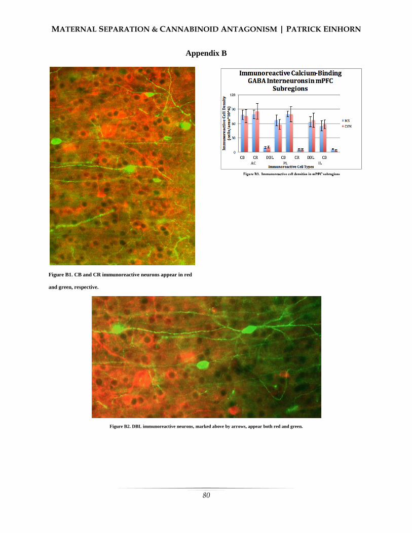
MATERNAL SEPARATION & CANNABINOID ANTAGONISM | PATRICK EINHORN
80
Appendix B
Figure B1. CB and CR immunoreactive neurons appear in red
and green, respective.
Figure B2. DBL immunoreactive neurons, marked above by arrows, appear both red and green.

81
Sumer Vaid
University of Chicago
Recent advances in cognitive neuroscience have challenged the conventional model of language
processing in the brain. Traditional theories maintain that all aspects of language, including
pragmatics and semantics are processed in amodal or modally non-specific areas of the brain. In
these models, the brain areas typically used to process modality-specific sensory information are
not employed to somaticize language. However, systematic physiological evidence has indicated
that the neural substrates responsible for modality-specific sensory processing and language
semantics overlap. How strong is this overlap between language and perceptual systems in the
brain? How can traditional theories of language processing be consolidated with this newer
evidence? By employing Barsalou’s (2001) framework of embodied cognition, this paper
reviews the literature to conclude that there is some connection between the neural
underpinnings of semantics and modality-specific perception. However, further research with
newer neuroimaging technologies is needed to elucidate the strength of this connection. In
particular, action-based language processing in the brain overlaps considerably with the
semanticization of literal but not metaphoric action. The paper concludes that current evidence
suggests that literal action language is majorly instantiated in the premotor and motor cortices
but more metaphorical action language is instantiated in traditional language regions.
This paper is inspired and guided by Professor Daniel Casasanto’s class, The Body in The Mind,
offered at The University of Chicago. Professor Anne Henly supervised my writing and assisted
me in the articulation of my argument. A special thanks to Benjamin Pitt, who helped me create
an understanding of the psychological literature and also offered editorial remarks on this
manuscript.
Language Processing Theories in
Psychology
anguage is a defining trait of the
human species as it allows us to
effortlessly and precisely shape the
thoughts of others (Pinker, 1995). Yet, we
struggle to answer the question: how do
humans understand language? Language can
vary widely in form (i.e grammar), structure
(i.e syntax) and mode (i.e verbal or written).
However, despite these differences, humans
are able to use language to take in the
thoughts of others and to communicate their
own thoughts to others. Of the two utilities,
Examining Language Processing Under an
Embodied Cognition Framework
L

EXAMINING LANGUAGE PROCESSING| SUMER VAID
this paper is focused on the former: how are
humans able to “make sense” of language?
Making sense of language requires several
mental and physiological processes. First,
one must “perceive” language, based on its
medium of transmission (i.e visual, audial,
tactile). Second, once one has “perceived”
the contents of the language, one must
derive meaning from these contents. The
mental and neural mechanism engaged in
the process of deriving meaning from
language is the central topic of this paper.
Hence, for the purposes of this paper,
language processing is defined as the
processes through which the meaning of
language is derived, after it has been
“perceived”. Crucially, this definition of
language processing will address those
processes that occur after language has been
“perceived”, which necessarily implies that
our discussions will not overtly concern the
modality through which the language is
communicated.
To examine the mental and neural
mechanisms of this process of
semanticization, we will survey the fields of
cognitive neuroscience and psychology.
There are two broad trends in literature –
one classical, and the other more recent and
controversial.
Traditional Models of Language
Processing
The traditional model of language
processing in the brain asserts that there is a
highly specialized brain region, which is
solely responsible for the understanding and
production of language. This view also
claims that this language-specialized brain
region is not engaged in neural processes
that constitute perception (i.e this brain
region is not utilized to see, hear, smell) but
only to understand language, regardless of
format (Fodor, 1975). In his classic The
Modularity of Mind (1983), Fodor lays the
criteria for a modular mind, and then
supports it with some psychological
evidence. The traditional model of language
perception is concomitant to a modular
mind, the extensive discussion of which lies
outside the scope of the paper. Importantly,
however, Fodor’s postulates hold that
several cognitive substrates rely on highly
localized neural-architecture, specialized
only to sustain that cognitive substrate.
Language is one such cognitive substrate,
and therefore relies on a specialized brain
area to be produced. To adhere to the
Fodorian criterion for modularity, this
language brain area must necessarily not
partake in processes contributing to
perception.
This modular model easily accounts
for why humans are unique in their ability
for language (because other animals lack
this highly specialized brain area) and how
humans slowly develop ability in language
(in-conjunction with the physiological
development of this organ) (Tulving, 1972;
Smith and Medin, 1981; Pylyshyn, 1984).
This model is also able to explain how we
can effortlessly understand language across
modalities – that is, our understanding of our
native language is not reliant on the medium
through which it is conveyed. The
traditional view asserts that there are two
core areas of language processing: the left
inferior fontal cortex and the superior
temporal cortex. Based on abundant
neuropsychological surveys of lesioned

EXAMINING LANGUAGE PROCESSING| SUMER VAID
patients, the traditional view postulates that
one of these areas “produces” language,
whereas the other area is responsible for
language comprehension. (Geschwind,
1970; Lichtheim, 1885).
Embodied Perspectives on Language
Processing
However, this traditional theory has
been recently challenged by embodied
cognition scientists, who posit that some
language is semanticizedin addition to the
processing of language in traditional brain
regions, language is also processed in those
brain areas that were traditionally thought to
be involved only in perception (Barsalou,
2001). These arguments stem from neuro-
imaging data that indicates distributed,
systematic activations of supplementary
brain regions, in addition to the activation of
traditional language regions. Some cognitive
scientists further argue that the evolutionary
time-frame required for the development of
this specialized brain organ is far too short
to allow for evolutionary changes that span
millions of years – language, in its present
form, has been estimated to have emerged
around 60,000 years ago (Christiansen,
2003; for a detailed review of the
evolutionary origins of language, see Bates,
1994).
Hence, the last decade of research in
cognitive neuroscience has re-ignited an
age-old question concerning the nature of
language processing. Traditional theories
maintain that language is processed brain
areas traditionally not associated with
perception (Tulving, 1972; Smith and
Medin, 1981; Fodor, 1975; Pylyshyn, 1984)
whereas relatively recent embodiment
theories assert that there exists some
connection between traditional language
processing systems and the sensorimotor
system in the brain (Pulvermüller, 2005;
Barsalou, 2001; Pulvermüller and Fadiga,
2010). Hence, the automatic, spontaneous
activation of sensory-motor systems during
language exposure is commonly referred to
as a simulation in the sensory-motor
systems. Weak embodiment theories assert
that only perceptually abundant language is
simulated in sensory-motor areas of the
brain, whereas stronger theories assert that
even abstract language make use of sensory-
motor simulations (Desai et al., 2011).
Perceptually abundant language utilizes
verbs to denote physical movement or action
(“I threw a ball), as opposed to abstract
language, which uses verbs in metaphorical
settings (“I threw a fit of rage”. It is also
crucial to note that this of language
simulation is automatic – the central claim
of embodied cognition theories assert that
processing language elicits sensory-motor
systems for representation automatically
rather than consciously. One other
clarification: for the purposes of this paper,
perceptually abundant language is that
which involves direct action (“I threw a
ball”) as opposed to abstract language,
which involved the use of action words to
denote metaphoric action (“I threw a
party”). We will now examine psychological
and physiological evidence for the modern
model of language processing.
Behavioral Paradigms
Several purely behavioral studies
have attempted to investigate the link
between language and perceptual systems.

EXAMINING LANGUAGE PROCESSING| SUMER VAID
For instance, a seminal study, participants
led by R.A Zwaan attempted to assess if
language comprehension shapes the visual
representation of actionable events.
Participants heard a perceptually loaded
sentence (“He threw the ball”) and were
then showed two sets of images separated by
175 ms. In the first condition, two sets of
pictures displayed visual nuances of the
implied movement in the actionable
sentence (“He threw the ball” implies the
ball moving away from the thrower, hence
the second picture displayed a slightly
smaller ball as a result of implied further
distance). In the second condition, there was
no congruence between visual nuances and
the aforementioned linguistic input. Subjects
in the first condition had a slower reaction
time when the implied movement of the ball
matched the visual nuances of the pictures.
The study concluded that this effect of
language comprehension resulted from
“dynamic mental representation”, but it did
not attempt to provide a neural basis for the
observations (Zwaan et al., 2004). The
researchers concluded that this was a result
of “simulating” the imagery induced by the
sentence cue, which provided evidence for a
link between the perceptual and language
processing mechanisms in the mind.
Another widely cited experiment
measured the precise eye-movements of
subjects while they were explicitly informed
to imagine something (Spivey and Geng,
2001). In the first experiment, the
researchers ensured that the participants
were unaware that their eye movements
were being tracked, while the participants
heard a story with spatial elements
(“Imagine that you are standing across the
street from a 40 story apartment building”).
Special care was taken to ensure that the
participants did not hear words that implied
explicit spatial cues such as “top” or
“bottom”. While the participants read this
story, their eye movements were discretely
analyzed. The results indicated
“unconscious” eye-movements of the
participants were congruent to supposed eye
movements when the story was taking place
in real life. that subjects systematically
looked at blank regions of space during the
process of In the second experiment,
participants were presented with four
distinct objects at four different corners of a
computer screen. In the testing task, they
were presented with three of the
aforementioned items, and were then asked
to remember one particular descriptor of the
missing item (i.e color or shape). While the
participants performed this task, their eye-
movements were discretely analyzed. The
results indicated that while “imagining
something. The study concluded that this
was a result of ” this object, the participants
eye movements focused the gaze to the
corner in which the missing object had
initially been present. The study concluded
that both these results originated from the
manipulating and organizing of spatial
relationships between virtual or retinal
images.
Both these studies were immensely
important in establishing a foundation for
further, more physiological intellectual
venture into embodied cognition theories.
However, though these studies establish a
link between perceptual and linguistic
systems in the mind, they offer no evidence
that this mental connection is subserved by a

EXAMINING LANGUAGE PROCESSING| SUMER VAID
physical link between the perisylvian
language regions and modality-specific
perceptual brain systems. The behavioral
data does not explicitly indicate that the
cued language was processed in modality-
specific sensory systems; it may originate
from varying patterns of brain activation that
cannot be accurately inferred from non-
neuroimaging experiments. However,
Barsalou’s theory of embodiment critically
relies on such a functional link as it asserts
that even the most basic perceptive
mechanisms in the brain play a role in
higher-order linguistic processing. Ergo,
Barsalou’s theory of embodiment can only
be tested by examining the brain directly or
by examining other physiological properties
of perception and action. Furthermore, the
second study (Spivey and Geng, 2010) tests
embodied cognition through experimental
paradigms that involve conscious
“imagining” , which violated Barsalou’s
notion of automaticity. To test Barsalou’s
theory of embodied cognition, one would
not only have to examine the brain in real
time during language processing, but also
take measures to ensure that participants are
not told to explicitly “imagine” anything.
Any sensorimotor activation should result
from an unconscious process, which the
participants have no control over.
Brain-Imaging Evidence
The anatomy of the brain provides
some motivation for investigations into the
neural underpinnings of embodied
cognition. Traditionally, the cortical systems
for action control and language were
believed to be classic examples of a modular
brain (Shalice, 1988). These regions are
spatially different and appear to be
behaviorally exclusive as revealed by
neurological disease - stroke patients are
unable to move contralateral body side but
have no major disruptions in language
ability) (Pulvermüllerr, 2005). However,
neuroanatomy has revealed the existence
neuronal connections between the dorsal and
ventral premotor cortex and the left
perisylvian regions of language (Makris et
al., 1999; Pandya and Yeterian, 1985),
indicating that information can pass in
between them. Following the Hebbian
learning model, these connections are
postulated to exist because of frequent
interactions between linguistic brain areas
and action perception brain areas.
Neuroimaging experiments further
indicate that language and action are linked
in the brain. One landmark study found that
reading action words describing bodily
movements activated brain areas congruent
to actually performing those movements
physically (Hauk et al,. 2004). Participants
were first required to move their feet, arms
and tongue on separate trials while their
brain metabolism was recorded using
functional MRI. The participants were then
required to read either leg-related words,
arm-related words or face related words
while their brain metabolism was recorded.
There was a significant intersection between
the brain areas used to perform specific
actions and the brain areas used to
understand words related to that specific
action, indicative of a strong relationship
between language and action in the brain. It
is crucial to note that this overlap in
activation maps was not complete – that is,
there were substantial differences in the

EXAMINING LANGUAGE PROCESSING| SUMER VAID
brain-maps for each of the two tasks. This
implies that extreme embodied cognition
theories, which posit that even the most
abstract language is entirely instantiated in
modality-specific brain-regions, is lacking
evidence.
Functional MRI, like any brain
imaging technique, has many weaknesses.
One of these is that the hemodynamic
metabolism process that this technique
measures occurs significantly after the
electric potential of neurons has “peaked”
(Glover, 2011). In other words, fMRI is not
a direct measure of brain-activity, instead it
relies on changes in blood-flow to indicate
activity. This delay of activity-reporting
raises many questions: for instance, it is
entirely possible that the motor cortex
activations may result from a mental plan of
action triggered by the linguistic input
(action word). In other words, when the
subject “reads” the action word, they are
consciously imagining performing that
action, instead of unconsciously deriving the
meaning of the word. Our topic of interest is
the latter, not the former – hence, fMRI data
may be providing support an alternative,
equally likely hypothesis. Hence, the
observe data would be a result of conscious
mental imagery, violating the postulates of
Barsalou’s embodied cognition theory,
which relies on the unconscious nature of
language processing.
Fortunately, other brain imaging
techniques can be used to get around the
problem caused by activity-reporting delays.
In one such study, Event-Related Potentials
were measured while participants were
asked to read action words (Hauk and
Pulvermüller, 2005) using
electroencephalography. This technique
offers high temporal resolution, as it directly
measures the electric activity of the neurons
located near the scalp – thereby avoiding
delays caused by measuring blood activity.
The study reported a category-specific
differential activation around 200 ms after
exposure to input. In other words, the study
found activations for perceptually abundant
stimuli at 200 ms but not for control stimuli.
Furthermore, this activation was proximate
to an inferior frontal region for face-related
words and to a superior central source for
leg-related words, indicating prompt body-
part specific activation following the
linguistic input presentation. Previous
research in word processing indicates that
language understanding elicits brain area-
specific activation after 200 ms, on average
(Sereno and Rayner, 2013). As the observed
brain activations occurred after 200 ms, the
results supported the idea that modality-
specific activations were instantiating
immediate language understanding. Hence,
these results support the idea that the brain
activation is a result of semantic processing
and not because of processing a “plan of
action” encompassed by post-semantic
processing. Nevertheless, there are a few
weaknesses with this mode of brain
imaging. While EEG improves temporal
resolution, it is not able to offer the high
spatial resolution of fMRI. Hence, while we
can be sure that the brain activations
occurred somewhere in the vicinity of our
targeted modality-specific areas, we cannot
be sure that they were completely elicited in
our areas of interest, which prevents us from
using this data to conclusively support
Barsalou’s embodiment theories.
EXAMINING LANGUAGE PROCESSING| SUMER VAID
To address issues related to temporal
and spatial resolution, a new and promising
brain-imaging technique was used -
magnetoencephalography. While the merits
of this technique remain hotly debated,
many experts believe that it offers high
temporal and spatial resolution. The goal of
this study was to investigate the activation
of brain regions while participants engaged
in a distracting task while listening to action
words (Pulvermüller et al., 2005). It was
hypothesized that spatiotemporal activations
would occur first in the perisylvian language
regions of the brain (due to the processing of
the form of the action word) followed by
immediate activation in the somatotopic
specific areas of the premotor and motor
cortex (due to the semantic processing of the
action word). The purpose of the distractor
was to direct the attention of participants
away from the target stimulus, as this would
allow for the measurement of brain-
activations of language processing that is not
consciously being attended to. The study
found that superior temporal areas
(responsible for leg-related movement)
activated after an average of 130 ms, and
activated more significantly for words
relating to leg related movements. Inferior
fronto-central areas (responsible for facial
movements) activated between 142-146 ms
reacted more significantly for words related
to facial movement words. This activation
occurred shortly after the words were
identified as lexical items in the traditional
perisylvian language region, as predicted by
previous literature. Hence, this study
addressed some of the weaknesses of
previous neuro-imaging studies, and still
found convincing evidence to support the
idea that part of language is processed in
modality-specific regions of the brains, even
when participants were not attending to this
word consciously. This lack of directed
attention further separated the likelihood of
the participants consciously simulating a
plan of action following the processing of a
perceptually abundant word, which further
offered support an embodied cognition
hypothesis.
We have evaluated encouraging
evidence for embodied cognition theories
from three different brain-imaging studies,
each of which had its own set of weakness.
However, these three techniques have one
common, debilitating weakness; brain-
imaging evidence, however abundant, only
implies a correlational link between
activation in primary motor areas of the
brain and understanding of action words.
We can only conclude that modality-specific
brain activity and language comprehension
occur in conjunction, but cannot assess
(from these studies), if one is causing the
others. To do this, we need to examine
evidence originating from brain-stimulation
techniques and language comprehension.
Assessing behavioral patterns in relation
with non-invasive brain activation cements
this link to a certain extent and hints at a
functional link between the perceptual and
linguistic systems in the brain.
Brain-Stimulation
In one such seminal study,
Pulvermüller et al. (2005) applied weak
magnetic pulses to hand or leg areas in the
motor cortex while participants performed to
a lexical decision task involving leg-related
or hand-related words appearing on a screen.
EXAMINING LANGUAGE PROCESSING| SUMER VAID
The aim of the study was to determine if
creating temporary lesions in the motor
cortices had any outcome on how action
language was understood. The study found
that arm-area stimulation led to a reduction
in reaction time of response to arm-related
action words (Pick) but did not effect the
reaction time of leg-related action words
(kick). The reverse was true for leg-area
stimulation, suggesting that motor cortex
played a functional role in understanding
action-related language. This study further
cemented the body of evidence supporting
embodied cognition theories – not only do
activations in the primary motor cortices
(traditionally associated only with the
production of movements) co-occur with
action-based language, but also that
disrupting activity in these areas can affect
the understanding of action-based language.
This encouraging evidence has
motivated other off-spring theories of
embodied cognition, the testing of which
can offer further support to their parent
theory. One such off-spring theory is the
body-specificity hypothesis. According to
the body-specificity hypothesis (Casasanto,
2009; 2011), if we use our bodily experience
to think, then people with different bodies
should think differently. For instance, a
large body literature indicates that the
interaction of people with their environment
limits their perceptions and actions
(Linkenauger et al., 2009). Hence, one line
of research attempted to test this theory
using brain imaging and stimulation
techniques. Willems et al. (2010, 2011) used
this paradigm to investigate the extent of
body-specific nature of language processing.
The first study (Willems et al., 2010) aimed
to discern if brain-activations resulting from
the reading of action-words differed across
participants with different handedness. As
people are more likely to perform manual
actions with the dominant hand, brain-
imaging evidence should indicate that the
processing of manual action-words activates
the contra-lateral hemisphere of the
dominant hand. Left-handed people and
right-handed people were scanned while
they read a manual action verb (“throw”) as
opposed to a non-manual action verb
(“read”). FMRI evidence indicated that
when participants read manual action words
typically performed by their dominant hand,
a contralateral activation is observed in the
premotor cortex of the participants (Willems
et al., 2010). To establish a functional link
between these contralateral activations and
language comprehension, Willems, Labruna
et al. (2011) activated the left motor cortex
(contralateral to dominant right hand) while
right-handed participants processed manual
action words. This activation led to a
reduction in reaction time of response to
manual action words typically performed by
the dominant hand. Such an interaction was
not observed for non-manual action words
(e.g “earn”, “learn”). Furthermore,
activation to the right motor cortex,
responsible for controlling the non-dominant
hand, did not produce any interaction with
reaction time and type of action
(manual/non-manual), implying that the
motor basis for language are body-specific.
Specifically, these results were ‘body-
specific’ because they displayed an
interaction between language processing and
the dominant hand of subjects, implying that
body-specific features may modulate brain

EXAMINING LANGUAGE PROCESSING| SUMER VAID
activation representing the processing of
language. Collectively, support for the body-
specificity hypothesis automatically offers
support for some versions of the embodied
cognition theories. Hence, the literature
indicates that there is concrete evidence for
at-least some versions of embodied
cognition theories. However, one crucial
postulate of embodied cognition theories
remains empirically ambiguous – that of
abstract language processing.
Sensorimotor Metaphors
A pressing question raised by critics
of the embodied cognition theory is: if we
use perceptual simulations to think then how
do we think about things that are not found
in the real world? One proposal aims to
tackle this question through the mechanisms
of metaphors. Metaphors have been shown
to be more than just devices of language,
they also appear to be ways of thinking
(Casasanto and Bottini 2014; Landau et al.,
2010). For instance, words that appear closer
in space are judged to be closer in meaning
(Casasanto, 2008). Similarly, people feel
more warmly towards their acquaintances
when they are actually experiencing
physical warmth (Ijzerman and Semin
2009;). Lakoff describes these metaphors as
resulting from links between the source
domain (the physical and perceptual real-
world experience) and the target domain (the
abstract idea not found in the real world)
(Lakoff and Johnson, 1999). Hence, it is
postulated that abstract thought relies on the
metaphorical link between the contents of
the thought and perceptual systems in the
brain (Desai et al., 2011).
However, not all mental metaphors
are necessarily “embodied” (Casasanto and
Gijessels, 2015). For a mental metaphor to
be embodied (as per Barsalou’s theory of
embodiment), it must rely on modality-
specific areas in the brain to be processed.
Therefore, a key method to verify the
embodiment of mental metaphors is by
examining brain activations during the
processing of metaphoric language.
Experimental evidence for such
embodied metaphors remains inconsistent.
One study reported somatotopic activation
in the premotor cortex for literal action
sentences (“I kicked a ball”) but not for
phrases with idiomatic elements (“I kicked
the habit”) (Aziz-Zadeh et al., 2006).
Another group of experimenters reported
that somatotopic activation along the motor
strip was observed during the presentation of
idiomatic and literal sentences alike, and
reflected the body-part reference of the
words in the stimulus. This activation was
greatest after the subjects had finished
analyzing the entire sentence suggesting that
the context played a crucial role in the
neural reflection of the semantics
(Boulenger, Hauk and Pulvermüller, 2008).
Desai et al. (2011) postulated that this
general inconsistency of experimental
evidence resulted from the absence of a
complexity control across literal and
metaphoric sentences. In a series of
experiments, they carefully controlled for
the complexity of literal and metaphoric
sentences and found that metaphoric and
literal sentences of comparable complexity
elicited a response in the left anterior
inferior partial lobule, an area responsible
for action planning. They also found that the
EXAMINING LANGUAGE PROCESSING| SUMER VAID
activation of the primary motor areas was
inversely co-related to the familiarity of the
metaphor. They hypothesize that this effect
results from a gradual abstraction process as
detailed simulations are vital to understand
unfamiliar metaphors whereas lesser
detailed simulations rely more on the
secondary motor regions due to the increase
in familiarity of the metaphor. However, this
evidence is still insufficient as it does not
display a robust, causal connection between
primary modality-specific brain regions and
metaphoric action-evoking language. In
another experiment, Desai et al. (2013)
found that the general abstractness of an
action word was inversely correlated with
the activation of modality-specific
sensorimotor regions.
The interaction between space and
musical pitch offers a promising test bed to
investigate the physical roots of
metaphorical understanding in the brain.
Dolscheid et al. (2014) examined brain
metabolism while participants judged
stimuli across the visual, tactile and auditory
modalities (high-pitch vs low-pitch). If the
judgments of pitch height involved
multimodal simulations, they would show a
pattern of activation resulting from the
combination of visual and tactile activations.
If the judgments relied on modality-specific
brain area activation, they would show a
hemodynamic pattern congruent to either
visual or tactile patterns. In support of the
latter, judgments of pitch height activated
unimodal visual areas, offering a one-of-its
kind evidence that abstract metaphorical
thinking relies on basic perceptual apparatus
for processing. Further research in exploring
the neural groundings of space-pitch
associations are required, in particular, a
causal relationship must be defined through
the use of brain-stimulation techniques.
The Big Picture
Converging evidence from brain-
imaging and brain-stimulation techniques
indicates that modality-specific brain areas
play some role in deriving the meaning of
language. Perceptually abundant language
(eg: “I threw a ball”) elicits large activations
in modality-specific brain areas such as the
primary motor cortex, as confirmed by
different types of neuro-imaging
technologies. Functional MRI, which has
high spatial resolution but poor temporal
resolution, indicates a significant overlap
between modality-specific brain areas when
specific actions are performed and
perceived. Similar evidence is also reported
using EEG – a technique with high temporal
resolution but poor spatial resolution. MEG,
a technique with reasonably high spatial and
temporal resolution, verifies these findings –
there is a localized and instantaneous brain
activation response in modality-specific
areas following the onset of perceptually
abundant stimuli. However, all this imaging
evidence is not causal – a confounding
variable (such as word-structre)
systematically varying with the independent
variable maybe causing the elicited brain
activations. However, brain-stimulation
techniques indicate that temporarily
lesioning modality-specific area interacts
with the processing of perceptually
language, confirming the hypothesis that
modality-specific brain areas play a causal
role in deriving the meaning of perceptually
abundant language. This causal evidence is

EXAMINING LANGUAGE PROCESSING| SUMER VAID
further strengthened by the evidence for
body-specificity hypothesis, which posits
that if bodily experience influences our
thinking, then people with different bodies
should think differently. When contralateral
hemisphere of right-handed people is
temporarily lesioned, a change in reaction
times to perceptually abundant stimuli is
noted. This variation in reaction times is
lacking when control sites or non-
contralateral brain areas of right-handed
subjects are lesioned, supporting the body-
specificity hypothesis.
However, despite the overwhelming
evidence for the embodiment of perceptually
abundant language, there is little proof for
embodiment regarding abstract language.
According to theorists, metaphors are not
just devices of language use but also
technologies of thinking – in other words,
we use metaphors to think about abstract
concepts. Nevertheless, this does not
necessarily imply that metaphors are
embodied – because there is little neural
evidence to indicate that mental metaphors
are instantiated in modality specific brain
regions. Neuro-imaging evidence indicates
that while perceptually abundant language
clearly elicits activation in modality-specific
brain areas, metaphoric language does not
elicit this type of activation. The sparse
evidence for the embodiment of metaphors
originates from the neural relationship
between musical pitch and space. Judgments
of musical pitch activate modality-specific
vision areas, implying that metaphors used
to describe “high” and “low” pitches are
instantiated in the primary visual areas of
the human brain.
Beyond Language: Conceptual
Processing
Existing evidence indicates that there
exists a functional link between the
traditional linguistic systems in the brain and
the perceptual systems of the brain. For
literal language, the motor cortex is
activated and plays a functional role in its
processing. For metaphoric action language,
we do not see a robust activation of the
motor cortex. However, across other cross-
modal paradigms such as space, we see a
potential for modality-specific groundings
for metaphors.
The reach and scope of embodied
cognition stretches far beyond language
processing. The theory’s broader
implications are tightly linked with
philosophical ideas about cognition, the
discussion of which was beyond the scope
of this review paper. A fundamental driving
aim of cognitive science is to gain a better
understanding of knowledge is organized,
stored and communicated in the mind.
Different types of embodied cognition
theories make different claims about how
each of these knowledge characteristics are
entrenched in the brain. This paper
examined how the communication of
knowledge through language is processed in
the brain under the framework of Barsalou’s
widely cited embodied cognition theory.
Research in this specific area should address
the following potential questions:
1. Is the link between pitch
metaphors and occipital lobe
activation in occipital lobe
functional?
2. How does embodied language
processing interact with the

EXAMINING LANGUAGE PROCESSING| SUMER VAID
organization and the storage of
knowledge?
References
Aziz-Zadeh, L., Wilson, S. M., Rizzolatti,
G., & Iacoboni, M. (2006). Congruent
Embodied Representations for Visually
Presented Actions and Linguistic
Phrases Describing Actions. Current
Biology, 16(18), 1818–1823.
http://doi.org/10.1016/j.cub.2006.07.060
Barsalou, L. W., Simmons, W. K., Barbey,
A. K., & Wilson, C. D. (2003).
Grounding conceptual knowledge in
modality-specific systems. Trends in
Cognitive Sciences.
http://doi.org/10.1016/S1364-
6613(02)00029-3
Bates, E. (1994). Modularity, Domain
Specificity and the development of
language. Discussions in Neuroscience,
10, 1/2, 136-149.
Boulenger V, Hauk O, Pulvermuller F
(2009) Grasping Ideas with the Motor
System: Semantic Somatotopy in Idiom
Comprehension. Cerebral Cortex
19:1905-1914.
Casasanto, D. (2008). Similarity and
proximity: When does close in space
mean close in mind? Memory &
Cognition, 36(6), 1047-1056.
Casasanto, D. (2011). Different Bodies,
Different Minds: The Body Specificity
of Language and Thought. Current
Directions in Psychological Science.
http://doi.org/10.1177/09637214114220
58
Casasanto, D., & Bottini, R. (2014). Mirror-
reading can reverse the flow of time.
Journal of Experimental Psychology:
General, 143(2), 473-9.
Casasanto, D., & Gijssels, T. (2015). What
makes a metaphor an embodied
metaphor? Linguistics Vanguard.
doi:10.1515/lingvan-2014-1015
Desai, R. H., Binder, J. R., Conant, L. L.,
Mano, Q. R., & Seidenberg, M. S.
(2011). The neural career of sensory-
motor metaphors. Journal of Cognitive
Neuroscience, 23(9), 2376–2386.
http://doi.org/10.1162/jocn.2010.21596
Desai, R., Conant, L., Binder, J.R., Park, H.,
Seidenberg, M.S. (2013). A piece of the
action: Modulation of sensory-motor
regions by action idioms and metaphors.
NeuroImage.
Dolscheid, S., Willems, R. M., Hagoort, P.,
& Casasanto, D. (2014). The relation of
space and musical pitch in the brain. In
P. Bello, M. Guarini, M. McShane, & B.
Scassellati (Eds.), Proceedings of the
36th Annual Meeting of the Cognitive
Science Society (CogSci 2014) (pp. 421-
426). Austin, Tx: Cognitive Science
Society.
Fodor, J.A. (1975) The Language of
Thought, Harvard University Press

EXAMINING LANGUAGE PROCESSING| SUMER VAID
Hauk, O., & Pulvermüller, F. (2004).
Neurophysiological Distinction of
Action Words in the Fronto-Central
Cortex. Human Brain Mapping, 21(3),
191–201.
http://doi.org/10.1002/hbm.10157
Hauk, O., Johnsrude, I., & Pulvermüller, F.
(2004). Somatotopic Representation of
Action Words in Human Motor and
Premotor Cortex. Neuron, 41(2), 301–
307. http://doi.org/10.1016/S0896-
6273(03)00838-9
Ijzerman, H., & Semin, G. R. (2009). The
thermometer of social relations:
Mapping social proximity on
temperature: Research article.
Psychological Science, 20(10), 1214–
1220. http://doi.org/10.1111/j.1467-
9280.2009.02434.x
Landau, M. J., Meier, B. P., & Keefer, L. A.
(2010). A metaphor-enriched social
cognition. Psychological Bulletin,
136(6), 1045–1067.
http://doi.org/10.1037/a0020970
Makris, N., Meyer, J. W., Bates, J. F.,
Yeterian, E. H., Kennedy, D. N., &
Caviness, V. S. (1999). MRI-Based
topographic parcellation of human
cerebral white matter and nuclei II.
Rationale and applications with
systematics of cerebral connectivity.
NeuroImage, 9(1), 18–45.
http://doi.org/10.1006/nimg.1998.0384
Pandya, D. N. & Yeterian, E. Architecture
and Connections of Cortical Association
Areas, in Cerebral Cortex. Vol. 4 (eds
Peters, A. & Jones, E. G.) 3–61 (Plenum,
London)
Pulvermüller, F. (2005). Brain mechanisms
linking language and action. Nature
Reviews Neuroscience, 6(7), 576–582.
http://doi.org/10.1038/nrn1706
Pulvermüller, F., & Fadiga, L. (2010).
Active perception: sensorimotor circuits
as a cortical basis for language. Nature
Reviews Neuroscience, 11(5), 351–360.
http://doi.org/10.1038/nrn2811
Pulvermüller, F., Hauk, O., Nikulin, V. V.,
& Ilmoniemi, R. J. (2005). Functional
links between motor and language
systems. European Journal of
Neuroscience, 21(3), 793–797.
http://doi.org/10.1111/j.1460-
9568.2005.03900.x
Pulvermüller, F., Shtyrov, Y., & Ilmoniemi,
R. (2005). Brain signatures of meaning
access in action word recognition.
Journal of Cognitive Neuroscience, 17.
Pylyshyn, Z.W. (1984) Computation and
Cognition, MIT Press
Sereno, S.C. & Rayner, K. (2003)
Measuring word recognition in reading:
eye movements and event-related
potentials. Trends in Cognitive Science.
7, 489-493
Shallice, T. (1988 ) From Neuropsychology
to Mental Structure, Cambridge Univ.
Press
EXAMINING LANGUAGE PROCESSING| SUMER VAID
Simmons, W. K., Pecher, D., Hamann, S. B.,
Zeelenberg, R., & Barsalou, L. W.
(2003). fMRI evidence for modality-
specific processing of conceptual
knowledge on six modalities, 1–16.
http://doi.org/10.1016/S1364-
6613(02)00029-3
Smith, E.E. and Medin, D.L. (1981)
Categories and Concepts, Harvard
University Press
Spivey, M. J., & Geng, J. J. (2001).
Oculomotor mechanisms activated by
imagery and memory: Eye movements
to absent objects. Psychological
Research, 65(4), 235–241.
http://doi.org/10.1007/s004260100059
Tulving, E. (1972) Episodic and semantic
memory. In Organization and Memory
(Tulving, E. and Donaldson, W., eds) pp.
381–403, Academic Press.
Willems, R. M., Labruna, L., D’Esposito,
M., Ivry, R., & Casasanto, D. (2011). A
functional role for the motor system in
language understanding: evidence from
theta-burst transcranial magnetic
stimulation. Psychological Science,
22(7), 849–54.
http://doi.org/10.1177/09567976114123
87
Wilson, B. (2001). Philosophy in the Flesh:
The Embodied Mind and Its Challenge
to Western Thought. English Linguistics.
Journal of the English Linguistic
Society of Japan.
http://doi.org/10.1590/S0102-
44502001000100008
Zwaan, R. A., Madden, C. J., Yaxley, R. H.,
& Aveyard, M. E. (2004).
Moving words: Dynamic
representations in language
comprehension. Cognitive Science,
28(4), 611–619.
http://doi.org/10.1016/j.cogsci.2004.
03.004