ucns review course: high yield images - cdn.ymaws.com · ucns review course: high yield images...
TRANSCRIPT

UCNS Review Course:
High Yield Images
Stephanie J. Nahas, MD, MSEd, FAHS, FAAN
Associate Professor of Neurology
Director, Headache Medicine Fellowship Program
Assistant Director, Neurology Residency Program
Department of Neurology
Jefferson Headache Center
Thomas Jefferson University
Philadelphia, Pennsylvania
@stephanieJnahas
Disclosures
• I have received author/editor honoraria from: Demos Medical, MedLink Neurology, and UpToDate
• I have received advising/consulting/speaking honoraria from: Allergan, Amgen, Electrocore, Eli Lilly, Supernus, Teva

Learning Objectives
• recognize normal and abnormal findings on diagnostic imaging in patients presenting with headache
• contextualize the significance of imaging findings to potential diagnoses in patients with headache
• identify visible findings on physical or neurologic exam that aid in diagnosis of patients with headache
Depressed, Demented Dolores
• 64 y.o. woman referred for depression and dementia
• Migraines since her 20s
• Memory troubles since her late 40s
• In her early 50s had left-sided weakness for a few weeks which resolved
• Was told she has multiple sclerosis
Depressed, Demented Dolores
• Increasing headache frequency
• Further cognitive decline over the past few years
• Depressed and at times agitated
• One period with psychosis
• Father had “Alzheimer’s”
• Multiple family members also with migraine, and a paternal aunt diagnosed with MS

Is this her MRI?
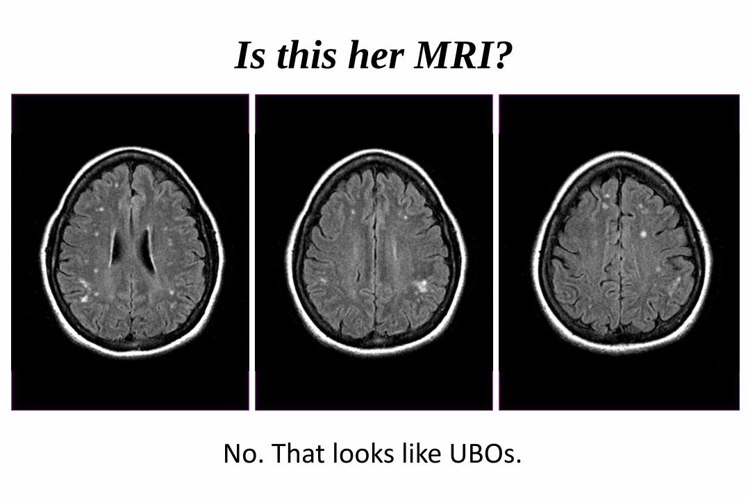
Is this her MRI?
No. That looks like UBOs.
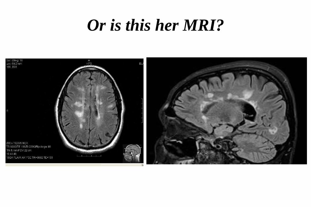
Or is this her MRI?
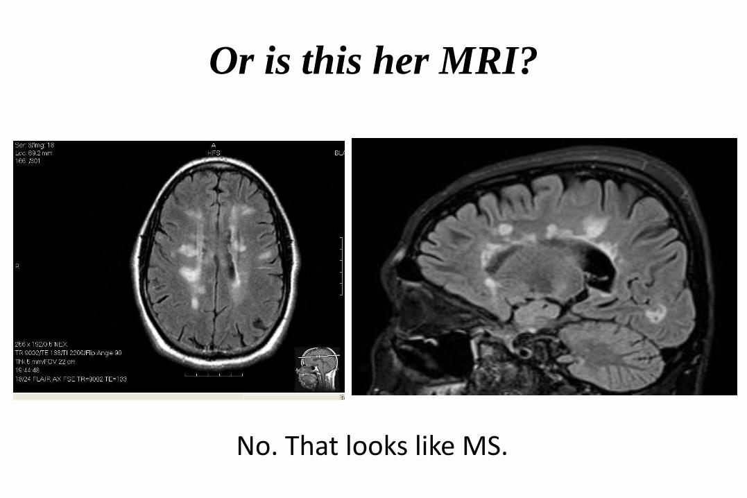
Or is this her MRI?
No. That looks like MS.

No, this is. Diagnosis?
Auer D P et al. Radiology 2001;218:443-451 ©2001 by Radiological Society of North America

CADASIL(Cerebral Autosomal Dominant Arteriopathy
with Subcortical Infarcts and Leukoencephalopathy)
Gene: NOTCH3Chromosome: 19
Weary, One-Eyed Wanda
• 85 y.o. woman 7 months of slowly progressive right-sided headache
• Throbbing temporal/parietal
• Ipsilateral photophobia, lacrimation, nasal congestions
• Nocturnal exacerbations
• Treated with CBZ for TN – no benefit
Weary, One-Eyed Wanda
• One week ago, increased tearing and eye swelling, given steroid eye drops after normal ophtho exam
• Three days ago, abrupt ptosis and diplopia
• PMHx: DM, HTN, cardiomyopathy
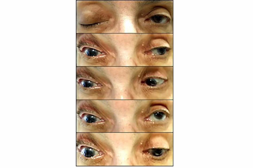
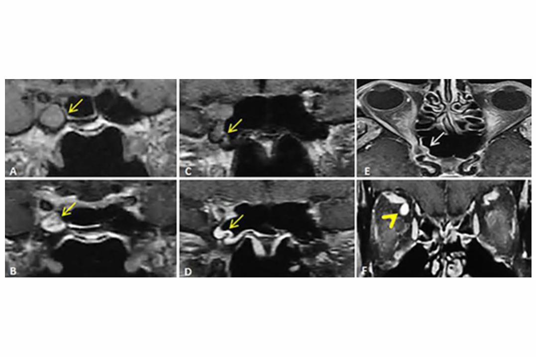

Cavernous sinus aspergillosis in uncontrolled diabetes
Roy B, Grosberg BM. Teaching images in headache: Cavernous sinus aspergillosis. Headache. 2016;56(10):1653-5.
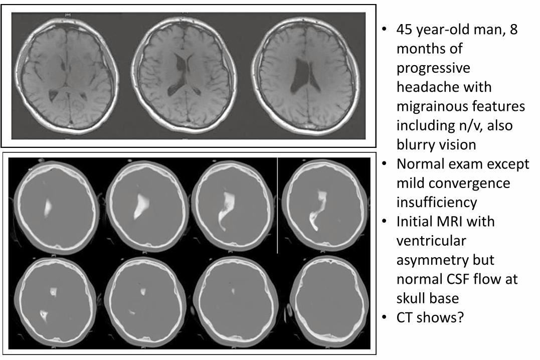
• 45 year-old man, 8 months of progressive headache with migrainous features including n/v, also blurry vision
• Normal exam except mild convergence insufficiency
• Initial MRI with ventricular asymmetry but normal CSF flow at skull base
• CT shows?
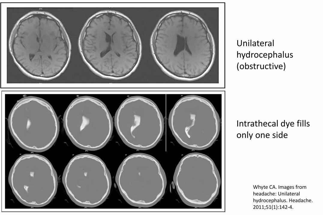
Unilateral hydrocephalus (obstructive)
Intrathecal dye fills only one side
Whyte CA. Images from headache: Unilateral hydrocephalus. Headache. 2011;51(1):142-4.
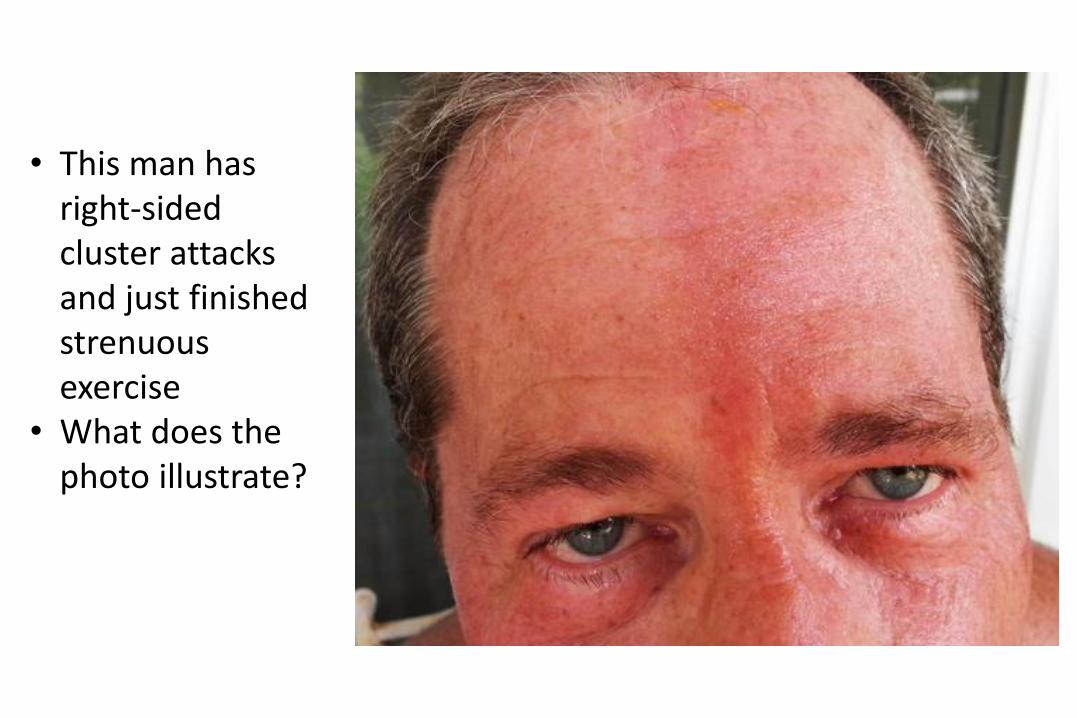
• This man has right-sided cluster attacks and just finished strenuous exercise
• What does the photo illustrate?
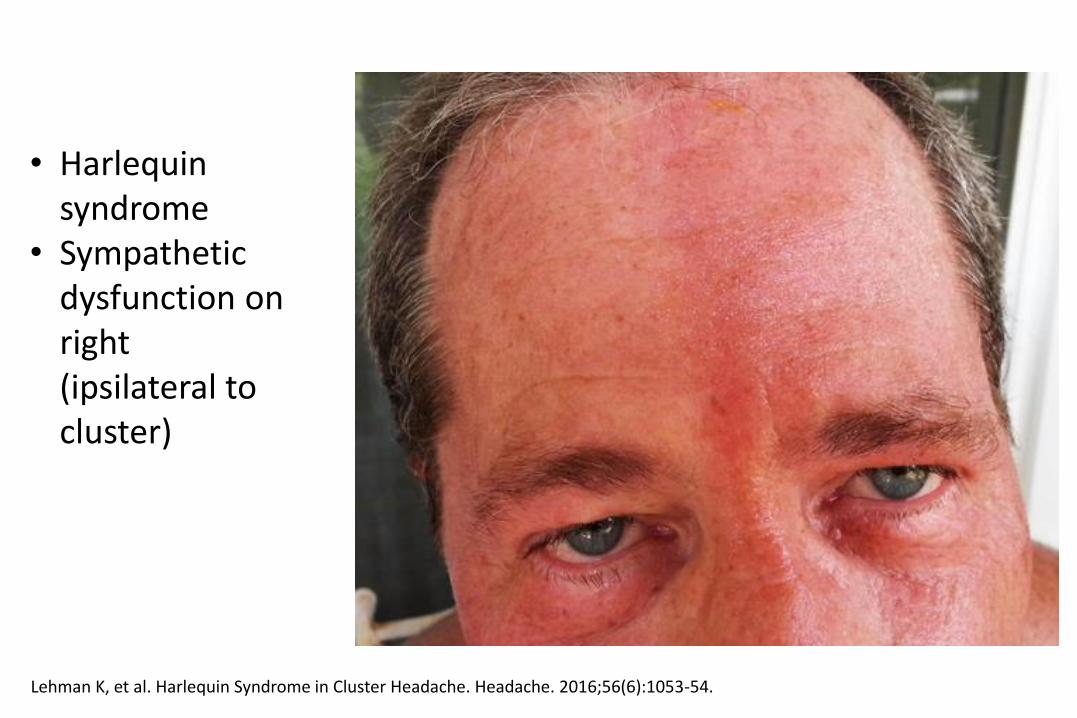
• Harlequin syndrome
• Sympathetic dysfunction on right (ipsilateral to cluster)
Lehman K, et al. Harlequin Syndrome in Cluster Headache. Headache. 2016;56(6):1053-54.
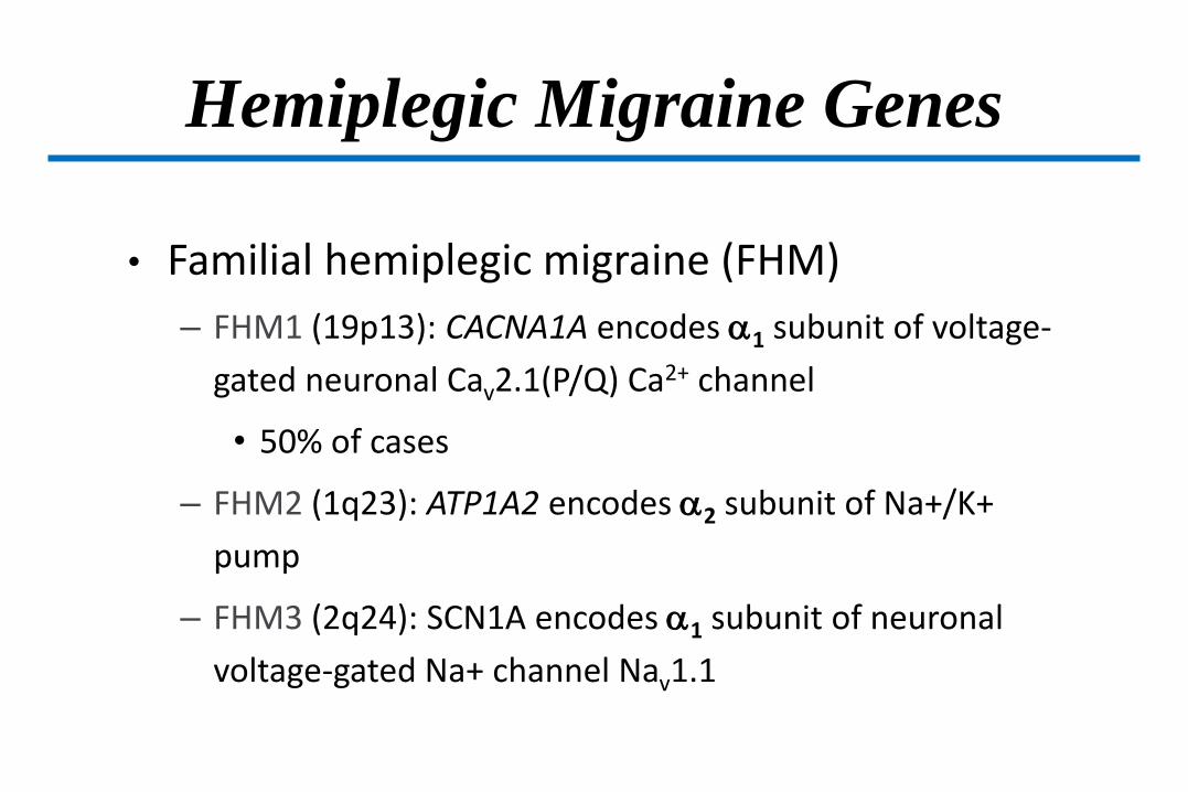
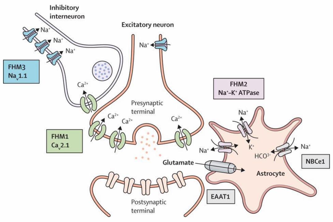
Hemiplegic Migraine Genes
• Familial hemiplegic migraine (FHM)
– FHM1 (19p13): CACNA1A encodes 1 subunit of voltage-
gated neuronal Cav2.1(P/Q) Ca2+ channel
• 50% of cases
– FHM2 (1q23): ATP1A2 encodes 2 subunit of Na+/K+
pump
– FHM3 (2q24): SCN1A encodes 1 subunit of neuronal
voltage-gated Na+ channel Nav1.1
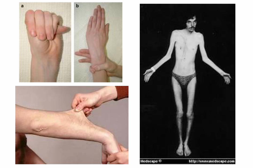
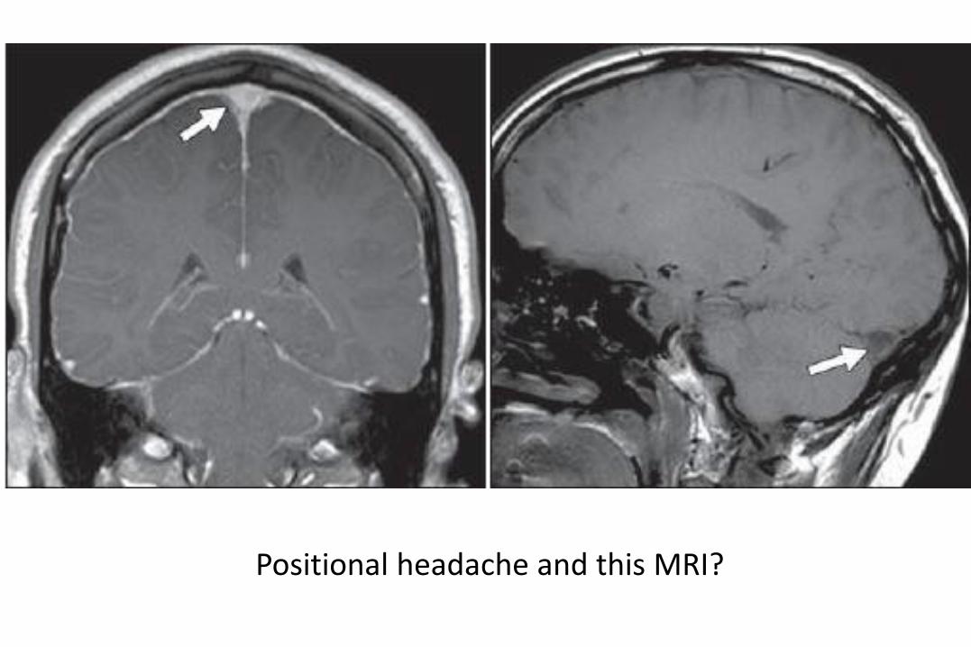
Positional headache and this MRI?

• Marfanoid or Ehlers-danlos
• At risk for spinal CSF leak
• Might not have positional headache
• Best initial step to localize is spine MRI
• May need CISS sequences (heavily T2-weighted) with thin cuts

• Myelogram if can’t get MRI or it’s unrevealing
• Extravasation of dye through nerve sheath diverticula into paraspinal spaces
• Radionucelide study another option

Normal radioisotope cisternogram Mokri B. Radioisotope cisternography in spontaneous CSF leaks: interpretations and misinterpretations. Headache. 2014;54(8):1358-68
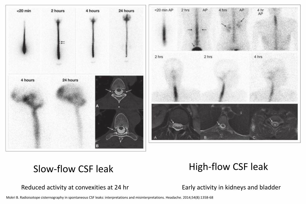
Slow-flow CSF leak High-flow CSF leak
Reduced activity at convexities at 24 hr Early activity in kidneys and bladder
Mokri B. Radioisotope cisternography in spontaneous CSF leaks: interpretations and misinterpretations. Headache. 2014;54(8):1358-68
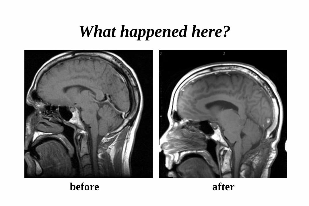
What happened here?
before after
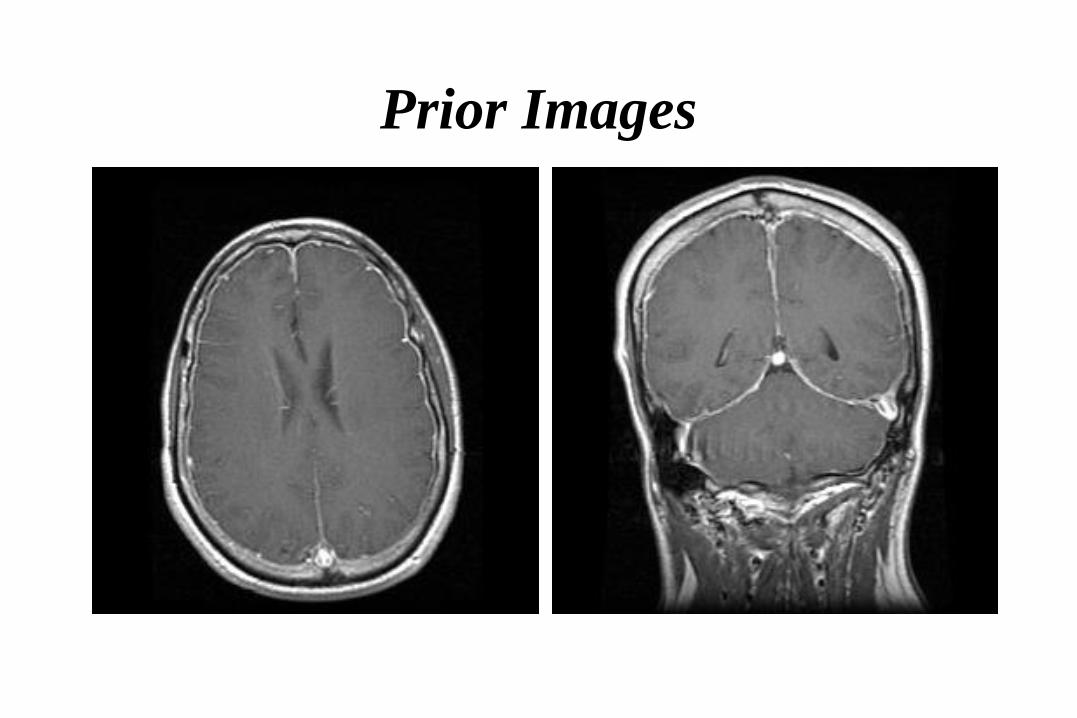
Prior Images
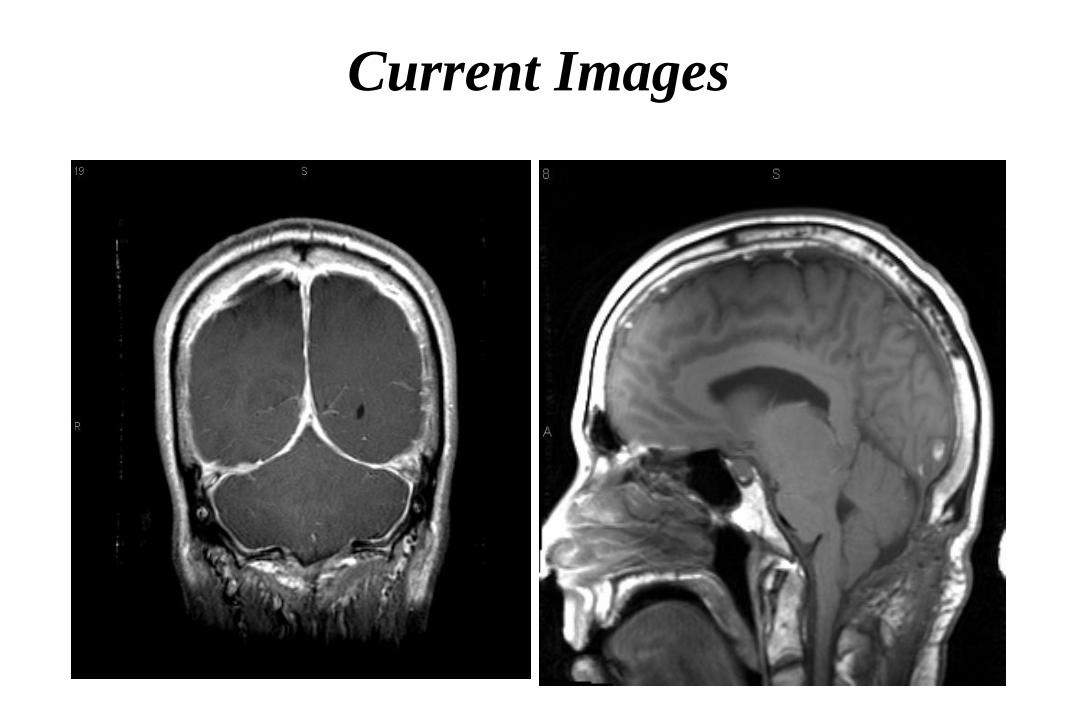
Current Images
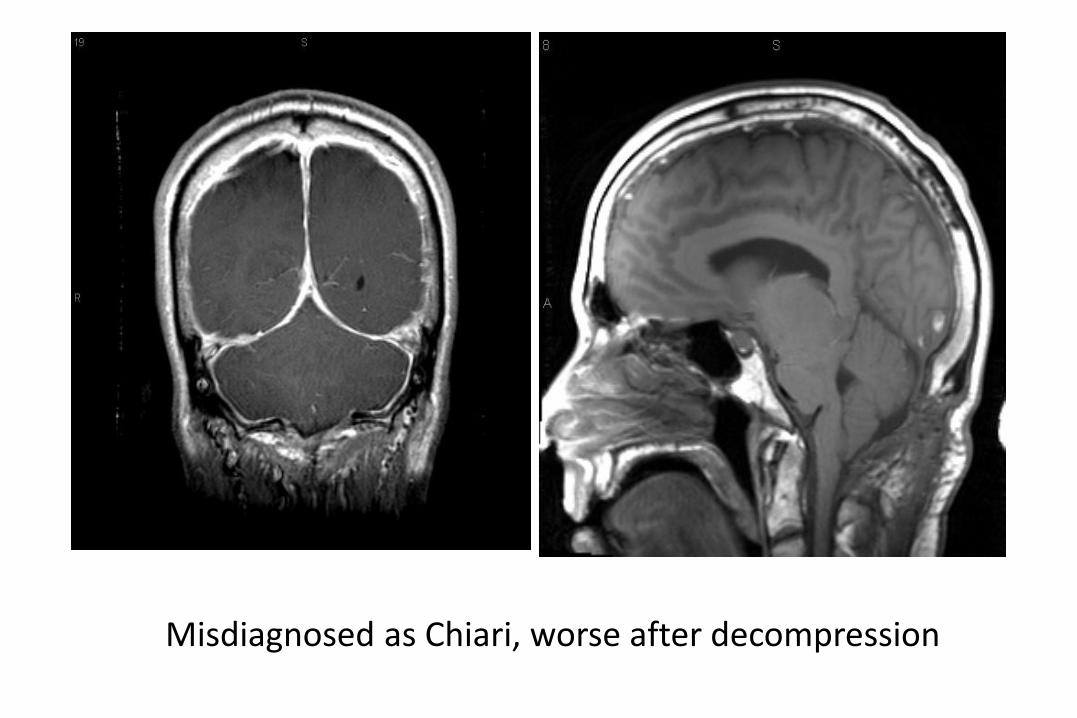
Misdiagnosed as Chiari, worse after decompression
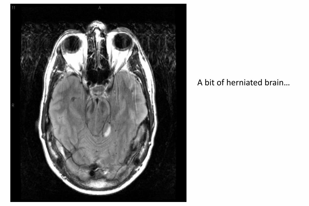
A bit of herniated brain…
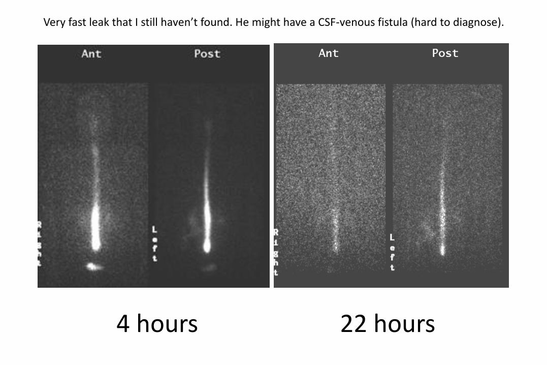
4 hours 22 hours
Very fast leak that I still haven’t found. He might have a CSF-venous fistula (hard to diagnose).

MRI findings in high CSF
pressure/volume states


White arrows: excess CSF in optic sheaths
Grey arrows: optic disc edema

• Flattened pituitary
• Or frank epmptysella
• Can have downward displacement of rounded cerebellar tonsils

Bilateral transverse sinus stenosis

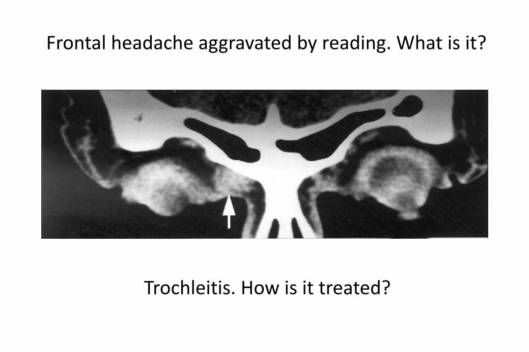
Frontal headache aggravated by reading. What is it?
Frontal headache aggravated by reading. What is it?
Trochleitis. How is it treated?
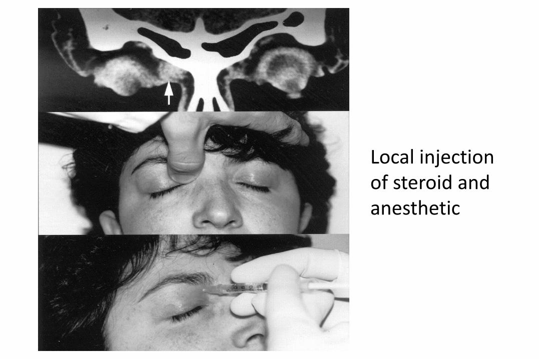
Local injection of steroid and anesthetic

Disorder Duration Frequency Gender
(F:M)
Acute
Treatment
Preventive/Bridge
Treatment
Cluster 15-180 min Every other
day to 8/day
1:3-7
(trending
towards
women)
Oxygen, SC
sumatriptan,
NS
sumatriptan
or
zolmitriptan
verapamil,
topiramate, lithium,
methylergonovine,
corticosteroids
PH 2-30 min 1-40/day 2-3:1
(trending
towards
men?)
None indomethacin
SUNCT/
SUNA
1-600 sec Dozens to
hundreds
per day
2:1
SUNA
1:2
SUNCT
None lamotrigine,
topirimate,
gabapentin,
indomethacin?
HC Constant
with spikes
Few to
many per
day
2:1 or
less
None indomethacin
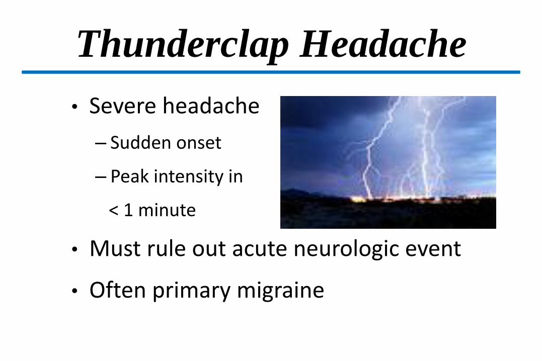
Thunderclap Headache
• Severe headache
– Sudden onset
– Peak intensity in
< 1 minute
• Must rule out acute neurologic event
• Often primary migraine

Thunderclap Headache
10 things that
cause thunderclap
headache?

Subarachnoid Hemorrhage
• Sudden and dramatic: “a blow on the head”
• Severe unilateral headache
– Becomes generalized
– Spreads to back of head
– May have backache
• Photophobia, neck stiffness, Kernig’s sign, focal
neurologic signs, and alterations in consciousness
• CT/LP usually diagnostic
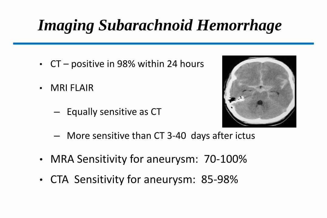
Imaging Subarachnoid Hemorrhage
• CT – positive in 98% within 24 hours
• MRI FLAIR
– Equally sensitive as CT
– More sensitive than CT 3-40 days after ictus
• MRA Sensitivity for aneurysm: 70-100%
• CTA Sensitivity for aneurysm: 85-98%
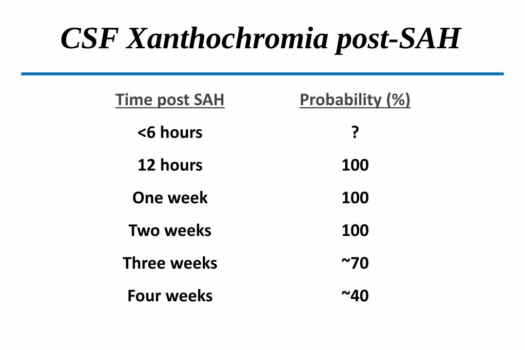
CSF Xanthochromia post-SAH
Time post SAH
<6 hours
12 hours
One week
Two weeks
Three weeks
Four weeks
Probability (%)
?
100
100
100
~70
~40

Thunderclap Headache
9 more things that
cause thunderclap
headache?
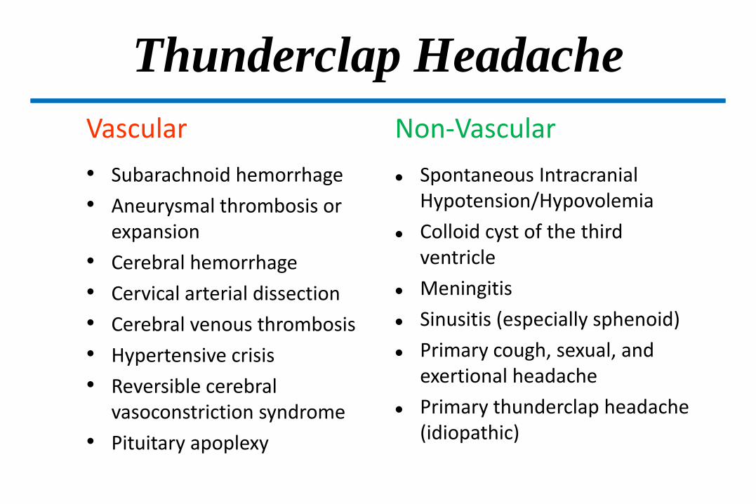
Thunderclap Headache
Vascular
• Subarachnoid hemorrhage
• Aneurysmal thrombosis or expansion
• Cerebral hemorrhage
• Cervical arterial dissection
• Cerebral venous thrombosis
• Hypertensive crisis
• Reversible cerebral vasoconstriction syndrome
• Pituitary apoplexy
Non-Vascular
Spontaneous Intracranial Hypotension/Hypovolemia
Colloid cyst of the third ventricle
Meningitis
Sinusitis (especially sphenoid)
Primary cough, sexual, and exertional headache
Primary thunderclap headache (idiopathic)

Some cerebrovascular causes of TCH
Cerebral hemorrhages: 5-10%
of all TCH (possible in infarcts)
Aneurysmal
warning leak
Dissection:TCH in 5%
Temporal
arteritis
Venous thrombosis:TCH in 3%
Pituitary
apoplexyRCVS
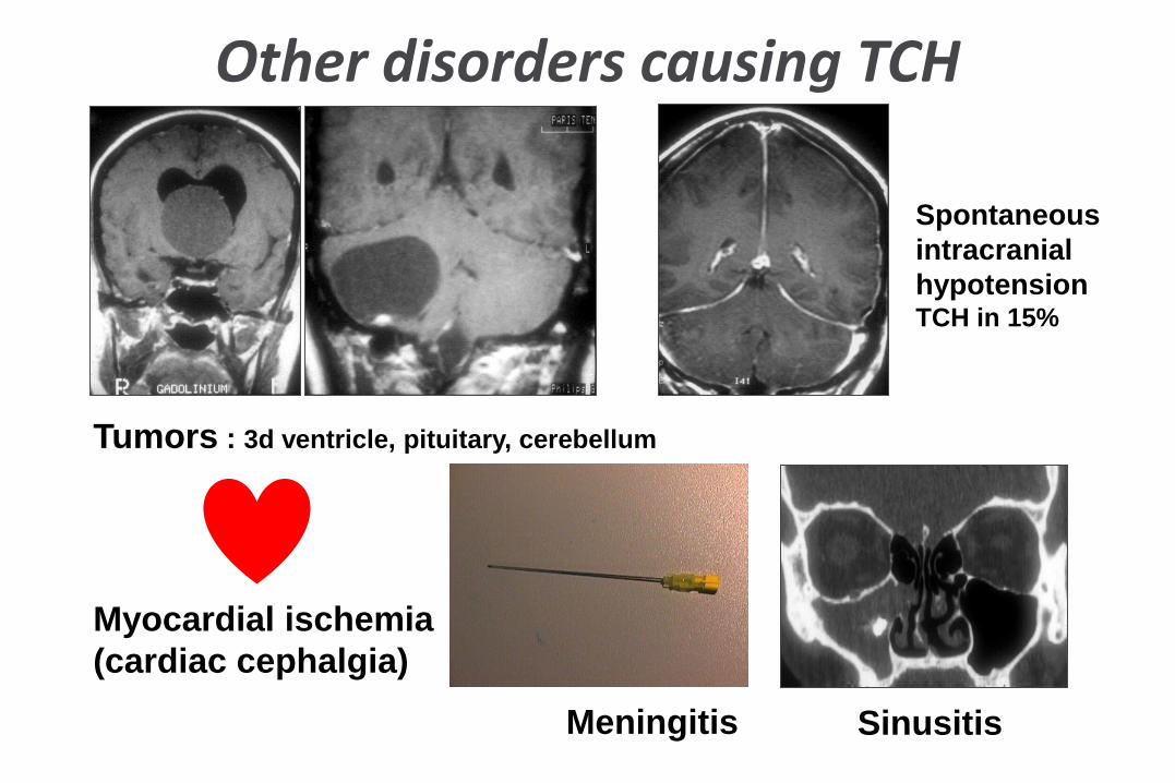
Other disorders causing TCH
Tumors : 3d ventricle, pituitary, cerebellum
Spontaneous
intracranial
hypotensionTCH in 15%
SinusitisMeningitis
Myocardial ischemia
(cardiac cephalgia)
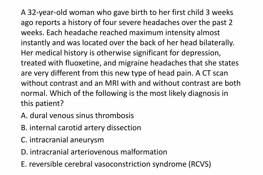
A 32-year-old woman who gave birth to her first child 3 weeks ago reports a history of four severe headaches over the past 2 weeks. Each headache reached maximum intensity almost instantly and was located over the back of her head bilaterally. Her medical history is otherwise significant for depression, treated with fluoxetine, and migraine headaches that she states are very different from this new type of head pain. A CT scan without contrast and an MRI with and without contrast are both normal. Which of the following is the most likely diagnosis in this patient?
A. dural venous sinus thrombosis
B. internal carotid artery dissection
C. intracranial aneurysm
D. intracranial arteriovenous malformation
E. reversible cerebral vasoconstriction syndrome (RCVS)
A. dural venous sinus thrombosis
B. internal carotid artery dissection
C. intracranial aneurysm
D. intracranial arteriovenous malformation
E. reversible cerebral vasoconstriction syndrome (RCVS)
The preferred response is E (reversible cerebral vasoconstriction syndrome [RCVS]). The recurrent nature of this patient’s thunderclap headaches together with her history of recently giving birth and a history of migraines would suggest the diagnosis of RCVS. Taking a serotonergic medication (fluoxetine) would also put her at risk for this condition. For more information, refer to page 1064 of the Continuum article ‘‘Thunderclap Headache.’’

Reversible Cerebral
Vasoconstriction Syndrome
Multifocal segmental stenosis (beading) of the major intracranial arteries
Calabrese, L.H., et al., Narrative review: Reversible cerebral
vasoconstriction syndromes. Annals of internal medicine, 2007
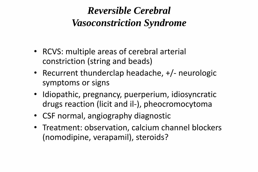
Reversible Cerebral
Vasoconstriction Syndrome
• RCVS: multiple areas of cerebral arterial constriction (string and beads)
• Recurrent thunderclap headache, +/- neurologic symptoms or signs
• Idiopathic, pregnancy, puerperium, idiosyncratic drugs reaction (licit and il-), pheocromocytoma
• CSF normal, angiography diagnostic
• Treatment: observation, calcium channel blockers (nomodipine, verapamil), steroids?
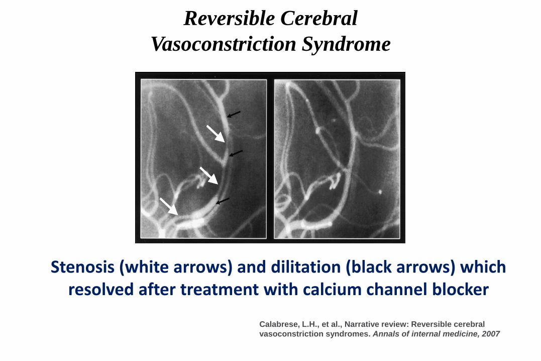
Reversible Cerebral
Vasoconstriction Syndrome
Stenosis (white arrows) and dilitation (black arrows) which resolved after treatment with calcium channel blocker
Calabrese, L.H., et al., Narrative review: Reversible cerebral
vasoconstriction syndromes. Annals of internal medicine, 2007
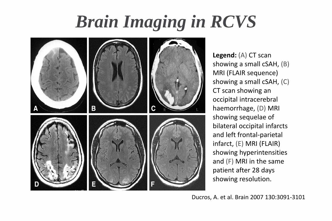
Brain Imaging in RCVS
Ducros, A. et al. Brain 2007 130:3091-3101
Legend: (A) CT scan showing a small cSAH, (B)MRI (FLAIR sequence) showing a small cSAH, (C)CT scan showing an occipital intracerebral haemorrhage, (D) MRI showing sequelae of bilateral occipital infarcts and left frontal-parietal infarct, (E) MRI (FLAIR) showing hyperintensities and (F) MRI in the same patient after 28 days showing resolution.

FSPGR MPR coronal reconstructed image (a) revealed the trigeminal nerve in its cisternal
tract, bilaterally (arrows); 3D-TOF MR angiography sequence MPR coronal reconstruction (b)
showed the contact between left superior cerebellar artery and the upper surface of the nerve
(arrowhead).FSPGR: fast spoiled gradient echo; MPR: multi-planar reconstruction; 3D-TOF
MR: three-dimensional time-of-flight magnetic resonance.
Valentina Favoni et al. Cephalalgia 2013;33:1337-1348Copyright © by International Headache Society

FISP MPR coronal reconstructed image (a) revealed the trigeminal nerve in its cisternal tract, bilaterally (arrows); 3D-TOF MR angiography sequence MPR coronal reconstruction (b) and MIP reconstruction (c) showed the contact between right superior cerebellar artery and the upper surface of the nerve (arrowhead).FISP: fast imaging with steady-state precession; MPR: multi-planar reconstruction; 3D-TOF MR: three-dimensional time-of-flight magnetic resonance; MIP: maximum intensity projection.
Valentina Favoni et al. Cephalalgia 2013;33:1337-1348Copyright © by International Headache Society

Questions?