uc san diego · pdf file1.28 radiation producing machines ... online regulatory guides . ......
TRANSCRIPT

Radiation Safety Manual
http://blink.ucsd.edu/menu/rad
UC San Diego ENVIRONMENT, HEALTH AND SAFETY

2
June, 1983
Revised, August, 1987
Revised, November, 1989
Revised, November, 1990
Revised, September, 1991
Revised, January, 1995
Revised, July, 1996
Revised, April, 1999
Revised, January, 2001
Revised, June, 2001
Revised, January, 2002
Revised, March, 2003
Revised, January, 2004
Revised, January, 2008
Revised, April 2010
Revised, April 2013
Revised, November 2013
Revised, June 2015
Revised August 2016

3
4
Introduction - Scope and Purpose of the Radiation Safety Manual ........................................ 6 Section 1: General Radiation Safety Requirements ............................................................... 7
1.0 UC San Diego Policy & Procedure Manual…………………………………...……7 1.2 Department Chairs’ Responsibilities ................................................................... 9 1.3 RSO Responsibilities .......................................................................................... 9 1.4 Notification of Regulatory Bodies ...................................................................... 10 1.5 Principal Investigators’ Responsibilities ............................................................ 11 1.6 Principal Investigator’s Responsibilities with Human Subjects ......................... 12 1.6A Dose Assessment and Consent Forms .......................................................... 12 1.6B Principal Investigator’s Responsibilities for Human Subjects, HERC ............. 17 1.6C Principal Investigators’ Responsibilities for Radioactive Drugs, RDRC ......... 18 1.7 Users’ Responsibilities ...................................................................................... 19 1.8 Radioisotope Use Authorization (RUA) ............................................................ 20 1.9 ALARA .............................................................................................................. 22 1.10 Pregnancy ....................................................................................................... 22 1.11 Audits .............................................................................................................. 23 1.12 Audit Violations ............................................................................................... 23 1.13 Ordering and Receipt ...................................................................................... 24 1.14 Security of Radioactive Materials ................................................................... 25 1.15 Posting and Labeling Work Areas .................................................................. 26 1.16 Surveys ........................................................................................................... 28 1.17 Inventory ......................................................................................................... 31 1.18 Waste .............................................................................................................. 31 1.19 Release of Potentially Contaminated Equipment ........................................... 39 1.20 Incidents ......................................................................................................... 39 1.21 Dose Limits ..................................................................................................... 41 1.22 Dosimetry Badges .......................................................................................... 42 1.23 Bioassay ......................................................................................................... 43 1.24 Sealed Source Leak Testing ........................................................................... 43 1.25 Calibrating Survey Instruments ....................................................................... 44 1.26 Maintenance on Potentially Contaminated Equipment ................................... 44 1.27 Decommissioning Radiologically Posted Facilities ......................................... 44 1.28 Radiation Producing Machines (non-medical) ................................................ 44 1.29 Irradiators ........................................................................................................ 52 2.1 Training Requirements for Research Users ..................................................... 53 2.2 Transfer or Shipping Packages of Radioactive Materials ................................. 53 2.3 Care and Handling of Animals Containing Radioactivity .................................. 54 2.4 Working with Volatile S-35 ................................................................................ 56 2.5 Working with P-32 ............................................................................................. 57 2.6 Iodinations ........................................................................................................ 58 2.7 Use of Radioisotopes on Vessels ..................................................................... 59
Section 3: Medical Use of Radiation .................................................................................... 61 3.1 Treatments Utilizing Radioactive Implants or Radiopharmaceuticals ............... 61 3.2 Inpatients Receiving Implants of Radioactive Sealed Sources ......................... 62 3.3 Inpatients Administered Radiopharmaceuticals ................................................ 63 3.4 Patients in the Operating Room ........................................................................ 63 3.5 Waste from Patients Treated with Radioactive Materials ................................. 64 3.6 Transportation of Patients ................................................................................. 64 3.7 Biopsies of Tissues Containing Radioactivity ................................................... 64 3.8 Radioactive Cadavers ....................................................................................... 64 3.9 Use of Radiation-Generating Devices .............................................................. 65

5
3.10 Use of Fluoroscopic Equipment ...................................................................... 65 3.11 Use of X-rays in Medical Diagnosis and Treatment ........................................ 68 3.12 Use of Radiation Therapy Machines ............................................................... 68 3.13 Training Requirements for Medical Personnel ................................................ 69 3.14 Medical Events ............................................................................................... 69 3.15 Fetal Radiation Exposure from Medical Procedures ...................................... 71 3.16 QA Program for Administered Radiopharmaceuticals .................................... 71 3.17 QA Program for Implants of Radioactive Sealed Sources .............................. 72 3.18 Audits of Nuclear Medicine and Radiation Oncology ...................................... 74 3.19 Minimizing Undue Radiation Dose to Patients ................................................ 74 3.20 Exposure of Individuals to Radiation .............................................................. 74
Section 4: Appendices .......................................................................................................... 75 4.1 Conversion Factors ........................................................................................... 75 4.2 Isotope Data ..................................................................................................... 76 4.3 Dose Rate Rules of Thumb .............................................................................. 77 4.4 Participation of Human Beings as Research Subjects ..................................... 77 4.5 Policy and Responsibilities of the RDRC .......................................................... 77
Section 5: Online Campus Forms
User Enrollment and Cancellation Forms Radioisotope Use Authorization Application Radioisotope Use Authorization Application (Sealed Source Only) Ancillary Personnel Researchers Using Radiation-Producing Machines Researchers Using Sealed Radioisotopes Researchers Using Unsealed Radioisotopes User Cancellation Form
Radioisotope Use Authorization Amendment Machine Use Registration Machine Registration Amendment Declaration of Pregnancy Application to the Radioactive Drug Research Committee (Human Use) Laboratory Survey Sheet Laboratory Meter Survey Record Transfer of Radioactive Material (Intracampus) Transfer of Radioactive Material (Off Campus) Iodination Log
Section 6: Online Medical Center Forms User Enrollment and Cancellation Forms Ancillary Personnel
Medical Workers Using Radiation-Producing Machines Nuclear Medicine Workers Radiation Oncology Workers User Cancellation Form
Declaration of Pregnancy Section 7: Online Regulatory Guides
Instruction Concerning Risks from Occupational Radiation Instruction Concerning Prenatal Radiation Exposure
6
Introduction - Scope and Purpose of the Radiation Safety Manual The Chancellor is responsible for the existence of a radiation safety program that provides for surveillance of departmental activities and radiological safety services. The radiation safety program ensures that all sources of ionizing radiation are handled in accordance with the official policies and procedures of the University and governmental agency requirements. The Chancellor is also responsible for the interpretation of University policies and the development of additional campus policies and procedures compatible with governmental regulations, licenses and national radiation protection standards. The Chancellor has delegated responsibility for the radiation safety program to the committees and individuals as stated below. UC San Diego uses radioactive materials under a Broad Scope Radioactive Material License issued by the State of California Department of Public Health and a reciprocity license from the Nuclear Regulatory Commission. UC San Diego also uses radiation producing machines under multiple Registered Facilities. This Manual represents the radiation safety program for all locations on those licenses and registrations including the Campus, UC San Diego Health System Hospitals (referred to as Medical Centers), Outpatient and Clinic locations, The Scripps Institution of Oceanography ( state facilities and ships in state, national and international waters), and the Sanford Consortium. The UC Health System – Nevada does not fall under the jurisdiction of the UC San Diego Radiation Safety Program. This Manual is incorporated by reference into UC San Diego’s license. All Principal Investigators and radioactive material users are required to comply with all of its provisions, as well as the conditions of the Radioisotope Use Authorization (RUA) and Machine Use Authorization (MUA).

7
Section 1: General Radiation Safety Requirements 1.0 UC San Diego Policy & Procedure Manual The Policy & Procedure Manual (PPM), along with the Academic and Staff Personnel Manuals, serves as the primary reference guide for campus operating policies and procedures which apply to academic, administrative, research, and service units. The policies connect the campuses mission to the everyday actions of its community, clarify the institutions expectations of its individual members, mitigate institutional risk, enhance efficiency, and support the university’s compliance with laws and regulations. UC San Diego’s Policy and Procedure Manual, PPM 516-22 gives the Radiation Safety Committee the responsibility for approving and monitoring the use of radioactive materials and radiation producing devices at UC San Diego facilities.
1.1 Committees Radiation Safety Committee The Radiation Safety Committee (RSC) advises the Chancellor of the university through the Vice Chancellor – Resource Management & Planning on all matters related to radiation safety and recommends such policies and procedures as it may deem appropriate to ensure an adequate radiation safety program.
The RSC consists of at least six members appointed by the Chancellor, and a Radiation Safety Officer (RSO) experienced in the use of radioisotopes and in protection against ionizing radiation. Activities of the Committee are directed by its Chair. The Chair of the RSC shall be full time UC San Diego faculty Member. The Chair shall convene the Committee as often as is necessary to consider all aspects pertinent to radiation safety.
A quorum shall consist of the Chair (or his/her designee), the RSO (or his/her alternate), the executive management (or his/her alternate), a representative from each area of use from which specific issues will be discussed, and any other member whose field of expertise is necessary for the discussion is considered acceptable. This requirements for the quorum are set by California Department of Public Health (CDPH) as part of its delegation of authority to UC San Diego to regulate its use of radioactive materials on campus (10 CFR 33.13). Committee business may be conducted via email ballot where members may vote to tentatively approve proposals, however, final approval voting is required at the next RSC meeting to formalize any tentative actions.
The RSC has the ultimate responsibility for the use of radioactive material at UC San Diego and shall be the ultimate reviewing and authorizing agent for the use of all ionizing radiation. It shall set policy to be carried out by the Radiation Safety Officer. It shall receive and review all pertinent reports and records of the Environment, Health and Safety (EH&S) Department, the subcommittee minutes, and shall keep and maintain a record of all its transactions and reports. The RSC shall consider the liabilities of the university in all activities involving radioisotopes.

8
Radioactive Drug Research Committee This Subcommittee provides authorization, surveillance, and oversight to the use of radioactive drugs in human subjects. The Committee is a subcommittee of, and reports to the Radiation Safety Committee, but members are vetted and approved annually by the Food and Drug Administration (FDA) according to Title 21 of the Code of Federal Regulations, Part 361. Chair of this Committee shall also serve as an ex officio member of the Radiation Safety Committee. This Committee serves on an ad hoc basis in response to active protocols that require FDA oversight.
A quorum consisting of more than 50 percent of the membership must be present with the appropriate representation of the required fields of specialization. Meetings are held as necessary to review and conduct the business of the Subcommittee, with at least one meeting per calendar quarter in which research activity has been authorized or conducted [21 CFR 316.1 (c) (2)]. Membership is approved by the Food and Drug Administration (FDA) following an annual submission process by Environment, Health and Safety staff that includes the FDA’s review of the applicant member’s qualifications as set forth on their website at: http://www.fda.gov/Drugs/ScienceResearch/ResearchAreas/Oncology/ucm196480.htm. Human Exposure Review Committee This Subcommittee reviews all uses of radioactive materials or radiation producing equipment which results in exposure to human subjects. The Committee is a subcommittee of, and reports to the Radiation Safety Committee. Chair of this committee also serves as an ex-officio member of the Radiation Safety Committee.
The Chair shall convene the Committee as often as is necessary to consider all aspects pertinent to radiation safety. A quorum shall consist of the chairperson of the committee (or his/her designee), the RSO, a representative from each area of use from which specific issues will be discussed, and any other member whose field of expertise is necessary for the discussion is considered acceptable. Committee business may be conducted via email ballot where members may vote to tentatively approve proposals.
Members are charged with assuring all such uses are within regulatory requirements, and that a net benefit results from the radiation exposure. Physiological studies which involve radioactive drug usage as defined in 21 CFR 361.1 are referred to the Radioactive Drug Research Subcommittee for approval.
Health System Radiation Safety Committee
9
The Health System Radiation Safety Committee (HSRSC) is a subcommittee of the RSC and the Environment of Care Committee whose purpose is to promote the safe practice in the handling and use of radiation sources within the area of medical usage. The HSRSC consists of physicians, managers, technologists or representatives of departments and divisions of the Medical Center, and the RSO. Activities of this subcommittee are directed by its Chair, who also serves on the RSC. The Chair, or Vice Chair, shall convene the subcommittee as often as is necessary to consider all aspects pertinent to radiation safety. A quorum shall consist of at least five members including the RSO or Alternate RSO. HSRSC members are selected by the Medical Center administration. 1.2 Department Chairs’ Responsibilities Department Chairs, or equivalent, are responsible for the review and approval of proposed uses of radioisotopes and ionizing radiation producing machines within their jurisdiction. Such approval signifies that the Department will provide the resources necessary to control hazards and will assist in the enforcement of pertinent University and governmental standards and regulations. 1.3 RSO Responsibilities The RSO directly supervises the Radiation Safety Program in technical and administrative issues. The RSO's duties and responsibilities are radiological safety, compliance with California and Department of Transportation (DOT), Nuclear Regulatory Commission (NRC) and International Air Transportation Association (IATA) regulations, and the conditions of the license. These duties and responsibilities include ensuring that:
Activities involving licensed material that the RSO considers unsafe are stopped. Radiation exposures are as low as reasonably achievable (ALARA). Up-to-date radiation protection procedures in the daily operation of the licensee's byproduct material program are developed, distributed, and implemented. Possession, use, and storage of licensed material are consistent with the limitations in the license, the regulations, the sealed source and device registration (SSDR) Certificate(s), and the manufacturer's recommendations and instructions. Individuals installing, relocating, maintaining, or repairing devices containing sealed sources are trained and authorized by an NRC or Agreement State license. Personnel training is conducted and is commensurate with the individual's duties regarding licensed material. Documentation is maintained to demonstrate that individuals are not likely to receive, in one year, a radiation dose in excess of 10% of the allowable limits or that personnel monitoring devices are provided.

10
When necessary, personnel monitoring devices are used and exchanged at the proper intervals, and records of the results of such monitoring are maintained. Licensed material is properly secured. Documentation is maintained to demonstrate, by measurement or calculation, that the total effective dose equivalent to the individual likely to receive the highest dose from the licensed operation does not exceed the annual limit for members of the public. Proper authorities are notified of incidents such as loss or theft of licensed material, damage to or malfunction of sealed sources, or fire. Medical Events are investigated and reported to the California Department of Public Health, Radiologic Health Branch in accordance with Title 17, California Code of Regulations, and Section 30295. Causes and appropriate corrective actions are identified, and timely corrective actions are taken. Audits of the radiation protection program are performed at least annually and documented. If violations of regulations or license conditions or program weaknesses are identified, effective corrective actions are developed, implemented, and documented. Licensed material is transported in accordance with all applicable DOT and IATA requirements. Licensed material is disposed of properly. Appropriate records are maintained. Up-to-date license is maintained and amendment and renewal requests are submitted in a timely manner.
Dose records and surveys are reviewed at least semiannually. ALARA practices are being followed. New users and uses of radioactive material are reviewed prior to first use.
1.4 Notification of Regulatory Bodies If an event exceeds the limits set forth in UC San Diego’s radioactive material license, 17 CCR 30295, 10 CFR 20 Subpart M or adopted sections of 10 CFR 35, the California Department of Public Health (CDPH) must be notified. In addition to the immediate notification, CDPH may require written reports of the incidents within 30 days. Such reports will be prepared by the RSO using information provided by the Principal Investigator (PI) and/or users.

11
1.5 Principal Investigators’ Responsibilities All Principal Investigators shall:
Attend the Radiation Safety Seminar. Complete and submit the Radioisotope Use Authorization and Researchers Using Unsealed/Sealed Radioisotopes Form. Ensure all users on their RUA/MUA receive appropriate training prior to any unsupervised work with radioactive material or radiation producing machines. Provide instruction and training to all users on their RUA/MUA in the practices and techniques required to ensure safety and to maintain radiation exposures ALARA. Supervise the safety performance of all users on their RUA/MUA to ensure that the required safety practices and techniques are employed. Correct or modify work procedures and conditions that may result in the release of radioactive materials and compromise the integrity of containment. Investigate and report in writing to the RSO any problems pertaining to the operation and implementation of safety procedures, equipment, or facilities. Notify the RSO immediately in case of a spill of radioactive material, if an unusual exposure of personnel to ionizing radiation occurs or if there has been a possible loss of radioactive material. Notify EH&S prior to the transfer of radioactive materials outside UC San Diego, as well as intra-campus transfers. Ensure that periodic surveys are performed and that an up-to-date record of surveys is maintained in the laboratory. Ensure that periodic inventory checks of radioactive material are completed and that an up-to-date inventory is maintained in the laboratory. Ensure survey meters in service are operable and that their calibration is current. Ensure stored radioactive materials are secure from unauthorized removal or access. Ensure control and constant surveillance are maintained over radioactive materials that are not in storage. Ensure all radioactive material/machine users in their laboratory are listed on their RUA/MUA. Maintain up-to-date RUA/MUA information by notifying EH&S of changes that affect the RUA/MUA, including but not limited to: locations, users, and inventory.

12
1.6 Principal Investigator’s Responsibilities with Human Subjects All studies that involve human subjects must be approved by UC San Diego’s Institutional Review Board called the Human Research Protection Program and either the Human Exposure Review Committee or the Radioactive Drug Research Committee prior to the start of the study. Human research is sub divided into two groups:
1. Radioactive drugs used to obtain basic research information and not intended for immediate therapeutic, diagnostic or similar purposes or to determine the safety and effectiveness of the drug in humans for such purposes (i.e., to carry out a clinical trial). These protocols are reviewed by the RDRC
2. Radiation producing machines procedures and radioactive drugs used for diagnosis or therapy or any Investigational New Drug or radioactive drug in a clinical trial. These protocols are reviewed by the HERC.
Examples of protocols that require approval by the HERC are:
1. CT or CAT scan 2. X-ray 3. Nuclear Medicine procedure 4. Fluoroscopy procedure 5. Dual energy X-ray Absorptiometry (DXA or DEXA) 6. Positron Emission Tomography (PET) 7. Clinical Trial 8. Investigational New Drug (INDs)
1.6A Dose Assessment and Consent Forms
The HRPP requires investigators who submit applications for protocols which involve exposure to ionizing radiation to inform study participants of the degree of risk involved due to radiation. The HRPP has rigorous requirements for what needs to be explained and how the explanations should be presented. The UC San Diego Radiation Risk Statement Calculator along with these notes will assist in the process of conforming to the required levels of explanation. Research protocols requiring exposure to radiation from imaging studies considered standard of care and would be done even if the participant were not taking part in a research protocol must be accounted for and exposure estimates and risks explained. When figuring radiation exposure no distinction is made between routine studies and studies done expressly for the research protocol. All exposure is dealt with cumulatively. Additionally, if the patient will be undergoing radiation therapy, the type of radiation therapy and the estimated radiation dose from the therapy should be included in both Section 14, Potential Risks of the Research Protocol, and the UC San Diego Consent to Act as a Research Subject.

13
The document titled “UC San Diego Human Research Protections Program New Biomedical Application Research Plan” serves as the official UC San Diego research protocol for a particular study. All explanations of exposure to radiation should appear in this document as well as in the UC San Diego Consent to act as a Research Subject. Please do not refer the HERC reviewer to the “Master” protocol for radiation exposure explanations. Risk explanations should appear in two places when submitting a protocol for radiation review:
1. Section 14, Potential Risks of the Research Protocol, should contain an explanation suitable for the professional person reviewing the study.
2. The “Risks and Discomforts” section of the “UC San Diego Consent to Act as a Research Subject”. The wording here should be suitable for the patient or guardian to understand.
The UC San Diego Radiation Risk Statement Calculator has its own directions and will guide you through the process of figuring out what studies are going to be performed and the amount of exposure each of those studies will contribute to a participant’s exposure. Once your data is entered into the calculator, you click on the, “Create Statement” button and the calculator will provide you with a total exposure for the mix of studies that were entered and a “Risk Statement” to explain that exposure. By appropriately cutting and pasting the risk statement into the correct sections of your protocol you will satisfy the radiation explanation requirements for the HERC Committee. Remember to submit the UC San Diego Radiation Risk Statement Calculator worksheet with your protocol so the HERC reviewer will know how you arrived at your stated exposure category and explanation of risk. At times arriving at a reasonable explanation of exposure will not be a straight forward exercise. For example, when a study may or may not be performed depending upon the clinical judgment of the investigator or attending physician. In these cases the best description of what may or may not happen will have to be made. It is better to estimate that more studies and exposure will be used than to estimate fewer studies will be used. The explanation given for the situation will have to be amended for each unique situation. It is suggested to cut and paste as much as possible from the risk statements generated by the UC San Diego Radiation Risk Statement Calculator and when a departure from the supplied wording must be made, to keep it simple and within the scope and spirit of the wording and comparisons customarily used at UC San Diego. The only reference we use is reference to background radiation which is given as 1.6 mSv. Please do not stray from the overall theme of the generated wording by adding such statements as comparing the amount of radiation to some percentage of background or a number of cross country plane flights. The examples below offer suggestions for some common variations which may not fit a normal situation in one way or another. Cutting and pasting from these examples as appropriate should satisfy UC San Diego explanation requirements. When necessary, original verbiage should also be used as long as it conforms to the general requirements as stated throughout this document. The “Risks and Discomforts” section of the “UC San Diego Consent to Act as a Research Subject” must contain a radiation risk statement.
14
In most cases following the directions on the UC San Diego Radiation Risk Statement Calculator will generate a risk statement with the total exposure stated in mSv. This may be cut and pasted into the consent as appropriate. For situations where the mix of tests and the number of times they may need to be repeated is not clear, such as when a test may or may not be performed based on clinician decision, you will have to use your judgment and amend the generated consent wording to accurately describe the situation. Please be as accurate as possible when listing the types of tests that may be performed based on clinical decision in conjunction with your study. Example (1): To explain and stay within the context of the UC San Diego consent statement, a study that will definitely require a chest x-ray and then may or may not require a mammogram, confirmatory chest x-rays, bone scans, or chest CT scans, can be amended to read as follows:
As a result of participating in this study, you will be exposed to radiation from scheduled x-rays and/or scans. The total exposure resulting from these imaging studies is calculated to be approximately 0.2 mSv. This amount is less than you would receive from one year of natural exposure in the San Diego area, which is approximately 1.6 mSv. If clinically indicated, other imaging studies may be required as deemed necessary by your doctor. Additional imaging studies may include a mammogram (0.33 mSv/scan), confirmatory chest x-rays (0.2 mSv/scan), bone scans (4.4 mSv/scan), or chest CT scans (5.4 mSv/scan) and will increase your exposure to radiation if they are required. Cumulative exposure from radiation may increase your risk of developing certain types of cancer in the future. The principal investigator for this research study has determined and verified that all/most/some of the x-rays and/or imaging scans prescribed for this study would typically be performed as part of the standard medical care required to adequately monitor your current illness. [Investigator may be specific here by listing the scans that are considered standard of care.] [In addition, non-radiation producing imaging alternatives should be included here if described in the research plan.] If you are especially concerned with radiation exposure or you have had a lot of x-rays or imaging scans already, you should discuss this with the principal investigator for this study, Dr. Principal Investigator, or with your regular doctor.
The radiation exposure values come directly from inputting 1 chest x-ray, 1 mammogram, 1 Tc-99m MDP bone scan and 1 chest CT into the UC San Diego Radiation Risk Statement Calculator. The risk statement was generated automatically by the UC San Diego Radiation Risk Statement Calculator. The italic was not generated by the UC San Diego Radiation Risk Statement Calculator, but is an acceptable suggestion as to how to explain the additional studies which may or may not be performed. Section 14, Potential Risks, Radiation Exposure, in the Research Protocol, must contain almost the same information concerning exposure to radiation as does the UC San Diego Consent to Act as a Research Subject. This section requires more detailed information and is speaking to the professional level reviewer. In most cases using the consent wording generated by the UC San Diego Radiation Risk Statement Calculator amended as necessary for this section should be appropriate. This section requires the researcher to describe the examinations to be used in the study, give

15
an estimate of how many times they may need to be repeated, and then state a total expected exposure in mSv. If a study is to run multiple years, and the imaging schedule changes from year to year, please account for the subsequent years in the explanation. Example (1 cont.): To explain within the context of section 14, Potential Risks, a study that that will definitely require a chest x-ray and then may or may not require a mammogram, chest x-rays, bone scans, or chest CT scans, can be presented to read as follows:
As a result of participating in this study, participants will be exposed to radiation from scheduled x-rays and/or imaging scans. The total exposure resulting from these imaging studies is calculated to be approximately 0.2 mSv. This exposure will come from a chest x-ray required for participation in this study. Additional imaging studies may be necessary as deemed appropriate by the investigator. Additional studies may include a mammogram, confirmatory chest x-rays, bone scans, or chest CT scans, resulting in the exposure increasing by 0.33 mSv/scan, 0.2 mSv/scan, 4.4 mSv/scan, and/or 5.4 mSv/scan, respectively.
The radiation exposure values comes directly from inputting 1 chest x-ray, 1 mammogram, 1 Tc-99m MDP bone scan and 1 chest CT into the UC San Diego Radiation Risk Statement Calculator. The italics were not generated by the UC San Diego Radiation Risk Statement Calculator, but represent revisions to the risk statement which explain the situation in acceptable terms. Note in the first sentence the word “you” was changed to “participants”. Example (2): This study requires a fluoroscopic procedure (Cardiac Catheterization). At nine months the procedure may need to be repeated. Section 13, Potential Risks Radiation Exposure
As a result of participating in this study, participants will be exposed to radiation from a scheduled cardiac angiogram. The total exposure resulting from this imaging study is calculated to be approximately 4.4mSv. There is a possibility of a second cardiac angiogram being performed at nine months. If this is done the exposure to radiation will be increased by an additional 4.4 mSv/scan...
The value 4.4 mSv come from inputting 1 cardiac catheterization into the UC San Diego Radiation Risk Statement Calculator. The italics were not generated by the UC San Diego Radiation Risk Statement Calculator but explain the situation in an acceptable manner. Note in the first sentence the word “you” was changed to “participants”. Note: The references given by the UC San Diego Radiation Risk Statement Calculator makes the assumption the procedure will use between 5.6 – 14.8 minutes of fluoroscopy time. If the procedure being performed as part of your protocol will fall outside of these given reference times you would be obligated to adjust the expected exposure in mSv to reflect the actual time for your fluoroscopic procedure. Example (2 cont.): To explain this situation in the UC San Diego Consent to Act as a Research Subject, what follow would be acceptable:
As a result of participating in this study, you will be exposed to radiation from a scheduled cardiac angiogram. The total exposure resulting from this imaging study is calculated to be approximately 4.4 mSv. There is a possibility a repeat angiogram will need to be performed at your nine month follow up visit, resulting in an additional 4.4 mSv. This amount is more than you would receive from one year of natural exposure in
16
the San Diego area, approximately 1.6 mSv. Cumulative exposure from radiation may increase your risk of developing certain types of cancer in the future. The principal investigator for this research study has determined and verified that all/most/some of the x-rays and/or imaging scans prescribed for this study would typically be performed as part of the standard medical care required to adequately monitor your current illness. [Investigator may be specific here by listing the scans that are considered standard of care.] [In addition, non-radiation producing imaging alternatives should be included here if described in the research plan.] If you are especially concerned with radiation exposure, or you have had a lot of x-rays or imaging scans already, you should discuss this with the principal investigator for this study, Dr. Principal Investigator, or with your regular doctor.
Except for the italic, the explanation of exposure and risk to radiation was generated by the UC San Diego Radiation Risk Statement Calculator. Example (3): This protocol calls for multiple imaging studies (PET/CT and chest/abdomen/pelvis CT) to be done. All patients will receive radiation therapy and will be treated with an experimental drug. Imaging studies occur at regularly scheduled times during the clinical trial: screening, post-radiation therapy, post-drug treatment, and during the follow-up phase, which lasts several years. Additionally, if clinically indicated, a head CT and/or bone scan may be ordered by the principal investigator. Some of the scans are considered standard of care. The following examples suggest how this situation could be handled: Section 13, Potential Risks Radiation Exposure:
Screening: 1 PET/CT (20.1 mSv) Post-Radiation Therapy: 1 Chest/abdomen/pelvis CT (13 mSv) Post-Treatment (24 weeks after start of treatment): 1 Chest/abdomen/pelvis CT (13 mSv) Follow-up: Every 8 weeks until the end of year 1 3 Chest/abdomen/pelvis CT (3 x 13 mSv = 39 mSv) Every 12 weeks in subsequent years 4 Chest/abdomen/pelvis CT (4 X 13 mSv = 52 mSv) If clinically indicated: Head CT (2.5 mSv/scan) Tc-99m MDP Bone Scan (4.4 mSv/scan) Year 1 total: 85.1 mSv Each subsequent year: 52 mSv MRIs may be performed if CT is contraindicated.

17
Radiation therapy: Total dose delivered is calculated to be XX Gy delivered in approximately XX fractions. Radiation therapy will be administered by {describe modality}.
The UC San Diego Radiation Risk Statement Calculator is not equipped to calculate the radiation exposure for radiation therapy... Therefore the researcher must explain the situation as accurately as possible by providing the estimated amount of exposure to radiation the patient will receive from undergoing therapy. Example (3 cont.): UC San Diego Consent to Act as a Research Subject: Risks of Imaging Scans
During your participation in this research study, you will be exposed to radiation from scheduled imaging scans. The total exposure resulting from these imaging studies is calculated to be approximately 85.1 mSv in the first year and 52 mSv each additional year. If clinically indicated, head CTs and/or bone scans may be performed, increasing your dose by 2.5 mSv per scan and 4.4 mSv per scan, respectively. This amount is more than you would receive from one year of natural exposure in the San Diego area, which is approximately 1.6 mSv. Cumulative exposure from radiation may increase you risk of developing certain types of cancer in the future. The principal investigator for this research study has determined and verified that some of the x-rays and/or imaging scans prescribed for this study would typically be performed as part of the standard medical care required to adequately monitor your current illness. Radiation exposure may be decreased of non-radiation imaging is used, such as MRI instead of CT. If you are especially concerned with radiation exposure, or you have had a lot of x-rays or imaging scans already, you should discuss this with the principal investigator for this study, Dr. Principal Investigator, or with your regular doctor. In addition, you will undergo radiation therapy. The total dose is calculated to be XX Gy delivered in approximately XX fractions.
This explanation follows what the UC San Diego Radiation Risk Statement Calculator would generate if a definite test menu were available to input, but has been modified to more clearly explain the imaging schedule, as shown in italics. Additionally, the risk explanation has been modified to include a statement regarding the radiation therapy the patient will be undergoing.
1.6B Principal Investigator’s Responsibilities for Human Subjects, HERC The investigator shall complete section four of the HRPP cover sheet and include:
1. Proper selection and inclusion of a radiographic procedure. 2. Description or name of nuclear medicine procedure if applicable.
18
3. Indication and Radioactive Use Authorization number of non-routine radioactive drugs if applicable.
The investigator shall ensure that the dose assessed for the protocol described and the description of risk described in the consent form is accurate and complete. The UC San Diego Radiation Risk Statement Calculator may be used to calculate the dose and provide language to describe the risk. 1.6C Principal Investigators’ Responsibilities for Radioactive Drugs, RDRC The investigator intending to use a radioactive drug on human subjects must complete the Application to the Radioactive Drug Research Committee (Human Use) and furnish the Radioactive Drug Research Committee the following information, in writing: Radiation dose to subjects To assure that the radiation dose to research subjects is as low as practicable to perform the study and meet the criteria of applicable regulations, the Radioactive Drug Research Committee shall require that:
The investigator provide absorbed dose calculations based on biologic distribution data available from published literature or from other valid studies,
The investigator provide for an acceptable method of radioassay of the radioactive drug prior to its use to assure that the dose calculations actually reflect the administered dose,
The radioactive drug chosen for the study has that combination of half-life, types of radiations, radiation energy, metabolism, chemical properties, etc., which results in the lowest dose to the whole body or specific organs with which it is possible to obtain the necessary information, and
The investigator utilizes adequate and appropriate instrumentation for the detection and measurement of the specific radionuclide.
Pharmacologic dosage
To determine that the amount of active ingredients to be administered does not exceed the limitations in the current FDA regulations, the committee shall require that the investigator provide pharmacological dose calculations based on data available from published literature or from other valid human studies.
Qualifications of investigators
Each investigator shall be qualified by training and experience to conduct the proposed research studies.
Human research subjects

19
Each investigator shall select appropriate human subjects and shall obtain the consent of such human beings or their representatives in accordance with FDA regulations. The research subjects shall be at least 18 years of age and legally competent. Exceptions are permitted only in those special situations when it can be demonstrated to the committee that the study presents a unique opportunity to gain information not presently available and requires the use of research subjects less than 18 years of age and is without significant risk to the subject. Studies involving minors shall be supported with review by qualified pediatric consultants to the Radioactive Drug Research Committee. Each female research subject of child bearing potential shall state in writing that she is not pregnant, or, on the basis of a pregnancy test, be confirmed as not pregnant before she may participate in any study.
Quality of radioactive drug
The radioactive drug used in the research study shall meet appropriate chemical, pharmaceutical, radiochemical, and radionuclide standards of identity, strength, quality, and purity as needed for safety and be of such uniform and reproducible quality as to give significance to the research study conducted. The Radioactive Drug Research Committee shall determine that radioactive materials for parenteral use are prepared in sterile and pyrogen-free form.
Research Protocol
No matter how small the amount of radioactivity, no study involving administration of a radioactive drug, as defined by FDA in this manual, to research subjects under this section, shall be permitted unless the Radioactive Drug Research Committee concludes, in its judgment, that scientific knowledge and benefit is likely to result from that study. Therefore, the protocol shall be based upon a sound rationale derived from appropriate animal studies or published literature and shall be of sound design such that information of scientific value may result. The radiation dose shall be both sufficient and no greater than necessary to obtain valid measurement. The projected number of subjects shall be sufficient but no greater than necessary for the purpose of the study. The number of subjects shall also reflect the fact that the study is intended to obtain basic research information and not intended for immediate therapeutic, diagnostic or similar purposes or to determine the safety and effectiveness of the drug in humans for such purposes (i.e., to carry out a clinical trial).
Adverse reactions
The investigator shall immediately report to the Radioactive Drug Research Committee all adverse effects associated with the use of the radioactive drug in the research study.
1.7 Users’ Responsibilities All radioactive material users are required to comply with the following good laboratory practices:
20
Attend UC San Diego radiation safety training as soon as possible, and prior to any use of radioactive materials that is not directly supervised by the PI or a trained user listed on the RUA. Complete the Researchers Using Unsealed/Sealed Radioisotopes enrollment form to be listed as an authorized user on the RUA. Wear protective clothing and impermeable gloves when working with unsealed sources. Use trays that will hold the contents in the event of spills or breakage of containers during storage or transport between laboratory areas and common hallways. Line trays and working surfaces with absorbent paper. Store liquid form of radioactive materials in sealed containers. Label all containers, storage and use areas. Store high-energy beta and all gamma emitters in properly shielded containers. Use remote handling tools when appropriate to minimize extremity exposures. Use assigned dosimetry to monitor exposure to radiation. Work with radioactive materials in accordance with radiation safety operating and emergency procedures. Monitor work areas, hands and clothing whenever there is a possibility of contamination and after each day of use. Clean up spills promptly in accordance with this manual. Do not eat drink, smoke, store food or apply cosmetics in areas where unsealed radioactive materials are used. Do not pipette by mouth. Avoid working in a radiologically controlled area if a nuclear medicine diagnostic or therapeutic procedure was recently performed on you. Contact EH&S at 858-534-6138 for advice.
1.8 Radioisotope Use Authorization (RUA) The PI will submit a Radioisotope Use Authorization Application form to EH&S after approval by the Department Chair, or equivalent. All personnel working under the proposed RUA must submit a Radioisotope Use Enrollment form and attend the Radiation Safety Seminar. Each RUA must have a Geiger counter and a liquid scintillation counter listed. EH&S will review and submit completed applications to the RSC for approval.

21
If a Radioisotope Use Authorization is submitted between RSC meetings, tentative approval can be done via email. The application will be formally reviewed and approved at the next RSC meeting. To be eligible for an RUA, the PI must be a Faculty member with independent research space and funding. The Faculty appointment may be in the Professorial or Research Scientist series. The space may be assigned by a University Space Committee or locally by the Department Chair, or equivalent. The funding may be extramural or University start-up funds. The PI must attend the UC San Diego Radiation Safety Seminar. The RSC will evaluate experience on a case by case basis. Certain EH&S managers are also granted RUAs for shipping, receiving and storing radioactive materials, calibrating survey instruments, performing training demonstrations and managing radioactive waste. RUAs will be classified as high use or low use depending on the Hazard Guide Value (HGV). RUAs with total HGVs less than 1000 are designated low use, while RUAs with total HGVs greater than or equal to 1000 are designated high use. The HGV is equal to Q x U x P x T, where:
Q = Quantity Factor, equal to the possession limit in mCi/year.
U = Use Factor, based on how much activity is used per experiment. < 1 mCi per experiment 0.1 > 1 but < 10 mCi per experiment 1 >10 mCi per experiment 10
P = Physical Form Factor, based on the physical form of material. solid 0.1 non-volatile liquid 1 volatile liquid, gas, or powder 10
T = Toxicity Factor, based on 10 CFR 20, Appendix C
App. C value > 100 0.1 App. C value < 100 but > 1 1 App. C value < 1 but > 0.01 10 App. C value < 0.01 100 In addition to the high use and low use RUAs, separate classifications are used for medical, waste management, sealed sources and at-sea activities. RUAS are billed as follows:

22
High Use $195/month Low Use $65/month Sealed Source $65/month The RUA may be amended subsequent to its initial approval. Minor changes are approved by the RSO. Amendments that result in changing the laboratory classification from low use to high use will be approved by the RSC. RUAs will be due for renewal every three years after initial approval. Renewals will be approved by the Department Chair (or equivalent), the RSC Chair, or Vice Chair, and the RSO. Part of the renewal process will be a refresher training session tailored to the RUA. Laboratory specific issues and audit results will be discussed with the PI and authorized users. This training session will be conducted as part of the weekly laboratory meeting when possible. An RUA may be extended for up to 3 months beyond the three-year renewal if due to extenuating circumstances the PI is not available, e.g., temporarily out of the country. The renewal will be processed at the first opportunity. An RUA may remain active during sabbatical leave of the PI as long as an acting PI is named in writing and approved by the RSO. An RUA will be deactivated upon request of the PI. The RUA number is archived and reserved for potential future use by the PI. Reactivation will be processed similar to an amendment. EH&S should be notified at least 30 days in advance of an RUA deactivation. A copy of the RUA should be available in the main radioactive materials use area. It can be in paper or electronic form. 1.9 ALARA The basic principles of time, distance and shielding shall be used as required to maintain doses from external sources of radiation ALARA (as low as reasonably achievable). Engineering controls such as filtration, ventilation and containment shall be used as required to maintain potential internal doses ALARA. UC San Diego encourages all users to review current practices and procedures, and to develop new procedures as appropriate to implement the ALARA concepts. EH&S will investigate all known instances of deviation from good ALARA practices. The Radiation Safety Program will be reviewed annually for adherence to ALARA concepts. Detailed information concerning risks from occupational radiation exposure is given in Regulatory Guide 8.29, included at the end of this Manual. 1.10 Pregnancy It is UC San Diego’s responsibility, as well as the responsibility of the user, to insure that the dose to the embryo/fetus from the occupational exposure of a declared pregnant worker not exceed 500 mrem during the entire pregnancy.
23
To declare a pregnancy, complete a Declaration of Pregnancy form, or equivalent, and submit it to the RSO (see section 5). The declaration of pregnancy is voluntary (i.e., the pregnant worker need not declare her pregnancy if she so chooses) and can be withdrawn at any time. If you have any questions or want further information, please call EH&S at 858-534-6418, or [email protected]. All information relating to the pregnancy is maintained confidential by EH&S. Once pregnancy has been declared, a review of the individual’s exposure history will be made. If it is determined likely that the embryo/fetus will receive in excess of 500 mrem during the entire gestation period, reassignment of work or restrictions may be necessary. If possible, laboratory workers who are pregnant or breast-feeding should not use millicurie quantities of volatile iodine, e.g., iodinating proteins. Declared pregnant workers may be assigned a dosimeter to be worn at waist level during their pregnancy to measure exposure to the embryo/fetus. More detailed information concerning pregnancy is given in Regulatory Guide 8.13, included at the end of this Manual.
1.11 Audits EH&S will periodically conduct audits of all activities and areas under the RUA. Audits of research laboratories will be scheduled initially on a quarterly or semiannual basis, depending on the use level of the RUA. This frequency may be increased or decreased based on the average violation points of the 3 most recent audits, as shown below. Audits of the RUAs at the Medical Center involved in the routine treatment and diagnosis of patients will be conducted quarterly. Audits of the EH&S waste management RUA will be conducted quarterly. The audit scoring system is based on accumulated violation points, with 1, 4, 16 or 32 points assigned to each issue, depending on the severity of the issue. Points are doubled if the same issue is found in consecutive audits. For example, a laboratory with one 4-point violation, one 4-point repeat violation, and two 1-point violations would receive a total score of 14 points. The goal for all laboratories is zero points.
Additional audits may be conducted at the discretion of the RSC, PI or RSO. 1.12 Audit Violations If an RUA fails an audit (violation points > 35), the PI will respond in writing with proposed or implemented corrective actions. There will be a follow-up audit scheduled within one month.
AverageScore High Use Labs Low Use Labs< 10 semi-annually annually
11 - 25 quarterly semi-annually26 - 35 monthly quarterly
Audit Frequency
24
The RSC will be informed of the failure at the next scheduled meeting. Every effort will be made by EH&S working with the PI to improve areas of non-compliance prior to the follow-up. Should an RUA receive a second failure within a three year period, the RSC will be informed immediately and the PI will receive a written notice from the RSC indicating that the RUA may be suspended. The PI will meet with the RSO to address all issues. Any PI receiving 2 consecutive failures, 3 failures within a 3-year period, or at the discretion of the RSC, will be required to appear before the RSC to prevent suspension of the RUA. The length of the suspension will be determined by the RSC. 1.13 Ordering and Receipt All orders must have a valid UC San Diego Purchase Order Number. Valid purchase order numbers are issued by the IFIS purchasing system. Only approved buyers with the appropriate IFIS training are authorized to place orders for radioactive materials. The PI's name and RUA should be clearly marked on the packing slip accompanying a shipment. Laboratory personnel who are listed on the RUA as authorized users or authorized by the Department to place orders, and authorized by the Purchasing office, are responsible for calling the vendor, placing the order, and entering the appropriate information into IFIS. For more information visit the Buying Radioactive Materials Overview webpage. Orders requested through Central Purchasing are initiated by submitting a purchase requisition to that office. These orders may be placed only by approved buyers in the Purchasing Office. Shipments received as free samples for which there will be no purchase order number are acceptable, provided that the packing slip clearly indicates that this is the case and the package is shipped to the Isotope Receiving Lab. Orders placed by other institutions for use by UC San Diego personnel are acceptable, provided that the purchase order number is valid for that institution. Blanket orders are orders for a predetermined quantity of a specific item for a predetermined time period. Standing orders are similar to blanket orders, but they have a designated repetitive delivery time. Each release of isotope under a blanket or standing order must reference the correct purchase order number. The individual order must not exceed the limits set on the purchase order or RUA. To order radioactive material, you must log on to http://marketplace.ucsd.edu. Search for the catalog number. Perkin Elmer and MP Biomedicals are approved vendors and have most isotopes available on marketplace. If the catalog number is not found you may have to submit an iRequest or miniRequest. Please see the Marketplace FAQ for more information on iRequests and miniRequests. If you found the material you are shopping for add it to the shopping cart. Process radioactive material orders separate from other supplies you are ordering. If you select “checkout” you will be brought to a screen asking to verify the various tabs. Verify your shipping address is for the EH&S Isotope Lab. On the Payment: Index(es) tab input your index #. On the UCID/FAB/WO & RUA tab in put your RUA #. Click on the Final Review tab for one last verification that all the information is correct then click Submit or

25
Assign Cart. For additional guidance on entering the correct shipping address in Marketplace please use the short walkthrough. Incoming radioisotopes shall be delivered to the EH&S Services Laboratory, University Center 301B, where they shall be monitored, checked for leakage and/or damage and entered into the inventory system. The user’s laboratory will be notified when the shipment is ready to be released. For free isotope delivery from EH&S to your lab, please see the Radioisotope Package Delivery Service webpage. For laboratories located on UC San Diego’s main campus, a person named as an authorized user on the RUA will pick-up the isotope. Those picking up radioactive materials should return directly to their laboratories and properly store the package. Radioactive materials should not be carried to any other locations on campus. Radioactive materials should not be carried on any campus shuttle bus, nor transported via personal motor vehicle. Although not required, those picking up radioactive shipments may bring clean, uncontaminated shielded secondary containers to carry the package. Food or drinks should not be brought to or from the EH&S Services Laboratory while carrying radioactive materials. For laboratories located off the main campus, shipments of radioactive materials will be delivered on the day after receipt. There is a two working day delay for packages that are delivered to the EH&S Services Laboratory on a Friday. Deliveries of isotopes to the UC San Diego Medical Center will be made directly to Nuclear Medicine hot labs at either the Hillcrest or Thornton locations. All such packages must be surveyed prior to use and records maintained for inspection. Any radioactive materials delivered directly to a user laboratory must be brought to the EH&S Services Laboratory for proper processing. The receipt of radioisotopes bound for visiting investigators on research vessels is acceptable, provided the requirements outlined in Section 2.7 are met. 1.14 Security of Radioactive Materials Federal regulations (10 CFR 20.1801) mandate that all radioactive material must be secured against unauthorized removal, meaning it must be within the direct sight of an authorized user or stored in a lab, refrigerator, locked container etc. An authorized user is someone on the RUA, a lab member who will challenge someone tampering with the material or an approved radio-pharmacy courier. Using a risk-based approach, UC San Diego has set security levels for isotopes requiring particular attention to security. Security levels for reporting losses of material to the State are set at low levels (see table below). These security level values are based on radiotoxicity of the isotope; the greater the radiotoxicity, the lower the reportable quantity. As radioactive shipments are received in the EH&S Services Lab, stock vials are now labeled if they exceed the security level.

26
Security levels for commonly used isotopes are:
------------------------------------------------------------------------ Radionuclide Quantity ([micro] Ci)
------------------------------------------------------------------------ Barium-133.................................................. 1,000 Barium-133m............................................... 1,000 Calcium-45.................................................. 1,000 Carbon-14................................................... 1,000 Cesium-137................................................... 100 Chromium-51............................................... 10,000 Copper-64.................................................... 10,000 Hydrogen-3.................................................. 10,000 Iodine-125....................................................... 10 Iodine-131....................................................... 10 Lead-210………………………..…………..….……..0.1 Manganese-54............................................. 1,000 Nickel-63...................................................... 1,000 Phosphorus-32.............................................. 100 Phosphorus-33............................................. 1,000 Polonium-210.................................................... 1 Strontium-90……………………….……......………….1 Sulfur-35....................................................... 1,000 Technetium-99m............................................. 10,000 Technetium-99............................................. 1,000 Uranium & Transuranics (excluding U-238)…..…0.01 Uranium-238............................................... . 1,000 All Others……………………………….…… ……..100
1.15 Posting and Labeling Work Areas Radiologically Controlled Areas Refrigerators, freezers and entrances to areas where radioactive materials are used or stored shall be posted with a Caution Radioactive Materials sign. Individual containers shall be labeled with a Caution Radioactive Materials tape or labels. Small containers, Eppendorf tubes, etc. containing less than 1 microcurie of activity need not be individually labeled as long as they are stored in a labeled secondary container or rack. Clean Areas When required by space considerations, areas within the laboratory containing desks, writing tables or food refrigerators may be designated as Clean Areas with the following stipulations:
The area must be approved by EH&S and posted as a Clean Area. The area must be physically separated from any laboratory work area by at least one meter, or by a substantial physical barrier such as a solid bookcase or an acrylic shield.
27
Laboratory personnel must remove gloves and wash hands after working with radioisotopes, and prior to handling any papers or working in a Clean Area. A waste receptacle must be provided within the Clean Area and used only for non-laboratory trash. Surveys within and at the boundary of Clean Areas shall be included as part of the periodic laboratory surveys. Eating, drinking, applying cosmetics and food storage are allowed in Clean Areas.

28
1.16 Surveys Periodic surveys for contamination are required in areas where unsealed sources are used. For direct (meter) surveys, the probe appropriate for the isotope should be used. The table below lists the efficiencies for some common isotopes and probes.
Isotope
Specific Probe Efficiencies @ 1 cm
Ludlum 44-9 GM (Pancake)
Alpha, Beta, Gamma
Ludlum 44-3 Thin Nal Most Efficient
Between 10 to 60 KeV photons
Ludlum 44-2 Thick Nal Most Efficient Between 60 to 125 KeV Photons
C-14 3% 0% 0% Ca-45 9% 0% 0% Cl-36 20% 0% 0% Co-60 9% 0% 1% Cr-51 3% 0% 3%
Cs-137 7% 0% 4% Cu-64 6% 0% 0% Cu-67 9% 0% 3% F-18 9% 0% 1%
Fe-55 0% 0% 0% Fe-59 11% 0% 1% Ga-67 0% 0% 3%
H-3 0% 0% 0% I-125 0% 5% 0% I-131 9% 0% 2% Na-22 11% 0% 14% Ni-63 0% 0% 0% P-32 13% 0% 0% P-33 4% 0% 0% S-35 3% 0% 0%
Se-75 1% 0% 17% Sm-153 9% 0% 3% Sr-85 0% 0% 1% Sr-90 13% 0% 0% Tc-99 4% 0% 0%
Tc-99m 1% 0% 3% Tl-201 0% 0% 1% Y-90 13% 0% 0%
29
To convert from counts per minute (cpm) to disintegrations per minute (dpm), divide by the efficiency. For example, 100 cpm of C-14 as measured with a pancake probe is 3,333 dpm. For wipe tests, an area of 16 square (sq) inches (100 sq cm) should be wiped. Filter paper or cloth wipes, wet or dry, may be used. The sample is analyzed using an LSC or gamma counter using the protocol appropriate for the isotopes in use. For H-3, only wipe testing is required. Documented surveys will be performed with a frequency based on the laboratory classification and activity in use. The term use means the amount of isotope listed on the RUA in the mCi/experiment column. It refers to the amount of activity initially withdrawn from a stock vial, not the amount of activity in the stock vial. For all isotopes except H-3, direct (meter) surveys will be performed. Wipe tests for these isotopes are only required in areas of locally high background, e.g., near or on the outside of a waste container, or in areas where direct meter monitoring is impractical, e.g., inside a microfuge. Wipe tests may be substituted for meter surveys when very small amounts of activity are used, i.e., less than 1 microcurie. Laboratory use Documented Survey Frequency
Low Monthly High Weekly >10 mCi/experiment Daily (or after each use)
Routine checks (before, during or after use) for personnel contamination and suspected facility contamination by individual users need not be documented, unless contamination is found. In these cases, documentation of the decontamination and subsequent survey is required. Adequate survey documentation includes the date of the survey, the name or initials of the person performing the survey, the background level in cpm or dpm, a map, sketch or description of the areas surveyed, and an indication of the results. Surveys should be taken in storage areas, work areas and clean areas (if any). Results may be noted as not statistically different from background if less than 3 times background or in dpm if the efficiencies are known. If contamination is found, a record of the survey taken after decontamination is also required. These records must be kept in the laboratory for a period of 3 years for State inspection. The Laboratory Survey form or any equivalent form may be used. The LSC print out may be attached to the form rather than transcribing the results. The Laboratory Survey Record form, or equivalent, may be used in conjunction with a map of sketch of the laboratory showing standard survey points. Any laboratory specific forms that include the above items are also acceptable. If isotopes are in storage, but not used, a survey of the storage area is required to detect the potential inadvertent spread of contamination from the stored container. If there is no reasonable chance to inadvertently spread contamination, e.g., the laboratory is locked and unoccupied, the stored isotopes are still in their original unopened shipping container, or the stored isotopes are kept in a separate sealed or locked secondary container, then no surveys are required. If the laboratory has no isotopes in inventory, surveys are not required. The survey documentation should indicate that surveys were not performed for

30
the time period that isotopes were securely stored or not in inventory; a single entry at the beginning and end of the time period is sufficient. Common areas, shared by more than one RUA, should be surveyed as follows: Area Use Documented Survey Frequency Equipment (no open containers; LSC waste only) Monthly Waste storage Weekly Routine laboratory (open containers) Weekly >10 mCi/experiment After each use The PIs sharing the common areas should agree among themselves on the survey responsibilities. The monthly or weekly surveys might be performed by the RUA using the area the most often, or on a rotating basis. For example, if RUA A, B and C all share one room, RUA A could be responsible for the surveys in January, April, July, and October, etc. Iodination rooms shall have a documented survey performed after each use. Surveys at the Environmental Management Facility shall be performed on a weekly basis. As a first approximation, an area is considered to be contaminated if the survey indicates activity statistically above background. If the efficiency is known, areas exceeding these values (in dpm/100 sq cm) are considered to be contaminated:
Contamination may be fixed (non-removable) or loose (removable). Total contamination is fixed plus loose. Wipe tests detect loose contamination, while meter surveys detect total contamination. Loose contamination can be removed by ordinary cleaning methods. Fixed contamination may be shielded for decay or removed by extraordinary methods, e.g., removing floor tiles. Contact EH&S at 858-534-6138 for advice on dealing with fixed contamination or for isotopes not listed above. It is acceptable to have detectable contamination within equipment that is difficult to decontaminate or that routinely gets contaminated, such as inside a microfuge. The equipment must be labeled with a Caution Radioactive Materials tape and internal
Loose Total
U-nat, U-235, U-238, and associated decay products 1000 5000
Transuranics, Ac-227, I-129, Pa-231, Ra-226, Ra-228, Th-228, Th-230 20 100
I-125, I-126, I-131, I-133, Ra-223, Ra-224, Sr-90, Th-nat, Th-232, U-232 200 1000
Beta-gamma emitters except others listed above (C-14, Ca-45, Cr-51, Mn-54, P-32, P-33, S-35, Mn-54)
1000 5000
Action Level (dpm per 100 cm2)Nuclide

31
contamination. This equipment must be decontaminated when levels exceed 10 times the dpm/100sq cm levels shown above. Shielding shall be used to reduce exposure rates to be ALARA, and less than 2 mR/hr at 30 cm (approximately 1 foot) from the source. Use plastic shielding for beta emitters and lead shielding for photon emitters. When shielding multiple millicuries of high-energy beta emitters, e.g., P-32, it is useful to add a thin sheet of lead shielding to absorb the low energy x-rays produced in the plastic shielding. See Section 4.2 for the shielding requirements for common isotopes. As a very rough approximation, use the following guidelines for 2 mr/hr. These probes are designed to measure contamination, and are neither designed nor calibrated to measure exposure rate. Contact EH&S if you need precise measurements taken of potential exposure rates over 2 mR/hr. Probe Example probe cpm per 2 mr/hr Pancake Ludlum 44-9 6,600 Thin NaI Ludlum 44-3 675,000 Thick NaI Ludlum 44-2 175,000 End Window GM Ludlum 44-7 4,200 1.17 Inventory Each stock vial of radioactive material received will have an inventory number and barcode assigned to it. The individual user will receive an inventory sheet with each stock vial. This sheet must be maintained either in a notebook, binder, or posted on or near the storage area for the vial while the vial is located in the laboratory. The user should keep a record of aliquots in millicuries taken from the stock vial. The laboratory must maintain a record of current stock vial inventory in units of millicuries. Upon completion of use of the stock vial, i.e., when the vial is empty, the inventory sheet may be returned to EH&S to update the electronic inventory records. Alternatively, the inventory sheets may be kept in the laboratory. Each PI will receive a quarterly printout of their radioactive materials inventory. The PI, or designee, shall perform a physical inventory to verify that the printout is current and return the updated inventory to EH&S. If the quarterly inventory update is not returned in a timely manner, EH&S may hold new isotope orders until it is returned. If a PI plans long-term storage (> 1 year) of significant amounts (> 1 mCi) of radioactive samples (not stock vials), EH&S should be contacted so that these materials may be added to the inventory. EH&S will inform the PI when their inventory exceeds 80% of their possession limit so that the inventory may be evaluated. 1.18 Waste

32
Radioactive waste is processed at the Environmental Management Facility. All radioactive waste materials received are segregated according to isotope and form (dry or liquid). A solid waste compaction unit is used to compact solid radioactive waste. Glass scintillation vials are crushed; plastic vials are shredded. Waste is decayed for at least ten half-lives, or until indistinguishable from background. It can then be disposed of as non-radioactive waste or retained at the facility for further processing. Segregation of radioactive wastes by the generator is an integral part of operating a safe and cost efficient waste handling program. Radioactive labels or markings must be removed or defaced if uncontaminated labeled items are disposed of in the regular trash. Safely Accumulating and Storing Radioactive Waste Only those listed as authorized users on an RUA are allowed to prepare radioactive waste for collection. To aid in the processing of radioactive waste, all isotope users should follow these general guidelines:
Radioactive waste must be transferred to EH&S for disposal. Do not dispose of radioactive waste into regular trashcans or by pouring it down drains. Designate a specific location for the storage and collection of radioactive waste. Post the area and label the collection container with a Caution Radioactive Materials sign. Containers should be easily distinguished from non-radioactive waste containers. Label waste containers with Caution Radioactive Materials tape and the isotope prior to use. Attach a completed Radioactive Waste Tag prior to pick-up. Shielding shall be used to reduce exposure rates to be ALARA, and less than 2 mR/hr at 30 cm (approximately 1 foot) from the source. Use plastic shielding for beta emitters and lead shielding for photon emitters. When shielding multiple millicuries of high-energy beta emitters, e.g., P-32, it is useful to add a thin sheet of lead shielding to absorb the low energy x-rays produced in the plastic shielding. Separate waste by isotope and physical form. Monitor waste items to prevent the introduction of non-contaminated items into waste containers. Use biodegradable scintillation cocktails when possible (available from the UC San Diego Storehouse).
Containers with contaminated exteriors will be refused by EH&S. Keep waste pickup areas clear of debris and loose or open containers. Minimize quantities and storage demand by using smaller animals and short-lived isotopes whenever possible. Contact EH&S for specific instructions on disposal of gels, high specific activity liquid wastes, or any other special wastes. If you have questions, call the EH&S Environmental Management Facility at 858-534-2753 before generating any waste.

33
Uranium and thorium compounds, such as nitrates and acetates, are radioactive. They should be contained separately from other radioactive wastes.
Filling out the Radioactive Waste Tag A UC San Diego Radioactive Waste Tag must be securely attached to each radioactive waste container storing waste. Tags must be completed before EH&S will accept containers. All of the following information must appear on the tag: Refer to the Online Waste Tag Program (OTP) to ensure tag is filled out completely.
Waste Generator Number Generator contact's name and phone number. Building and room number where the waste is stored for collection. List each isotope and its total activity in millicuries separately. Give a full description of hazardous constituents other than radionuclides (chemicals, biohazards and infectious agents). Use brand names for scintillation cocktails. Liquid waste must have 100% of its composition listed. Acceptable abbreviations are given in Appendix 4.6. List the method used to disinfect biohazardous components. Contact the Biosafety Officer (BSO) at 858-534-6059 for appropriate methods.
Dry Radioactive Waste Dry waste includes paper, gloves, empty vials, disposable glassware, microcentrifuge tubes, and other contaminated material. Dry waste bags should not contain stock vials, lead, needles, razor blades or any liquid. Lead is a toxic metal and is considered a hazardous chemical waste. Lead contaminated with radioactive isotope is a mixed waste and should not to be included in dry waste (see Mixed Waste below).
Package your dry waste separately by isotope (H-3 and C-14 may be combined). Contain the waste in a clear plastic bag. When the plastic bags are full, seal and remove it from the work area. Place a second clear plastic bag around the waste and seal. Make sure the exterior is free of contamination. Attach a completed Radioactive Waste Tag to the waste bag. Store in the radioactive waste storage area of the lab. To request EH&S waste pickup see the How to Contact EH&S for Collection section below.
34
Liquid Radioactive Waste Liquid radioactive waste includes radioactive solutions and any subsequent rinses of each container or process that contain detectable activity (not statistically different than background). A liquid scintillation counter, gamma counter or equivalent instrument should be used to verify that a solution does not contain detectable activity; use of a portable survey meter is not appropriate. If you plan to perform research that could result in the generation of liquid waste containing more than 50 mCi, contact the EH&S Environmental Management Facility 858-534-2753 for proper handling guidelines prior to beginning the project. EH&S provides containers for liquid radioactive wastes. The standard containers are 5 gallon (large) and 2 gallon (small) Nalgene carboys. Any other containers must be specifically approved by EH&S. Carboys are replaced by EH&S at time of collection. To acquire these containers, contact EMF or send in a request as you would with hazardous waste. Carboys are for liquids only; solids are not acceptable in carboys, i.e., no pipettes, plastic pipette tips, microcentrifuge tubes, electrophoresis gels, or organic matter. Solids and gels damage equipment utilized in the waste management process.
Do not pour liquid radioactive wastes down the laboratory sink drains. Keep different isotopes, aqueous liquids, and non-aqueous liquids in separate containers (H-3 and C-14 may be combined). Minimize the generation of mixed waste. Example: Don't mix a P-32 buffer solution with a P-32 acetonitrile solution. Use a waste container provided by EH&S. Containers must be kept in a secondary container capable of holding the entire contents. Do not overfill carboys. Leave at least four inches of space at the top to prevent the caps from leaking during transportation and keep exterior of each container free of contamination. Complete the Radioactive Waste Tag. Liquid waste must have 100% of its composition listed. Store in the radioactive waste storage area of the lab. To request EH&S waste pickup see the How to Contact EH&S for Collection section below.
Liquid Scintillation Vials The use of biodegradable scintillation cocktails is recommended when possible.

35
Separate vials by isotope; H-3/C-14 may be combined. Separate glass vials from plastic vials. Ensure the caps are securely in place. Place vials in cardboard crate or double clear plastic bags. Complete a Radioactive Waste Tag and attach to waste. Specify the brand name of scintillation cocktail used. Vials from wipe tests or dual-labeled samples should list all suspected isotopes. Store in the radioactive waste storage area of the lab. To request EH&S waste pickup see the How to Contact EH&S for Collection section below.
Mixed Biohazardous/Radioactive Waste Biohazardous Materials are hazardous biological materials or organisms, and include: a) infectious organisms (bacteria, fungi, parasites, prions, rickettsias, viruses, etc.) which can cause disease in healthy humans and/or significant environmental or agricultural impact; b) human or primate tissues, fluids, cells, or cell culture; c) recombinant DNA; and d) animals known to be vectors of zoonotic diseases. Contact EH&S at 534-5366 prior to commencing any new protocol that may produce a mixed Biohazardous / Radioactive waste stream. EH&S will require specific procedures (including use of proper disinfectant) for any mixed waste. These will be determined on a case-by-case basis.
Whenever possible, disinfect liquid waste at the source of generation using a disinfectant compatible with the radioactive material. Avoid disinfecting radioiodine labeled infectious agents with compounds containing chlorine. Keep mixed waste separate from other waste (i.e., other biological or chemical waste). Package waste per designated waste stream (liquid, dry, etc.). If dry waste contains liquid residue or cultures, add absorbent material such as Red-Z or equivalent absorbent disinfecting material to bag to prevent liquid from accumulating at the bottom. Additional information about Red-Z can be found at: https://shop.life-assist.com/CatalogItemDetail.aspx?IGRN=659 Place the primary red biohazard bag into a secondary red biohazard bag and seal. Ensure that the exterior of the secondary bag is wiped down with disinfectant. Complete and attach a Radioactive Waste tag. On the lower section of the tag, indicate that the bag contains mixed biohazard/radioactive waste. List the biohazard, the isotope and disinfectant used (e.g., Salmonella and Tc-99m waste disinfected with 5% Wescodyne).
36
If the material contained is putrescible or is capable of producing odors of decay, place in freezer until EH&S is contacted for pick-up and disposal. To request EH&S waste pickup see the How to Contact EH&S for Collection section below.
Mixed Radioactive/Chemical Waste Whenever possible, hazardous chemicals and radioisotopes should not be used in combination. Contact EH&S for recommended nonhazardous chemical substitutes and ideas for minimizing generation of mixed wastes.
If the chemical waste is compatible with the containment described for radioactive waste, the container used for radioactive waste should be used. If not, select a similar containment compatible with the chemical. Label the waste with a Radioactive Waste Tag. Fill out the chemical composition portion of the tag, listing all chemicals and their percentages. Store the container in the radioactive waste storage area of the lab. To request EH&S waste pickup see the How to Contact EH&S for Collection section below.
Radioactive Animal Waste Animal waste may consist of carcasses, material left over from necropsies, including bedding and/or excreta contaminated with radioactive isotopes.
Place animal carcass and related necropsy waste (bench coat, Kimwipes, gauze, gloves) in a primary clear plastic bag. Place a sufficient amount of absorbent material such as Red-Z or equivalent absorbent disinfecting material in the bag to prevent liquid from accumulating at the bottom as it separates from the solids. Additional information about Red-Z can be found at: https://shop.life-assist.com/CatalogItemDetail.aspx?IGRN=659 Place the primary clear bag into a secondary clear plastic bag and seal. Ensure that the exterior of the secondary bag is wiped down with disinfectant.
Complete and attach a Radioactive Waste tag. On the lower portion of the tag, clearly indicate biological hazard, isotope and disinfectant used (i.e., Salmonella and Tc-99m infected mice disinfected with 5% Wescodyne).
Place bags in freezer until EH&S is contacted for pick-up and disposal.
Call the EH&S Environmental Management Facility at 4-2753 with any questions.
To request EH&S waste pickup see the How to Contact EH&S for Collection section below.

37
Radioactive Gels EH&S provides five-gallon bucket containers for the collection of agarose gels used for electrophoresis. Other small leak-proof containers are acceptable, but these containers will not be returned to the lab.
Line the container with two clear plastic bags. Complete and attach a Radioactive Waste Tag to the container, specifying the type of gel used and any other hazardous constituents. Store only gel waste in the container. When the container is 3/4 full, seal the plastic bags. Complete the Radioactive Waste Tag (estimate activity in mCi) and place the container in the radioactive waste storage area of the lab. To request EH&S waste pickup see the How to Contact EH&S for Collection section below.
Radioactive Sharps Sharps are any object or device having acute rigid corners, edges or protuberances capable of cutting or piercing, including: glass pipettes (small and large), plastic pipettes, pipette tips, hypodermic needles, blades, slides, scalpels and broken glass. Needles and syringes shall not be clipped, bent, broken, sheared or recapped prior to disposal. Sharps must be disposed of in the red biohazard containers that are leak-proof, rigid, puncture-resistant and "tamper-proof" (made so that they cannot be reopened without great difficulty). These containers may be purchased at the campus storehouse.
Sharps and piercing objects contaminated with both chemicals and radioactivity, or both radioactivity and infectious agents, are handled separately from those contaminated with radioactivity only. Since they will create both a physical hazard and contamination problems if placed in regular waste bags, sharps and piercing objects must be put in a specially designed rigid, polyethylene container. Sharps containers can be purchased from the Storehouse in a variety of sizes. New sharps containers will have a BIOHAZARDOUS MATERIALS sticker on them. If the biohazard symbol does not apply, remove or deface it. Label the container radioactive waste only, identify the isotope and place a UC San Diego Radioactive Waste Tag on the container. Plastic sharps contaminated with radioactivity only may be stored in double plastic bags within a strong cardboard box.
38
EH&S will collect biohazardous waste, or any materials bearing the international biohazard symbol, only if it is cross contaminated with radioisotopes and/or chemicals. Sharps containers must not contain free liquids (such as full syringes). When the container is full, seal and remove it from the collection area. Place the sharps container in a clear plastic bag and seal it. Make sure the exterior of the bag is not contaminated. Complete and attach a Radioactive Waste Tag to the bag. Mark the waste type as other and write SHARPS on the tag. List any additional chemical or biological hazards and the method of deactivation/disinfection used. Store the bag in the radioactive waste storage area of the lab. To request EH&S waste pickup see the How to Contact EH&S for Collection section below.
How to Contact EH&S for Collection Online Waste Tag Program
The Online Waste Tag Program (OTP) is the preferred way to request waste pickup. The OTP will let you notify EH&S when you need a waste pick up and also display a history of your collected wastes.
Request a copy of the Electronic Mail Request Form by clicking [email protected] and typing "Waste Form" into the subject line. Send the e-mail. A copy of the request form will be e-mailed back to you. When you receive the form, "save as" to a convenient folder so you can re-use it for each collection request. Complete and e-mail the form to [email protected] to request collection.
FAX or Campus Mail
Request a copy of the Electronic Mail Request Form via [email protected] and typing "Waste Form" into the subject line. Send the e-mail. A copy of the request form will be e-mailed back to you. When you receive the form, "save as" to a convenient folder so you can re-use it for each collection request. Complete and print out the form. Fax or mail the form to Environment, Health & Safety (EH&S) at: Fax (858) 534-9708 (no cover page is necessary) or Mail Code 0958.

39
1.19 Release of Potentially Contaminated Equipment All equipment, appliances or lab ware that is contaminated or may be contaminated should be labeled with a caution radioactive materials label or tape. Prior to removing such equipment for calibration, maintenance, repair, temporary storage or for UC San Diego Surplus Sales, it must be released by EH&S. The item should first be thoroughly cleaned and all extraneous material removed. Thoroughly survey both interior and exterior surface using a radiation survey meter and wipes. Once the item is clean and the preliminary survey has been performed, contact EH&S for final clearance. To request a clearance survey for your equipment, fill out the online Equipment Free Release Request Form. If the item is found free of contamination by EH&S, all radioactive material labels will be removed and a Clearance Sticker will be attached to the item. The item is then released from all radioactive material requirements or controls. 1.20 Incidents The Radiation Safety Office is to be notified as soon as possible of any unplanned occurrence involving ionizing radiation. This includes, but is not limited to: accidental direct radiation exposure, suspected exposure, extensive contamination of floors and work surfaces, or contamination of laboratory personnel. It is recommended that laboratories maintain supplies such as disposable shoe covers, gloves, paper towels and decontamination solutions ready for use in the event of a spill. Major Incidents (laboratory staff feels that assistance is required)
Stop the spill, warn others, isolate the area, and minimize personnel exposure.
Vacate the spill area, minimizing the spread of contamination by remaining in the vicinity and leaving behind clothing and other articles that may be contaminated.
Keep all persons out of the area, except for monitoring and rescue teams.
Call EH&S at 858-534-3660 (campus) or 619-543-7575 (Hillcrest) immediately. After hours call 858-534-HELP.
Do not attempt to decontaminate the area except as expressly directed by EH&S. Personnel decontamination should begin immediately. Please refer to the section below regarding the specifics of personnel decontamination.
EH&S will forward a report of the incident to the PI. The RSO will report incidents to the State of California in accordance with 17 CCR 30295 or 17 CCR 30297.
Personnel Contamination
Administer first aid measures, as necessary.
Remove the person from the contaminated area and hold at a transfer point.
40
Report the incident immediately to EH&S at 858-534-3660 (campus) or 858-543-7575 (Hillcrest), after hours dial 858-534-HELP.
Flush the contaminated skin area with lukewarm water using care not to abrade the skin. Use mild soap only. Vinegar may be useful in removing P-32 contamination.
Refer suspected internal contamination immediately to EH&S.
Ingestion
Persons swallowing radioactive solutions should be transported immediately to the nearest hospital emergency room and treated for poisoning. Call the UC San Diego Poison Center at 800-876-4766.
Injuries
Persons cut by glassware, injured by hypodermic needles, etc., should immediately wash the injured part under a strong stream of lukewarm water.
If it is possible that an article bearing a hazardous material caused the injury, the employee should report to the nearest hospital emergency room. For campus and SIO, use Thornton Hospital ER/Urgent Care (657-6111); for the Hillcrest area, use UC San Diego Medical Center ER (543-6400).
Notify Radiation Safety immediately if it is suspected that radioactivity was on the skin or entered the blood stream.
Minor Incidents (microcurie amounts in a single room)
Stop the spill, warn others, isolate the area, and minimize personnel exposure.
Monitor personnel before they leave and then change clothes or laboratory coats, as necessary. Notify Radiation Safety immediately if anyone has radioactivity directly on the skin or if ingestion is suspected. Begin decontamination immediately as described above.
Wear double gloves, laboratory coats, shoe covers, appropriate dosimetry, and other protective equipment as needed. Wash your hands first if they are contaminated following the Radiation Safety Manual procedures for decontamination of the hands and skin (above).
Survey, mark, or block off the contaminated area with warning signs or labels.
Use absorbent paper or absorbent material on the spill to limit the spread of contamination.
Call EH&S at 858-534-3660 (campus) or 858-543-7575 (Hillcrest) for assistance as needed.
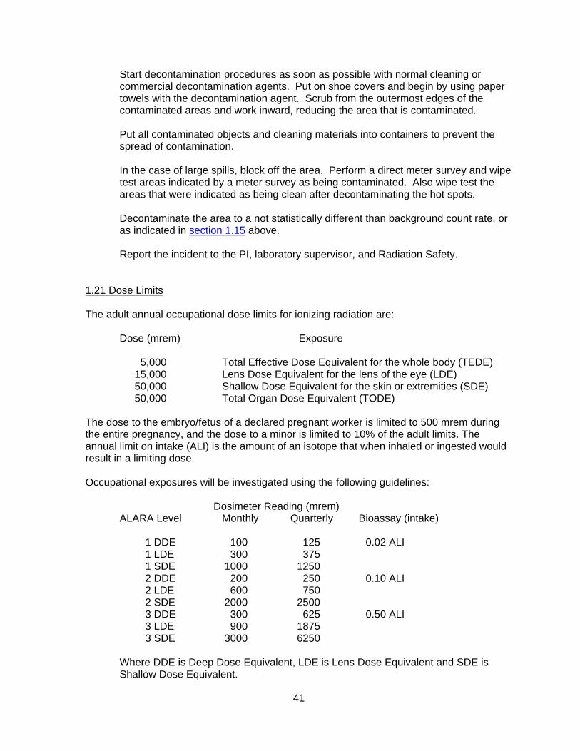
41
Start decontamination procedures as soon as possible with normal cleaning or commercial decontamination agents. Put on shoe covers and begin by using paper towels with the decontamination agent. Scrub from the outermost edges of the contaminated areas and work inward, reducing the area that is contaminated.
Put all contaminated objects and cleaning materials into containers to prevent the spread of contamination.
In the case of large spills, block off the area. Perform a direct meter survey and wipe test areas indicated by a meter survey as being contaminated. Also wipe test the areas that were indicated as being clean after decontaminating the hot spots.
Decontaminate the area to a not statistically different than background count rate, or as indicated in section 1.15 above.
Report the incident to the PI, laboratory supervisor, and Radiation Safety.
1.21 Dose Limits The adult annual occupational dose limits for ionizing radiation are: Dose (mrem) Exposure 5,000 Total Effective Dose Equivalent for the whole body (TEDE) 15,000 Lens Dose Equivalent for the lens of the eye (LDE) 50,000 Shallow Dose Equivalent for the skin or extremities (SDE) 50,000 Total Organ Dose Equivalent (TODE) The dose to the embryo/fetus of a declared pregnant worker is limited to 500 mrem during the entire pregnancy, and the dose to a minor is limited to 10% of the adult limits. The annual limit on intake (ALI) is the amount of an isotope that when inhaled or ingested would result in a limiting dose. Occupational exposures will be investigated using the following guidelines: Dosimeter Reading (mrem) ALARA Level Monthly Quarterly Bioassay (intake) 1 DDE 100 125 0.02 ALI 1 LDE 300 375 1 SDE 1000 1250 2 DDE 200 250 0.10 ALI 2 LDE 600 750 2 SDE 2000 2500 3 DDE 300 625 0.50 ALI 3 LDE 900 1875 3 SDE 3000 6250
Where DDE is Deep Dose Equivalent, LDE is Lens Dose Equivalent and SDE is Shallow Dose Equivalent.

42
Level 1: A written notification is sent to the user and PI. EH&S will conduct an interview with the user to determine if ALARA practices can be improved; a written report is placed in the user and RUA/MUA file. The RSC and HSRSC, if applicable, will be informed at next regularly scheduled meeting. Level 2: EH&S will conduct an interview with the user to determine if ALARA practices can be improved. A written report is sent to user and PI. The PI must sign to acknowledge receipt of the report. The RSC and HSRSC, if applicable, will be notified after the report is completed. Level 3: A verbal and written notification is sent to user and PI. The RSC and HSRSC, if applicable, will be notified immediately by email. The user must suspend radiation work pending an investigation by EH&S. A written report of the investigation is sent to user, PI, RSC and HSRSC, if applicable. The PI must sign to acknowledge receipt of the report. The RSC may authorize continuing radiation work after review of the report. These levels do not apply to declared pregnant workers or minors, whose exposure will be investigated on a case specific basis. Investigations do not need to be repeated if the ALARA level remains the same from period to period and the dose is ALARA. No user will be allowed to work with radiation if there has been an actual or suspected overexposure. In the case of a suspected overexposure that has been shown to be false, the RSC may authorize continuing radiation work after review of the report. 1.22 Dosimetry Badges Dosimetry will be issued as follows:
The term use means the amount of isotope listed on the Researchers Using Sealed/Unsealed Radioisotope User Enrollment form in the mCi/experiment column. It
Rad Use Description Dosimetry IssuedBeam Alignment Quarterly RingFluoroscopy Monthly or Quarterly CollarNuclear Medicine Quarterly Body and RingPET Monthly Body and Left and Right RingPortable X-Ray Quarterly CollarRadiation Oncology - Machines Quarterly BodyRadiation Oncology - Materials Quarterly Body and RingResearch - Any amount of Low Energy Beta *** NoneResearch - Greater than 1 mCi/Exp of High Energy Beta** Quarterly Body and RingResearch - Greater than 10 mCi/Exp of High Energy Beta** Monthly Body and RingResearch - Greater than 10 mCi/Exp of Low or Mid Energy Gamma* Quarterly Body and RingResearch - Greater than 100 mCi/Exp of High Energy Beta** Monthly Body and Left and Right RingResearch - Greater than 100 mCi/Exp of Low or Mid Energy Gamma* Monthly Body and Ring
* I-125 or Tc-99m** P-32, PET Isotopes*** H-3, C-14, S-35, P-33

43
refers to the amount of activity initially withdrawn from a stock vial, not the amount of activity in the stock vial. Rings should be worn under the gloves, with the label on the palm side of the hand, on the hand expected to receive the most exposure. Whole body dosimetry should be worn between the neck and waist. Fluoroscope users shall wear the badge at the collar, outside the lead apron. Fluoroscope users assigned two badges shall wear the collar badge on the collar, outside the lead apron, and the waist badge on the torso, underneath the lead apron. Based on the requirements of State of California Radiation Safety Advisory 96-5, the deep dose of fluoroscopists is automatically corrected by multiplying it by 0.3. This correction gives appropriate credit for the protection afforded by the lead apron. EH&S will periodically send dosimetry reports to the PIs or users. These reports may be either posted in the laboratory or otherwise made available to users. The PI should review the reports and will be involved in any investigations as outlined above. 1.23 Bioassay H-3 users will have their urine assayed every two weeks if 100 mCi or more is used per experiment or per month of a volatile form of tritium. EH&S will report positive analytical results to individuals as soon as results are available. The ALARA level 1 actions apply if the intake is in excess of 800 microcuries. Users performing iodinations or other procedures with volatile forms of iodine may voluntarily participate in the thyroid-monitoring program. I-123 users will have thyroid bioassays at least 6 hours after, but within 2 days of a procedure. I-125 users will have thyroid bioassays at least 6 hours after, but within 30 days of a procedure. I-131 users will have thyroid bioassays at least 6 hours after, but within 5 days of a procedure. Note that performing multiple iodinations for a single bioassay is acceptable, as long as the time from the first iodination to the time of the bioassay does not exceed the limits listed above, and provided that the time from the last iodination to the time of the bioassay is at least 6 hours. Users performing procedures using 10 mCi or more of I-125 will have thyroid bioassays at least 6 hours after, but within 5 days of the procedure. The ALARA Level 1 actions apply if the intake is in excess of 400 microcuries for I-123, 4 microcuries for I-125, and 4 microcuries for I-131. 1.24 Sealed Source Leak Testing Beta and/or gamma emitting sources with an activity greater than 100 microcuries, and alpha and/or neutron emitting sources with an activity greater than 10 microcuries, must be leak tested. EH&S will leak test these sealed sources every 6 months unless otherwise directed by the manufacture’s specific instructions. Beta, gamma and/or neutron emitting sources in storage are exempt from this requirement. These sources will be leak tested prior to use upon removal from storage. PIs shall inform EH&S prior to any change in the status of their sealed sources.

44
1.25 Calibrating Survey Instruments EH&S will calibrate portable survey instruments annually. Users should bring their instruments to the Services Laboratory at University Center 301B. Instruments must be free of contamination prior to being brought to the Services Laboratory. If there are questions or concerns regarding a contaminated meter, call 858-534-6418 for instructions. Instruments are calibrated by EH&S at a cost to cover time and materials. PIs may have their instruments calibrated by an outside certified calibration laboratory if they wish, and should inform EH&S of the calibration date. Loaner instruments are available for use while your instrument is being calibrated. The Services Laboratory can also supply replacement parts and perform minor repairs. The manuals and catalogs of various instrument manufacturers are also available. The calibration cost for each instrument is $81.46. 1.26 Maintenance on Potentially Contaminated Equipment Prior to maintenance or repair activities in radioactive material labs, areas, or rooms, EH&S should be contacted. These activities include painting, electrical work, or plumbing. EH&S will ensure the area is clear for general access. 1.27 Decommissioning Radiologically Posted Facilities EH&S shall be contacted prior to any demolition, renovation or modification of a radiologically posted facility. 1.28 Radiation Producing Machines (non-medical) Procedures for Purchase, Use, Transfer At the acquisition or fabrication of a machine that produces ionizing radiation, the PI must notify EH&S before the machine goes into service and provide the following information:
Description of machine (type of machine, manufacturer, model, year of manufacture, maximum operating parameters, energy and beam current). Operating protocol and typical operating parameters.
EH&S will perform an initial survey of the machine, register each x-ray tube with the State and recharge the tube’s owner. The current State rate is $344/tube billed biennially (once every two years).

45
Any change in the use, design or location of a radiation producing machine must be approved by EH&S. EH&S must be notified before a radiation producing machine is sold, traded, transferred or discarded. Training Regulatory requirements state that all persons who will be using a radiation producing machine (including PI’s) are an Authorized User. Authorized Users are those who have enrolled as a UC San Diego Radiation Machine User and have taken our Research Radiography tutorial and assessment (or pertinent test depending on the type of machine being used, see below) as well as training required by each lab for their specific machines and policies. The training shall be completed before machine use. Online enrollment and training is available. Please see EH&S website for details. Note: Electron microscope users are exempt from this assessment, but must still comply with all training and safety procedures issued by the scope manufacturer and/or the lab management responsible for the device. Personnel Dosimetry Researchers solely using non-medical radiation producing machines performing beam alignment or working with fluoroscopy will need a collar badge and/or a ring dosimeter When EH&S requires dosimetry all personnel must wear their dosimeters while working with or near the operating machine. The PI responsible for the machine must ensure that the appropriate dosimeters are worn and returned to EH&S for assessment. These dosimeters must not be used to measure beam output nor deliberately placed in the beam. Forms for dosimetry can be found on the EH&S website. Safety Devices Certain safety devices are required by State and Federal agencies for each radiation producing machine, e.g., warning lights, beam enclosures, interlocks, shielding and radiation survey meters etc. All safety devices must be maintained in good working order and must not be replaced or modified without specific EH&S approval. The safety device should always act as a backup rather than a replacement for proper procedure. A safety device must never be purposely defeated. If the design of a device makes a certain desired operation inconvenient or impossible, another device providing the same degree of protection must be substituted and approved by EH&S. Serious injuries have occurred when safety devices were bypassed. If a required safety device becomes nonfunctional, the machine must not be operated until it is repaired. EH&S must be notified immediately in the event of a failure in any radiation protection system or of suspected personnel radiation exposure. Contact EH&S for information regarding radiation safety or radiation safety survey instrumentation. A copy of the California Radiation Control Regulations is available at EH&S. X-Ray Diffraction/Fluorescence Units

46
X-ray diffraction units can be very hazardous because of extremely high primary beam exposure rates (several 100,000 R/minute) at the x-ray tube ports. Serious damage can result to an individual's eyes and skin, even if exposed to this intense radiation level for a very short period of time. Extreme caution must be exercised in the use of x-ray diffraction equipment. The following are general requirements for the safe use of x-ray diffraction units:
Only authorized individuals may operate x-ray diffraction/fluorescence equipment. Authorized users are those who have enrolled as a UC San Diego Radiation Machines User and have completed the UC San Diego Research Radiography Tutorial and Test. Always confirm that the high voltage is OFF or that the shutter is CLOSED before changing samples. When using systems with shutters, a survey meter is required to confirm that the shutter is closed before changing samples. DO NOT use the safety interlock to discontinue x-ray production; use the main switch. Operations involving removal of covers, shielding materials, tube housings, or modifications to shutters, collimators, or beam stops must be performed by qualified personnel only. Ring dosimetry is required for personnel performing beam alignment. Check radiation leakage with a survey meter after each beam re-alignment. Diffraction/fluorescence units must be secured against unauthorized use. A log of ALL machine operations shall be maintained and be available and include user name, date, kVp/mA rating, and total beam on time. Changes in the location or disposition of diffraction/fluorescence units must have the approval of Radiation Safety. Notify Radiation Safety prior to the acquisition, disposal, or transfer of any diffraction/fluorescence x-ray equipment.
Cabinet X-ray Systems The following are general procedures for the safe use of cabinet x-ray systems:
Only authorized individuals may operate Cabinet x-ray equipment. Authorized users are those who have enrolled as a UC San Diego Radiation Machines User and have completed the UC San Diego Research Radiography Tutorial and Test. A log of ALL machine operations shall be maintained and be available and include user name, date, kVp/mA rating, and total beam on time. DO NOT override the safety interlock. DO NOT use the safety interlock to turn the machine off; use the main switch.

47
Make sure the machine is OFF before changing samples. Always check the current and voltage meters and/or use a survey meter to detect x-rays. Do not modify the built-in shielding. If modifications must be made, contact Radiation Safety to request a unit and operation safety survey. Cabinet x-ray systems must be secured against unauthorized use. Changes in the location or disposition of x-ray units must have the approval Radiation Safety. Notify Radiation Safety prior to the acquisition, disposal, or transfer of any x-ray producing device.
DEXA Scanners for Human Use The following are general procedures for the safe use of DEXA x-ray scanners performed on humans:
Only individuals possessing a valid Diagnostic Technologist Certificate, X-ray Technician Limited Permit, or currently enrolled in UC San Diego School of Medicine may perform bone densitometry. Only authorized individuals may operate DEXA scanner equipment. Authorized users are those who have enrolled as a UC San Diego Radiation Machines User and have completed the UC San Diego DEXA Safety Tutorial and Test. Deliberate x-ray exposure of an individual for training or demonstration purposes is not permitted unless there is a medical indication and the exposure is prescribed by a physician. Never scan anyone who is pregnant, or who suspects she may be pregnant. Keep door closed while scanning. Only individuals required to be present shall be in the room while scans are being performed. Do not hold patients during scans. Avoid re-scans by positioning the patient correctly and instructing them not to move. Keep the patient comfortable and under constant observation. The operator should be located as far as practical from the scanner. Secure the DEXA scanner against unauthorized use by ensuring that the door remains locked when unattended. Maintain patient records of each scan performed. Conspicuously post a copy of the Supervisor/Operator Certificate as well as the valid Certificate/Permit of every person performing bone densitometry excluding students enrolled in the UC San Diego School of Medicine.

48
Notify Radiation Safety for any location changes, modifications, significant increases in workload, or prior to the acquisition, disposal, or transfer of any x-ray equipment.
DEXA Scanners for Veterinary Use The following are general procedures for the safe use of DEXA scanners in veterinary applications:
Only authorized individuals may operate DEXA scanner equipment. Authorized users are those who have enrolled as a UC San Diego Radiation Machines User and have completed the UC San Diego Veterinary DEXA Safety Tutorial and Test. Always stand behind the line marked on floor during scans. Maintain direct surveillance and control of DEXA machine during scans. Keep door(s) closed while scanning. Only individuals required to be present shall be in the room while scans are being performed. Never put any part of the body in the path of the useful beam during scans. Do not hold or support animals during scans. Plan scans carefully to avoid unnecessary retakes. Secure units against unauthorized use. Only qualified individuals should attempt to make repairs or remedy malfunctions. Notify Radiation Safety for any location changes or modifications, significant increases in workload, or prior to the acquisition, disposal, or transfer of any x-ray equipment.
Medical X-Ray Machines To control the radiation exposure hazard from medical x-ray sources, and maintain radiation exposure to individuals as low as reasonably achievable (within dose limits), authorized users and operating personnel must comply with the following procedures:
All personnel shall wear the required personnel monitors assigned to them. Any employee who is required to be in the room during operation of the x-ray unit shall wear a protective lead apron (recommended thickness 0.5 mm lead equivalent, minimum 0.25 mm lead equivalent) or stand behind a protective barrier. Any individual who holds a patient during an x-ray examination shall wear a lead apron and lead gloves. Whenever possible, no individual shall, as any part of his/her job, regularly hold patients during x-ray examinations.

49
Lead aprons, gloves, and other protective devices should be inspected once every year to detect cracks and breaks in the shielding and should be replaced immediately if defects are detected. Fluoroscopists should never place their hands in the primary beam, and should wear lead gloves when hands are positioned near the primary beam. Because of high radiation exposure to the patient during fluoroscopy, and especially during cineradiography, special care should be taken to minimize the time the beam is on. All fluoroscopic units should be equipped with a manually reset timing device. This device should turn the x-ray machine off or indicate the termination of the elapsed time when the timer reaches a preset limit for the examination. Such timing devices shall not exceed a maximum setting of five minutes.
Gonadal shielding shall be used for examinations where the primary beam will include or is near the patient gonadal area, except when the physician determines that this will interfere with the x-ray examination. In the case of pregnant women, protective shielding shall be provided for the fetus, except when the physician determines that such shielding will interfere with the x-ray examination. Gonadal and fetal shields should be a minimum of 0.5 mm lead equivalent. All x-ray machines must have properly installed and approved primary beam collimation devices, and the operator shall collimate the primary radiation beam to include only that area required in the examination. Any individual operating portable x-ray machines shall wear a lead apron and stand as far as possible from the radiation field during the exposure. The operator shall make certain that all other individuals are clear of the area before the exposure is made. For medical X-ray machines located at a satellite facility, there shall be a written signed agreement between the facility and the X-ray supervisor, designating the latter as the person having responsibility for, and control of, quality, radiation safety and technical aspects of all X-ray examinations and procedures. All off-site medical use X-ray producing equipment shall have a written X-ray policy and procedures manual approved by the offsite supervisor in place in accordance with 10 CFR 20.1101.
Veterinary Radiography The following are general procedures and requirements for the safe use of x-ray systems used in veterinary radiography:
Only authorized individuals may operate x-ray machines. Authorized users are those who have enrolled as a UC San Diego Radiation Machine User and have completed the UC San Diego Veterinary Radiography Tutorial & Test. Online training is available. Please see EH&S website for details. Only individuals required for the procedure shall be in the room during exposures; all such individuals shall stand behind a protective barrier or wear 0.25 mm lead equivalent protective garments.

50
Keep the number of exposures to a minimum. Avoid re-takes by planning the exposures carefully and position the animal correctly the first time. Verify by direct surveillance that no one can gain access to the primary beam or high scatter radiation areas during exposures. Collar dosimeters are required for all personnel not standing behind protective barriers and are to be worn on the outside of protective garments. Do not hold or support animals during exposures. Use immobilization devices to position animals for exposures if needed. Always collimate the useful beam to the area of clinical interest. X-ray equipment must be secured against unauthorized use. Only qualified individuals should attempt to make repairs or remedy malfunctions. Notify Radiation Safety for any location changes or modifications, significant increases in workload, or prior to the acquisition, disposal, or transfer of any x-ray equipment.
Veterinary Fluoroscopy The following are general rules and procedures for the safe use of x-ray equipment used in veterinary fluoroscopy:
Only authorized individuals may operate fluoroscopic equipment. Authorized users are those who have enrolled as a UC San Diego Radiation Machine User and have completed the UC San Diego Veterinary Fluoroscopy Tutorial & Test. Online training is available. Please see EH&S website for details. Only individuals required to be present during fluoroscopic procedures shall be in the room during the exposure, and all such individuals must wear lead aprons or stand behind a protective barrier. Collar dosimetry is required for all personnel working with fluoroscopic units. The dosimeters must be worn on the outside of the lead aprons. Fluoroscopy should not be used as a substitute for radiography. Limit use to necessary evaluation of moving structures using the shortest irradiation or “beam on” time. Do not hold or support animals during exposures. The fluoroscopic field must be restricted to the area of clinical interest.

51
The tube kVp, filtration, and SDD (source to detector distance) should be as large as practical. Fluoroscopic units must be secured against unauthorized use. An operating log must be maintained that includes the following information for each use of the unit: Date, Operator, kVp/mA settings, and total beam on time. Only qualified individuals should attempt to make repairs or remedy malfunctions. Notify Radiation Safety for any locations changes, modifications, significant increases in workload, or prior to the acquisition, disposal, or transfer of any x-ray equipment.
Dental X-Ray Machines The following are general procedures for the safe use of dental x-ray equipment:
Only licensed dentists or a person who is under the supervision of a licensed dentist may operate x-ray equipment including positioning patients for exposure. All such operators shall pass a radiation safety course approved by the board. Deliberate x-ray exposure of an individual for training or demonstration purposes is not permitted unless there is a medical indication and the exposure is prescribed by a dentist. Do not hold patients during exposures. Carefully position the patient, tube and detector correctly. Keep the patient comfortable and under constant observation. Only individuals required for the procedure shall be within 6 feet of the patient during the exposure. The operator shall stand at least 6 feet from the patient or behind a protective barrier during exposures. Each patient undergoing dental radiography shall be draped with a protective apron of not less than 0.25 mm lead equivalent to cover the gonadal area. Women should be verbally screened for the possibility of pregnancy prior to any x-ray examination. If dental care is to be delayed until after delivery, then exposure should also be delayed. Maintain patient records of each exposure performed. The x-ray unit must be secured against unauthorized use. Only qualified individuals should attempt to make repairs or remedy malfunctions. Notify Radiation Safety for any location changes, modifications, significant increases in workload or prior to the acquisition, disposal, or transfer of any x-ray equipment.

52
Electron Microscopes The following are general procedures for the safe use of electron microscopes:
Electron microscopes must be secured against unauthorized use. This can be accomplished through key control of the unit or the room. Do not modify the built-in shielding and viewing ports. If modifications must be made, contact Radiation Safety to have a survey done of the unit prior to and after modifications. Changes in the location or disposition of electron microscopes must have the approval of EH&S. Notify Radiation Safety prior to the acquisition, disposal, or transfer of any electron microscope.
Radiation Therapy Accelerators and Therapy X-Ray Machines Written operating instructions and emergency procedures, that are approved by EH&S must be available for each accelerator facility. All individuals who use an accelerator must be familiar with and operate the facility in compliance with the procedures. All radiation protection interlock and warning systems will be checked once every six months by the radiation physicist. 1.29 Irradiators Increased Controls All regulations must be followed for the increased security and control of radioactive materials that constitute Quantities of Concern as defined and required by United States Nuclear Regulatory Commission (USNRC) and California Department of Public Health – Radiologic Health Branch (CDPH-RHB). .All personnel with unescorted access to irradiators must be enrolled in the Increased Controls program with EH&S. All operating procedures for the irradiators must be approved by EH&S and all operators of the equipment must be thoroughly familiar with the procedures prior to operating the equipment.

53
Section 2: Research Use of Radioactivity 2.1 Training Requirements for Research Users All users shall attend the appropriate training: Use Training Any radioactive materials Radiation Safety Seminar Iodination, unbound use of NaI Iodine Safety Seminar P-32 use > 10 mCi per experiment P-32 Safety Seminar S-35 use > 10 mCi per experiment S-35 Safety Seminar Radiation producing machines Radiation Producing Machines Safety Fundamentals The Radiation Safety Seminar is given monthly and must be attended as soon as possible, and prior to any use of radioactive materials that is not directly supervised by the PI or a trained user listed on the RUA. Other seminars are scheduled periodically, and any seminar may also be given on request. Tailored versions of the Radiation Safety Seminar are given for students using radioactive materials as part of laboratory courses. Refresher training will be part of the RUA renewal process, and will be tailored to the RUA. Laboratory specific issues and audit results will be discussed with the PI and authorized users. This refresher training will be conducted as part of a laboratory meeting when possible. For those workers who are authorized users at other universities or research institutions and rendezvous with UC San Diego PIs at remote sites, e.g., Antarctica or vessels, special arrangements will be made. They must either provide a statement of training from their home institution, or sign a statement that they have read, understood and will comply with the provisions of this Manual. These individuals will also receive on-the-job training by the UC San Diego PI or the chief scientist at the remote site. 2.2 Transfer or Shipping Packages of Radioactive Materials Between Locations on Campus or Medical Center Radioactive materials may not be transferred from one person or laboratory to another unless the recipient has a valid RUA for the isotopes and quantities to be transferred. A Request for Intracampus Transfer of Radioactive Material form or equivalent must be completed by the transferring parties and submitted to EH&S. The use of shared isotopes in common use areas is permitted as long as all RUAs involved are authorized for the isotopes and inventory records are maintained. The use of the Transfer form in these cases is not required.
54
The means of transportation and container must be adequate to ensure safety during transfer. All movement of radioactive material off University property will be in conformance with U.S. Department of Transportation (DOT) regulations. Certified containers are available from EH&S. Transfer of Radioactive Materials Off-Campus All transfers of radioactive material off-campus must have specific prior approval from EH&S. EH&S will obtain approval for transfer from the receiving institution's RSO as well as a copy of their radioactive material license. For transfer within the San Diego area, a Transfer of Radioactive Material form or equivalent must be filled out, and the transfer coordinated with EH&S. For transfer via a common carrier, a Shipping Memo form or equivalent must be filled out and delivered to EH&S, along with the isotope(s) to be shipped. The following accompanying information will be required: material description, chemical form, physical form, radioisotope, activity in mCi and any special storage or temperature requirements Radioactive material must be packaged and transported according to DOT specifications for packaging, labeling, and hazardous goods shipments. EH&S will provide appropriate packaging. 2.3 Care and Handling of Animals Containing Radioactivity Approval to use radioisotopes in animals requires written authorization from both the Institutional Animal Care and Use Committee (UC San Diego Animal Subjects Committee) and EH&S. Before an authorization is granted for use of radioisotopes in animals, EH&S will review procedures with the applicant. The applicant must have adequate facilities to use and care for animals, and must make provisions for collection and storage of animal carcasses and associated waste. Additional information concerning the use of animals in research is available by calling 858-822-2494. General instructions for the use of radioisotopes in animals are listed below. Specific instructions and recommendations will depend on the protocol, isotope(s), quantities of radioactivity, frequency of use, number of animals, and type of animal(s). Injecting or Feeding Animals Radioactive materials shall be administered only to animals owned by the University. Injections or gastric lavage of radioactive materials into animals shall be done in a manner, which will control and limit accidental spillages. A laboratory coat and protective gloves shall be worn.
55
Animal Cages Metabolic cages in which animals containing radioisotopes are housed must be labeled with a radiation caution tag that lists the type and quantity of radioisotopes in each animal, date of administration, and name of PI. The contaminated absorbent material or bedding must be discarded as radioactive waste in the animal care facility. The researcher shall ensure that routine surveys are made of the cages and rooms where such animals are housed. Areas that indicate removable contamination must be immediately decontaminated. Cages must be surveyed and decontaminated before they are sent to a cage wash facility. The RSO may specify that animals containing radioactive materials be kept in cages apart from other animals. Animal Waste Animal excreta should be regarded as radioactive unless appropriate monitoring indicates there is no radioactivity present. Disposal of excreta should be in a manner similar to that for animal carcasses. Radioactive animal carcasses shall be double-bagged, properly labeled, and stored in a freezer to await pick up by EH&S. Contact EH&S for waste pickup. Animal waste containing 0.05 microcurie or less of H-3 or C-14 per gram of animal tissue, averaged over the weight of the entire animal, may be disposed of as if it were not radioactive. Ventilation If there is a possibility of airborne radioactive contamination, sufficient ventilation or air cleaning must be provided for animal rooms such that the derived air concentration values are not exceeded. Training of Caretakers and Custodians Principal investigators are responsible for assuring that animal caretakers and custodians are aware of potential hazards and are adequately trained and supervised in the observance of necessary precautions. If any assistance is needed in the training of animal care personnel or the monitoring of use facilities, contact EH&S. The general animal husbandry staff of the Office of Animal Resources are not authorized as radioisotope users and do not provide husbandry for radioactive animals. Principal investigators should ensure that the Supervisor of the animal care facility is notified when radioactive experiments are begun. Radiation Protection Instructions for Animal Caretakers The animal care supervisor or attendant must be informed and advised when animals under his/her care contain radioisotopes. This is the responsibility of the PI who is director of the research project.
Cages or cage cards must be posted with a Caution Radioactive Material sign. Information on the outside of the cage must include the date of administration, the isotope and the quantity administered.

56
Radiation surveys must be made around the cages to determine levels of external radiation. If the PI cannot provide these surveys, contact EH&S. A contamination survey must be made of all cage facilities following use. Animals that have been irradiated by x-rays or gamma rays do not present a radiation hazard. If the radioisotopes will be excreted in the urine or feces, absorbent material in a tray must be provided below or within each cage. The absorbent material must be changed periodically and disposed of as radioactive waste. If dogs or other large animals will excrete radioisotopes in the urine or feces, a metabolic cage must be used, and the excrement collected and properly stored prior to pick up as radioactive waste. Contact EH&S for cage washing instructions. Small animal cages may be washed in the laboratory sink if this procedure is approved by EH&S. In centralized animal facilities, animal care supervisors should be fully apprised of the radioisotopes in use so that an animal husbandry procedure that includes appropriate cage cleaning and sanitation may be initiated. Laboratory coats, appropriate eye protection and disposable gloves must be worn during cage cleaning and when handling the animals. When there is a possibility of airborne radioactivity being present, respiratory protection should be used. Contact EH&S for more information. Animal carcasses containing radioisotopes must be properly disposed of in accordance with the requirements of EH&S. Radioactive animal carcasses and associated waste must be placed in double plastic bags. The bag must be labeled with the type and number of animals and what radioisotope(s) they contained and the activity of each radioisotope. The animals will require temporary storage in a laboratory freezer or refrigerator to prevent biodegradation until picked up by EH&S.
In case of radiation emergencies such as a spill of contaminated waste, contact EH&S at 858-534-3660. 2.4 Working with Volatile S-35 Many sulfur compounds present the risk of volatility. When working with these isotopes, the following precautions should be followed: Wear impermeable gloves, such as nitrile, co-polymer, neoprene or PVC. Latex gloves should not be used. Use fresh, highly purified S-35 labeled amino acids. Whenever possible, use stabilized solutions of these amino acids. Thaw vials of S-35 amino acids in a fume hood. Off-gas the volatile component that is formed during defrosting with a syringe filled with activated charcoal by placing the needle through the septum and drawing off the air in the top of the vial.

57
If possible, dedicate an incubator in the laboratory just for cell culture with S-35 labeled amino acids. Double glove whenever handling materials that have been placed in this incubator. Remove the outer glove after you have handled the cultures to prevent the spread of contamination from your glove to other laboratory surfaces. Be aware that the condensation in the incubator will be contaminated with S-35. Placing an activated charcoal filter on the lid of your culture, or inside of a plastic bag containing your cultures, will reduce the amount of free S-35 in the incubator. A shallow tray containing activated charcoal placed in the incubator is also an effective method for trapping free S-35. The condensation that forms on the interior walls and doors of an incubator can be a source of contamination if the free S-35 is not trapped by these means. Monitor the incubator, floor below the incubator and your workstation with a pancake GM survey meter after each use of S-35 amino acids. Decontaminate any areas reading not statistically different than background and document the survey after clean up. 2.5 Working with P-32 Since P-32 emits a very energetic beta particle, significant skin doses can occur unless special precautions are followed. Handling of uncovered or un-shielded containers presents a potential for excessive and unnecessary radiation dose to the hands and face. Never place hands or other parts of the body over an open unshielded vessel containing millicurie quantities of P-32 in relatively small volumes of liquid. Dose rates from 1 mCi of P-32 in a glass vial can be as high as1200 mrem/hour on contact, 300 mrem/hour at 1 cm and 5 mrem/hour at 10 cm. A drop containing 1 microcurie of P-32 in contact with the bare skin can give up to 2,000 mrem/hour. All personnel using greater than 1mCi are required to wear dosimetry badges and finger rings. The finger ring should be worn under the gloves with the label on the palm side of the hand. Dosimeters should also be worn by laboratory personnel when receiving shipments from the EH&S isotope lab. P-32 stock solution vials should not be handled with the hands. Use remote handling tools. Lucite shields should be used when handling Petri dishes containing more than 10 mCi of P-32. Impermeable gloves, such as nitrile, co-polymer, neoprene or PVC, and laboratory coats should be worn. Wear two pairs of gloves and change them frequently. Safety glasses with side panels provide protection from beta radiation and should be worn when handling P-32. The stock solution vials must be stored in a shielded container, approximately 1/2 inch (1 cm) of Lucite or equivalent. Due to the production of low energy x-rays in the shielding, use of a few millimeters of lead or steel shielding outside the Lucite is recommended when multiple millicuries of P-32 are stored. In the event of contamination, most P-32 labeled compounds can be cleaned with standard decontamination solutions. P-32 as a phosphate adheres tightly to glass and usually needs an acid rinse. If skin contamination is detected, decontaminate by using mild soap and
58
lukewarm water. Do not irritate or break the skin by using any type of scrub brush. A dilute vinegar solution is useful for skin decontamination. If you are unable to decontaminate an area to not statistically different than background, label the area and contact EH&S for assistance. Fixed contamination may be shielded and allowed to decay. 2.6 Iodinations Individuals performing iodinations must be authorized by EH&S, and should contact EH&S prior to commencing the experiments in order to obtain a baseline bioassay measurement. Training of newly authorized individuals must be conducted under the direct supervision of an individual authorized to conduct iodinations. In addition, attendance at the Iodine Safety Seminar is required prior to performing an unsupervised iodination. The user will complete the Iodination Log or equivalent. The room and hood should be surveyed before use with the survey meter with a NaI Probe. Contamination is considered to be a reading statistically different from background at a distance of approximately three inches from the surface. Any area showing contamination before use should be handled by contacting the last individual listed on the Iodination Log. The following protective clothing must be worn during the iodination procedure: laboratory coat, safety glasses, dosimetry and impermeable gloves, such as nitrile, co-polymer, neoprene or PVC. A lead apron is optional. Latex gloves should not be used. Never handle stock vials directly. Use tongs to hold the vial if it is removed from the lead pig and use remote handling devices when possible. Line the bottom of the hood with bench paper without blocking airflow. Use at least 0.5 mm lead or 12 mm leaded acrylic to attenuate the I-125 gamma radiation. Wrap sample containers with lead tape to decrease the radiation exposure. Vent the free radioiodine from the vial into a vendor supplied charcoal trap. All procedures shall be done in an iodination designated fume hood. Acid will liberate free iodine from Nal solution. Use extreme caution to avoid inhalation if acid is added to the radioiodine solution. After use, contaminated bench paper should be disposed of in the radioactive waste container. All surfaces and equipment should be cleaned with Count Off, or equivalent, and surveyed. No counts statistically different than background should be detected in the room after cleanup. Cleanup should take place immediately after room use. Results of the post iodination survey should be documented on the Iodination Log Sheet, or equivalent. Users must always remove their gloves and survey their laboratory coat before leaving the iodination hood area. The results of the survey are to be recorded on the Iodination Log Sheet under the Personnel Survey Column. Contaminated laboratory coats must not be worn elsewhere in the building. Dry contaminated waste will be disposed of in labeled lead-lined waste containers. Liquid waste should be placed in labeled, sealed containers inside the hood behind lead shielding. Notify EH&S for pickup of radioactive waste upon completion of the iodination procedure.
59
The following are the thyroid bioassay requirements for individuals conducting iodinations using more than 10 mCi: I-125 At least 6 hours after, but within 30 days I-131 At least 6 hours after, but within 5 days I-123 At least 6 hours after, but within 2 days In the case of an accidental or suspected uptake of iodine, the user must contact EH&S immediately for possible emergency treatment. 2.7 Use of Radioisotopes on Vessels The Scripps Institution of Oceanography (SIO) vessels provide a multi-purpose research platform for a variety of oceanographic research. The use of radioactive materials on vessels is highly controlled by State and Federal regulations. Each researcher wishing to use radioisotopes at sea must apply for approval through the SIO Ship Scheduling Office. Ship Scheduling will provide the proper forms and instructions. Alternatively, forms and instructions can be obtained from the SIO, Marine Operations Home Page. Ship Scheduling distributes and collects the radioisotope related forms. UC San Diego researchers are authorized via the UC San Diego RUA process. They may either hold an RUA themselves or be listed on another PI’s RUA. Non-UC San Diego researchers must be authorized for the type, quantity, and experimental procedure at their home institution and proof of authorization provided to EH&S. The SIO Marine Operations Committee (MOC) Isotope Usage Panel, the UC San Diego Radiation Safety Officer or person designated by the RSO, and the Director of Marine Operations and Shipboard Technical Support will review the application forms and the proposed protocol. Procurement or Transfer of Radioactive Materials Radioisotopes may not be purchased or transferred until the investigator has a valid RUA for the isotopes and quantities to be used. An authorization statement from the RSO at the user’s home institution may be substituted for the UC San Diego authorization. EH&S can assist in moving radioactive materials to or from the research vessels. All transportation must be consistent with federal and international transportation regulations. Radioisotope Usage The PI is responsible for compliance with University and governmental regulations. At sea, the cruise’s Chief Scientist is responsible to be sure the PI and radioisotope users are practicing regulatory compliance. When at sea, the vessel’s Master retains ultimate responsibility and authority for all personnel and research on board the ship. Radioisotopes should be used inside portable laboratories called Radioisotope Isolation Vans. Individuals wishing to be excepted from this restriction must request permission from

60
the MOC Isotope Usage Panel. Care must be taken to prevent the movement of externally contaminated items from the isolation van to the ship’s decks or laboratories. Where this practice is necessary, approval must be obtained from the MOC Isotope Usage Panel. In addition, the Radiation Safety Officer, the Resident Marine Technician, and the Ship’s Master must be informed. Contamination Surveys Researchers must survey their work areas and decontaminate as necessary. Monitoring of work areas to assure control of potential radioactive contamination is to be conducted by the research staff. The radioisotope work areas shall be wipe tested at least monthly. The SIO Shipboard Isotope Usage Panel, at their discretion, may require wipe tests more frequently. Documentation of location in the van (a map), the date, and results should be maintained. If a liquid scintillation counter is on board, it should be used to analyze wipe tests and the printout retained for review. Decontamination efforts and re-wipes should also be documented. If no liquid scintillation counter is available, the wipe tests can be completed and mailed to EH&S for analysis. Where sufficiently energetic radioisotopes (e.g., C-14 and S-35) are in use, a portable survey meter should be used to monitor the work area periodically. As with wipe tests, documentation should be retained for review. Radioisotope Waste Containment and Disposal EH&S can be of assistance in determining appropriate waste containment during the cruise, and practical disposal procedures after the cruise. Prior to the cruise, a description of the types and quantities of wastes expected to be generated is required. All radioactive wastes must be retained inside the radioisotope isolation vans. No wastes may be stored on the vessel decks. If waste storage presents an extreme space problem, special permission may be requested from the MOC Isotope Usage Panel to store packaged radioactive wastes inside the ship. All radioactive wastes must be disposed of according to UC San Diego policy, State and Federal regulations. There may be no discharge of radioactive matter, liquid or solid, into the ocean. Radioactive waste is to be collected and stored for disposal at the end of the cruise. When the vessel returns directly to the port of San Diego, dry waste may be collected and retained in double plastic bags. When wastes are to be off loaded at ports other than San Diego, specific supplies (e.g., 30-gallon drums) will be arranged for by EH&S. Shipment of these wastes will also be arranged for by EH&S. Post Cruise Procedures At the end of the cruise, post-cruise forms should be completed, signed and forwarded to the SIO Ship Scheduling Office. The SIO Ship Scheduling office will forward the forms to the Radiation Safety Officer.

61
Section 3: Medical Use of Radiation 3.1 Treatments Utilizing Radioactive Implants or Radiopharmaceuticals Permission to use radionuclides in patients for experimental diagnostic or therapeutic purposes must be obtained from the Human Research Protections Program in addition to the application submitted to Radiation Safety. All physicians using radioactive materials for diagnostic or therapeutic procedures must be approved by the RSC prior to prescribing these procedures. Radiation Safety maintains the procedure for such approval. Radiation Safety requires no notifications of outpatient treatments unless medical events or accidents such as spills or unexpected personnel exposure to radiation occur. When the total effective dose equivalent to any individual from the release of a patient is likely to exceed 100 mrem, the patient or patient's responsible relative or guardian shall be provided with written information on risks of radiation and methods for reducing the exposure of individuals. At no time may a patient be provided outpatient treatment when a member of the general public may be reasonably expected to receive a dose in excess of 500 mrem from the treated patient. In no case shall a nurse, attendant, or Resident from other than Nuclear Medicine or Radiation Oncology attend the administration of therapeutic doses of radioactive material that are implanted, injected, or swallowed. Either Nuclear Medicine or Radiation Oncology will provide the patient or patient's responsible relative or guardian information regarding radiation safety precautions to be followed during treatment and after release from the hospital. Personnel from either Nuclear Medicine or Radiation Oncology will measure and document radiation levels near the patient to determine stay times for visitors and nursing staff. Either Nuclear Medicine or Radiation Oncology will ensure that the patient’s room is properly posted, measure radiation levels in adjacent rooms and post as necessary. The patient’s room should be as far away from the nursing station and heavy traffic hallways as is consistent with good medical care. It will be a private room with private sanitary facilities and no carpet. A visitor’s safe line should be marked on the floor with tape as far from the patient as possible. A member of either Nuclear Medicine or Radiation Oncology will perform a radiation survey following each administration of therapeutic doses of radiopharmaceuticals or implanted radioactive sources. Surveys should be done at the bedside, one meter away and at the doorway. Either Nuclear Medicine or Radiation Oncology shall post the patient’s room door with a Caution Radioactive Materials sign, a Radiation Safety Notice for nurses that lists important phone numbers. Adjacent rooms and hallways should be surveyed to ensure that the dose in any unrestricted area does not exceed 2 mrem in any one hour. Patients, the patient's room and all items contained therein shall not be released to unrestricted use until they are monitored for radioactive contamination. Before release, the monitoring must reveal that no residual radioactivity is located in the patient’s belongings, or anywhere within the patient’s room. Either Nuclear Medicine or Radiation Oncology will make a note in the patient’s chart after this monitoring has occurred, authorizing release of the patient, the patient's room and all items contained therein.

62
In an emergency situation, life safety issues will always take priority over any radiological control issues detailed in this Manual. 3.2 Inpatients Receiving Implants of Radioactive Sealed Sources If an implant is performed in other than the patient’s room, the area used for the procedure will be surveyed immediately afterward. In the case of seeds, any area where the seeds were handled (e.g., sterilization areas, source storage room, etc.) shall also be surveyed immediately after use. Declared pregnant nurses shall not be assigned to the care of these patients. Bed baths should be omitted while the sources are in place. If Nursing feels that a source could have been inadvertently displaced, Radiation Oncology should be contacted immediately to perform a survey. Patients must stay in bed unless orders to the contrary are written. In any event, patients will remain in their assigned rooms during the treatment period. Visitors are limited to those 18 years of age or over unless other instructions are noted on the precaution sheet on the patient’s chart. Visitors should sit at least 3 feet from the patient and should remain no longer than the time specified on the form posted on the patient’s door or on his or her chart. The visitor’s safe line should be observed. At the conclusion of treatment, a survey will be performed by Radiation Oncology to ensure that all sources other than permanent implants have been removed from the patient and that no sources remain in the patient’s room or in any other area occupied by the patient. At the same time, all radiological postings will be removed from the patient’s door. If any applicators or sources are found fully or partially dislodged from a patient, or the position of the applicator has moved, the nurse or attendant shall leave the room immediately and notify Radiation Oncology. In no case shall a nurse or attendant, or a Resident other than from Radiation Oncology, attempt to remove, replace, or otherwise transport any source or applicator. Radiation Safety will render assistance when requested. In the event a patient containing radioactive sealed sources becomes uncooperative or disruptive, to the point of requiring significant close contact (>5 minutes) with any hospital personnel, the radioactive sources shall be removed from the patient by the attending radiation oncologist as soon as possible. These sources will be stored in the emergency pig in the patient’s room until they can be reloaded into the patient or removed by radiation oncology personnel who are authorized to do so. All linen and dressings shall be saved in the patient’s room. These will be checked prior to disposal in order to avoid misplacement of any radioactive source. Following removal of temporarily implanted sources, Radiation Oncology shall perform a radiation survey of the patient to ensure that all sources were properly removed from the patient and the room. The results of the post-source withdrawal survey shall be recorded on the patient information sheet and retained by Radiation Oncology.

63
3.3 Inpatients Administered Radiopharmaceuticals Nuclear Medicine is required to keep records of all patients receiving radiopharmaceutical therapy (time of application, activity of the radioactive material, unit number, etc.). Therapy rooms shall be set up to minimize the potential for contamination during the patient’s stay by covering the floor, doorknobs, chairs, and other objects likely to be touched with plastic or plastic-backed absorbent material. Patients should be discouraged from bringing personal items into the room with them since they may become contaminated. Street clothing must remain in a closet until the patient dresses for discharge. Disposable table service should be used for the duration of the patient’s stay. Housekeeping personnel should be informed to stay out of the room when the room is posted with the radiation symbol. For patients containing therapeutic quantities of radioactive material, the following applies: Separate plastic-lined containers for linen and disposable waste contaminated items will be provided. Patients will remain hospitalized until their exposure to anyone else is less than 500 millirem Total Effective Dose Equivalent. An exposure worksheet will be completed and signed by the Chief of Nuclear Medicine Nuclear Medicine or Radiation Safety shall brief Nurses working with these patients annually on radiation safety procedures. Nuclear Medicine shall brief the patient on radiation safety procedures for the dosage administration, visitor control, radioactive waste, and other applicable items. All persons entering the patient’s room shall follow standard Entrance Precautions: Gloves, a disposable gown and shoe covers shall be put on prior to entering the room. When exiting the patient’s room, the gown and shoe covers shall be removed while standing in the doorway followed by the gloves. The person may then step unto the protective pad just outside the door. Gloves and shoe covers shall be placed in a small waste container maintained at the patient’s door for this purpose. Hands must be washed after exiting the room. Hands and feet must be monitored for radioactivity if a radiation monitor is present. Only persons needed for medical, safety, or training purposes shall be present during the administration of radiopharmaceutical therapy. Personnel shall wear gloves and work within fume hoods when opening containers of volatile radiopharmaceuticals such as I-131. Personnel who administer I-131 shall monitor their thyroids at least 6 hours after, but within 5 days of the procedure. Nursing personnel are not required to receive thyroid monitoring provided they observe proper radiation safety precautions. Before using the patient’s room for general occupancy, it must be decontaminated and released by Nuclear Medicine or Radiation Safety. The room may only be released for unrestricted use by Radiation Safety. 3.4 Patients in the Operating Room When radioactive seeds are implanted in an Operating Room (OR), Radiation Oncology personnel are required to survey the room after patient departure. During such procedures, all waste containers, rinse waters, fluids, linens, surgical equipment, the OR floor and table, and other areas and items shall be checked for the presence of radioactive seeds during the
64
procedure and after the patient is removed from the OR. The purpose of this survey is to ensure that no radioactive sources are inadvertently disposed of in an improper manner. 3.5 Waste from Patients Treated with Radioactive Materials Excreta from individuals undergoing medical diagnosis or therapy with radioactive material may be disposed of into the sanitary sewerage without being subject to any limitations. Persons handling body fluids should follow Universal Precautions. Nuclear Medicine hot labs are authorized to hold short lived radioactive materials for decay in storage and to dispose of aqueous radioactive liquids to the sanitary sewer in accordance with procedures approved by EH&S. All other radioactive waste shall be treated as described in section 1.17. Portal monitors are located at the entrance to waste docks to ensure that no radioactive waste from patients leaves the hospital. If the monitor alarm goes off, workers transporting the waste are required to place it in a secure area and notify Radiation Safety immediately. 3.6 Transportation of Patients A person from either Nuclear Medicine or Radiation Oncology should accompany any patient containing over 30 millicuries of any radioactive material. Elevators used for transport shall be cleared of all personnel not essential to the care or transport of the patient. 3.7 Biopsies of Tissues Containing Radioactivity Some tissues, such as sentinel nodes, that contain radioactive materials may be removed. The radiation dose and risk to OR and Pathology staff is minimal during such procedures, and the amount of radioactive materials used during these procedures is similarly small. Accordingly, such tissue samples may be transported from the OR to the laboratory without radiological precautions. Analysis of such tissues shall be conducted over nonporous surfaces (such as stainless steel) that are covered with plastic-backed absorbent paper. In the event the tissue sample is found to contain radioactive sources (e.g., a sample of prostate tissue containing implanted seeds), Radiation Safety shall be contacted immediately. Under supervision from Radiation Oncology or Radiation Safety, a member of the laboratory staff shall remove the adhering tissue from such seeds to the maximum extent possible. Following analysis, Radiation Safety will take possession of the radioactive seeds. 3.8 Radioactive Cadavers If any patient containing radioactive material expires, it shall be the responsibility of the physician who pronounces such patient as dead to notify the physician in charge of the case of the presence of radioactive material. No autopsy of any cadaver containing in excess of

65
5 millicuries of any radioactive element shall be performed without prior consultation with Radiation Safety. 3.9 Use of Radiation-Generating Devices Any medical facility with permanently installed radiation-generating devices shall meet the shielding requirements of section 12-31C-101, Part 12 of the California Referenced Standards Code. Radiation-generating devices include, but are not limited to, x-ray machines, fluoroscopy units, CT scanners, linear accelerators, x-ray diffraction units, electron microscopes, and other devices that produce ionizing radiation as a result of their normal operation. Operators shall wear radiation badges if annual exposures greater than 500 mrem are possible, or if the equipment is capable of producing greater than 5 millirem in 1 hour in an accessible area at a distance of 30 centimeters from the tube. Radiation-generating devices shall undergo physics acceptance testing before use and whenever any alterations or repairs are made that might change the radiation level to which a person could be exposed. Diagnostic X-ray machines shall be subject to periodic physics performance testing. Therapeutic radiation generating devices shall be calibrated before the system is first used for irradiation of a patient, and thereafter at intervals not to exceed 24 months. A written record of all surveys will be presented to the individual responsible for each unit within one week after the survey unless the survey indicates abnormal values in which case the responsible individual should be informed immediately. All serious violations, i.e., those that endanger patients or personnel, must be corrected immediately or the unit must be shut down until such corrections can be made. Other, less serious violations must be corrected within a 60-day period. 3.10 Use of Fluoroscopic Equipment Scope This policy applies to all individuals who operate fluoroscopic equipment. Definitions 1. Fluoroscopy means a radiological examination utilizing fluorescence for the observation
of a transient image. 2. Direct Supervision means the supervising physician is physically present in the same room
and is able to observe, and correct as needed, the individual who is performing the procedure.
3. General Supervision means the supervising physician is physically present at the procedure facility, and is immediately available either in-person or by telephonic/electronic modalities to provide guidance during the procedure.
4. Oversight means the supervising physician is available to review procedures and provide feedback after the procedure has been performed.
Required Licenses, Certificates, and Permits 1. Physicians
66
a. A current and valid Radiology Supervisor and Operator certificate or Fluoroscopy Supervisor and Operator permit (SupOp) shall be required of any physician who does one or more of the following:
i. Actuates or energizes fluoroscopy equipment; ii. Directly controls radiation exposure to the patient during fluoroscopy
procedures (e.g. determines when the administration of radiation to the patient is appropriate, directs the operation of the fluoroscopy equipment, determines the appropriate exposure factors);
iii. Supervises the use of fluoroscopy by one or more persons who hold Radiologic Technology Fluoroscopy permits (CRT-F);
iv. Supervises the use of fluoroscopy by individuals (i.e. residents and fellows) under an approved medical training program.
b. Physicians of equal stature may participate in a procedure as a collaborative activity; however, the individual(s) directing the use of fluoroscopy during the procedure shall possess a valid SupOp.
2. Residents and Fellows
a. Individuals in a medical training program may use fluoroscopy on human beings prior to obtaining a valid SupOp, provided the following:
i. Individuals in a training program shall have fluoroscopy privileges as defined by a written training program;
ii. The training program shall include, for each procedure, a written description of the required competencies and level of supervision necessary for the individual to operate x-ray equipment in a fluoroscopy mode;
iii. The successful completion of competencies shall be documented, and available for inspection, prior to the use of fluoroscopy by the individual.
iv. Unless otherwise documented in writing, all procedures conducted by a trainee shall be under Direct Supervision.
v. Regardless of i-iv, no individual using fluoroscopy shall direct a CRT-F unless said individual possesses a valid Sup/Op.
3. Technologists
a. A current and valid Radiologic Technology Fluoroscopy (CRT-F) permit shall be required of any certified radiologic technologist who does one or more of the following:
i. Exposes patients to x-rays in a fluoroscopy mode ii. Positions the patient iii. Positions the fluoroscopy equipment iv. Selects exposure factors (e.g. mA, kVp, exposure time)
Weekly Fluoroscopic Output Monitoring 1. Non-certified, non-permitted personnel may operate x-ray equipment in a fluoroscopy
mode, solely for the purposes of weekly fluoroscopic output (i.e. tube current and potential) monitoring, provided the following:
a. The person performing the monitoring has received appropriate training prior to operation of the fluoroscope. Training shall include hands-on instruction using the same make/model of fluoroscope that will be monitored. The individual performing the testing must be familiar with the equipment used, operating and emergency procedures, and documentation requirements. This training shall be documented and records must be readily available for inspection.

67
b. Only the phantom is exposed to the useful beam of the fluoroscope; human exposure is not allowed.
c. If monitoring results indicate that the tube current or potential vary in such a way that could increase the patient radiation exposure rate by more than 25% from the most recent annual exposure rate measurement, the person performing the monitoring will promptly notify all operators of the fluoroscope and the medical physicist, or designee, responsible for the annual exposure rate measurement as appropriate.
d. Personal dosimetry is available and worn as indicated. e. The individual responsible for the fluoroscopy equipment retains all responsibility
for ensuring that testing is performed correctly. Supervision 1. Residents and Fellows
a. Individuals in a medical training program shall only operate x-ray equipment in a fluoroscopy mode under the supervision of a physician who possesses a valid SupOp.
b. The required level of supervision, as listed below, shall be determined by the program director and faculty members and included as part of the written training program description.
i. Direct Supervision ii. General Supervision iii. Oversight
c. Faculty supervision assignments should be of sufficient duration to assess the knowledge and skills of each resident/fellow and delegate to him/her the appropriate level of patient care authority and responsibility. Written documentation of supervising faculty assessment and review of resident/fellow performance and competency to perform specified fluoroscopic procedures shall be maintained by the training program director, and made available for inspection, prior to use of x-ray equipment in a fluoroscopy mode.
d. Absent a written description of the required level of supervision in the training program, and absent written documentation of the resident’s competency to perform a specific procedure, residents and fellows shall only operate x-ray equipment in a fluoroscopy mode under Direct Supervision.
e. Residents and fellows without a valid SupOp shall not supervise the use of fluoroscopy by a CTR-F.
2. Technologists
a. Certified radiologic technologists with a fluoroscopy endorsement (CRT-F) shall not operate x-ray equipment in a fluoroscopy mode except under the Direct Supervision of a physician who possesses a valid SupOp. Prior to the initiation of each fluoroscopic exposure by a CRT-F, the supervising physician with a valid SupOp shall be responsible for reviewing the set-up, making any necessary adjustments to the patient or equipment, and confirming exposure factors.
i. Exception: The CRT-F may use fluoroscopy under the General Supervision of a physician who possesses a valid SupOp only if performing a standardized procedure or protocol (e.g. barium swallow) for which there is a documented procedure available, and in which case, no deviation is permitted.
b. The CRT-F shall be directly supervised by a physician with a valid SupOp for the length of the fluoroscopic procedure, which is defined as beginning with the
68
initiating exposure in a fluoroscopy mode and ending with any one of the following: the final exposure in a fluoroscopy mode, or the CRT-F leaving the procedure room, or the fluoroscopic equipment leaving the procedure room.
Radiation Safety 1. Only individuals required for the fluoroscopic procedure shall be in the room during the
exposure; individuals shall wear aprons of at least 0.25 mm lead equivalent or stand behind a leaded shield during the procedure. Thyroid shields and leaded glasses are strongly recommended for individuals working within a few feet of the fluoroscope.
2. A whole body dosimeter shall be worn by all individuals operating x-ray equipment in a fluoroscopy mode. The dosimeter shall be worn on the collar, outside of the lead apron.
3. No individual shall be permitted to hold patients during fluoroscopy, except during emergencies, nor shall any individual be regularly used for this service.
4. The fluoroscopic field shall be restricted to the area of interest. 5. Fluoroscopic units shall be secured to prevent unauthorized use.
The exposure to the patient shall be kept at a minimum consistent with clinical requirements and standard of care. Methods of reducing patient exposure include, but are not limited to, increasing voltage, increasing beam filtration, and reducing exposure time. Radiation badges shall be worn at the level of the neck and outside of the lead apron. Based on the requirements of State of California Radiation Safety Advisory 96-5, the deep dose of fluoroscopists is automatically corrected by multiplying it by 0.3. This correction gives appropriate credit for the protection afforded by the lead apron. Physicians who may also receive extremity exposures over 5,000 mrem in a year shall wear extremity dosimeters. 3.11 Use of X-rays in Medical Diagnosis and Treatment Normally, no person should hold patients during x-ray exposure. If circumstances require such action, however, the person holding the patient shall wear protective aprons and gloves and avoidance of exposure to the direct beam is mandatory. The exposure to the patient should be carefully restricted to the part under investigation. Radiation badges shall be worn by technologists at the level of the neck and outside of the apron. Technologists shall be provided with references on average patient radiation doses for the examinations that they are expected to perform. Technique charts shall be available for technologist use and reference. 3.12 Use of Radiation Therapy Machines Only technologist possessing a valid California Therapeutic Radiologic Technologist Certificate may operate therapeutic radiation producing machines. All such users must be operating under the supervision of a physician possessing a valid Radiology Supervisor &

69
Operator Certificate. No person other than the patient shall be in the treatment room during the exposure. The facility shall be operated in compliance with limitations indicated by Radiation Safety inspections. The patient and the control panel shall be kept under observation during patient treatment. Technologists shall wear dosimeters. 3.13 Training Requirements for Medical Personnel Initial training is required for all non-board certified radiation workers and ancillary personnel (e.g., nursing, clerical, housekeeping, security) whose duties may require them to work with or in the vicinity of radioactive material. This training is typically a radiation safety in service. Supervisors are required to inform workers about site-specific radiation hazards and appropriate precautions as stated in CCR Title 17, section 30255. Personnel shall be instructed before assuming duties with, or in the vicinity of, radioactive materials and whenever there is a significant change in duties, regulations, or the terms of the University’s Radioactive Material License. All radiation workers are required to receive annual refresher training. Any human use of radioactive material (i.e., the internal or external administration of radioactive material to human beings) must be carried out under the supervision of an Authorized User. Such application of or order to apply radiation shall be in the course of the Authorized Users professional practice and shall comply with the provisions of the University’s radioactive material license. The RSC shall approve Authorized Users wishing to prescribe the use of radiation or radioactivity for the diagnosis or treatment of diseases in humans. Physicians who have not been approved for such use by the Radiation Safety Committee may work only under the supervision of an Authorized User. 3.14 Medical Events Radiation Safety must be immediately notified of any event, except for an event that results from patient intervention, in which the administration of byproduct material or radiation from byproduct material results in –
(1) A dose that differs from the prescribed dose or dose that would have resulted from the prescribed dosage by more than 0.05 Sv (5 rem) effective dose equivalent, 0.5 Sv (50 rem) to an organ or tissue, or 0.5 Sv (50 rem) shallow dose equivalent to the skin; and
(i) The total dose delivered differs from the prescribed dose by 20 percent or more;
(ii) The total dosage delivered differs from the prescribed dosage by 20 percent or more or falls outside the prescribed dosage range; or
(iii) The fractionated dose delivered differs from the prescribed dose, for a single fraction, by 50 percent or more.

70
(2) A dose that exceeds 0.05 Sv (5 rem) effective dose equivalent, 0.5 Sv (50 rem) to an organ or tissue, or 0.5 Sv (50 rem) shallow dose equivalent to the skin from any of the following—
(i) An administration of a wrong radioactive drug containing byproduct material;
(ii) An administration of a radioactive drug containing byproduct material by the wrong route of administration;
(iii) An administration of a dose or dosage to the wrong individual or human research subject;
(iv) An administration of a dose or dosage delivered by the wrong mode of treatment; or
(v) A leaking sealed source.
(3) A dose to the skin or an organ or tissue other than the treatment site that exceeds by 0.5 Sv (50 rem) to an organ or tissue and 50 percent or more of the dose expected from the administration defined in the written directive (excluding, for permanent implants, seeds that were implanted in the correct site but migrated outside the treatment site).
Any event resulting from intervention of a patient or human research subject in which the administration of byproduct material or radiation from byproduct material results or will result in unintended permanent functional damage to an organ or a physiological system, as determined by a physician.
Radiation Safety will notify the appropriate regulating agency. The referring physician of the affected patient must also be notified unless the referring physician personally informs the licensee either that he or she will inform the individual or that, based on medical judgment, telling the individual would be harmful. To meet this requirement the notification may be made to the individual’s responsible relative or guardian. Notification of parties must be done within 24 hours. If the referring physician, patient, or the patient's responsible relative or guardian cannot be reached within 24 hours, notification of the medical event will be made as soon as practicable. Radiation Safety must be immediately notified of any event, except for an event that results from patient movement or interference in which the administration of radiation delivered by a radiation producing machine results in the following:
(1) Repeating of a CT examination, unless otherwise ordered by a physician or a radiologist, if the following dose values are exceeded:
(A) 0.05Sv (5 rem) effective dose equivalent. (B) 0.5 Sv (50 rem) to an organ or tissue. (C) 0.5 Sv (50 rem) shallow dose equivalent to the skin.
(2) CT X-ray irradiation of a body part other than that intended by the ordering physician or a radiologist if one of the following dose values are exceeded:
(A) 0.05 Sv (5 rem) effective dose equivalent. (B) 0.5 Sv (50 rem) to an organ or tissue. (C) 0.5 Sv (50 rem) shallow dose equivalent to the skin.

71
(3) CT or therapeutic exposure that results in unanticipated permanent functional damage to an organ or a physiological system, hair loss, or erythema, as determined by a qualified physician. (4) A CT or therapeutic dose to an embryo or fetus that is greater than 50 mSv (5 rem) dose equivalent, that is a result of radiation to a known pregnant individual unless the dose to the embryo or fetus was specifically approved, in advance, by a qualified physician. (5) Therapeutic ionizing irradiation of the wrong individual, or wrong treatment site. (6) The total dose from therapeutic ionizing radiation delivered differs from the prescribed dose by 20 percent or more. A report shall not be required pursuant to this paragraph in any instance where the dose administered exceeds 20 percent of the amount prescribed in a situation where the radiation was utilized for palliative care for the specific patient. The radiation oncologist shall notify the referring physician that the dose was exceeded.
Radiation Safety will notify the appropriate regulating agency within five days of discovery by or notification to the Radiation Safety’s office. Notification of the referring physician must be done within five business days after the discovery of the event. The person subject to the event shall be provided a written notification within 15 business days after discovery. 3.15 Fetal Radiation Exposure from Medical Procedures Radiation Safety must be immediately notified of any dose to an embryo/fetus that is greater than 50 mSv (5 rem) dose equivalent that is a result of an administration of byproduct material or radiation from byproduct material to a pregnant individual unless the dose to the embryo/fetus was specifically approved, in advance, by the authorized user. Radiation Safety must be immediately notified of any dose to a nursing child that is a result of an administration of byproduct material to a breast-feeding individual that is greater than 50 mSv (5 rem) total effective dose equivalent; or has resulted in unintended permanent functional damage to an organ or a physiological system of the child, as determined by a physician. Radiation Safety must be immediately notified of any dose to an embryo/fetus greater than 50 mSv (5 rem) dose equivalent that is a result of irradiation from a CT or Therapeutic radiation producing machine to a known pregnant individual, unless the dose was specifically approved, in advance, by a qualified physician. 3.16 QA Program for Administered Radiopharmaceuticals A written directive, which identifies the patient, radiopharmaceutical, dose and route of administration, shall be signed and dated by an Authorized User prior to preparation or administration of any therapeutic radiopharmaceutical, or doses of I-125 and I-131 sodium iodide over 30 microcuries. Records will be retained of written directives and administration of radiopharmaceutical dosage for a period of three years after the date of administration. Such records should be kept readily available for inspection. Prior to administering the radiopharmaceutical, the person who is to administer the radiopharmaceutical will verify the identity of the patient in the written directive by at least

72
two methods. The patient should be asked their name, if possible; other methods of identification that can be used are recorded birth date, social security number, ID bracelet, other hospital identification, address, or personal knowledge of the physician. Prior to administering the radiopharmaceutical, the person who will do the administration is to verify that the details of the administration are in accordance with the written directive. In addition to verifying the patient's identity, the radiopharmaceutical, dose (activity), and the route of administration are to be verified to be in agreement with the written directive, or with the oral revision made by the Authorized User. The radiopharmaceutical photon activity is to be measured in a dose calibrator, and the results recorded and compared to the written directive. The Authorized Users, or a qualified individual under supervision of an Authorized User (physicist or technologist), shall make a record of the administration. The record will include the date, patient's name, radiopharmaceutical dose, and signature or initials of the person administering the dose. The administration record is included on the same form with the written directive. 3.17 QA Program for Implants of Radioactive Sealed Sources Prior to any administration of radiation from sealed sources, an Authorized User will date and sign a written directive and a treatment plan for the procedure. The written directive will include the patient's name, treatment site, radionuclide, number and sequence of sources, source strength, and total radiation dose to be delivered to the target area. Records of written directives and administration of each administered dose shall be retained by Radiation Oncology for a period of three years after the date of administration. Such records should be kept readily available for inspection. Before administering a dose, at least two methods will be used to verify that the patient is the individual named in the written directive. The patient should be asked their name, if possible. Other identification methods that may be used include birth date, address, social security number, name on patient's ID bracelet or hospital card, name on the patient's medical insurance card, or the photograph of the patient's face made during the examination and planning portion of the patient's radiation therapy care. Before sealed radioactive sources are administered, a qualified person (such as a radiation therapy physicist, oncology physician, dosimetrist, or radiation therapy technologist), working under the supervision of an Authorized User, will verify that the radioisotope, number of sources, and source strengths of sealed sources described in the plan of treatment are in agreement with the written directive. The person verifying agreement between the plan of treatment and the written directive will sign or initial and date the plan of treatment. Before the total prescribed dose is administered, an Authorized User or a qualified person will check the dose calculations for the procedure. This will not be the same person who made the original calculations. Manual dose calculations will be checked for: arithmetic errors; appropriate transfer of data from the written directive, plan of treatment, tables, and graphs; appropriate use of nomograms; appropriate use of all pertinent data in the calculations. Printouts from computer-generated dose calculations will be checked to verify

73
the correct data were used in the calculations, including position of the applicator or sealed sources, isotope, number of sources, total source strength, or source loading sequence. Prior to administering a sealed source, a qualified person under the supervision of an Authorized User will verify that the radioisotopes, number of sources, source strengths, and if applicable, loading sequence of the sources to be used are in agreement with the written directive and plan of treatment. The verification method will be appropriate to the sources used, and may include checking the serial number of sealed sources behind an appropriate shield, using a dose calibrator, observing color codes on the sources, monitoring radiation output of the sources with a radiation detector, or using clearly marked storage locations where separate storage areas are used for different source strengths. For temporary sealed source implants, the position of the sources will be verified using x-ray imaging procedures. When possible, the x-ray imaging will be done with dummy sources before the radioactive sources are inserted. These x-ray images of source position will be used to calculate the exposure time needed to achieve the prescribed radiation doses, or to calculate the total dose. In the cases where fixed geometry applicators, appliances or templates are used to calculate an exposure time or total dose, x-ray imaging of the source positions is not mandatory. Immediately after implanting sources in the patient, surveys of the patient and the surrounding area will be made to confirm no sources are unintentionally misplaced outside of the patient. Daily surveys of the patient and the surrounding area will be made to confirm that no sources have subsequently become misplaced outside of the patient. After insertion of temporary implant brachytherapy sources, an authorized physician will promptly record the isotope and number of sources on the appropriate form. This form will be in the patient's chart or other appropriate record, and will be signed or initialed by the authorized physician. For permanent sealed source implants, imaging of the sources in place will be performed as a basis for verifying the position of the source and calculating the total dose. If applicable, this imaging will be done after inserting the sources into the patient. In cases where fixed geometry applicators or templates are used, imaging of the source positions in the patient is not mandatory. Immediately after implanting sources in the patient, survey of the patient and the surrounding area will be made to confirm no sources are unintentionally located outside of the patient. After insertion of permanent or temporary implant brachytherapy sources, an Authorized User will promptly record the isotope and number of sources implanted on the appropriate form. This form will be in the patient's chart or other appropriate record, and will be signed or initialed by the Authorized User. Verified electronic signatures are acceptable. If an Authorized User determines that delaying brachytherapy treatment in order to perform the checks of dose calculations would jeopardize the patient's health because of the emergent nature of the patient's medical condition, the checks of the calculations will be performed within two working days of completion of the treatment. Before the first use of treatment planning or dose calculating computer programs for dose calculations, a qualified person will perform acceptance testing of the programs. The functions of the computer programs evaluated during acceptance testing will include the
74
dose and spatial accuracy of calculations in tissue for at least those brachytherapy procedures clinically performed by authorized physicians under this license. 3.18 Audits of Nuclear Medicine and Radiation Oncology In order to ensure that safety rules are observed and that radioactive materials have been adequately controlled, Radiation Safety conducts quarterly audits of both the Nuclear Medicine and Radiation Oncology programs. Areas reviewed at this time include inventory, quality assurance and contamination survey records. It is the responsibility of the department to have such records well organized, updated and readily available. Any issues encountered by Radiation Safety during the audit are discussed with the manager and, when necessary, with the Department Head. 3.19 Minimizing Undue Radiation Dose to Patients It is the responsibility of Nuclear Medicine, Radiation Oncology and Interventional Radiology to maintain undue radiation exposure to patients as low as possible and to ensure that proper radiological techniques are administered to the right patients. The exposure to the patient should be carefully restricted to the part under investigation. Whenever practical, gonadal shielding shall be provided to the patient. Proper collimation of the x-ray beam should be verified before exposures to restrict the field to the area of interest. In fluoroscopy, the time in which the beam is energized should be minimized as much as practical. 3.20 Exposure of Individuals to Radiation UC San Diego Policy Statement Regarding Exposure of Individuals to Radiation No individual shall be deliberately exposed to radiation from radiation-producing machines or radioactive materials unless they are: 1) Under the personal care of a physician and a written prescription or order as part of the standard of care for the radiological study is present prior to the radiological examination. The prescribing physician shall be involved in and responsible for the routine care of the individual. The prescribing physician and the physician conducting the radiological examination shall not be the same individual. 2) A participant in a research study that has been reviewed and approved by the Human Subjects Protection Program (HRPP), which includes a review and approval of the radiological studies in the research protocol by the Human Subjects Exposure Committee (HERC).

75
Section 4: Appendices 4.1 Conversion Factors Standard activity units
1 curie (Ci) = 3.70 X 10¹⁰ dps 1 curie (Ci) = 2.22 X 10¹² dpm 1 millicurie (mCi) = 2.22 X 10⁹ dpm 1 microcurie (µCi) = 2.22 X 10⁶ dpm 1 nanocurie (nCi) = 2.22 X 10³ dpm 1 picocurie (pCi) = 2.22 dpm
International activity units
1 Megabecquerel (MBq) = 1 X 10⁶ dps 1 becquerel (Bq) = 1 dps
Curie to Becquerel conversion 1 curie (Ci) = 37 Gigabecquerel (GBq) 1 millicurie (mCi) = 37 Megabecquerel (MBq) 1 microcurie (µCi) = 37 kilobecquerel (kBq) 1 nanocurie (nCi) = 37 becquerel (Bq) 1 picocurie (pCi) = 37 millibecquerel (mBq)
Becquerel to Curie conversion
1 Terabecquerel (TBq) = 27 curie (Ci) 1 Gigabecquerel (GBq) = 27 millicurie (mCi) 1 Megabecquerel (MBq) = 27 microcurie (µCi) 1 kilobecquerel (kBq) = 27 nanocurie (nCi) 1 becquerel (Bq) = 27 picocurie (pCi)
Rem to Sievert conversion (Unit of Dose Equivalent)
100 rem = 1 sievert (Sv) 1 rem = 10 millisievert (mSv) 1 millirem (mrem) = 10 microsievert (µSv) 1 microrem (µrem) = 10 nanosievert (nSv)
Rad to Gray conversion (Unit of Absorbed Dose) 100 rad = 1 gray (Gy) 1 rad = 10 milligray (mGy) 1 millirad (mrad) = 10 microgray (µGy)
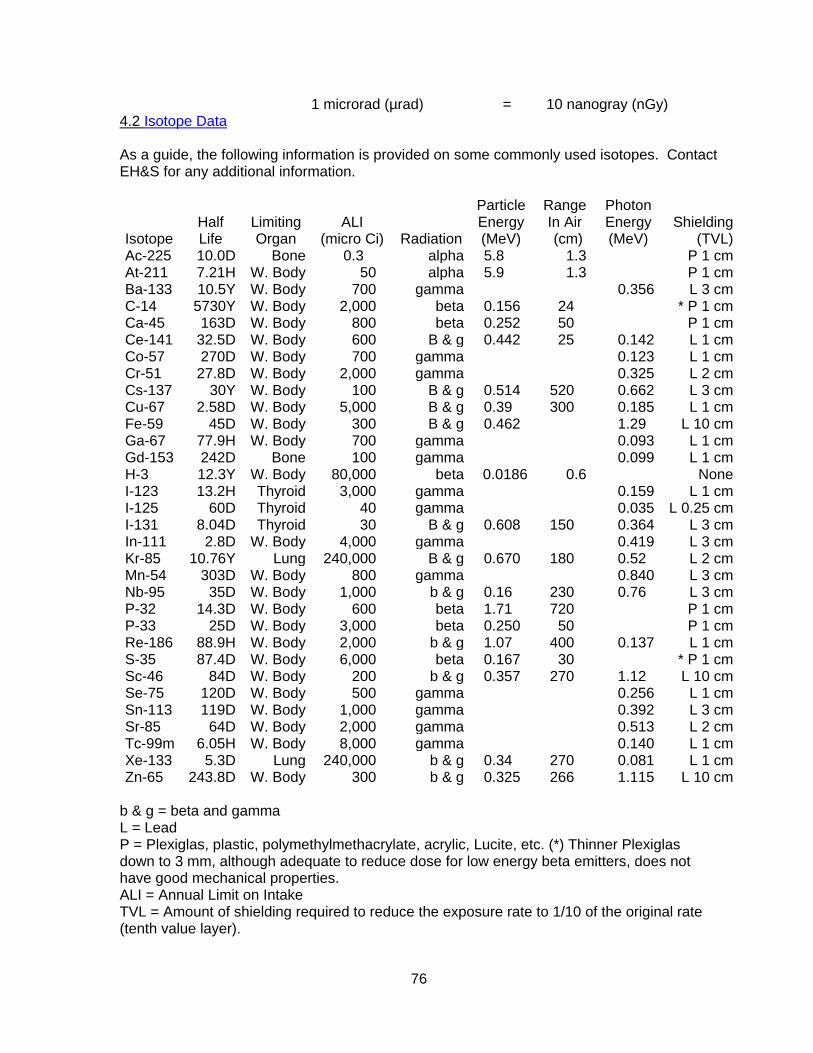
76
1 microrad (µrad) = 10 nanogray (nGy) 4.2 Isotope Data As a guide, the following information is provided on some commonly used isotopes. Contact EH&S for any additional information. Isotope
Half Life
Limiting Organ
ALI
(micro Ci)
Radiation
Particle Energy (MeV)
Range In Air (cm)
Photon Energy (MeV)
Shielding
(TVL) Ac-225 10.0D Bone 0.3 alpha 5.8 1.3 P 1 cm At-211 7.21H W. Body 50 alpha 5.9 1.3 P 1 cm Ba-133 10.5Y W. Body 700 gamma 0.356 L 3 cm C-14 5730Y W. Body 2,000 beta 0.156 24 * P 1 cm Ca-45 163D W. Body 800 beta 0.252 50 P 1 cm Ce-141 32.5D W. Body 600 B & g 0.442 25 0.142 L 1 cm Co-57 270D W. Body 700 gamma 0.123 L 1 cm Cr-51 27.8D W. Body 2,000 gamma 0.325 L 2 cm Cs-137 30Y W. Body 100 B & g 0.514 520 0.662 L 3 cm Cu-67 2.58D W. Body 5,000 B & g 0.39 300 0.185 L 1 cm Fe-59 45D W. Body 300 B & g 0.462 1.29 L 10 cm Ga-67 77.9H W. Body 700 gamma 0.093 L 1 cm Gd-153 242D Bone 100 gamma 0.099 L 1 cm H-3 12.3Y W. Body 80,000 beta 0.0186 0.6 None I-123 13.2H Thyroid 3,000 gamma 0.159 L 1 cm I-125 60D Thyroid 40 gamma 0.035 L 0.25 cm I-131 8.04D Thyroid 30 B & g 0.608 150 0.364 L 3 cm In-111 2.8D W. Body 4,000 gamma 0.419 L 3 cm Kr-85 10.76Y Lung 240,000 B & g 0.670 180 0.52 L 2 cm Mn-54 303D W. Body 800 gamma 0.840 L 3 cm Nb-95 35D W. Body 1,000 b & g 0.16 230 0.76 L 3 cm P-32 14.3D W. Body 600 beta 1.71 720 P 1 cm P-33 25D W. Body 3,000 beta 0.250 50 P 1 cm Re-186 88.9H W. Body 2,000 b & g 1.07 400 0.137 L 1 cm S-35 87.4D W. Body 6,000 beta 0.167 30 * P 1 cm Sc-46 84D W. Body 200 b & g 0.357 270 1.12 L 10 cm Se-75 120D W. Body 500 gamma 0.256 L 1 cm Sn-113 119D W. Body 1,000 gamma 0.392 L 3 cm Sr-85 64D W. Body 2,000 gamma 0.513 L 2 cm Tc-99m 6.05H W. Body 8,000 gamma 0.140 L 1 cm Xe-133 5.3D Lung 240,000 b & g 0.34 270 0.081 L 1 cm Zn-65 243.8D W. Body 300 b & g 0.325 266 1.115 L 10 cm
b & g = beta and gamma L = Lead P = Plexiglas, plastic, polymethylmethacrylate, acrylic, Lucite, etc. (*) Thinner Plexiglas down to 3 mm, although adequate to reduce dose for low energy beta emitters, does not have good mechanical properties. ALI = Annual Limit on Intake TVL = Amount of shielding required to reduce the exposure rate to 1/10 of the original rate (tenth value layer).

77
4.3 Dose Rate Rules of Thumb The dose rate at 30 cm from 1 mCi of an unshielded high-energy beta source such as P-32 is about 300 mR/hour.
The average gamma radiation levels at 30 cm from 1 mCi of some commonly used isotopes are: Isotope Radiation Level (mR/hr)
Ba-133 3 Ce-141 1 Co-57 1 Cr-51 0.2 Cs-137 4 Fe-59 7 Ga-67 1 I-123 1 I-125 1 I-131 2 In-111 1.5 Mn-54 5 Nb-95 5 Re-186 0.2 Sn-113 2 Sr-85 3 Tc-99m 1 Xe-133 0.1 Zn-65 3
4.4 Participation of Human Beings as Research Subjects All studies that involve human subjects must be approved by the Human Research Protection Program and either the Human Exposure Review Committee or the Radioactive Drug Research Committee prior to the start of the study. 4.5 Policy and Responsibilities of the RDRC Purpose The purpose of the Radioactive Drug Research Committee is to guarantee patients who take part in either research protocols or clinical trials the highest degree of both radiation and pharmacological safety. It is also their responsibility to determine the intrinsic value of the research and weigh risk versus benefit considerations before approving such studies. Federal law defines this committee, and the FDA must individually approve its members. Organization and Operation
The FDA also specifies its composition. By law the committee must be composed of:

78
a physician recognized as a specialist in nuclear medicine.
a person qualified by both training and experience to formulate radioactive drugs.
a person with special competence in radiation safety and radiation dosimetry.
The remaining members of the committee shall be selected from the pertinent disciplines that may be required to carry out the provisions of the law. The chairman of this committee shall sign all applications, minutes, and reports of the committee. The committee must meet at least four times per year and a quorum shall consist of more than 50% of its membership. Its minutes and records shall include the numerical results of the votes on protocols involving using radioactive drugs in human subjects. No member of this committee may vote on a protocol with which he is associated as an investigator. The committee must submit an annual report on or before January 31 of each year. This report shall include the names and qualifications of the committee members and of any consultants used by the committee. This report shall also incorporate the reports from the individual institutional users and supply statistical information showing the number of applications, the number of investigators, and pertinent information on any applications not approved for investigational study. The committee is also obligated to report immediately the approval of any study that will involve the exposure of more than 30 research subjects or if any subjects were expected to be under the age of 18. The FDA will conduct periodic reviews of the approved committee by reviewing the annual reports, reviewing the minutes, and by examination of the full protocols for pertinent studies that have been approved by the committee. They may also institute on-site inspections. Responsibilities of the RDRC The RDRC is required to carry out its purpose by acting as the on-site representative of the FDA. The RDRC is subject to scrutiny by the FDA by examination of detailed reports of the committee as well as by on-site inspection. Two types of evaluation for radioactive drugs have been defined. The first pertains to research and the second to clinical trials. Research with radioactive drugs is interpreted by the FDA to mean that these pharmaceuticals will be used to obtain basic information regarding drug metabolism, kinetics, distribution, localization, etc., of a radioactively labeled pharmaceutical or physiological and/or biochemical studies which are not intended for diagnostic or therapeutic purposes. They define clinical trials as those intended to determine the safety and/or effectiveness of the radioactive pharmaceutical. Therefore, the use of the so-called tagged compound under this definition involves all studies concerned with the gathering of basic information on drug metabolism, specific physiological and pathological processes that determine kinetics and distribution, and localization of such compounds in man. All such new diagnostic agents must be studied under an IND. The FDA has also adopted a general philosophy with respect to both radiation dose and pharmacological dose associated with the use of radioactive pharmaceuticals. With respect to radiation they expect any studies utilizing radiopharmaceuticals will produce radiation doses that are as low as possible. The responsibility for monitoring and approval of such studies lies in a newly defined institutional committee called The Radioactive Drug Research Committee.

79
Their philosophy concerning the limit of pharmacological dose states that the amount of active ingredient or combination of active ingredients is such that no non-clinically detectable pharmacological effects have been reported in humans. (For additional background information, refer to NCRP Report No. 70.) The project involving human subjects must have the overall approval of the UC San Diego Human Research Protections Program. In addition, prior approval by the RDRC is required.