two-stage optical system for colorectal polyp assessments
TRANSCRIPT
Two-stage optical system for colorectal polyp assessments
Mirosław Szura1• Artur Pasternak1,2
• Krzysztof Bucki3 • Katarzyna Urbanczyk4•
Andrzej Matyja1
Received: 4 January 2015 / Accepted: 23 March 2015 / Published online: 4 April 2015
� The Author(s) 2015. This article is published with open access at Springerlink.com
Abstract
Background and aims Macroscopic real-time evaluations
of the histopathology and degree of invasion of colorectal
polyps help to select the most suitable endoscopic treat-
ment method. Dual-focus (DF) narrow-band imaging (NBI)
is a new imaging enhancement system that uses digital and
optical methods to enhance the view of blood vessels on
mucosal surfaces. However, the superiority of this tech-
nique over standard imaging techniques has not been pre-
viously reported. The aim of this study was to determine
whether the two-stage optical systems in a new generation
of endoscopes will increase the diagnostic accuracy of
colorectal polyp recognition.
Methods The study included 270 patients, and 386 col-
orectal polyps were diagnosed and removed. The polyps
were assessed with white light and NBI using one- and
two-stage optical systems, respectively. After being
classified according to the Kudo pit pattern schemes, the
polyps were removed and histopathologically verified.
Results Regarding non-neoplastic lesions (Kudo I and II),
no difference was observed in the recognition of polyps
when using the NBI-DF function. We observed improved
accuracy in the preliminary diagnoses of Kudo IIIL lesions
(from 87.16 to 90.09 %, p\ 0.05) and Kudo IIIS lesions
(from 87.29 to 92.79 %, p\ 0.01). NBI-DF also increased
the accuracy of preliminary diagnoses of Kudo IV lesions
(from 88.24 to 94.12 %, p\ 0.01). The Kudo V pit pat-
terns were also more distinct with NBI-DF imaging, in-
creasing the diagnostic accuracy from 91.67 to 100 %.
Conclusions Using a two-stage optical system with
electronic colorization of the mucosa increased diagnostic
accuracy for differentiating colorectal polyps with neo-
plastic potential.
Keywords Colonoscopy � Colorectal cancer � Narrow-
band imaging � Dual-focus magnification
Colorectal cancer (CRC) is the third most common cancer
and the second leading cause of cancer deaths worldwide
[1]. Most colorectal cancers stem from preexisting adeno-
matous polyps, following the adenoma–carcinoma se-
quence [2, 3]. Colonoscopy is widely used to diagnose
colorectal cancer, as well as to detect and remove adeno-
matous polyps. Optical endoscopy has been endorsed as the
preferred CRC screening strategy for CRC prevention be-
ginning at age 50 [4, 5]. The ability to distinguish between
benign and malignant lesions using endoscopy is crucial.
However, endoscopy using conventional white light (WL)
imaging alone is frequently insufficient for a preliminary,
real-time diagnosis. Narrow-band imaging (NBI), also
known as ‘‘electronic chromoendoscopy,’’ is a newly
ClinicalTrials.gov number, NCT01688557.
& Mirosław Szura
Artur Pasternak
1 First Department, General, Oncological and Gastrointestinal
Surgery, Jagiellonian University Medical College, 40th
Kopernika St., 31-501 Krakow, Poland
2 Department of Anatomy, Jagiellonian University Medical
College, 12th Kopernika St., 31-034 Krakow, Poland
3 MEDICINA - Specialist Diagnostic & Therapeutic Centre,
5th Rogozinskiego St., 31-559 Krakow, Poland
4 Department of Pathomorphology, Jagiellonian University
Medical College, 16th Grzegorzecka St., 31-531 Krakow,
Poland
123
Surg Endosc (2016) 30:204–214
DOI 10.1007/s00464-015-4186-x
and Other Interventional Techniques
developed technology that provides a unique opportunity to
assess surface mucosal and vascular patterns on polyps,
potentially providing an in vivo histological diagnosis [6].
NBI uses optical filters for red, green, blue (RGB) se-
quential illumination and narrows the bandwidth of spec-
tral transmittance [7]. This technique enables the
observation of fine capillaries in the superficial mucosa of
the gastrointestinal tract. NBI may have the potential to
improve the detection rate of colorectal polyps compared to
conventional WL colonoscopy, particularly for small and
flat lesions [8]. Kudo et al. described a pit pattern classi-
fication system for colorectal neoplasia (type I to type V)
[9–15]. In the colorectal field, pit pattern diagnosis is
clinically significant because it can differentiate between
neoplasia and non-neoplasia, diagnose the degree of his-
tological atypia in a tumor, diagnose the invasion depths of
early carcinomas, detect minute residual tumors after en-
doscopic resection, estimate the degree of histological in-
flammation in ulcerative colitis, and diagnose
dysplasia-/colitis-associated carcinomas in ulcerative coli-
tis [16]. Usually, standard magnification requires the use of
chromoagents (e.g., indigo carmine, crystal violet, or
methylene blue) to clarify the pit structures in these diag-
nostic procedures. Simpler and more convenient proce-
dures are desirable for magnifying procedures. It has been
suggested that NBI colonoscopy is as effective as chro-
moendoscopy in differentiating between neoplastic and
non-neoplastic colorectal lesions using pit pattern classifi-
cation, and its diagnostic accuracy is much greater than that
of conventional WL colonoscopy.
In addition to NBI, a novel diagnostic technique has
recently emerged, the so-called dual-focus (DF) magnifi-
cation (Olympus Optical Co. Ltd, Tokyo, Japan). DF two-
stage optical lens technology from Olympus allows
physicians to switch from a normal focus mode to the near
focus (NF) mode, enabling up to 1009 magnification with
the single push of a button. This visualization allows for
the close examination of mucosal tissue and capillary
networks.
The aim of this study was to evaluate and compare the
diagnostic characteristics of a novel NBI system with DF
magnification function in differentiating colorectal polyps.
Materials and methods
The study was conducted at MEDICINA Specialist Diag-
nostic & Therapeutic Centre (a private hospital that per-
forms approximately 6000 colonoscopies each year), in
Cracow, Poland. The study was approved by the local
ethics committee and was conducted in accordance with
the principles of the Declaration of Helsinki. As a clinical
trial, the study was registered in a centralized clinical trials
registry (ClinicalTrials.gov - NCT01688557). All authors
of this study had access to the study data and had reviewed
and approved the final manuscript.
We included patients 40–65 years old who underwent the
procedure in the context of an opportunistic colorectal can-
cer screening. In addition to age, the following exclusion
criteria were applied: symptoms of colon cancer (e.g.,
bleeding unrelated to hemorrhoids), changes in bowel
movement regularity, and unexplained weight loss. Patients
who had already received a colonoscopy within the last
10 years were also excluded. A total of 2806 patients were
referred and scheduled for outpatient colonoscopy in 2012 as
a part of a national colorectal cancer-screening program. Of
these patients, 842 were examined using a 190 series Exera
III NBI system (CF-HQ190L, Olympus Co. Ltd, Tokyo,
Japan) with DF capability. The cecum was successfully in-
tubated in 771 patients, whereas the remaining 71 subjects
were excluded due to inadequate bowel preparation or
neoplastic infiltration occluding the lumen. Colorectal
polyps were detected in 270 patients (35 %) who were
prospectively enrolled in the study (Fig. 1).
All patients were given the same bowel preparation
guidelines based on the oral ingestion of liquid propulsive
agents (i.e., 420 g of polyethylene glycol (PEG) in 4 L of
water taken in 4 doses every 6 h one day before the
colonoscopy). Each colonoscopy was performed by one of
the three experienced endoscopists (each of whom has
performed more than 5000 colonoscopies).
A standard, commercially available high-definition
colonoscope (190 series Exera III NBI system with DF
capability) was used for all procedures in this study.
Moreover, all examinations were performed with magnetic
endoscope imaging (ScopeGuide, Olympus Optical Co.
Ltd, Tokyo, Japan), which helped to avoid loop formation
upon insertion and improved the accuracy of anatomical
localization and endoscope positioning. These changes
were crucial not only for conducting an effective procedure
but also for localizing the pathological lesions more pre-
cisely within the large intestine. After cecal intubation, the
colonic mucosa was carefully visualized with WL while
withdrawing the colonoscope. All polyps detected during
the procedure were documented for size, location, and
morphology (i.e., pedunculated, sessile, or flat). Each polyp
was routinely evaluated in real time, initially with WL and
later with NBI and NBI-DF. All images were captured and
stored as high-definition JPEG files (200–300 kb,
1280 9 1024 pixel array, 32-bit RGB representation).
Histology was predicted for all polyps in vivo using WL,
NBI, and NBI-DF based on the surface mucosal and vas-
cular patterns identified by the respective techniques. Fol-
lowing these evaluations, the lesions were classified
according to Kudo’s pit pattern classification and subse-
quently resected endoscopically or surgically. Kudo
Surg Endosc (2016) 30:204–214 205
123
proposed a gross classification of pit patterns into seven
types. It has been suggested that type I and II pit patterns
are characteristic of non-neoplastic lesions, such as normal
mucosa or hyperplastic polyps. However, most lesions
showing pattern types IIIS, IIIL, or IV, as well as a subset of
VI, are intramucosal neoplastic lesions (e.g., adenoma or
intramucosal carcinoma). Lesions with a type VN pattern
and a subset of type VI are suggestive of deep invasive
carcinoma (Fig. 2) [17, 18]. Thus, type I and II lesions
were designated as non-neoplastic patterns, and all other
types were neoplastic. All polyps detected during the ex-
amination were removed after the endoscope was
withdrawn. Polyps with a diameter of\ 3 mm were re-
sected using biopsy forceps, without diathermy; polyps
measuring 4–7 mm were resected by endoscopic loop,
without diathermy; and the larger lesions were removed by
endoscopic loop, with diathermy, or endoscopic mucosal
resection. Resected specimens were stained with hema-
toxylin and eosin (HE) and reviewed by an expert gas-
trointestinal pathologist with more than 10 years of
gastrointestinal pathology experience. The pathologist was
blinded to the endoscopic findings. The WL, NBI, and
NBI-DF predictions were then compared with the final
histopathological diagnosis (Figs. 3, 4, 5, 6, 7, 8).
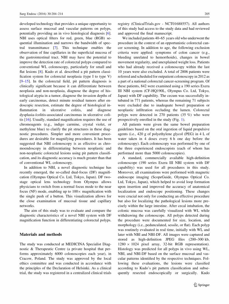
2806 patients – CRC screening in 2012
842 colonoscopies usingOlympus CF-HQ190L
71 incomplete colonoscopies(no cecal intubation)
- poor bowel preparation- neoplastic stenosis
771 complete colonoscopiesperformed
501 colonoscopies – no polypdetected
270 colonoscopies with polypdetection
1964 colonoscopiesusing otherinstruments
Fig. 1 CONSORT diagram of patient enrollment
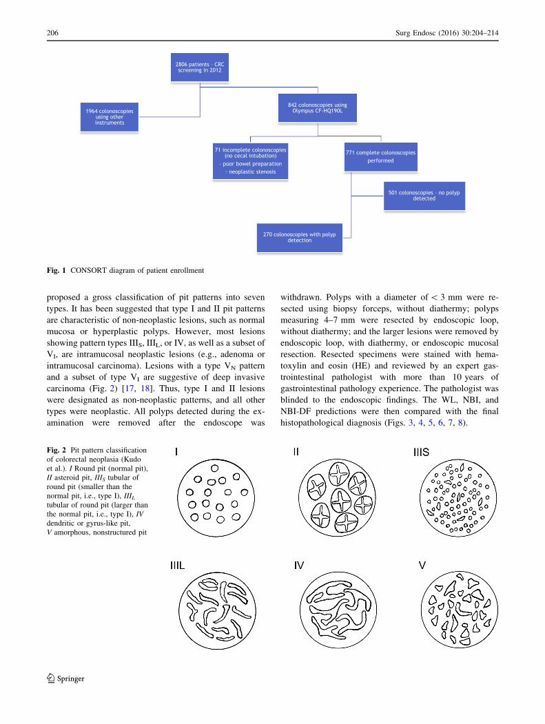
Fig. 2 Pit pattern classification
of colorectal neoplasia (Kudo
et al.). I Round pit (normal pit),
II asteroid pit, IIIS tubular of
round pit (smaller than the
normal pit, i.e., type I), IIILtubular of round pit (larger than
the normal pit, i.e., type I), IV
dendritic or gyrus-like pit,
V amorphous, nonstructured pit
206 Surg Endosc (2016) 30:204–214
123
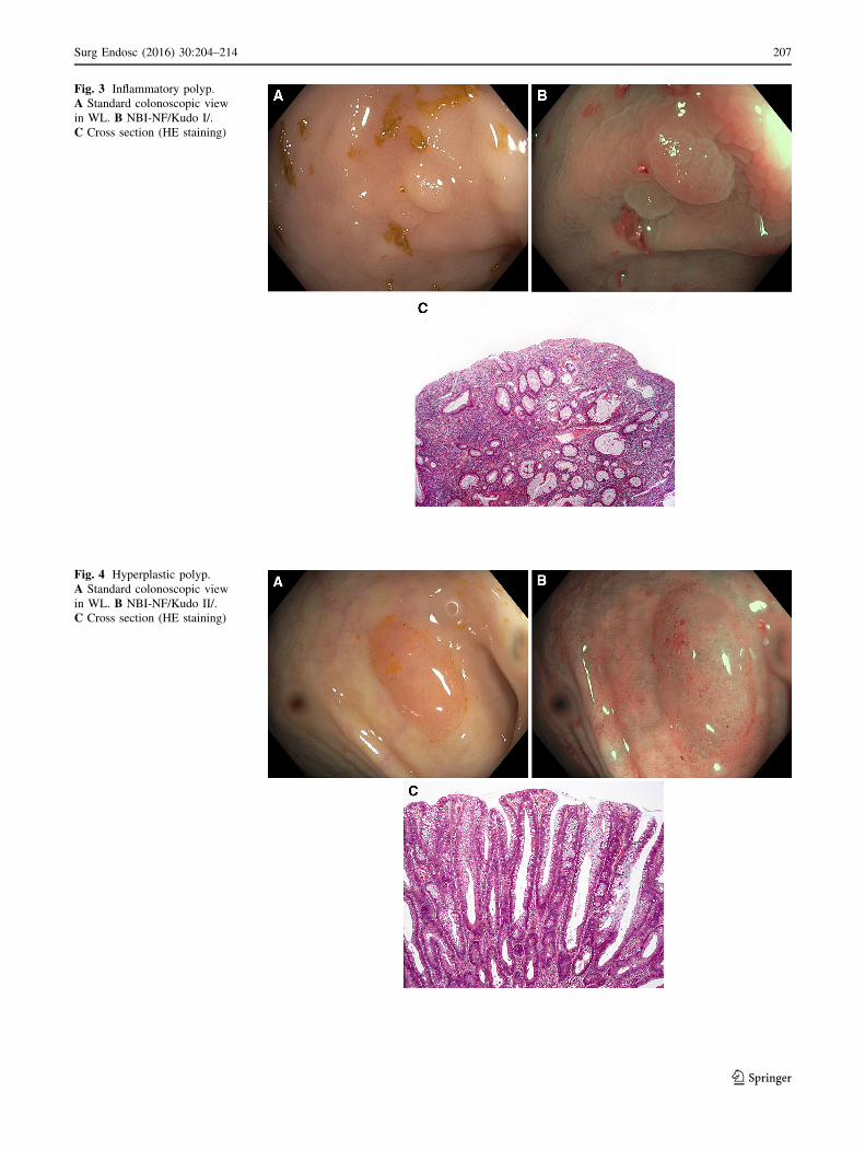
Fig. 3 Inflammatory polyp.
A Standard colonoscopic view
in WL. B NBI-NF/Kudo I/.
C Cross section (HE staining)
Fig. 4 Hyperplastic polyp.
A Standard colonoscopic view
in WL. B NBI-NF/Kudo II/.
C Cross section (HE staining)
Surg Endosc (2016) 30:204–214 207
123
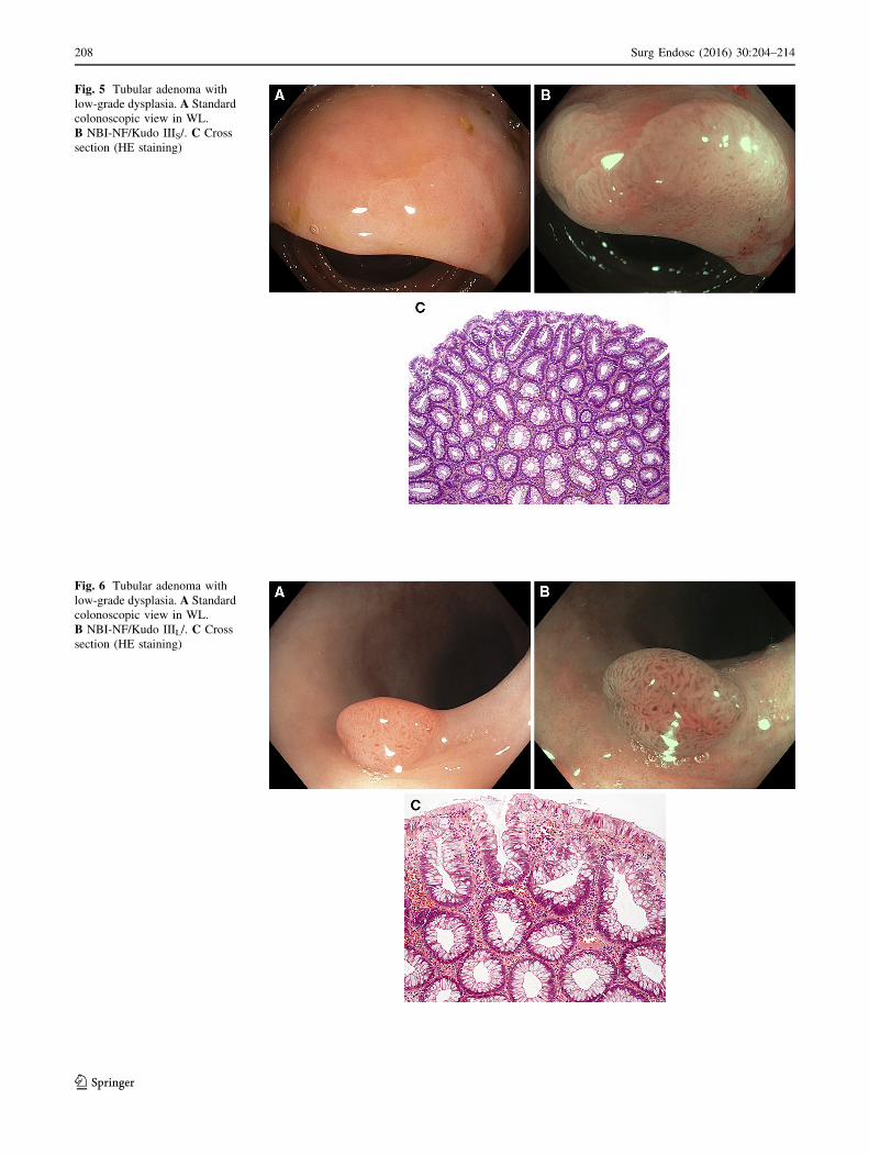
Fig. 5 Tubular adenoma with
low-grade dysplasia. A Standard
colonoscopic view in WL.
B NBI-NF/Kudo IIIS/. C Cross
section (HE staining)
Fig. 6 Tubular adenoma with
low-grade dysplasia. A Standard
colonoscopic view in WL.
B NBI-NF/Kudo IIIL/. C Cross
section (HE staining)
208 Surg Endosc (2016) 30:204–214
123
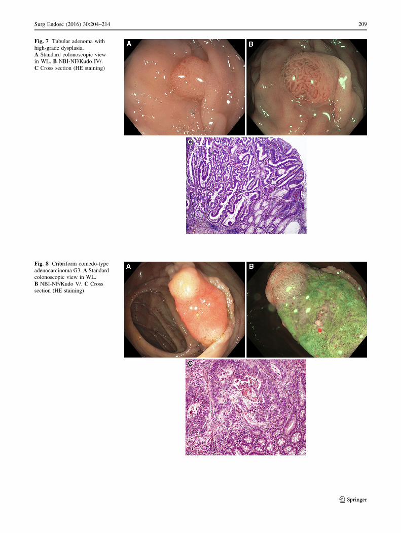
Fig. 7 Tubular adenoma with
high-grade dysplasia.
A Standard colonoscopic view
in WL. B NBI-NF/Kudo IV/.
C Cross section (HE staining)
Fig. 8 Cribriform comedo-type
adenocarcinoma G3. A Standard
colonoscopic view in WL.
B NBI-NF/Kudo V/. C Cross
section (HE staining)
Surg Endosc (2016) 30:204–214 209
123
Statistics
The materials acquired in this study were systematized and
analyzed, and a distribution of the variables was estab-
lished. Because the analyzed parameters do not have a
normal distribution, nonparametric tests were used in the
analysis. Qualitative variables were compared using the
independent v2 test. The Mann–Whitney U test was used to
compare quantitative variables between two groups. The
Kruskal–Wallis test was used for comparisons of quanti-
tative data in more than two groups. The materiality
threshold was established at p B 0.05.
Results
A total of 270 patients [128 males (47.41 %) and 142 fe-
males (52.59 %); mean age 55.04 years, ±standard de-
viation (SD) 4.06 years; range 40–65 years] were
recruited. The number of patients in each age group is
summarized in Table 1. The mean body mass index (BMI)
was 27.69 ± SD 4.06. The cecum was successfully intu-
bated in all patients (100 %), and the total time required to
reach the cecum was 55–470 s (mean 234 s). The with-
drawal time was[6 min in each case. Patients had 1 to 5
polyps detected (mean 1.43 ± SD 0.75) (Table 2). A total
of 386 polyps were detected and further analyzed macro-
scopically. The anatomical localization of the polyps was
determined based on the endoscopic image and location
presented on a ScopeGuide navigation system display.
Among the analyzed patients, the polyp detection rate in
particular locations of the large intestine was similar in
males and females. The most frequent polyp locations were
the sigmoid colon and sigmorectal junction [177 lesions
(45.85 %) in total]. The distribution of the remaining le-
sions was as follows: rectum, 86 (22.28 %); descending
colon and splenic flexure, 34 (8.81 %); transverse colon, 26
(6.74 %); ascending colon and hepatic flexure, 51
(13.21 %); and cecum, 12 (3.11 %) (Table 3). The ma-
jority of detected lesions were small polyps with a diameter
measuring \10 mm (87.82 %). The morphological shape
of the polyps was evaluated using the Paris classification
[19]. A total of 143 polyps (37 %) were pedunculated or
subpedunculated (Paris types Ip and Ips, respectively); 188
(49 %) polyps were sessile (Paris type Is); and the re-
maining 55 polyps (14 %) were superficial and elevated
(Paris types IIa, b, c) (Table 4). The most numerous group
consisted of sessile lesions with diameters up to 6 mm
(Paris type Is), representing 44 % of the total number of
lesions (170) (Table 4). All detected polyps were removed
and subsequently evaluated histopathologically. Final his-
tology showed that 142 polyps (36.79 %) were hyper-
plastic; 214 polyps were low-grade adenomas (55.44 %);
19 were high-grade adenomas (4.92 %); and 11 were
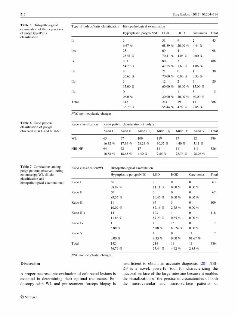
adenocarcinomas (2.85 %) (Table 5). The histopatho-
logical distribution of polyps in terms of their macroscopic
appearance is shown in Table 5.
During the endoscopic examination, we evaluated the
polyp structure visible by standard WL imaging and then
by NBI. Next, we assessed the polyp structure using optical
magnification, the DF option. The structural evaluation was
based on Kudo’s pit pattern classification. After WL
imaging, 33.7 % of the detected polyps were potentially
non-neoplastic (Kudo I, 16.32 % and Kudo II, 17.36 %);
63.2 % of the polyps were noninvasive (Kudo IIIL,
28.24 %; Kudo IIIS, 30.57 %; and Kudo IV, 4.4 %); and
3.11 % of the polyps were invasive (Kudo V). Using NBI
imaging together with the DF function (NBI-DF), we de-
tected 35.2 % potentially non-neoplastic polyps (Kudo I,
16.58 % and Kudo II, 18.65 %), 36 % noninvasive (Kudo
IIIL, 28.76 %; Kudo IIIS, 28.76 %; and Kudo IV, 4.4 %),
and 2.85 % invasive polyps (Kudo V) (Table 6).
Table 1 Number of patients by age groups
Sex Age groups (years)
40–45 46–50 51–55 56–60 61–65 Total
Male 15 20 25 40 28 128
11.72 % 15.62 % 19.53 % 31.25 % 21.88 %
Female 13 20 36 34 39 142
9.15 % 14.09 % 25.35 % 23.94 % 27.47 %
Total 28 40 61 74 67 270
10.37 % 14.81 % 22.59 % 27.42 % 24.81 %
Table 2 Number of polyps
detected during colonoscopy for
males and females
Sex Number of polyps detected during colonoscopy
1 polyp 2 polyps 3 polyps 4 polyps 5 polyps Total
Male 78 35 10 4 1 128
60.94 % 27.34 % 7.81 % 3.13 % 0.78 %
Female 108 25 8 0 1 142
76.06 % 17.61 % 5.63 % 0.00 % 0.70 %
Total 186 60 18 4 2 270
68.89 % 22.22 % 6.67 % 1.48 % 0.74 %
210 Surg Endosc (2016) 30:204–214
123
We performed a comparative analysis of the polyps
observed during colonoscopies and the histopathological
results. Regarding the histopathological diagnosis of hy-
perplastic polyps in WL, we noticed a predominant ma-
jority of polyps with Kudo I (88.89 %) or Kudo II
(89.55 %) pit patterns, and low-grade adenomas were pri-
marily predicted as Kudo IIIL (87.16 %) and Kudo IIIS
(87.29 %). High-grade dysplasia polyps (4.92 %) were
described in vivo as Kudo IV (88.24 %). For malignant
lesions, the mucosal pattern was usually designated as in-
vasive pit pattern Kudo V (91.67 %) (Table 7).
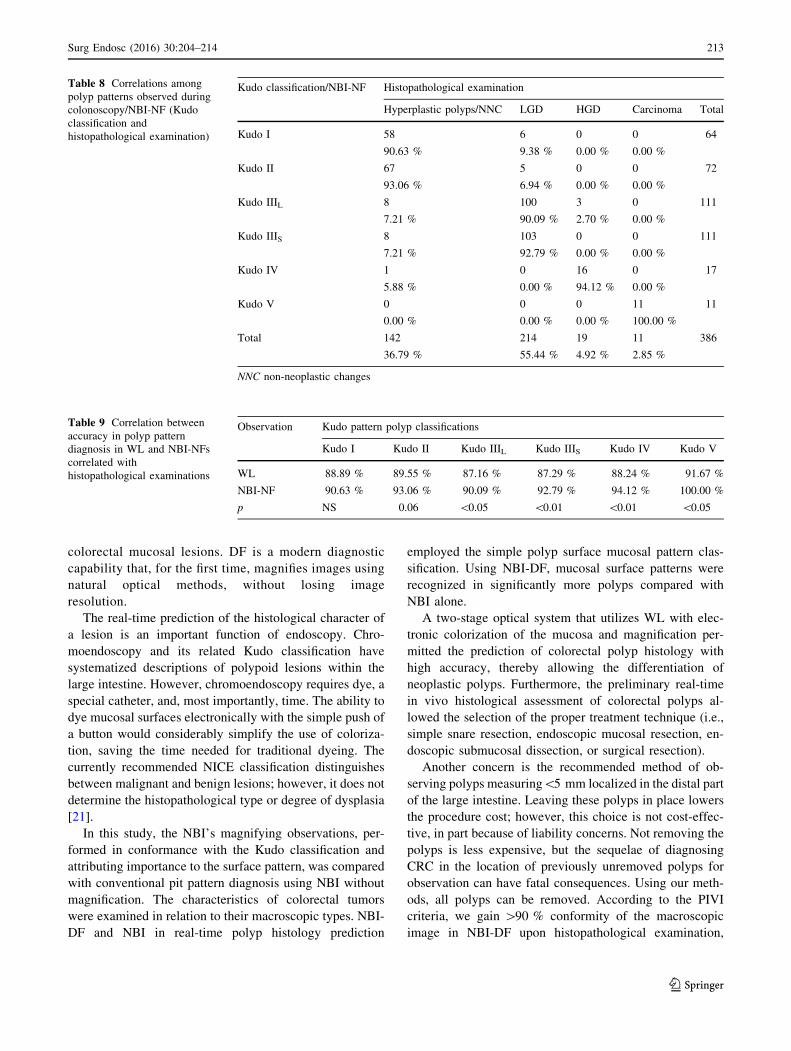
On NBI imaging with magnification, 90.63 % of the polyps
designated as Kudo I were hyperplastic. Furthermore, 93.06 %
of the polyps classified as Kudo II were non-neoplastic;
90.09 % of Kudo IIIL and 92 % of Kudo IIIS polyps were low-
grade adenomas; and 94.12 % polyps with noninvasive pit
patterns (Kudo IV) were high-grade adenomas. All Kudo V
polyps with an invasive pit pattern had a structure of adeno-
carcinoma upon histopathological examination (Table 8).
We evaluated the accuracy of real-time polyp type
recognition with WL imaging compared to NBI-NF
imaging. We noticed no difference between the diagnostic
accuracy of Kudo I and II polyps. We observed improved
accuracy in the preliminary diagnoses of the Kudo IIIL
lesions (from 87.16 to 90.09 %, p\ 0.05) and the Kudo
IIIS lesions (from 87.29 to 92.79 %, p\ 0.01). In the case
of Kudo IV pit patterns, NBI-DF increased the accuracy of
preliminary diagnoses from 88.24 to 94.12 % (p\ 0.01).
Kudo V pit patterns were also more distinct on NBI-DF
imaging, improving from 91.67 to 100 % (Table 9).
Table 3 Number of polyps by
anatomical localizationAnatomical localization in the colon and rectum Sex
Male Female Total
Cecum 7 5 12
3.52 % 2.67 %
Ascending colon and hepatic flexure 25 26 51
12.56 % 13.90 %
Transverse colon 15 11 26
7.54 % 5.88 %
Descending colon and splenic flexure 12 22 34
6.03 % 11.76 %
Sigmoid colon and sigmorectal junction 96 81 177
48.24 % 43.32 %
Rectum 44 42 86
22.11 % 22.46 %
Total 199 187 386
51.55 % 48.45 %
Table 4 Number of polyps
depending on the type of
polyps/Paris classification
Type of polyps/Paris classification Size of polyps in mm
1–3 mm 4–6 mm 7–9 mm [= 10 Total
Ip 1 12 13 19 45
2.22 % 26.67 % 28.89 % 42.22 %
Ips 9 70 14 5 98
9.18 % 71.43 % 14.29 % 5.10 %
Is 81 89 8 10 188
43.09 % 47.34 % 4.26 % 5.32 %
IIa 2 18 7 3 30
6.67 % 60.00 % 23.33 % 10.00 %
IIb 2 7 3 8 20
10.00 % 35.00 % 15.00 % 40.00 %
IIc 0 1 2 2 5
0.00 % 20.00 % 40.00 % 40.00 %
Total 95 197 47 47 386
24.61 % 51.04 % 12.18 % 12.18 %
Surg Endosc (2016) 30:204–214 211
123
Discussion
A proper macroscopic evaluation of colorectal lesions is
essential in determining their optimal treatments. En-
doscopy with WL and pretreatment forceps biopsy is
insufficient to obtain an accurate diagnosis [20]. NBI-
DF is a novel, powerful tool for characterizing the
mucosal surface of the large intestine because it enables
the visualization of the precise microanatomies of both
the microvascular and micro-surface patterns of
Table 5 Histopathological
examination of the dependence
of polyp type/Paris
classification
Type of polyps/Paris classification Histopathological examination
Hyperplastic polyps/NNC LGD HGD carcinoma Total
Ip 3 31 9 2 45
6.67 % 68.89 % 20.00 % 4.44 %
Ips 25 69 4 0 98
25.51 % 70.41 % 4.08 % 0.00 %
Is 103 80 3 2 188
54.79 % 42.55 % 1.60 % 1.06 %
IIa 8 21 0 1 30
26.67 % 70.00 % 0.00 % 3.33 %
IIb 3 12 2 3 20
15.00 % 60.00 % 10.00 % 15.00 %
IIc 0 1 1 3 5
0.00 % 20.00 % 20.00 % 60.00 %
Total 142 214 19 11 386
36.79 % 55.44 % 4.92 % 2.85 %
NNC non-neoplastic changes
Table 6 Kudo pattern
classification of polyps
observed in WL and NBI-NF
Kudo classification Kudo pattern classification of polyps
Kudo I Kudo II Kudo IIIL Kudo IIIS Kudo IV Kudo V Total
WL 63 67 109 118 17 12 386
16.32 % 17.36 % 28.24 % 30.57 % 4.40 % 3.11 %
NBI-NF 64 72 17 11 111 111 386
16.58 % 18.65 % 4.40 % 2.85 % 28.76 % 28.76 %
Table 7 Correlations among
polyp patterns observed during
colonoscopy/WL (Kudo
classification and
histopathological examinations)
Kudo classification/WL Histopathological examination
Hyperplastic polyps/NNC LGD HGD Carcinoma Total
Kudo I 56 7 0 0 63
88.89 % 11.11 % 0.00 % 0.00 %
Kudo II 60 7 0 0 67
89.55 % 10.45 % 0.00 % 0.00 %
Kudo IIIL 11 95 3 0 109
10.09 % 87.16 % 2.75 % 0.00 %
Kudo IIIs 14 103 1 0 118
11.86 % 87.29 % 0.85 % 0.00 %
Kudo IV 1 1 15 0 17
5.88 % 5.88 % 88.24 % 0.00 %
Kudo V 0 1 0 11 12
0.00 % 8.33 % 0.00 % 91.67 %
Total 142 214 19 11 386
36.79 % 55.44 % 4.92 % 2.85 %
NNC non-neoplastic changes
212 Surg Endosc (2016) 30:204–214
123
colorectal mucosal lesions. DF is a modern diagnostic
capability that, for the first time, magnifies images using
natural optical methods, without losing image
resolution.
The real-time prediction of the histological character of
a lesion is an important function of endoscopy. Chro-
moendoscopy and its related Kudo classification have
systematized descriptions of polypoid lesions within the
large intestine. However, chromoendoscopy requires dye, a
special catheter, and, most importantly, time. The ability to
dye mucosal surfaces electronically with the simple push of
a button would considerably simplify the use of coloriza-
tion, saving the time needed for traditional dyeing. The
currently recommended NICE classification distinguishes
between malignant and benign lesions; however, it does not
determine the histopathological type or degree of dysplasia
[21].
In this study, the NBI’s magnifying observations, per-
formed in conformance with the Kudo classification and
attributing importance to the surface pattern, was compared
with conventional pit pattern diagnosis using NBI without
magnification. The characteristics of colorectal tumors
were examined in relation to their macroscopic types. NBI-
DF and NBI in real-time polyp histology prediction
employed the simple polyp surface mucosal pattern clas-
sification. Using NBI-DF, mucosal surface patterns were
recognized in significantly more polyps compared with
NBI alone.
A two-stage optical system that utilizes WL with elec-
tronic colorization of the mucosa and magnification per-
mitted the prediction of colorectal polyp histology with
high accuracy, thereby allowing the differentiation of
neoplastic polyps. Furthermore, the preliminary real-time
in vivo histological assessment of colorectal polyps al-
lowed the selection of the proper treatment technique (i.e.,
simple snare resection, endoscopic mucosal resection, en-
doscopic submucosal dissection, or surgical resection).
Another concern is the recommended method of ob-
serving polyps measuring\5 mm localized in the distal part
of the large intestine. Leaving these polyps in place lowers
the procedure cost; however, this choice is not cost-effec-
tive, in part because of liability concerns. Not removing the
polyps is less expensive, but the sequelae of diagnosing
CRC in the location of previously unremoved polyps for
observation can have fatal consequences. Using our meth-
ods, all polyps can be removed. According to the PIVI
criteria, we gain [90 % conformity of the macroscopic
image in NBI-DF upon histopathological examination,
Table 8 Correlations among
polyp patterns observed during
colonoscopy/NBI-NF (Kudo
classification and
histopathological examination)
Kudo classification/NBI-NF Histopathological examination
Hyperplastic polyps/NNC LGD HGD Carcinoma Total
Kudo I 58 6 0 0 64
90.63 % 9.38 % 0.00 % 0.00 %
Kudo II 67 5 0 0 72
93.06 % 6.94 % 0.00 % 0.00 %
Kudo IIIL 8 100 3 0 111
7.21 % 90.09 % 2.70 % 0.00 %
Kudo IIIS 8 103 0 0 111
7.21 % 92.79 % 0.00 % 0.00 %
Kudo IV 1 0 16 0 17
5.88 % 0.00 % 94.12 % 0.00 %
Kudo V 0 0 0 11 11
0.00 % 0.00 % 0.00 % 100.00 %
Total 142 214 19 11 386
36.79 % 55.44 % 4.92 % 2.85 %
NNC non-neoplastic changes
Table 9 Correlation between
accuracy in polyp pattern
diagnosis in WL and NBI-NFs
correlated with
histopathological examinations
Observation Kudo pattern polyp classifications
Kudo I Kudo II Kudo IIIL Kudo IIIS Kudo IV Kudo V
WL 88.89 % 89.55 % 87.16 % 87.29 % 88.24 % 91.67 %
NBI-NF 90.63 % 93.06 % 90.09 % 92.79 % 94.12 % 100.00 %
p NS 0.06 \0.05 \0.01 \0.01 \0.05
Surg Endosc (2016) 30:204–214 213
123
regardless of the polyp size [22]. Nevertheless, the authors
admit that this result does not sufficiently support the ter-
mination of endoscopic polypectomy.
In conclusion, this study shows that NBI with optical
magnification is highly accurate in predicting polyp his-
tology in real time using a simple pattern classification
system. Characteristic pit patterns obtained by magnifying
NBI endoscopy provide useful clues regarding the differ-
entiation of adenomatous from non-adenomatous polyps
in vivo, without using dye. The pit pattern observations
using magnifying NBI colonoscopy were also useful for
assessing the resected margins after polypectomy or en-
doscopic mucosal resection. It may be necessary to perform
subsequent management procedures, such as hot biopsies
or argon plasma coagulation procedures, when neoplastic
pit patterns (Kudo’s IIIL or IV pits) are recognized at the
margins of the resected tumor. In the future, NBI might
contribute to real-time histological analysis during
colonoscopy, which could substantially reduce the risk of
polypectomy and the costs of histological evaluation by
allowing adenomatous polyps to be resected and discarded.
Used appropriately in experienced hands, this technique
has potential as a valuable adjunct to standard colonoscopy
in predicting the histological characteristics of colorectal
polyps.
Acknowledgments The research project reported in this manuscript
has been fully sponsored by the authors.
Disclosures Mirosław Szura declares no conflict of interests. Artur
Pasternak declares no conflict of interests. Krzysztof Bucki declares
no conflict of interests. Katarzyna Urbanczyk declares no conflict of
interests. Andrzej Matyja declares no conflict of interests.
Open Access This article is distributed under the terms of the
Creative Commons Attribution 4.0 International License (http://
creativecommons.org/licenses/by/4.0/), which permits unrestricted
use, distribution, and reproduction in any medium, provided you give
appropriate credit to the original author(s) and the source, provide a
link to the Creative Commons license, and indicate if changes were
made.
References
1. Jemal A, Bray F, Center MM, Ferlay J, Ward E, Forman D (2011)
Global cancer statistics. CA Cancer J Clin 61:69–90
2. Allen JI (1995) Molecular biology of colon polyps and colon
cancer. Semin Surg Oncol 11:399–405
3. Carethers JM (1996) The cellular and molecular pathogenesis of
colorectal cancer. Gastroenterol Clin North Am 25:737–754
4. US Preventive Services Task Force (2008) Screening for col-
orectal cancer: US Preventive Services Task Force recommen-
dation statement. Ann Intern Med 149:627–637
5. McFarland EG, Levin B, Lieberman DA, Pickhardt PJ, Johnson
CD, Glick SN, Brooks D, Smith RA (2008) Revised colorectal
screening guidelines: joint effort of the American Cancer Society,
US Multisociety Task Force on Colorectal Cancer, and American
College of Radiology. Radiology 248:717–720
6. Rastogi A, Keighley J, Singh V, Callahan P, Bansal A, Wani S,
Sharma P (2009) High accuracy of narrow band imaging without
magnification for the real-time characterization of polyp histology
and its comparison with high-definition white light colonoscopy: a
prospective study. Am J Gastroenterol 104:2422–2430
7. Gono K, Obi T, Yamaguchi M, Ohyama N, Machida H, Sano Y,
Yoshida S, Hamamoto Y, Endo T (2004) Appearance of en-
hanced tissue features in narrow-band endoscopic imaging.
J Biomed Opt 9:568–577
8. Byeon JS, Kim JS, Lee CK, Park JM, Chang DK, Kim YB (2008)
Narrow band imaging in the detection of colorectal polyp:
Korean experience. Digest Endosc 20:61–66
9. Kudo S, Hirota S, Nakajima T, Hosobe S, Kusaka H, Kobayashi
T, Himori M, Yagyuu A (1994) Colorectal tumors and pit pattern.
J Clin Pathol 47:880–885
10. Kudo S, Tamura S, Nakajima T, Yamano H, Kusaka H, Watan-
abe H (1996) Diagnosis of colorectal tumour lesions by magni-
fying endoscopy. Gastrointest Endosc 44:8–14
11. Tsuji Y (1998) Usefulness of magnifying endoscopy for diag-
nosing tumorous lesions of the colorectum. Kurume Med J
45:87–94
12. Tanaka S, Haruma K, Nagata S, Oka S, Chayama K (2001) Di-
agnosis of invasion depth in early colorectal carcinoma by pit
pattern analysis with magnifying endoscopy. Digest Endosc
13:S2–5
13. Tanaka S, Haruma K, Ito M, Nagata S, Oh-e H, Hirota Y, Ku-
nihiro M, Kitadai Y, Yosihara M, Sumii K, Kajiyama G (2000)
Detailed colonoscopy for detecting early superficial carcinoma:
recent developments. J Gastroenterol 35:121–125
14. Nagata S, Tanaka S, Haruma K, Yoshihara M, Sumii K, Ka-
jiyama G, Shimamoto F (2000) Pit pattern diagnosis of colorectal
carcinoma by magnifying colonoscopy: clinical and histological
implications. Int J Oncol 16:927–934
15. Oka S, Tanaka S, Nagata S (2005) Relationship between
histopathological features and type V pit pattern determined by
magnifying videocolonoscopy in early colorectal carcinoma.
Digest Endosc 17:117–122
16. Tanaka S, Oka S, Hirata M (2006) Pit pattern diagnosis for col-
orectal neoplasia using narrow band imaging magnification.
Digest Endosc 18:S52–56
17. Tanaka S, Yoshida S, Chayama K (2004) Clinical usefulness of
high-frequency ultrasound probes for new invasion diagnosis in
submucosal colorectal carcinoma. Digest Endosc 16:S161–164
18. Tanaka S, Kaltenbach T, Chayama K, Soetikno R (2006) High-
magnification colonoscopy (with videos). Gastrointest Endosc
64:604–613
19. Endoscopic Classification Review Group (2005) Update on the
Paris classification of superficial neoplastic lesions in the diges-
tive tract. Endoscopy 37:570–578
20. Miwa K, Doyama H, Ito R, Nakanishi H, Hirano K, Inagaki S,
Tominaga K, Yoshida N, Takemura K, Yamada S, Kaneko Y,
Katayanagi K, Kurumaya H, Okada T, Yamagishi M (2012) Can
magnifying endoscopy with narrow band imaging be useful for
low grade adenomas in preoperative biopsy specimens? Gastric
Cancer 15:170–178
21. Hewett DG, Kaltenbach T, Sano Y, Tanaka S, Saunders BP, Pon-
chon T, Soetikno R, Rex DK (2012) Validation of a simple classi-
fication system for endoscopic diagnosis of small colorectal polyps
using narrow-band imaging. Gastroenterology 143:599–607
22. Rex DK, Kahi C, O’Brien M, Levin TR, Pohl H, Rastogi A,
Burgart L, Imperiale T, Ladabaum U, Cohen J, Lieberman DA
(2011) The American Society for Gastrointestinal Endoscopy
PIVI on real-time endoscopic assessment of the histology of
diminutive colorectal polyps. Gastrointest Endosc 73:419–422
214 Surg Endosc (2016) 30:204–214
123