two new faces of amifostine: protector from dna damage in ...€¦ · doi:...
TRANSCRIPT

Seediscussions,stats,andauthorprofilesforthispublicationat:https://www.researchgate.net/publication/303008336
TwoNewFacesofAmifostine:ProtectorfromDNADamageinNormalCellsandInhibitorofDNARepairinCancerCells
ArticleinJournalofMedicinalChemistry·April2016
DOI:10.1021/acs,jmedchem.5b01628
CITATION
1
READS
33
13authors,including:
MichalHofer
InstituteofBiophysicsPrague
111PUBLICATIONS1,156CITATIONS
SEEPROFILE
MartinFalk
InstituteofBiophysics,AcademyofScienceso…
46PUBLICATIONS590CITATIONS
SEEPROFILE
BorivojKlejdus
MendelUniversityinBrno
70PUBLICATIONS2,412CITATIONS
SEEPROFILE
KarelJAngelis
AcademyofSciencesoftheCzechRepublic
69PUBLICATIONS1,354CITATIONS
SEEPROFILE
Allin-textreferencesunderlinedinbluearelinkedtopublicationsonResearchGate,
lettingyouaccessandreadthemimmediately.
Availablefrom:MartinFalk
Retrievedon:26October2016

Two New Faces of Amifostine: Protector from DNA Damage inNormal Cells and Inhibitor of DNA Repair in Cancer CellsMichal Hofer,† Martin Falk,*,† Denisa Komurkova,† Iva Falkova,†,‡ Alena Bacíkova,† Borivoj Klejdus,§,∥
Eva Pagacova,† Lenka Stefancíkova,† Lenka Weiterova,† Karel J. Angelis,⊥ Stanislav Kozubek,†
Ladislav Dusek,# and Stefan Galbavy ‡
†Department of Cell Biology and Radiobiology, Institute of Biophysics, v.v.i., Czech Academy of Sciences, Kralovopolska 135, CZ-61265 Brno, Czech Republic‡Department of Medical Technology, St. Elisabeth University of Health and Social Sciences, Palackeho 1, SK-810 00 Bratislava,Slovak Republic§Institute of Chemistry and Biochemistry, Faculty of Agronomy, and ∥CEITEC-Central European Institute of Technology, MendelUniversity in Brno, Zemedelska 1, CZ-613 00 Brno, Czech Republic⊥Institute of Experimental Botany, v.v.i., Czech Academy of Sciences, Na Karlovce 1, CZ-160 00 Prague 6, Czech Republic#Institute of Biostatistics and Analyses, Masaryk University, Kamenice 126/3, CZ-625 00 Brno, Czech Republic
*S Supporting Information
ABSTRACT: Amifostine protects normal cells from DNA damage induction byionizing radiation or chemotherapeutics, whereas cancer cells typically remainuninfluenced. While confirming this phenomenon, we have revealed by comet assayand currently the most sensitive method of DNA double strand break (DSB)quantification (based on γH2AX/53BP1 high-resolution immunofluorescencemicroscopy) that amifostine treatment supports DSB repair in γ-irradiated normalNHDF fibroblasts but alters it in MCF7 carcinoma cells. These effects follow from thesignificantly lower activity of alkaline phosphatase measured in MCF7 cells and theirsupernatants as compared with NHDF fibroblasts. Liquid chromatography−massspectrometry confirmed that the amifostine conversion to WR-1065 was significantlymore intensive in normal NHDF cells than in tumor MCF cells. In conclusion, due tocommon differences between normal and cancer cells in their abilities to convert amifostine to its active metabolite WR-1065,amifostine may not only protect in multiple ways normal cells from radiation-induced DNA damage but also make cancer cellssuffer from DSB repair alteration.
1. INTRODUCTIONAmifostine (ethanethiol, 2-[(3-aminopropyl)amino]dihydrogenphosphate), also known as WR-2721, is an organicthiophosphate agent; it is rapidly dephosphorylated by alkalinephosphatase (ALP) at the cell surface of healthy tissues, givingrise to its clinically active metabolite, WR-1065.1−3 Whenactivated, amifostine protects cells from radiation- andchemotherapy-induced DNA damage, mostly by competingwith oxygens and preventing their interactions with DNAradicals and donating hydrogen to repair the already existingDNA damage.4,5 Currently, amifostine is the only radio-protective drug approved for clinical use.Concerning the practical application of amifostine in human
medicine, a key role has been ascribed to its differential effecton cancer and normal cells, respectively: Whereas in normalcells or tissues amifostine clearly acts as a radio- andchemoprotective agent, this property of the drug is lost incancer cells.6−12 This cell type-specific behavior of amifostinehas been largely attributed to low levels of ALP in cancer cellsas compared with normal cells;13 however, the situation is stillnot that clear because a variety of human cancers ectopically
express high levels of ALP, thus leading some scientists to the(opposite) suggestion that ALP might be critically involved intumor development.14−16 Indeed, a comprehensive comparisonof ALP mRNA and protein expression and activity in cancercells in the literature is missing. This uncertainty points to thecaution with which each model of disease (e.g., different celltypes) should be tested.In addition, it is still not obvious how the four main classes of
ALP (tissue nonspecific TNAP, intestinal IAP, placental PLAP,and placental-like GCAP)15 participate in amifostine con-version in various normal and especially cancer cells whereexpression of ALP isoenzymes may be altered.17 ALP genes arealso highly inducible by many agents.15 Finally, thoughcovalently anchored to the outer surface of the plasmamembrane,16 ALP can be released into the serum (orextracellular medium) by the GPI-dependent phospholipaseD under stress and some medical conditions, such as cancer.While previous reports showed that the membrane-bound ALP
Received: October 16, 2015Published: March 15, 2016
Article
pubs.acs.org/jmc
© 2016 American Chemical Society 3003 DOI: 10.1021/acs.jmedchem.5b01628J. Med. Chem. 2016, 59, 3003−3017
has different enzymatic kinetics and molecular properties ascompared to the soluble enzyme,16,18 the question remains ofhow this finding is reflected in amifostine metabolism in normaland cancer tissues.The most serious radiation-induced DNA damage is the
double strand break (DSB) that causes a loss of the DNAmolecule integrity. As DSB formation is also modulated byamifostine,19,20 DSBs represent the most relevant type of DNAlesions in the context of amifostine-mediated cell radio-protection and radiosensitization. Molecular events followingthe ionizing radiation-induced DNA breakage include animmediate phosphorylation of histone H2AX (ser139),21,22
which can be nowadays used to immunologically visualize theextent of DSB damage induction in intact cells as the so-calledγH2AX foci within minutes after the DSB induction.23−25 Onthe other hand, assessment of γH2AX foci disappearance duringthe postirradiation (PI) time allows monitoring of DSB repair.In this work, we advantageously employ this method,
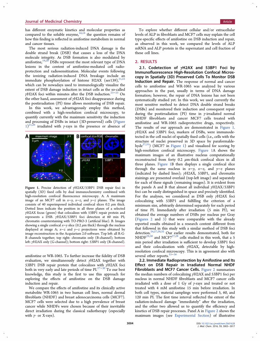
combined with a high-resolution confocal microscopy, toquantify currently with the maximum sensitivity the inductionand processing of DSBs in intact (3D-preserved) cells (Figure1)21,26 irradiated with γ-rays in the presence or absence of
amifostine or WR-1065. To further increase the fidelity of DSBevaluation, we simultaneously detect γH2AX together with53BP1 DSB repair protein that colocalizes with γH2AX fociboth in very early and late periods of time PI.27−30 To our bestknowledge, this study is the first to use this approach forexploring the effects of amifostine on the DSB damageinduction and repair.We compare the effects of amifostine and its clinically active
metabolite WR-1065 in two human cell lines, normal dermalfibroblasts (NHDF) and breast adenocarcinoma cells (MCF7).MCF7 cells were selected due to a high prevalence of breastcancer while NHDFs were chosen because of their inevitabledirect irradiation during the classical radiotherapy (especiallywith γ- or X-rays).
To explore whether different cellular and/or extracellularlevels of ALP in fibroblasts and MCF7 cells may explain the celltype-specific effects of amifostine on DSB induction and repair,as observed in this work, we compared the levels of ALPmRNA and ALP protein in the supernatant and cell fraction ofthese cell lines.
2. RESULTS2.1. Codetection of γH2AX and 53BP1 Foci by
Immunofluorescence High-Resolution Confocal Micros-copy in Spatially (3D) Preserved Cells To Monitor DSBInduction and Repair. The response of normal and cancercells to amifostine and WR-1065 was analyzed by variousapproaches in the past, usually in terms of DNA damageformation; however, the repair of DNA lesions has not beensystematically studied yet. In this work, we used currently themost sensitive method to detect DNA double strand breaks(DSBs) and monitored their induction and consequent repairduring the postirradiation (PI) time in γ-irradiated normalNHDF fibroblasts and cancer MCF7 cells treated withamifostine and WR-1065 radioprotective drugs, respectively.The results of our approach are demonstrated in Figure 1.γH2AX and 53BP1 foci, markers of DSBs, were immunode-tected in the cell nuclei of spatially fixed cells (i.e., cells with thestructure of nuclei preserved in 3D space by paraformalde-hyde27,31) (MCF7 in Figure 1) and visualized for scoring byhigh-resolution confocal microscopy. Figure 1A shows themaximum images of an illustrative nucleus computationallyreconstructed from forty 0.2 μm-thick confocal slices in allthree planes. Figure 1B then displays a single confocal slicethrough the same nucleus in x−y, x−z, and y−z planes(indicated by dashed lines); γH2AX, 53BP1, and chromatinstainings are presented overlaid (top-left image) and separatelyfor each of these signals (remaining images). It is evident fromthe panels A and B that almost all individual γH2AX/53BP1foci can be easily distinguished in space and precisely identified.For the analyses, we considered as DSB only γH2AX focicolocalizing with 53BP1 and fulfilling the criterion of aminimum size, arbitrarily determined separately for each periodof time PI. Immediately after irradiation (5 min PI), weobtained the average numbers of DSBs per nucleus per Gray(Figures 2 and 3) that were comparable with the alreadyreported results obtained in a research context different fromthat followed in this study with a similar method of DSB focidetection.24,27,28,32 Our earlier results demonstrated, both forNHDF29,30 and MCF727,28 cells studied in this work, that a 5min period after irradiation is sufficient to develop 53BP1 fociand their colocalization with γH2AX, detectable by high-resolution confocal microscopy. This is in agreement also withseveral other reports.33−36
2.2. Immediate Radioprotection by Amifostine and ItsEffect on DSB Repair in Irradiated Normal NHDFFibroblasts and MCF7 Cancer Cells. Figure 2 summarizesthe median numbers of colocalizing γH2AX and 53BP1 foci pernucleus in normal NHDF fibroblasts and MCF7 cancer cellsirradiated with a dose of 1 Gy of γ-rays and treated or nottreated with 4 mM amifostine 15 min before irradiation. Inboth cell types, material samplings were performed 5, 60, and120 min PI. The first time interval reflected the extent of theradiation-induced damage “immediately” after the irradiation,and the other two allowed us to quantify the efficiency andkinetics of DSB repair processes. Panel A in Figure 2 shows themaximum images (see Experimental Section) of illustrative
Figure 1. Precise detection of γH2AX/53BP1 DSB repair foci inspatially (3D) fixed cells by dual immunochemistry combined withhigh-resolution confocal fluorescence microscopy. A. A maximumimage of an MCF7 cell in x−y, x−z, and y−z planes. The imageconsists of 40 superimposed individual confocal slices 0.2 μm thick.Dotted lines indicate the position in all three planes of a selectedγH2AX focus (green) that colocalizes with 53BP1 repair protein andrepresents a DSB. γH2AX/53BP1 foci detection at 60 min PI;chromatin counterstaining with TO-PRO-3 (artificial blue). B. Imagesshowing a single confocal x−y slice (0.2 μm thick) through the nucleusdisplayed at image A; x−z and y−z projections were obtained byimage reconstruction in the Acquiarium 2.0 software. Top left: all R-G-B channels together; top right: chromatin only (B-channel); bottomleft: γH2AX only (G-channel); bottom right: 53BP1 only (R-channel).
Journal of Medicinal Chemistry Article
DOI: 10.1021/acs.jmedchem.5b01628J. Med. Chem. 2016, 59, 3003−3017
3004

nuclei of NHDF and MCF7 cells for each treatment and thefirst two periods of time PI analyzed (data for 120 min PI arepresented and discussed in more detail later in section 2.3,Figure 5). In normal NHDF fibroblasts, amifostine induced asignificant (P < 0.001) decrease in the median numbers ofγH2AX/53BP1 foci per nucleus in all the time intervals, for 5and 60 min PI, namely up to 75.8% and 61.9% of the values inuntreated cells, respectively. The repair efficiency determined asthe difference between the median DSB values at 5 and 60 minPI (Figure 4) corresponded to 7 DSBs in cells irradiated in theplain medium and 8 DSBs in cells irradiated in the mediumsupplemented with amifostine. This result reflects an ongoingDSB repair slightly accelerated (about 10%) by amifostine(and/or its active metabolite WR-1065, discussed later; seeFigure 4).On the other hand, cancer MCF7 cells provided a totally
different picture as compared with normal fibroblasts.Amifostine did not significantly decrease the median numbersof γH2AX/53BP1 foci per nucleus in MCF7 cells at 5 min PI
(Figure 2). In addition, as we report here for the first time for γ-irradiated intact cells, a significantly (P < 0.001) higher numberof the foci (137.5% of the value in untreated cells) surprisingly
Figure 2. Numbers of ionizing radiation-induced repair foci (γH2AX,green; 53BP1, red) demonstrating the effects of amifostine on theextent of DSB induction (5 min PI) and efficiency of DSB repair (60min PI) in normal human skin fibroblasts (NHDF) and mammarycarcinoma cells (MCF7). A. Maximum images composed of 40confocal slices (0.2 μm thick) through spatially (3D) fixed cells byparaformaldehyde are shown; chromatin counterstaining with TO-PRO-3 (artificial blue). B. Median DSB values with standard errors;statistical significance determined by the Mann−Whitney U test isindicated (*, P < 0.05; **, P < 0.01; ***, P < 0.001).
Figure 3. Numbers of ionizing radiation-induced repair foci (γH2AX,green; 53BP1, red) demonstrating the effects of WR1065 on the extentof DSB induction (5 min PI) and efficiency of DSB repair (60 min PI)in normal human skin fibroblasts (NHDF) and mammary carcinomacells (MCF7). A. Maximum images composed of 40 confocal slices(0.2 μm thick) through spatially (3D) fixed cells by paraformaldehydeare shown; chromatin counterstaining with TO-PRO-3 (artificialblue). B. Median DSB values with standard errors; statisticalsignificance determined by the Mann−Whitney U test is indicated(*, P < 0.05; **, P < 0.01; ***, P < 0.001).
Figure 4. Kinetics of DSB repair in irradiated (1 Gy of γ-rays; 1 Gy/min) NHDF and MCF7 cells treated or not with 4 mM amifostine orWR-1065 for 15 min prior to irradiation. The graph shows averagefractions of γH2AX/53BP1 foci persisting in cells at 60 min PI,determined as % of corresponding values measured at 5 min PI. Meanvalues with SEM are shown.
Journal of Medicinal Chemistry Article
DOI: 10.1021/acs.jmedchem.5b01628J. Med. Chem. 2016, 59, 3003−3017
3005

persisted in amifostine-treated MCF7 cells at 60 min PI(Figures 2 and 4) and 120 min PI (Figure 5; discussed later).Hence, amifostine treatment not only selectively protectednormal cells from DSB damage but it also markedly sloweddown the repair of lesions already induced by irradiation incancer cells (Figures 2 and 4).2.3. Immediate and Later Effects of Amifostine
Metabolite WR-1065 in Irradiated Normal NHDFFibroblasts and MCF7 Cancer Cells. In the next step, weused the same experimental approach to reveal the effects of anisomolar concentration of WR-1065 (Figure 3), an amifostinemetabolite that is supposed to mediate the radioprotectiveeffects of amifostine. Unlike in cells treated with amifostine,WR-1065 in the cultures reduced the numbers of γH2AX/53BP1 foci per nucleus in both normal and cancer cell typesand both the time intervals of 5 and 60 min PI. The percentagedecreases produced by WR-1065 for MCF7 cells amounted to75.0% (P < 0.001) and 82.9% (P < 0.05) at 5 min PI and 60min PI, respectively; for NHDF fibroblasts, these valuescorresponded to 65.2% (P < 0.05) and 50.0% (P < 0.001) at5 min PI and 60 min PI, respectively.Altogether, the immediate (5 min PI) and late (60 min PI)
response to the irradiation was comparable for normal NHDFand cancer MCF7 cells only in cultures incubated with WR-1065 (Figure 3). For amifostine, the response basicallydepended on the normal or cancer cell status. Thoughamifostine at 5 min PI induced a response similar to that ofWR-1065 (radioprotection) in the case of NHDF fibroblasts,we observed no effect of this prodrug in MCF7 cells (Figure 2).At 60 min PI, as compared with the averaged value foruntreated irradiated controls, we observed a small acceleration(about 9%) of DSB repair in NHDF fibroblasts incubated withamifostine (Figure 4). On the other hand, the speed of repairmarkedly decreased (−20.7%) in cancer MCF7 cells relative tothe average control. WR-1065 then also speeded up (17.7%)DSB repair in NHDF fibroblasts relative to the averaged
untreated control but exerted slightly opposite (−7.7%)changes in MCF7 cells.Figure 5 shows how DSB foci continued until 120 min PI in
NHDF and MCF7 cells irradiated with 1 Gy of γ-rays andtreated or not treated with amifostine, as described previously.As follows from both the inserted images and graphs showingthe distributions of γH2AX/53BP1 foci per nucleus, amifostinesupported DSB repair in normal NHDF fibroblasts while it hadthe opposite influence on cancer MCF7 cells. The differencesfrom the untreated cells were statistically significant in both celltypes though the enhancement of DSB repair in NHDFfibroblasts was less prominent (P = 0.019) than its decelerationin MCF7 cells (P < 0.001). The repair of untreated NHDF andMCF7 cells proceeded with similar efficiency (P = 0.579).
2.4. DSB Induction and Repair Analyzed by CometAssay. To verify our results obtained above with γH2AX/53BP1 foci immunostaining, we quantified DSB induction andrepair in irradiated normal NHDF and cancer MCF7 cellstreated or not treated with amifostine or WR-1065 also by thecomet assay, the method accepted in radiobiology as a “goldstandard” for this purpose. While the comet assay provides amore direct view on DNA breakage and rejoining than γH2AX/53BP1 foci immunodetection, it suffers from lower sensitivity.Therefore, we irradiated the cells with 6 Gy of γ-rays (1 Gy/min) instead of 1.0 Gy used in the immunodetectionexperiments.As demonstrated by Figure 6 and Figures S1-A and S1-B in
Supporting Information, the measurements of comet tailmoments (TM = L(T) × IF(T)/100, where L(T) = length ofthe tail and IF(T) = the fluorescence intensity of the tail given asa percentage; Figure 6B) well correlated at all the periods oftime postirradiation with the results that followed fromγH2AX/53BP1 foci immunodetection. Shortly (5 min) afterirradiation, WR-1065 markedly decreased DSB induction inNHDF (p < 0.01) and MCF7 (p < 0.001) cells but amifostineonly protected the first cells (p < 0.01 for NHDF vs p = 0.553for MCF7). In later periods of time PI (60 and 120 min PI)
Figure 5. Distributions of the numbers of ionizing radiation-induced repair foci (γH2AX, green; 53BP1, red) persisting 2 h postirradiation in nucleiof NHDF and MCF7 cells, respectively, exposed to 1 Gy of γ-rays and treated or not treated with amifostine. Illustrative images of nuclei areinserted. Chromatin counterstaining with TO-PRO-3. P-values indicate the results of the Mann−Whitney U test.
Journal of Medicinal Chemistry Article
DOI: 10.1021/acs.jmedchem.5b01628J. Med. Chem. 2016, 59, 3003−3017
3006

amifostine and WR1065 supported DSB repair in normalNHDF cells (p < 0.001 for amifostine, p < 0.01 for WR-1065)but decreased the kinetics of this process in MCF7 cancer cells.When compared as a difference between the average tailmoments of untreated irradiated controls and cells treated withamifostine or WR-1065, the delay of DSB repair was significant(p < 0.001) for amifostine in both periods of time but
insignificant (P = 0.722, 60 min PI; P = 0.063, 120 min PI) forWR-1065. This lack of significance in case of WR-1065 inMCF7 cells clearly follows from the “averaging” of the twoopposite effects observed with this drug in this cell type: theslower repair kinetics and the (already described) lowerinduction of DSBs at 5 min PI (p < 0.001) in MCF7 cellstreated with WR-1065 but not amifostine (Figure 6). Hence,the repair in MCF7 cells treated with WR-1065 proceeds with asimilarly slower kinetics to that observed for cell cultures withamifostine. The obtained results agreed well with bothanalytical strategies used in this work (see ExperimentalSection, section 4.6.2): the high-throughput analysis of “low-resolution” comets (Figures S1-A and S1-B) and thecomparison of detailed comets acquired with a high resolution(63× oil immersion lens; Figure 6A).Our results coming from DNA comet measurements and
γH2AX/53BP1 foci counting thus correspondingly support thehypothesis that while amifostine administration protects normalcells from radiation damage through its metabolite WR-1065, itis rather toxic for cancer cells, where it negatively influencesDSB repair. Moreover, WR-1065 in normal cells has a double-positive effect; it protects them from radiation-induced DSBlesions and consequently supports their repair.
2.5. Activity of ALP in Cell Lysates of Normal NHDFFibroblasts and MCF7 Cancer Cells and in TheirExtracellular Environment. To confirm that our observa-tions described above correlate with the metabolic conversionof amifostine to WR-1065, we determined the activity ofalkaline phosphatase (ALP) in NHDF and MCF7 cells. Toreveal a potential importance of ALP localization, we comparedthe enzyme activity in both the cells themselves (themembrane-bound ALP) and in the supernatants of theircultures (excreted ALP). This approach was used because ofuncertainty in the literature about some data showing thatrather extracellular ALP is responsible for the conversion ofamifostine to WR-1065 in in vivo conditions.37 The results aresummarized in Figure 7. It is evident that when we measured
the ALP activity in the cells themselves, its value in the normalNHDF cells was 124.8% of that in the tumor MCF7 cells (P <0.01); when determining its values in the supernatants, the ALPactivity in NHDF cells was 148.9% of that in MCF7 cells (P <0.001).Next, we addressed (ALP-mediated) conversion of amifos-
tine to WR-1065 in NHDF and MCF7 cells directly byquantifying the ratio of these compounds in cells and cellsupernatants by liquid chromatography−mass spectrometry(LC-MS/MS). Figure 8A proves a high percentage (>90%) of
Figure 6. DSB induction and repair quantified by the neutral cometassay. A. Illustrative DNA comets of NHDF and MCF7 cells treated ornot treated for 15 min with 4 mM amifostine or WR-1065 prior toirradiation with 6 Gy of γ-rays (1 Gy/min). Detailed DNA comets at 5,60, and 120 min PI are shown as the maximum images obtained bysuperimposition of 30−50 individual confocal slices taken with a z-stepof 0.3 μm using the 63× oil-immersion lens. Inserted comet imagesshow the comet tails and amounts of DNA in these tails (the purple toblue gradient reflects increasing DNA staining) as detected by theCASP Lab Software (see Experimental Section). B. Comet analysis bythe CASP Lab Software. Top image: an example of DNA cometstained with Gel Red and obtained by superimposition of a confocal3D z-stack (described in A). Middle image: the comet head and tail asdetected by the CASP software. Bottom image: red fluorescence(606−708 nm) profiles quantifying DNA amounts in the comet head(red line) and tail (green line); white line indicates the cell nucleus. C.Average comet tail moments with standard errors calculated forparticular cells and cell treatments (described in A); statisticalsignificance of differences between the samples was determined bythe Mann−Whitney U test (*, P < 0.05; **, P < 0.01; ***, P < 0.001).For wide-field microscopic images of comets see Figures S1-A and S1-B.
Figure 7. Alkaline phosphatase (ALP) activity in tumor MCF7 cells,normal NHDF cells, and their supernatants as determined bycolorimetric conversion of the ALP substrate, 4-p-nitrophenylphosphate. Mean values with standard errors are shown. Statisticalsignificance of differences between the samples (*, P < 0.05; **, P <0.01; ***, P < 0.001) was determined by the two-sample t testperformed on the data of merged experiments after standardizationusing z-score; all analyses were repeated six times.
Journal of Medicinal Chemistry Article
DOI: 10.1021/acs.jmedchem.5b01628J. Med. Chem. 2016, 59, 3003−3017
3007

WR-1065 in both NHDF and MCF7 cells directly treated withthis compound (4 mM, 37 °C, pH 7.1, at the beginning ofincubation) and at both periods of time studied, i.e. at 20 and75 min after the drug administration. Figure 8B then comparespercentages of WR-1065 in cell cultures incubated with 4 mMamifostine under the same experimental conditions. It isevident from the figure that the levels of WR-1065, theamifostine metabolite, are substantially higher in normalNHDF fibroblasts and their culture supernatants than inMCF7 cells and their culture supernatants, both at 20 min(2.56× for the cells; 1.62× for the supernatants) and 75 min(1.47× for the cells; 2.17× for the supernatants) of incubation.Twenty and 75 min drug incubations used here correspond to 5and 60 min time periods in γH2AX/53BP1 immunodetectionand comet assay experiments (15 min drug preincubation + 5or 60 min postirradiation time).Figure 8C shows illustrative LC-MS/MS total-ion compound
chromatograms for amifostine and WR-1065 in NHDF andMCF7 cells after 20 min incubation. It should be noted that thechromatograms show the raw data before their correction forcell concentrations and vitalities. As determined by flow
cytometry, the concentration of MCF cells was 2.1 timeshigher as compared with NHDF fibroblasts, and this ratio was1.8 when only nonapoptotic cells were considered (the laterconversion factor was used to obtain Figure 8A,B). The total-ion chromatogram of MCF7 cells in plain DMEM mediumshown in Figure 8D (with x-axis resolution increased by severalorders of magnitude as compared with Figure 8C) revealed nosignals in amifostine and WR-1065 positions.The LC-MS/MS results thus correlate with our colorimetric
measurements on ALP activity (Figure 7) and directly proveconversion of amifostine to WR-1065, which is high in NHDFcells and their supernatants and substantially lower (but stillmeasurable) in MCF7 cells and their supernatants. The lowerlevel of WR-1065 in NHDF cells observed for the 75 mintreatment as compared with the 20 min treatment probablyreflects WR-1065 decomposition into its metabolites (seeDiscussion).
2.6. Transcription Levels of ALP Isoenzyme Genes inNormal NHDF Fibroblasts and MCF7 Cancer Cells. Atleast four main classes of ALP, namely tissue nonspecific ALP(TNAP), intestinal ALP (IAP), placental ALP (PLAP), and
Figure 8. Relative amifostine and WR-1065 amounts in tumor MCF7 cells, normal NHDF cells, and their supernatants as determined by LC-MS/MS at 20 and 75 min after the treatment (PT, post-treatment) with these compounds. Mean values with standard errors are shown; 4 mM drugconcentration, incubation at 37 °C, pH 7.1 (A, B). C, D. LC-MS/MS chromatograms of amifostine and WR-1065 in NHDF and MCF7 cells(NHDF: 2.16 × 10⁵ cells mL−1, MCF7:4.54 × 10⁵ cells mL−1). Total-ion compound chromatogram of amifostine and WR-1065 in NHDF andMCF7 cells after 20 min incubation (C); total-ion chromatogram of MCF7 cells in plain DMEM medium (D).
Journal of Medicinal Chemistry Article
DOI: 10.1021/acs.jmedchem.5b01628J. Med. Chem. 2016, 59, 3003−3017
3008

placental-like ALP (GCAP), have been distinguished,15 buttheir involvement in amifostine conversion in normal andcancer cells remains to be systematically studied. Theexpression patterns of ALP isoenzymes may be altered incancer cells,17 and ALP genes are highly inducible by a varietyof agents.15 Hence, in addition to the overall ALP activity andWR-1065 levels in cells and supernatants of their cultures, wealso determined mRNA expression for individual ALPisoenzymes in both the tumor MCF7 and normal NHDFcells. The values for TNAP, IAP, and PLAP are shown in Figure9; the GCAP isoenzyme mRNA expression was too low to
allow for quantification. Unexpectedly and in contrary to theALP activity, the mRNA expression levels of all threequantifiable isoenzymes including IAP, which has beensuggested as predominantly responsible for the conversion ofamifostine to WR-1065,38 were always higher in MCF7 cellsthan in NHDF cells, the differences being significant (P < 0.05)in the case of the TNAP and IAP isoenzymes.
3. DISCUSSION AND CONCLUSIONS3.1. Mysterious Amifostine as a Still Controversial
Paradigm in Radiotherapy and Radioprotection. Cancertherapy always balances on a sharp edge between the killing ofthe tumor and the survival of (critically important) adjacentnormal tissues. Chemical compounds specifically interactingwith altered biochemical and genetic processes in cancer cells inthe way they support cancer cell eradication while leavingnormal cells uninfluenced or even protected may thussignificantly shift therapeutic outcomes to benefits. However,there is still only one radioprotectant fulfilling these criteria thathas been approved for clinical use: amifostine6−12,39,40 (see ref41 for a comprehensive review).Not surprisingly, amifostine is the best characterized
radioprotective agent; however, reassessment of its mechanismof action by newly emerged molecular-genetic methods broughtabout interesting findings, showing that biological effects ofamifostine are more complex and difficult to explain thanpreviously thought. This is documented by a number ofconclusions published in the literature on various aspects ofamifostine and WR-1065 and on the activities of theirmetabolites (e.g., ref 42 and citations therein; comprehensively
reviewed in ref 41). Hence, our better understanding of theamifostine paradigm may pave the way for a more efficientsearch of new selective radiomodifiers, so needed in light of thestill increasing incidence of cancers. Current reports and alsoour results discussed here suggest that amifostine could be (atleast) in some therapeutic circumstances more advantageousthan it seemed previously. On the other hand, some discoverieswarn that the clinical use of amifostine might also becounterproductive. For instance, Andreassen et al.43 demon-strated undesirable radioprotection of some tumors uponamifostine treatment. Thus, what remains unclear is thephenomenon of why amifostine selectively protects non-transformed and some transformed cells while it does noteliminate the cytotoxicity delivered by ionizing radiation toother cancer cells.
3.2. Radioprotection versus Radiosensitization byAmifostine in Normal and Cancer Cells. With the use oftwo independent methods, the comet assay and γH2AX/53BP1foci immunofluorescence microscopy, we systematicallycompared under the same conditions the effects of amifostineand its active metabolite WR-1065 on the induction and repairof DNA double strand breaks (DSBs) in normal human skinfibroblasts and mammary carcinoma MCF7 cells irradiated withγ-rays.It follows from our results obtained by both the methods
(the first representing a gold standard in radiobiology and thesecond the most sensitive method of DSB detection at present)that, in contrast to the earlier opinion simply considering(activated) amifostine as a selective radioprotector of normalcells,6−12 the drug has also pronounced negative effects onirradiated cancer (MCF7) cells. We have also found that bothamifostine and WR-1065, its metabolite, not only protectnormal (NHDF) cells from acute radiation damage but alsosignificantly support DSB repair in these cells. Because the DSBrepair-supporting effects were higher for WR-1065 than foramifostine, it could be supposed that amifostine was notcompletely converted to WR-1065 even in normal cells, thoughthe “equilibrium” was largely shifted to WR-1065. Theseassumptions have been confirmed in our study by a directassessment of the amifostine-to-WR-1065 conversion by LC-MS/MS in the two cell lines (see Figure 8).Contrary to normal cells, the radioprotective effect of
amifostine on DSB induction (5 min PI) was absent in cancerMCF7 cells as detected by comet assay (see Figure 6) as well asby γH2AX/53BP1 foci immunodetection (see Figure 2). Evenmore, the addition of the drug increased the persistence ofDSBs in the nuclei at 60 and 120 min PI (Figures 2 and 6).Therefore, amifostine (or its metabolites) not only missed itsradioprotective effect in cancer (MCF7) cells, it even alteredthe DSB repair process and acted in a radiosensitizing manner.The lower DSB induction measured at 5 min PI in normal
cells incubated with amifostine prior to their irradiation(relative to untreated cells) can be easily explained by theradical scavenging ability of the metabolized drug (reviewed inref 44). However, the necessity of converting amifostine into itsactive thiolic form (WR1065) can account (at least partially,Mitchell et al.45,46) for the absence of the amifostine-mediatedradioprotection in cancer cells; the activation is mediated byalkaline phosphatase which is in low abundance in the majorityof cancer cells3 including the MCF7 cells, as we also show inthis study.
3.3. What is the Mechanism of How Amifostine ExertsIts Negative Effects on DSB Repair in Cancer Cells?
Figure 9. Expression of mRNA for various types of alkalinephosphatase (ALP) isoenzymes in tumor MCF7 and normal NHDFcells. Mean values with standard errors are shown. Statisticalsignificance of differences between the samples (*, P < 0.05; **, P< 0.01; ***, P < 0.001) was determined by the two-sample t testperformed on the data of merged experiments after standardizationusing z-score; all measurements were performed in biologicaltriplicates. PLAP (placental ALP); IAP (intestinal ALP); TNAP(tissue nonspecific ALP).
Journal of Medicinal Chemistry Article
DOI: 10.1021/acs.jmedchem.5b01628J. Med. Chem. 2016, 59, 3003−3017
3009

However, how amifostine exerts its negative effect on DSBrepair in cancer cells remains disputable (Figure 10). Wesuppose that amifostine and/or its metabolites interfere inmalignant cells in some detrimental way with the processes ofDNA repair, having no significant influence on the process ofthe radiation-induced formation of DSBs itself. Some authorsreported that only the active metabolite of amifostine, WR1065,was taken up into the cells.37 This finding opens up a paradox:Nonmetabolized amifostine, if considered responsible for thealteration of DSB repair, cannot penetrate into the cells; WR-1065, when suspect, is not produced in cancer cells in largeramounts.Theoretically, low amounts of amifostine itself might enter
the cells, which could be enough to exert negative effects of thisdrug on DSB repair in cancer cells. Inside the cells, amifostinemay influence the repair directly or indirectly by interferingwith vital cell processes. Differences in signaling pathways in
normal and cancer cells, and their cell type-specific interactionswith amifostine, can explain how the negative influence on DSBrepair is mediated in cancer cells. Because the benefits fornormal cells, the immediate radioprotection and improvementof DSB repair, are exerted by amifostine metabolites, we termedthis scenario The Good and the Bad Hypothesis (Figure 10B).Though this mechanism might be supported by our LC-MS/
MS data (see Figure 8) revealing non negligible concentrationsof amifostine also inside the cells, the sensitivity of the methoddoes not exclude the possibility that amifostine contained in thecell medium contributed to the values measured, despite ourcareful cell washing. In any case, because we observed thealteration of DSB repair also in MCF7 cells directly incubatedwith WR-1065 and a reverse conversion of this metabolite backto amifostine has not been described, we consider as moreprobable The Jekyll and Hyde Hypothesis, where WR-1065supports DSB repair in normal cells but disturbs it in cancer
Figure 10. The original hypothesis (A) and the current “Good and Bad” (B), “Jekyll and Hyde” (C), and “Third Player” (D) hypotheses onamifostine effects in normal and cancer cells. B: In cancer cells, amifostine is almost not converted to WR-1065 (because of low levels of ALP andacidic pH) and behaves as “Bad”. While amifostine was considered as biologically inactive in previous works (panel A), some authors55,86 show thatthis prodrug per se is rather toxic, with direct and/or indirect negative effects on DSB repair and cell survival. On the other hand, amifostine innormal cells is converted to WR-1065, its “Good” active metabolite. WR-1065 primarily ensures protection of normal cells against immediatecytoplasmic and DNA radiation-induced damage by scavenging free radicals (ROS). However, as also shown here, it supports the repair of DSBs too,directly by (physicochemical) interactions with damaged DNA and/or indirectly by modifying gene expression and biochemical cell regulatorypathways (see main text for more detailed discussion). C: As for (B) but the negative effect on cancer cells is exerted by WR-1065 (instead ofamifostine). Low amounts of WR-1065 in cancer cells cannot protect these cells from DSB induction but are sufficient to negatively influence theirDSB repair (and potentially other functions). The opposite effects of WR-1065 on DSB repair in normal and cancer cells follow from different WR-1065 levels and/or genetic backgrounds of these cells. WR-1065 thus only shows its “Mr. Hyde” face in cancer cells but “Mr. Jekyll” face in normalcells. D: As for B and C but varying mixtures of amifostine, WR-1065 and their metabolites are produced in normal and cancer cells, respectively;these mixtures interact with processes in normal and cancer cells in specific ways.
Journal of Medicinal Chemistry Article
DOI: 10.1021/acs.jmedchem.5b01628J. Med. Chem. 2016, 59, 3003−3017
3010

cells (Figure 10C). Only low amounts of WR-1065 produced incancer cells, which does not allow for efficient radioprotection,are probably sufficient to modify the expression of genes thatalter directly or indirectly DSB repair.Alternatively, other metabolites of amifostine or WR-1065
might cooperate in a plethora of different effects, including thedeceleration of DSB repair observed in this work (The Third-Player Ef fect Hypothesis; Figure 10D). In support of thishypothesis, McKibbin et al.47 showed that amifostine and WR-1065 are rapidly metabolized and distributed in the tissues,whereas the excretion of their metabolic products is very slow.WR1065 could be eliminated by several pathways47 and, inturn, the metabolites might interact with various cellularcompounds and processes in their specific ways. In vitro studiesshowed that oxidation of WR-1065 to its polyamine-likedisulfide metabolite (WR-33278) is followed by a rapidconsumption of oxygen in the culture medium; through thishypoxia, WR-33278 may up-regulate the expression of a varietyof proteins participating in DNA repair and apoptosis, such asBcl-2 and the hypoxia-inducible factor-1α.48−50 Bcl-2 may theninitiate or inhibit apoptosis depending on its cell type- and cellstatus-specific interactions (reviewed in refs 51 and 52). Similardual effects might thus also influence DSB repair.Concerning all the presented hypotheses, the different
response of normal (NHDF) and cancer (MCF7) cells toamifostine treatment most probably and primarily reflects (1) adifferent rate of ALP-mediated conversion of amifostine to WR-1065 (and consequently other metabolites), resulting inqualitatively and quantitatively varying “mixtures” of bio-logically active compounds influencing the cells, and (2) adifferent genetic background of the cells that is responsible forcell type- and cell status-specific interactions of cells with thesemixtures. In in vivo conditions, physiological parameters(hypoxia, pH, etc.) play an additional important role (reviewedin ref 41).3.4. What is the Mechanism of How Amifostine Exerts
Its Positive Effect on DSB Repair in Normal Cells? Afurther question remains, namely how amifostine metabolitesstimulate DSB repair in normal cells. As already discussed inthe previous paragraphs, (metabolized) amifostine influencesnormal cells via multiple pathways. It has been known thatamifostine/WR-1065 can directly support DNA repair (Figure10) by donating hydrogen atoms and depleting oxygen to asinglet state.53 Further, Almeida et al.20 revealed that theprotection by amifostine against radiation-induced genotoxicityin Escherichia coli cells depends on the functional recN gene thatis essential for the SOS response and repair of DSBs byhomologous recombination. Dziegielewski et al.54 showed thatWR-1065 prevents delayed genomic instability though, contraryto Almeida et al.,20 these authors suppose that WR-1065disrupts homologous recombination and prevents its dangeroushyperstimulation by ionizing radiation. In any case, directinteractions of the drug with p53, NFκB, ATM, and Tip60 (Xuet al.55), together with the modified expression of genesinvolved in DNA repair, cell cycle regulation, and apopto-sis56−58 confirm a more general influence of amifostine (and itsmetabolites) on DSB repair. In amifostine-treated cells, thisinfluence is also evidenced by a reduced frequency ofchromosomal aberrations59,60 which represent late effects ofionizing radiation.Multiple direct and indirect mechanisms therefore seem to
cooperate on the final positive effect of WR1065 on DSB repairin normal cells (Figure 10). However, it remains yet
undisclosed how individual processes interconnected in thisamifostine response network interplay in providing the finaleffect. For instance, because cancer cells are usually resistant tocell-cycle checkpoint arrests, only normal cells can profit fromprolonged times available for DSBs repair (reviewed in ref 61).At the same time, DSB repair can be stimulated directly54 andby initiating other (still unknown) processes. The morepronounced effect on DSB repair observed in this work at 60min PI for WR-1065 as compared with amifostine (Figures2−4) could be explained by an incomplete amifostinemetabolic conversion, though in normal cells.
3.6. Do Amifostine Effects Correlate with Extracellularand Cellular Activity of Alkaline Phosphatase in NormalNHDF Fibroblasts and MCF7 Cancer Cells? Howextracellular amifostine is converted into its active metabolitesand how these metabolites are transported into the cells areintensely disputed questions. Our findings revealed asignificantly higher ALP activity in the normal NHDFfibroblasts compared to cancer MCF7 cells. By LC-MS/MSin cells treated with amifostine, we also directly measuredsubstantially higher levels of WR-1065 in normal NHDFfibroblasts and their culture supernatants than in cancer MCF7cells and their supernatants. In accordance with our datadiscussed above, these observations support the concept bywhich ALP is necessary to metabolize amifostine to WR-1065in order to attain radioprotective effectiveness (Figure10).3,13,44,62 However, the differences in ALP activity betweennormal and cancer cells were only about 25% for the cell massand 50% for cell supernatants. The quantification of WR-1065in cells incubated with amifostine for 20 min by LC-MS/MSthen revealed a more efficient amifostine conversion and WR-1065 accumulation in normal NHDF cells by the factor 2.6when compared to cancer MCF7 cells. Higher though than inthe case of ALP activity measurements, this difference stillleaves open the question of why the radioprotective effect at thelevel of DNA damage is totally absent in MCF7 cells. Anexplanation could consist of the fact that we only quantifiedWR-1065 levels in total cell lysates and not solely in the cellnuclei. Moreover, our measurements were limited to the freethiolic form of amifostine, WR-1065. While WR-1065 has beenbroadly studied and known to passively diffuse in cells, the stillmysterious disulfide form of amifostine, WR-33278, which alsoexerts potent radioprotective properties,63 is transportedactively via the polyamine transport system.45,46 Therefore,we could not exclude that yet other mechanisms, such as thepolyamine transport system, regulate the cellular intake orexclusion of amifostine and its metabolites.45,46,64 Indeed,Quinones et al.63 demonstrated that selective exclusion ofamifostine derivatives by this system could account fordifferential radio- or chemoprotection in normal versus cancertissues. Furthermore, intracellular redox-mediated reactions,dependent on the cell type and condition, can significantlyaffect the interconversion between the thiol and disulfide formsof amifostine64 where the disulfide form can concentrate itselfover 10-fold within the nucleus as the consequence of its activesequestration by this organelle.64,65 Our LC-MS/MS data alsosupport earlier findings66 that WR-1065 is quite fastdecomposed into its metabolites, in our hands only by thecells themselves and not the supernatants of their cultures (seeFigure 8). The different extent and kinetics of WR-1065conversion in NHDF and MCF7 cells might suggest that thisphenomenon further contributes to selective effects of theamifostine prodrug, as presupposed by The Third-Player Ef fect
Journal of Medicinal Chemistry Article
DOI: 10.1021/acs.jmedchem.5b01628J. Med. Chem. 2016, 59, 3003−3017
3011

Hypothesis discussed in section 3.3. Taken together, the levels ofactive radioprotective compounds in the cell nucleus might bemuch higher in normal (NHDF) cells and much lower incancer (MCF7) cells than indicated by our ALP-activity andLC-MS/MS measurements, in accordance with the pronouncedradioprotective effect in NHDF fibroblasts but its absence inMCF7 cells.The measurement of ALP activity (and WR-1065 levels) also
in the culture supernatants and the finding of high ALP levelsalso in the cell milieu suggest that the conversion of amifostineto WR-1065 can, in principle, take place both extracellularlyand during the passage through the cell membrane (Figure 10).Of interest is also the observation that whereas the ALP
activity is significantly higher in normal cells compared withcancer cells, the opposite is true for the levels of ALP mRNAs.The finding of a high ALP mRNA concentration in the cancerMCF cells suggests that some malfunction of the translation ofALP mRNA into the respective functional protein exists inthese cells, which unsuccessfully try to compensate for it by anincreased mRNA production. This is in good agreement withthe fact that ALP is suspected to play an important role incarcinogenesis, because some tumors showed a strong activityof alkaline phosphatase.67 ALP ensures the cellular absorptionof complex molecules, regulates the activity of other enzymes,and provides phosphate groups for various cellular functions.Cancer cells would be expected to preferentially suffer fromALP insufficiency because they are fast proliferating and have ahigh metabolic activity. In our study, the mRNA levels of tissuenonspecific ALP (TNAP) were the highest and 1 order higherin cancer MCF7 cells than in normal fibroblasts. Nevertheless,further studies are needed to understand the role of individualALP isoenzymes in amifostine processing and in carcinogenesis.3.7. Reassessment of Old Problems with New Tools.
Methodological Progress in the Research of Radio-protective Agents and Novelty of This Work. Concerningour methodology, we followed DSB damage induction andrepair in intact normal and cancer cells treated with amifostineor WR-1065 by double immunodetection of DSB repair foci incombination with high-resolution fluorescence microscopy. Toour best knowledge, the only similar work was published byKataoka et al.68 (discussed in ref 69) who, however, used flowcytometry to quantify fractions of cells positive for DSB damageat 1 h PI. Instead, we scored individual γH2AX and 53BP1 foci(Figure 1) at 3D-high resolution microscopic imagesimmediately (5 min) postirradiation and then at 1 and 2 hPI. This allowed us to separately evaluate the effect ofamifostine or WR-1065 on DSB induction and repair. Ourmethod also currently provides the highest sensitivity of DSBdetection (in principle down to a single lesion)69,70 and, incombination with confocal microscopy, the ability to directlyscore DSBs in situ. The combined immunolabeling of γH2AXand 53BP1 foci allowed us to further increase the fidelity ofDSB detection and to distinguish unrepaired DSBs from thebackground or “relicts” of already rejoined lesions.DSB induction in amifostine-treated cells was already
evaluated by a number of techniques, including neutral cometassay71 also employed in this work, pulsed-field gel electro-phoresis (PFGE),11 counting of chromosomal aberrations,72
measuring of apoptosis activation,73 cytokinesis-blocked micro-nucleus assay,74 or methods assessing genomic instability.54 Allthese methods have their benefits but also suffer from someunderlying limits. The immunofluorescence microscopy that weused here allowed us to quantify DSBs from minutes up to
hours postirradiation, in intact cells, with the maximumsensitivity, and upon the (relatively low) radiation dosestypically used in radiotherapy30,75 (reviewed in ref 76). Cometassay enabled us to study DSB repair more directly, thoughhigher doses (6 Gy of γ-rays) were required to obtain moreprominent differences between the samples. The results of bothmethods well correlated; nevertheless, our results still have tobe generalized only with caution: Kataoka et al.68 found thatwhile an increased γH2AX signal corresponded with asubsequent cell survival in the case of WR-1065, this was nottrue for some other radioprotectors included in their study.Leaving thus the detection of γH2AX as a method of searchingfor novel radioprotectors as disputable, these findings indicatethat amifostine/WR-1065 influences more aspects of thecellular response to irradiation than only DSB induction; thisis retrospectively in accordance with our results.
3.8. Conclusions. Taken altogether, our results putamifostine in an entirely new light. In the treatment of somemalignancies, this drug can not only selectively protect normaltissues but can also act as a radiosensitizer which in parallelimproves the killing of the cancer cells by disrupting DNA DSBrepair. This finding highlights separated effects of amifostine onDSB induction and repair. Nevertheless, the complexity ofcellular processes potentially specifically influenced by amifos-tine and its metabolites in various normal and cancer cellsprevents a simple extension of our results to normal and cancercells in general. Hence, the action of amifostine and otherradiomodifiers should be studied carefully for each particularcell type. The rapidly growing repertoire of new molecular-genetic, genomic, and other “omic” methods77,78 now opensnew dimensions of further research on radiomodifying drugsand the mechanisms of their action.
4. EXPERIMENTAL SECTIONAll materials were obtained from common commercial suppliers andused without further purification. Amifostine and WR-1065, thecompounds studied in this article, were of ≥97% purity determined byTLC and ≥98% purity determined by HPLC, respectively.
4.1. Cell Lines and Their Cultivation. Certified normal humandermal fibroblasts (NHDF) and human breast adenocarcinoma cells(MCF7) were obtained from CLS Cell Lines Service GmbH(Eppelheim, Germany), cultivated in DMEM medium (PAN Biotech,Aidenbach, Germany, cat. no: P03-0710), supplemented with 10%fetal calf serum (PAA Laboratories GmbH, Pasching, Austria) and 1%penicillin + streptomycin (stock solution mixture 10 000 U/mLpenicillin + 10 mg/mL streptomycin; PAN Biotech, cat. no.: P06-07100), at 37 °C in a humidified atmosphere of 5% CO2. The cellsobtained (at passage 2) were multiplied and frozen, and the “young”passages 5 and 6 used for experiments to prevent possibleaccumulation of chromosomal aberrations and mutations and theirpotential effects on results.
4.2. Amifostine and WR-1065. Amifostine (Sigma, St. Louis,MO, product no.: A5922) was added to the cell cultures 15 min beforeirradiation. The concentration of amifostine in the cultures was 4 mM.This concentration was chosen as the most effective on the basis ofliterature data.12,14 WR-1065 (Tocris, Abingdon, UK, catalogue no.:3356) was used in an isomolar concentration and applied in the sametime interval as amifostine.
4.3. Irradiation. The cells were irradiated in the culture medium at37 °C from a 60Co source (Chisostat, Chirana, Prague, CzechRepublic) with a dose of 1 Gy (γH2AX/53BP1 foci immunofluor-escence) or 6 Gy (comet assay); the dose rates were 0.5 Gy/min and1.0 Gy/min, respectively. During the irradiation, the cells were kept ina thermostable box, ensuring a constant temperature and preventionfrom infection during the whole procedure. After irradiation, the cells
Journal of Medicinal Chemistry Article
DOI: 10.1021/acs.jmedchem.5b01628J. Med. Chem. 2016, 59, 3003−3017
3012

were immediately placed back into the incubator (37 °C/5% CO2)until the fixation.4.4. γH2AX and 53BP1 Foci Immunostaining. Nonirradiated
cells (0 min PI) and cells irradiated with 1 Gy of γ-rays (60Co; 0.5 Gy/min) were washed in PBS, spatially (3D) fixed with 4%paraformaldehyde (10 min, room temperature (RT)) in early andlater time intervals PI (5 and 60 min PI), permeabilized in 0.2% TritonX-100/PBS (14 min, RT), and immunoassayed as described in refs 26and 75. The primary mouse antibodies, antiphospho-H2AX (serine139) (Upstate Biotechnology, Lake Placid, NY, cat. no.: 05-636) andrabbit anti-53BP1 (Cell Signaling Technology, Danvers, MA, cat. no.:4937), were used simultaneously to detect the γH2AX and 53BP1ionizing radiation-induced DSB-repair foci (IRIFs). The antibodiesbound were visualized with the secondary FITC-conjugated donkeyantimouse and Cy3-conjugated donkey antirabbit antibodies (bothJackson Laboratory, West Grove, PA, cat. no.: 715-095-150 and 711-165-152); nuclear chromatin was counterstained with 1 μM TO-PRO-3 (Molecular Probes, Eugene, OR) in 2× saline sodium citrate (SSC)prepared fresh from a stock solution. After brief washing in 2× SSC,Vectashield medium (Vector Laboratories, Burlington, Canada) wasused for the final mounting of the samples.4.5. Evaluation of DNA Double Strand Break (DSB) Damage
and Repair by Comet Assay. Cell cultures were washed with freshDMEM medium (described in section 4.1) to remove dead cells andsupplemented with new medium containing or not 4 mM amifostine(Sigma) or 4 mM WR1065 (Tocris), both prepared fresh from 100mM stock solution. After 15 min treatment with these compounds inan incubator (37 °C, 5% CO2), cells were irradiated as described(section 4.3) and immediately harvested (5 min PI) or allowed torepair their DNA for next 60 or 120 min. For the comet assay, cellswere deprived of medium and scratched (by cell scrapers, Biotech) in800 μL of 1× PBS. The cell suspension (100 μL) was mixed with 400μL of melted 0.7% LMT-agarose (Ultra Pure Low Melting PointAgarose, ThermoFisher Scientific) at 40 °C, and 80 μL of thissuspension was immediately spread over preheated microscopic slides(cut edges, frosted ends slides from P-lab) coated with 0.5% LMT-agarose. The slides were covered by coverslips and placed on ice for 2min. Then coverslips were removed and slides submersed in lysisbuffer79 for 90 min, equilibrated in TA buffer for 5 min, and subjectedto electrophoresis for 15 min at 25 V (1.06 V/cm), 6 °C, in darkness.For fluorescence microscopy, DNA comets were stained with Gel RedNucleic Acid Stain (Biotium). The detailed procedure was describedpreviously.79
4.6. Confocal Microscopy and Image Analysis. 4.6.1. Imagingand Quantification of γH2AX and 53BP1 Foci. An automated high-resolution confocal fluorescence microscopic system Leica DM RXA,73
equipped with a CSU10a Nipkow disc (Yokogawa, Tokyo, Japan), anoil immersion Plan Fluotar objective (100×/NA1.3), a CoolSnap HQCCD camera (Photometrix, Tucson, AZ), and an Ar/Kr laser (Innova70C Spectrum, Coherent, Santa Clara, CA), was used for imageacquisition.80 Forty serial optical sections were captured at 0.2 μmintervals along the z-axis to reconstruct 3D images of the nuclei,81 at aconstant temperature of 26 °C. The maximal images representcomputational superimpositions of individual confocal slices. Theexposure time and the dynamic range of the camera in the red, green,and blue channels were adjusted to the same values for all slides toobtain quantitatively comparable images. Automated exposure, imagequality control, and other procedures were performed usingAcquiarium software.82
4.6.2. Imaging and Quantification of DNA Comets (cometassay). A Leica SP5 microscopy system equipped with UV-lasers (355and 405 nm), a white laser, and sensitive hybrid detectors (Leica) wasused for acquisition29 of comet images stained with Gel Red NucleicAcid Stain (Biotium). Two kinds of confocal image z-stacks (3Dimages) were acquired to ensure the maximum sensitivity and fidelityof comet analyses: (1) “Low-magnification” images were obtained withthe N PLAN 10× 0.25 DRY lens and (2) “high-magnification” images(1 to 5 comets per slide) using the HCX PL APO lambda blue 63× 1.4OIL lens. One hundred to 200 confocal slices with a z-step of 1.01 μm(low-magnification images) or 30−50 confocal slices with a z-step of
0.3 μm (detailed comets) were captured and superimposed tomaximum images for consequent analyses. The same conditions werekept for all slides (image size 1024 × 1024, scanning frequency 400 Hzin bidirectional mode, pinhole 208.01 μm for low-magnificationimages and 95 μm for high-magnification images, constant laser power,and hybrid detector acquisition range 606−708 nm). Two hundred to1000 comets per sample and about 50 comets per sample were usedfor the “high-throughput” and the “detailed” comets analyses,respectively.
Maximum images were computed and converted to TIFF files byLeica LAS AF software (ver. 2.6.0, Leica Microsystems). CASP Labsoftware (ver. 1.2.3beta1, Comet Assay Software Project Lab;Krzysztof Kon ca, 2003)83 was consequently used to quantify cometsparameters with the same settings for all samples compared. The slideswere prepared and analyzed in duplicates. The data were analyzed inSigma Plot Scientific Software (SPSS, ver. 12.5, Systat Software);statistical significance of tail moment differences was determined usingthe Mann−Whitney U test.
4.7. Flow Cytometry. Flow cytometry was used to quantify cellconcentrations and vitality prior to LC-MS/MS and colorimetricmeasurements. The Muse Cell Analyzer (Merck Millipore) and MuseAnnexin V & Dead Cell Assay Kit (MCH100105, Millipore) wereused according to the manufacturer’s instructions. The status of thecells was analyzed in three measurements.
4.8. Alkaline Phosphatase (ALP) Activity. The ALP activity inNHDF and MCF7 cells was determined in a lysate of sonicated cells(5 × 10⁵ per sample) and cell supernatants, respectively, afterincubation with the ALP substrate (4-p-nitrophenyl phosphate, Fluka,Buchs, Switzerland). The incubation took place in a 96-well plate at 37°C for 30 min, as described previously.84 The optical densities weremeasured at 405 nm (DigiScan Reader). The same approach was usedfor the measurement of ALP activity in the cell lysates andsupernatants of the cell cultures.
4.9. Liquid Chromatography−Mass Spectrometry (LC-MS/MS). Preparation of cells and cell supernatants: The cells in cultureflasks were provided with fresh DMEM medium (section 4.1)supplemented (37 °C, pH 7.1 at the beginning of incubation) with4 mM amifostine (Sigma) or 4 mM WR1065 (Tocris) prepared freshfrom 100 mM stock solution. The cells treated were placed into a cellincubator (37 °C) for 20 or 75 min until harvested for themeasurement (i.e., scratched, resuspended in 800 μL of 1× PBS,and disintegrated by sonication (2 min) and 3× repeated freezing inliquid nitrogen. Filtered suspension with disintegrated cells was usedfor LC-MS/MS.
LC-MS/MS instrumentation and conditions: The analyses wereperformed on an Agilent 1200 series (Agilent, Waldbronn, Germany)equipped with a G1312B binary pump, a G1367D-HiPALS SLautosampler, and a G1316B column oven. The sample was separatedusing a Zorbax Poroshell 120 EC 18 column (I.D. 3.0 mm × 50 mm,2.7 μm particle size; Agilent, Santa Clara, CA) and the columntemperature was 25 °C. Isocratic elution was applied with 0.2% aceticacid and methanol (2:98, v/v). The flow rate was 0.60 mL min−1, andthe injection volume was 1 μL.
Determination was performed using an Agilent Technologies 6460Triple Quadrupole LC/MS system with Jet Stream Technologies withelectrospray ionization (ESI). The compounds were ionized in thepositive ion polarity mode. The ionization source conditions were asfollows: capillary voltage 400 V, gas temperature 350 °C, gas flow 12 Lmin−1, nebulizer pressure 50 psi, sheath gas temperature 350 °C,sheath gas flow 12 L min−1. Quantification was performed usingmultiple reaction (MRM) modes. Precursor ion → product ion,fragmentor voltage and collision energy were selected for eachcompound individually for amifostine: 215 → 135, 90 V, 8 eV; for WR1065: 135 → 58, 90 V, 16 eV.
4.10. Quantitative Real-Time Reverse Transcription-PCR(qRT-PCR) analyses. cDNA was prepared using the TranscriptorFirst Strand cDNA Synthesis Kit (Roche Diagnostics, Mannheim,Germany). One microgram of RNA was used per each reversetranscription. The mRNA levels of selected genes (four isotypes ofALP) were measured by a real-time reverse-transcription-polymerase
Journal of Medicinal Chemistry Article
DOI: 10.1021/acs.jmedchem.5b01628J. Med. Chem. 2016, 59, 3003−3017
3013

chain reaction (qRT-PCR) in triplicates on a RotorGene 6000 cycler(Cobett Research, Sydney, Australia) using the FastStart SYBR GreenMaster (Rox) (Roche Diagnostics, Mannheim, Germany). The finalreaction volume (20 μL) included 10 μL of FastStart SYBR GreenMaster (Rox), 2 μL of cDNA, and 100 nM concentration of eachprimer. The primer sequences (see Table S1, Supporting Information)were obtained from Schar et al.85 The initial reaction cycle carried outat 95 °C for 10 min was followed by 40 cycles, each consisting of 15 sdenaturation at 95 °C, 20 s annealing at 60 °C, and 20 s extension at72 °C. To determine the relative gene expression levels, we used thedelta−delta Ct method based on the difference of the threshold cycles(Ct) of the target gene and β-actin housekeeping.4.11. Statistics. For the testing of statistical significance of the
differences in the numbers of γH2AX foci between pairs ofexperimental groups, the Mann−Whitney U test was used. For thetesting of statistical significance of the differences in the ALP activityor the mRNA expression of the individual ALP isoenzymes, the two-sample t test was used; these analyses were performed on data ofmerged experiments after standardization using z-score. All thestatistical analyses were performed using the IBM SPSS Statistics 19for Windows software (Release 19.0.1, IBM Corporation 2010).
■ ASSOCIATED CONTENT*S Supporting InformationThe Supporting Information is available free of charge on theACS Publications website at DOI: 10.1021/acs.jmed-chem.5b01628.
Sequence of primers used in RT-PCR for amplifyingalkaline phosphatase (ALP) isoenzyme mRNAs; DSBinduction and repair quantified by the neutral cometassay−wide-field images (extending Figure 6); HPLC-MS/MS fragmentation spectra of WR-1065 andamifostine (for Figure 8) (PDF)
■ AUTHOR INFORMATIONCorresponding Author*E-mail: [email protected]. Phone: +420-541517116 (work), +420-728084060 (mobile).Author ContributionsMichal Hofer and Martin Falk contributed equally. Theydesigned the experiments, analyzed data, and prepared themanuscript. Martin Falk summarized and proposed thehypotheses on amifostine action displayed in Figure 5. MartinFalk also participated in confocal microscopy. DenisaKomurkova, Lenka Weiterova, and Lenka Stefanc ıkova performed γH2AX/53BP1 foci immunostaining, quantification,and confocal microscopy; Denisa Komurkova also analyzed theALP activity and mRNA levels. Iva Falkova, Alena Bacıkova,and Borivoj Klejdus determined amifostine conversion to WR-1065 by LC-MS/MS. Iva Falkova, Alena Bacıkova, KarelAngelis, and Stefan Galbavy performed comet assay experi-ments, confocal image acquisition, and data analyses. IF alsocontributed to manuscript preparation. Eva Pagacova took partin immunofluorescence experiments and image acquisition andanalyses; she also maintained cell cultures. Stanislav Kozubekparticipated in confocal microscopy, data acquisition andanalyses, and in manuscript preparation. Ladislav Dusekperformed statistical evaluations.NotesThe authors declare no competing financial interest.
■ ACKNOWLEDGMENTSThis work was supported by the Czech Science Foundation(projects 16-12454S, P302/12/G157, and 16-01137S), Minis-
try of Health of the Czech Republic, AZV grant no. 16-29835A(all rights reserved) and from the Czech Republic contributionto the Joint Institute for Nuclear Research, Dubna (Project ofthe Czech Plenipotentiary and the 3 + 3 Project for 2015,2016). LC-MS/MS experiments were realized in CEITEC-Central European Institute of Technology with researchinfrastructure supported by the project CZ.1.05./1.1.00/02.0068 financed from European Regional Development Fund.
■ ABBREVIATIONS USED
ALP, alkaline phosphatase; DSB, DNA double strand breaks;GCAP, placental-like ALP; IAP, intestinal ALP; IR, ionizingradiation; MCF7, human mammary carcinoma cell line;NHDF, normal human dermal fibroblasts; PI, postirradiation;PLAP, placental ALP; TNAP, tissue nonspecific ALP
■ REFERENCES(1) Calabro-Jones, P. M.; Fahey, R. C.; Smoluk, G. D.; Ward, J. F.Alkaline Phosphatase Promotes Radioprotection and Accumulation ofWR-1065 in V79−171 Cells Incubated in Medium Containing WR-2721. Int. J. Radiat. Biol. Relat. Stud. Phys., Chem. Med. 1985, 47, 23−27.(2) Calabro-Jones, P. M.; Aguilera, J. A.; Ward, J. F.; Smoluk, G. D.;Fahey, R. C. Uptake of WR-2721 Derivatives by Cells in Culture:Identification of the Transported Form of the Drug. Cancer Res. 1988,48, 3634−3640.(3) Levi, M.; Knol, J. A.; Ensminger, W. D.; DeRemer, S. J.; Dou, C.;Lunte, S. M.; Bonner, H. S.; Shaw, L. M.; Smith, D. E. RegionalPharmacokinetics of Amifostine in Anesthetized Dogs: Role of theLiver, Gastrointestinal Tract, Lungs, and Kidneys. Drug Metab. Dispos.2002, 30, 1425−1430.(4) Tahsildar, H. I.; Biaglow, J. E.; Kligerman, M. M.; Varnes, M. E.Factors Influencing the Oxidation of Radioprotector WR-1065. Radiat.Res. 1988, 113, 243−251.(5) Durand, R. E.; Olive, P. L. Radiosensitisation and Radio-protection by BSO and WR-2721: the Role of Oxygenation. Br. J.Cancer 1989, 60, 517−522.(6) Taylor, C. W.; Wang, L. M.; List, A. F.; Fernandes, D.; Paine-Murrieta, G. D.; Johnson, C. S.; Capizzi, R. L. Amifostine ProtectsNormal Tissues from Paclitaxel Toxicity While Cytotoxicity againstTumour Cells Is Maintained. Eur. J. Cancer 1997, 33, 1693−1698.(7) Kurbacher, C. M.; Mallmann, P. K. Chemoprotection inAnticancer Therapy: The Emerging Role of Amifostine (WR-2721).Anticancer Res. 1998, 18, 2203−2210.(8) Orditura, M.; de Vita, F.; Roscigno, A.; Infusino, S.; Auriemma,A.; Iodice, P.; Ciaramella, F.; Abbate, G.; Catalano, G. Amifostine: aSelective Cytoprotective Agent of Normal Tissues from Chemo-Radiotherapy Induced Toxicity (Review). Oncol. Rep. 1999, 6, 1357−1362.(9) Buschini, A.; Aneschi, E.; Carlo-Stella, C.; Regazzi, E.; Rizzoli, V.;Poli, P.; Rossi, C. Amifostine (WR-2721) Selective Protection againstMelphalan Toxicity. Leukemia 2000, 14, 1642−1651.(10) Buschini, A.; Aneschi, E.; Carlo-Stella, C.; Regazzi, E.; Rizzoli,V.; Poli, P.; Rossi, C. Bleomycin Genotoxicity and Amifostine (WR-2721) Cell Protection in Normal Leukocytes vs. K562 Tumoral Cells.Biochem. Pharmacol. 2002, 63, 967−975.(11) Majsterek, I.; Gloc, E.; Blasiak, J.; Reiter, R. J. A Comparison ofthe Action of Amifostine and Melatonin on DNA-Damaging Effectsand Apoptosis Induced by Idarubicin in Normal and Cancer Cells. J.Pineal Res. 2005, 38, 254−263.(12) Margulies, B. S.; Damron, T. A.; Allen, M. J. The DifferentialEffects of the Radioprotectant Drugs Amifostine and Sodium SeleniteTreatment in Combination with Radiation Therapy on ConstituentBone Cells, Ewing’s Sarcoma or Bone Tumor Cells, andRhabdomyosarcoma Tumor Cells in Vitro. J. Orthop. Res. 2008, 26,1512−1519.
Journal of Medicinal Chemistry Article
DOI: 10.1021/acs.jmedchem.5b01628J. Med. Chem. 2016, 59, 3003−3017
3014
(13) Eisbruch, A. Amifostine in the Treatment of Head and NeckCancer: Intravenous Administration, Subcutaneous Administration, orNone of the Above. J. Clin. Oncol. 2011, 29, 119−121.(14) Brenner, B.; Wasserman, L.; Beery, E.; Nordenberg, J.;Schlechter, J.; Gutman, H.; Fenig, E. Variable Cytotoxicity ofAmifostine in Malignant and Non-Malignant Cell Lines. Oncol. Rep.2003, 10, 1609−1613.(15) Tsai, L. C.; Hung, M. W.; Chen, Y. H.; Su, W. C.; Chang, G. G.;Chang, T. C. Expression and Regulation of Alkaline Phosphatases inHuman Breast Cancer MCF-7 Cells. Eur. J. Biochem. 2000, 267, 1330−1339.(16) Sadeghirizi, A.; Yazdanparast, R. Plasma Membrane Homing ofTissue Nonspecific Alkaline Phosphatase under the Influence of 3-Hydrogenkwadaphnin, an Antiproliferative Agent from Dendrostelleralessertii. Acta Biochim. Polym. 2007, 54, 323−329.(17) Chang, T. C.; Wang, J. K.; Hung, M. W.; Chiao, C. H.; Tsai, L.C.; Chang, G. G. Regulation of the Expression of Alkaline Phosphatasein a Human Breast-Cancer Cell Line. Biochem. J. 1994, 303, 199−205.(18) Chan, J. R.; Stinson, R. A. Dephosphorylation of Phosphopro-teins of Human Liver Plasma Membranes by Endogenous and PurifiedLiver Alkaline Phosphatases. J. Biol. Chem. 1986, 261, 7635−7639.(19) Muller, A. C.; Pigorsch, S. U.; Dunst, J. Radioprotection withActivated Amifostine via Trans-Dominant Inhibition of PARP: DSB-Misrejoining and Chromosome Aberrations. Strahlenther. Onkol. 2003,179, 657−657.(20) Almeida, E.; Fuentes, J. L.; Cuetara, E.; Prieto, E.; Llagostera, M.Amifostine Protection against Induced DNA Damage in Gamma-Irradiated Escherichia coli Cells Depend on recN DNA Repair GeneProduct Activity. Environ. Toxicol. 2010, 25, 130−136.(21) Rogakou, E. P.; Pilch, D. R.; Orr, A. H.; Ivanova, V. S.; Bonner,W. M. DNA Double Strand Breaks Induce Histone H2AXPhosphorylation on Serine 139. J. Biol. Chem. 1998, 273, 5858−5868.(22) Turinetto, V.; Giachino, C. Multiple Facets of Histone VariantH2AX: a DNA Double-Strand-Break Marker with Several BiologicalFunctions. Nucleic Acids Res. 2015, 43, 2489−2498.(23) Ivashkevich, A.; Redon, C. E.; Nakamura, A. J.; Martin, R. F.;Martin, O. A. Use of the γ-H2AX Assay to Monitor DNA Damage andRepair in Translational Cancer Research. Cancer Lett. 2012, 327, 123−133.(24) Mariotti, L. G.; Pirovano, G.; Savage, K. I.; Ghita, M.;Ottolenghi, A.; Prise, K. M.; Schettino, G. Use of the γ-H2AX Assay toInvestigate DNA Repair Dynamics Following Multiple RadiationExposures. PLoS One 2013, 8, e79541.(25) Cornelissen, B.; Kersemans, V.; Darbar, S.; Thompson, J.; Shah,K.; Sleeth, K.; Hill, M. A.; Vallis, K. A. Imaging DNA Damage in VivoUsing GammaH2AX-Targeted Immunoconjugates. Cancer Res. 2011,71, 4539−4549.(26) Falk, M.; Lukasova, E.; Gabrielova, B.; Ondrej, V.; Kozubek, S.Chromatin Dynamics during DSB Repair. Biochim. Biophys. Acta, Mol.Cell Res. 2007, 1773, 1534−1545.(27) Sevcik, J.; Falk, M.; Kleiblova, P.; Lhota, F.; Stefancikova, L.;Janatova, M.; Weiterova, L.; Lukasova, E.; Kozubek, S.; Pohlreich, P.;Kleibl, Z. The BRCA1 Alternative Splicing Variant Δ14−15 with anIn-Frame Deletion of Part of the Regulatory Serine-ContainingDomain (SCD) Impairs the DNA Repair Capacity in MCF-7 Cells.Cell. Signalling 2012, 24, 1023−1030.(28) Sevcik, J.; Falk, M.; Macurek, L.; Kleiblova, P.; Lhota, F.; Hojny,J.; Stefancikova, L.; Janatova, M.; Bartek, J.; Stribrna, J.; Hodny, Z.;Jezkova, L.; Pohlreich, P.; Kleibl, Z. Expression of HumanBRCA1Δ17−19 Alternative Splicing Variant with a TruncatedBRCT Domain in MCF-7 Cells Results in Impaired Assembly ofDNA Repair Complexes and Aberrant DNA Damage Response. Cell.Signalling 2013, 25, 1186−1193.(29) Falk, M.; Lukasova, E.; Stefancíkova, L.; Baranova, E.; Falkova,I.; Jez kova, L.; Davídkova, M.; Bac íkova, A.; Vachelova, J.;Michaelidesova, A.; Kozubek, S. Heterochromatinization Associatedwith Cell Differentiation As a Model to Study DNA Double StrandInduction and Repair in the Context of Higher-Order ChromatinStructure. Appl. Radiat. Isot. 2014, 83, 177−185.
(30) Jezkova, L.; Falk, M.; Falkova, I.; Davídkova, M.; Bacíkova, A.;Stefancíkova, L.; Vachelova, J.; Michaelidesova, A.; Lukasova, E.;Boreyko, A.; Krasavin, E.; Kozubek, S. Function of ChromatinStructure and Dynamics in DNA Damage, Repair and Misrepair: γ-Rays and Protons in Action. Appl. Radiat. Isot. 2014, 83, 128−136.(31) Kozubek, S.; Lukasova, E.; Amrichova, J.; Kozubek, M.; Liskova,A.; Slotova, J. Influence of Cell Fixation on Chromatin Topography.Anal. Biochem. 2000, 282, 29−38.(32) Vasireddy, R. S.; Sprung, C. N.; Cempaka, N. L.; Chao, M.;McKay, M. J. H2AX Phosphorylation Screen of Cells fromRadiosensitive Cancer Patients Reveals a Novel DNA Double-StrandBreak Repair Cellular Phenotype. Br. J. Cancer 2010, 102, 1511−1518.(33) Rappold, I.; Iwabuchi, K.; Date, K.; Chen, J. Tumor Suppressorp53 Binding Protein 1 (53BP1) Is Involved in DNA Damage-SignalingPathways. J. Cell Biol. 2001, 153, 613−620; J. Cell Biol. 2001, 154, 469(erratum).(34) Fernandez-Capetillo, O.; Chen, H. T.; Celeste, A.; Ward, I.;Romanienko, P. J.; Morales, J. C.; Naka, K.; Xia, Z.; Camerini-Otero,R. D.; Motoyama, N.; Carpenter, P. B.; Bonner, W. M.; Chen, J.;Nussenzweig, A. DNA Damage-Induced G2-M Checkpoint Activationby Histone H2AX and 53BP1. Nat. Cell Biol. 2002, 4, 993−997.(35) Liu, B.; Wang, J.; Chan, K. M.; Tjia, W. M.; Deng, W.; Guan, X.;Huang, J. D.; Li, K. M.; Chau, P. Y.; Chen, D. J.; Pei, D.; Pendas, A.M.; Cadinanos, J.; Lopez-Otín, C.; Tse, H. F.; Hutchison, C.; Chen, J.;Cao, Y.; Cheah, K. S.; Tryggvason, K.; Zhou, Z. Genomic Instability inLaminopathy-Based Premature Aging. Nat. Med. 2005, 11, 780−785.(36) Chronis, F.; Rogakou, E. P. Interplay Between gH2 and 53BP1Pathways in DNA Double-Strand Break Repair Processes. In CancerDrug Discovery And Development Apoptosis, Senescence and Cancer;Gevirtz, D. A., Holt, S. E., Grant, S., Eds.; Humana Press: Totowa, NJ,2007; p 253.(37) Capizzi, R. L. The Preclinical Basis for Broad-Spectrum SelectiveCytoprotection of Normal Tissues from Cytotoxic Therapies byAmifostine. Semin. Oncol. 1999, 26, 3−21.(38) Giatromanolaki, A.; Sivridis, E.; Maltezos, E.; Koukourakis, M. I.Down-Regulation of Intestinal-Type Alkaline Phosphatase in theTumor Vasculature and Stroma Provides a Strong Basis for ExplainingAmifostine Selectivity. Semin. Oncol. 2002, 29, 14−21.(39) Seed, T. M.; Inal, C. E.; Singh, V. K. Radioprotection ofHematopoietic Progenitors by Low Dose Amifostine Prophylaxis. Int.J. Radiat. Biol. 2014, 90, 594−604.(40) Brizel, D. M.; Overgaard, J. Does Amifostine Have a Role inChemoradiation Treatment? Lancet Oncol. 2003, 4, 378−381.(41) Grochova, D.; Smardova, J. The Antimutagenic andCytoprotective Effects of Amifostine: the Role of p53. J. Appl. Biomed.2007, 5, 171−178.(42) Meier, T.; Issels, R. D. Degradation of 2-(3-aminopropylamino)-ethanethiol (WR-1065) by Cu-Dependent Amine Oxidases andInfluence on Glutathione Status of Chinese Hamster Ovary Cells.Biochem. Pharmacol. 1995, 50, 489−496.(43) Andreassen, C. N.; Grau, C.; Lindegaard, J. C. ChemicalRadioprotection: A Critical Review of Amifostine as a Cytoprotectorin Radiotherapy. Semin. Radiat. Oncol. 2003, 13, 62−72.(44) Kouvaris, J. R.; Kouloulias, V. E.; Vlahos, L. J. Amifostine: TheFirst Selective Target and Broad-Spectrum Radioprotector. Oncologist2007, 12, 738−747.(45) Mitchell, J. L.; Judd, G. G.; Diveley, R. R.; Choe, C. Y., Jr.;Leyser, A. Involvement of the Polyamide Transport System in CellularUptake of the Radioprotectants WR-1065 and WR-33278. Carcino-genesis 1995, 16, 3063−3068.(46) Mitchell, J. L.; Rupert, J.; Leyser, A.; Judd, G. G. MammalianCell Polyamine Homeostasis is Altered by the RadioprotectorWR1065. Biochem. J. 1998, 335, 329−334.(47) McKibbin, T.; Panetta, J. C.; Fouladi, M.; Gajjar, A.; Bai, F.;Okcu, M. F.; Stewart, C. F. Clinical Pharmacokinetics of Amifostineand WR 1065 in Pediatric Patients with Medulloblastoma. Clin. CancerRes. 2010, 16, 1049−1057.(48) Carmeliet, P.; Dor, Y.; Herbert, J. M.; Fukumura, D.;Brusselmans, K.; Dewerchin, M.; Neeman, M.; Bono, F.;
Journal of Medicinal Chemistry Article
DOI: 10.1021/acs.jmedchem.5b01628J. Med. Chem. 2016, 59, 3003−3017
3015

Abramovitch, R.; Maxwell, P.; Koch, C. J.; Ratcliffe, P.; Moons, L.; Jain,R. K.; Collen, D.; Keshet, E. Role of HIF-1α in Hypoxia-MediatedApoptosis, Cell Proliferation and Tumour Angiogenesis. Nature 1998,394, 485−490.(49) Kajstura, J.; Cheng, W.; Reiss, K.; Clark, W. A.; Sonneblick, E.H.; Krajewski, S.; Reed, J. C.; Olivetti, G.; Anversa, P. Apoptotic andNecrotic Myocyte Cell Deaths Are Independent ContributingVariables of Infarct Size in Rats. Lab. Invest. 1996, 74, 86−107.(50) Shimizu, S.; Eguchi, Y.; Kamiike, W.; Itoh, Y.; Hasegawa, J.;Yamabe, K.; Otsuki, Y.; Matsuda, H.; Tsujimoto, Y. Induction ofApoptosis As Well As Necrosis by Hypoxia Are PredominantPrevention of Apoptosis by Bcl-2 and Bcl-XL. Cancer Res. 1996, 56,2161−2166.(51) Akl, H.; Vervloessem, T.; Kiviluoto, S.; Bittremieux, M.; Parys, J.B.; De Smedt, H.; Bultynck, G. A Dual Role for the Anti-ApoptoticBcl-2 Protein in Cancer: Mitochondria versus Endoplasmic Reticulum.Biochim. Biophys. Acta, Mol. Cell Res. 2014, 1843, 2240−2252.(52) Weyhenmeyer, B.; Murphy, A. C.; Prehn, J. H.; Murphy, B. M.Targeting the Anti-Apoptotic Bcl-2 Family Members for theTreatment of Cancer. Exp. Oncol. 2012, 34, 192−199.(53) Grdina, D. J.; Kataoka, Y.; Murley, J. S. Amifostine: MechanismsUnderlying Cytoprotection and Chemoprevention. Drug Metab. DrugInteract. 2000, 16, 237−279.(54) Dziegielewski, J.; Goetz, W.; Murley, J. S.; Grdina, D. J.;Morgan, W. F.; Baulch, J. E. Amifostine Metabolite WR-1065 DisruptsHomologous Recombination in Mammalian Cells. Radiat. Res. 2010,173, 175−183.(55) Xu, Y.; Parmar, K.; Du, F.; Price, B. D.; Sun, Y. TheRadioprotective Agent WR1065 Protects Cells from RadiationDamage by Regulating the Activity of the Tip60 Acetyltransferase. J.Biochem. Mol. Biol. 2011, 2, 295−302.(56) Wozniak, K.; Gloc, E.; Morawiec, Z.; Blasiak, J. Amifostine CanDifferentially Modulate DNA Double-Strand Breaks and ApoptosisInduced by Idarubicin in Normal and Cancer Cells. Exp. Oncol. 2008,30, 22−28.(57) Khodarev, N. N.; Kataoka, Y.; Murley, J. S.; Weichselbaum, R.R.; Grdina, D. J. Interaction of Amifostine and Ionizing Radiation onTranscriptional Patterns of Apoptotic Genes Expressed in HumanMicrovascular Endothelial Cells (HMEC). Int. J. Radiat. Oncol., Biol.,Phys. 2004, 60, 553−563.(58) North, S.; Pluquet, O.; Maurici, D.; El-Ghissassi, F.; Hainaut, P.Restoration of Wild-Type Conformation and Activity of a Temper-ature-Sensitive Mutant of p53 (p53(V272M)) by the CytoprotectiveAminothiol WR1065 in the Esophageal Cancer Cell Line TE-1. Mol.Carcinog. 2002, 33, 181−188.(59) Hoffmann, G. R.; Sayer, A. M.; Littlefield, L. G. Potentiation byBleomycin by the Aminothiol WR-1065 in Assays for ChromosomalDamage in G0 Human Lymphocytes. Mutat. Res., Fundam. Mol. Mech.Mutagen. 1994, 307, 273−283.(60) Camelo, R. M.; Kehdy, F. S.; Salas, C. E.; Lopes, M. T.Amifostine Protection Against Mitomycin-Induced ChromosomalBreakage in Fanconi Anaemia Lymphocytes. Molecules 2008, 13,1759−1772.(61) Kastan, M. B.; Bartek, J. Cell-Cycle Checkpoints and Cancer.Nature 2004, 432, 316−323.(62) Koukourakis, M. I. Amifostine in Clinical Oncology: CurrentUse and Future Applications. Anti-Cancer Drugs 2002, 13, 181−209.(63) Quinones, H. I.; List, A. F.; Gerner, A. W. Selective Exclusion bythe Polyamine Transporter as a Mechanism for Differential Radio-protection of Amifostine Derivatives. Clin. Cancer Res. 2002, 8, 1295−1300.(64) Murley, J. S.; Kataoka, Y.; and Grdina, D. J. Amifostine and theEndogenous Cellular Antioxidant Enzyme Manganese SuperoxideDismutase in Radioprotection. In Oxidative Stress in Cancer Biology andTherapy; Oxidative Stress in Applied Research and Clinical Practice;Spitz, D. R., Dornfeld, K. J., Krishnan, K., Gius, D., Eds.; SpringerScience & Business Media: New York, 2011; p 152.
(65) Newton, G. L.; Aguilera, J. A.; Ward, J. F.; Fahey, R. C. Bindingof Radioprotective Thiols and Disulfides in Chinese Hamster V79 CellNuclei. Radiat. Res. 1996, 146, 298−305.(66) Meier, T.; Issels, R. D. Degradation of 2-(3-aminopropylamino)-ethanethiol (WR-1065) by Cu-Dependent Amine Oxidases andInfluence on Glutathione Status of Chinese Hamster Ovary Cells.Biochem. Pharmacol. 1995, 50, 489−496.(67) Zhang, Z.; Zhou, Y.; Qian, H.; Shao, G.; Lu, X.; Chen, Q.; Sun,X.; Chen, D.; Yin, R.; Zhu, H.; Shao, Q.; Xu, W. Stemness andInducing Differentiation of Small Cell Lung Cancer NCi-H446 cells.Cell Death Dis. 2013, 4, e633.(68) Kataoka, Y.; Murley, J. S.; Baker, K. L.; Grdina, D. J.Relationship Between Phosphorylated Histone H2AX Formationand Cell Survival in Human Microvascular Endothelial Cells(HMEC) As a Function of Ionizing Radiation Exposure in thePresence or Absence of Thiol-Containing Drugs. Radiat. Res. 2007,168, 106−114.(69) Mah, L. J.; Orlowski, C.; Ververis, K.; Vasireddy, R. S.; El-Osta,A.; Karagiannis, T. C. Evaluation of the Efficacy of Radiation-Modifying Compounds Using γH2AX As a Molecular Marker of DNADouble-Strand Breaks. Genome Integr. 2011, 2, 3.(70) Rothkamm, K.; Lobrich, M. Evidence for a Lack of DNADouble-Strand Break Repair in Human Cells Exposed to Very Low X-Ray Doses. Proc. Natl. Acad. Sci. U. S. A. 2003, 100, 5057−5062.(71) Littlefield, L. G.; Joiner, E. E.; Colyer, S. P.; Sallam, F.; Frome,L. Concentration-Dependent Protection Against X-Ray-InducedChromosome Aberrations in Human Lymphocytes by the AminothiolWR-1065. Radiat. Res. 1993, 133, 88−93.(72) Petrovecki, M.; Prager, A.; Terry, N. H. A.; Murray, D.Relationships between DNA Damage and the Survival of Murine BoneMarrow Cells Irradiated In Situ. Radiat. Res. 1994, 138, 443−450.(73) Mazur, L.; Augustynek, A.; Halicka, H. D.; Deptala, A. Inductionof Apoptosis in Bone Marrow Cells After Treatment of Mice with WR-2721 and Gamma-Rays: Relationship to the Cell Cycle. Cell Biol.Toxicol. 2003, 19, 13−27.(74) Mozdarani, H.; Taheri, A.; Haeri, S. A. Assessment ofRadioprotective Effects of Amifostine on Human LymphocytesIrradiated in Vitro by Gamma-Rays Using Cytokinesis-BlockedMicronucleus Assay. Iran. J. Radiat. Res. 2007, 5, 9−16.(75) Falk, M.; Lukasova, E.; Kozubek, S. Chromatin StructureInfluences the Sensitivity of DNA to γ-Radiation. Biochim. Biophys.Acta, Mol. Cell Res. 2008, 1783, 2398−2414.(76) Falk, M.; Lukasova, E.; Kozubek, S. Higher-Order ChromatinStructures in DSB Induction, Repair and Misrepair. Mutat. Res., Rev.Mutat. Res. 2010, 704, 88−100.(77) Falk, M.; Hausmann, M.; Lukasova, E.; Biswas, A.; Hildebrand,G.; Davídkova, M.; Krasavin, E.; Kleibl, Z.; Jezkova, L.; Stefancíkova,L.; Sevcík, J.; Hofer, M.; Bacíkova, A.; Matula, P.; Boreyko, A.;Vachelova, J.; Michaelidisova, A.; Kozubek, S. Determining OmicsSpatiotemporal Dimensions Using Exciting New Nanoscopy Techni-ques to Assess Complex Cell Responses to DNA Damage. Part A −Radiomic. Crit. Rev. Eukaryotic Gene Expression 2014, 24, 205−223.(78) Falk, M.; Hausmann, M.; Lukasova, E.; Biswas, A.; Hildebrand,G.; Davídkova, M.; Krasavin, E.; Kleibl, Z.; Jezkova, L.; Stefancíkova,L.; Sevcík, J.; Hofer, M.; Bacíkova, A.; Matula, P.; Boreyko, A.;Vachelova, J.; Michaelidisova, A.; Kozubek, S. Determining OmicsSpatiotemporal Dimensions Using Exciting New Nanoscopy Techni-ques to Assess Complex Cell Responses to DNA Damage. Part B −Structuromics. Crit. Rev. Eukaryotic Gene Expression 2014, 24, 225−247.(79) Hola, M.; Vagnerova, R.; Angelis, K. J. Mutagenesis DuringPlant Responses to UVB Radiation. Plant Physiol. Biochem. 2015, 93,29−33.(80) Kozubek, M.; Kozubek, S.; Lukasova, E.; Bartova, E.; Skalnikova,M.; Matula, P.; Matula, P.; Jirsova, P.; Cafourkova, A.; Koutna, I.Combined Confocal and Wide-Field High Resolution Cytometry ofFluorescent In Situ Hybridization-Stained Cells. Cytometry 2001, 45,1−12.
Journal of Medicinal Chemistry Article
DOI: 10.1021/acs.jmedchem.5b01628J. Med. Chem. 2016, 59, 3003−3017
3016

(81) Falk, M.; Lukasova, E.; Gabrielova, B.; Ondrej, V.; Kozubek, S.Local changes of higher-order chromatin structure during DSB-repair.JPCS 2008, 101, 12018−12035.(82) Matula, P.; Maska, M.; Danek, O.; Matula, P.; Kozubek, M.Acquiarium: Free Software for the Acquisition and Analysis of 3DImages of Cells in Fluorescence Microscopy; IEEE InternationalSymposium on Biomedical Imaging, Boston, MA, June 28−July 1,2009; IEEE: Picataway, NJ, 2009, pp 1138−1141.(83) Kon ca, K.; Lankoff, A.; Banasik, A.; Lisowska, H.; Kuszewski, T.;Gozdz, S.; Koza, T.; Wojcik, A. A. Cross-Platform Public Domain PCImage-Analysis Program for the Comet Assay. Mutat. Res., Genet.Toxicol. Environ. Mutagen. 2003, 534, 15−20.(84) Fajkus, J.; Borsky, M.; Kunicka, Z.; Kovaríkova, M. Changes inTelomerase Activity, Expression and Splicing in Response toDifferentiation of Normal and Carcinoma Colon Cells. AnticancerRes. 2003, 23, 1605−1612.(85) Schar, B. K.; Otto, V. I.; Hanseler, E. Simultaneous Detection ofFour Alkaline Phosphatase Isoenzymes in Human Germ Cell TumorsUsing Reverse Transcription-PCR. Cancer Res. 1997, 57, 3841−3846.(86) Gloc, E.; Warszawski, M.; Mlynarski, W.; Stolarska, M.; Hoser,G.; Skorski, T.; Blasiak, J. TEL/JA2 Tyrosine Kinase Inhibits DNARepair in the Presence of Amifostine. Biochim. Polym. 2002, 49, 121−128.
Journal of Medicinal Chemistry Article
DOI: 10.1021/acs.jmedchem.5b01628J. Med. Chem. 2016, 59, 3003−3017
3017