twin to twin transfusion syndrome (ttts) · twin to twin transfusion syndrome ... is for the two...
TRANSCRIPT
Twin to Twin Transfusion Syndrome (TTTS)
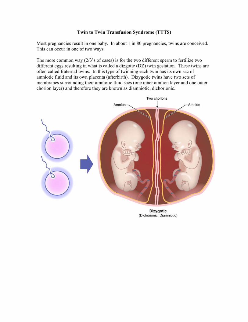
Most pregnancies result in one baby. In about 1 in 80 pregnancies, twins are conceived. This can occur in one of two ways. The more common way (2/3’s of cases) is for the two different sperm to fertilize two different eggs resulting in what is called a dizgotic (DZ) twin gestation. These twins are often called fraternal twins. In this type of twinning each twin has its own sac of amniotic fluid and its own placenta (afterbirth). Dizygotic twins have two sets of membranes surrounding their amniotic fluid sacs (one inner amnion layer and one outer chorion layer) and therefore they are known as diamniotic, dichorionic.

In about 1/3 of twin pregnancies, one sperm fertilizes one egg but this splits into two embryos resulting in what is known as monozygotic (MZ) twins. These twins are often referred to as identical twins since they have the same genetic material. Approximately 1/3 of MZ twins look just like fraternal twins on prenatal ultrasound since there are two separate amniotic sacs and two separate placentas. However in 2/3’s of identical twins, each twin has its own amniotic sac but the twins share a common placenta. This type of MZ twinning is called monochorionic, diamniotic since there is an inner layer surrounding the amniotic sac of each twin but there is only one common outer layer (chorion) surrounding both of the sacs. This type of twinning occurs in approximately one in 360 pregnancies. Monochorionic twins are at higher risk for complications since they share a common placenta.
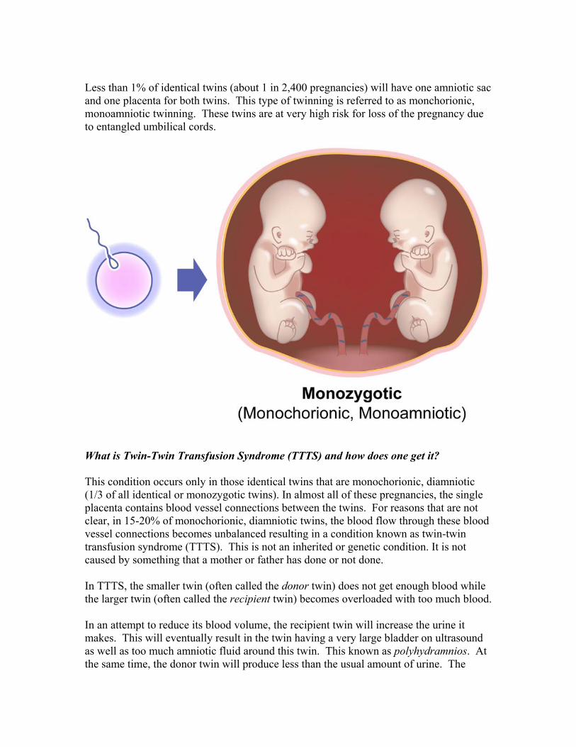
Less than 1% of identical twins (about 1 in 2,400 pregnancies) will have one amniotic sac and one placenta for both twins. This type of twinning is referred to as monchorionic, monoamniotic twinning. These twins are at very high risk for loss of the pregnancy due to entangled umbilical cords.
What is Twin-Twin Transfusion Syndrome (TTTS) and how does one get it? This condition occurs only in those identical twins that are monochorionic, diamniotic (1/3 of all identical or monozygotic twins). In almost all of these pregnancies, the single placenta contains blood vessel connections between the twins. For reasons that are not clear, in 15-20% of monochorionic, diamniotic twins, the blood flow through these blood vessel connections becomes unbalanced resulting in a condition known as twin-twin transfusion syndrome (TTTS). This is not an inherited or genetic condition. It is not caused by something that a mother or father has done or not done. In TTTS, the smaller twin (often called the donor twin) does not get enough blood while the larger twin (often called the recipient twin) becomes overloaded with too much blood. In an attempt to reduce its blood volume, the recipient twin will increase the urine it makes. This will eventually result in the twin having a very large bladder on ultrasound as well as too much amniotic fluid around this twin. This known as polyhydramnios. At the same time, the donor twin will produce less than the usual amount of urine. The
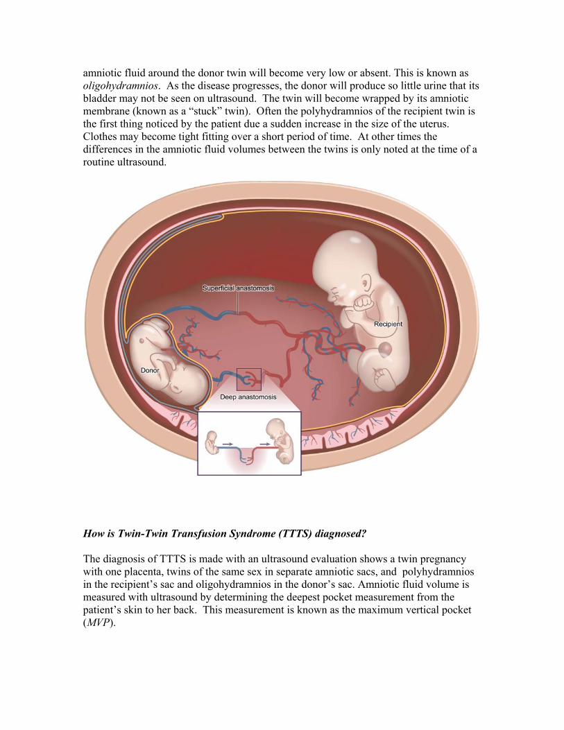
amniotic fluid around the donor twin will become very low or absent. This is known as oligohydramnios. As the disease progresses, the donor will produce so little urine that its bladder may not be seen on ultrasound. The twin will become wrapped by its amniotic membrane (known as a “stuck” twin). Often the polyhydramnios of the recipient twin is the first thing noticed by the patient due a sudden increase in the size of the uterus. Clothes may become tight fitting over a short period of time. At other times the differences in the amniotic fluid volumes between the twins is only noted at the time of a routine ultrasound.
How is Twin-Twin Transfusion Syndrome (TTTS) diagnosed? The diagnosis of TTTS is made with an ultrasound evaluation shows a twin pregnancy with one placenta, twins of the same sex in separate amniotic sacs, and polyhydramnios in the recipient’s sac and oligohydramnios in the donor’s sac. Amniotic fluid volume is measured with ultrasound by determining the deepest pocket measurement from the patient’s skin to her back. This measurement is known as the maximum vertical pocket (MVP).
What are the Twin-Twin Transfusion Syndrome (TTTS) five stages of classification? Quintero1 has proposed 5 stages of TTTS based on ultrasound findings:
Stage I: This is the initial way that TTTS is seen on ultrasound. In stage I, there is oligohydramnios in the donor’s sac with an MVP of 2 centimeters or less (3/4 inch) and polyhydramnios in the recipient’s sac with a maximum vertical pocket of fluid of 8 centimeters or more (just over 3 inches). The bladder of the donor baby is still seen.
Stage II: As defined above, there is polyhydramnios and oligohydramnios but the bladder is no longer seen in the donor twin during the ultrasound evaluation.
Stage III: Blood flow in the fetus can be measured with a special type of ultrasound called Doppler. In addition to the findings of Stages I and II, careful study of the blood flow in the umbilical cord and fetal ductus venosus (the large blood vessel in the fetus that returns blood to the heart from the placenta) reveals abnormal patterns in Stage III. These patterns can occur in either or both fetuses.
In the umbilical cord, the diastolic flow can be either absent or reversed in the umbilical artery. This pattern is usually seen in the donor twin.
In the ductus venosus, the diastolic flow can either be absent or reversed. This pattern is usually see in the recipient twin due to early heart failure. The recipient twin can also exhibit leakage across the main valve on the right side of the heart – this is known as tricuspid regurgitation.
Stage IV: One or both babies shows signs of hydrops. This means there is excess fluid in parts of the baby such as swelling of the skin around the head (scalp edema), fluid in the abdomen (ascites), fluid around the lungs (pleural effusions) or fluid around the heart (pericardial effusion). These findings are evidence of heart failure and are typically seen in the recipient twin.
Stage V: One or both babies have died.
The survival of the twins is poorer when there is progression to a higher stage over time. It has been estimated that half of patients will progress to a higher stage, 30% will remain at the same stage and 20% will improve to a lower stage.2, 3
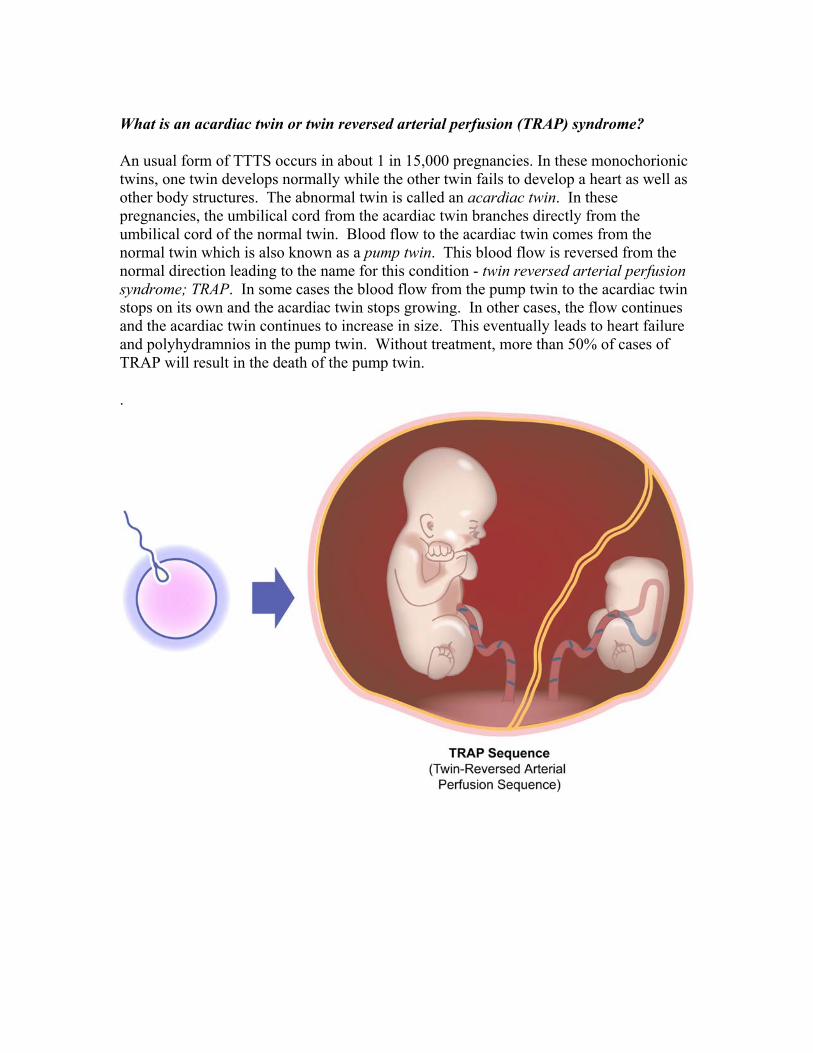
What is an acardiac twin or twin reversed arterial perfusion (TRAP) syndrome?
An usual form of TTTS occurs in about 1 in 15,000 pregnancies. In these monochorionic twins, one twin develops normally while the other twin fails to develop a heart as well as other body structures. The abnormal twin is called an acardiac twin. In these pregnancies, the umbilical cord from the acardiac twin branches directly from the umbilical cord of the normal twin. Blood flow to the acardiac twin comes from the normal twin which is also known as a pump twin. This blood flow is reversed from the normal direction leading to the name for this condition - twin reversed arterial perfusion syndrome; TRAP. In some cases the blood flow from the pump twin to the acardiac twin stops on its own and the acardiac twin stops growing. In other cases, the flow continues and the acardiac twin continues to increase in size. This eventually leads to heart failure and polyhydramnios in the pump twin. Without treatment, more than 50% of cases of TRAP will result in the death of the pump twin.
.
What is the outcome for Twin-Twin Transfusion Syndrome (TTTS)?
There are a number of ways to treat TTTS, any of which many be the correct method depending on ultrasound findings, the gestational age of the pregnancy and a couple’s specific needs. Left untreated, TTTS prior to 24 weeks’ gestation (6 months of pregnancy), 80 – 90% of cases are associated with the loss of one or both twins. If one of the twins should die, the blood vessel connections in the placenta can place the surviving twin at risk for long-term brain damage in as many as 1/3 of cases. In general, more advanced stages of TTTS have a worse prognosis than the earlier stages. When severe TTTS occurs at a very early gestational age (prior to 16 weeks or the fourth month of pregnancy), the option of termination of the pregnancy can be considered due to the grim prognosis. The various therapies that are available target either the unequal fluid between the twins’ sacs or interrupt the blood vessel communications between the twins on the single placenta. The successful outcome of these treatments has been based on the number of babies that survive as well as the number of babies who do not have brain damage. The treatments that are currently available are described below:
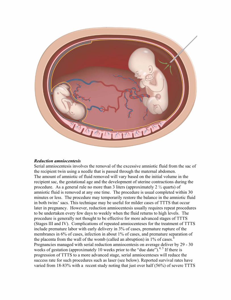
Reduction amniocentesis Serial amniocentesis involves the removal of the excessive amniotic fluid from the sac of the recipient twin using a needle that is passed through the maternal abdomen. The amount of amniotic of fluid removed will vary based on the initial volume in the recipient sac, the gestational age and the development of uterine contractions during the procedure. As a general rule no more than 3 liters (approximately 2 ½ quarts) of amniotic fluid is removed at any one time. The procedure is usual completed within 30 minutes or less. The procedure may temporarily restore the balance in the amniotic fluid in both twins’ sacs. This technique may be useful for milder cases of TTTS that occur later in pregnancy. However, reduction amniocentesis usually requires repeat procedures to be undertaken every few days to weekly when the fluid returns to high levels. The procedure is generally not thought to be effective for more advanced stages of TTTS (Stages III and IV). Complications of repeated amniocenteses for the treatment of TTTS include premature labor with early delivery in 3% of cases, premature rupture of the membranes in 6% of cases, infection in about 1% of cases, and premature separation of the placenta from the wall of the womb (called an abruption) in 1% of cases.4 Pregnancies managed with serial reduction amniocentesis on average deliver by 29 - 30 weeks of gestation (approximately 10 weeks prior to the “due date”).4, 5 If there is progression of TTTS to a more advanced stage, serial amniocenteses will reduce the success rate for such procedures such as laser (see below). Reported survival rates have varied from 18-83% with a recent study noting that just over half (56%) of severe TTTS
cases managed with reduction amniocentesis will end with at least one infant without brain damage.5 Approximately, 20 - 25% of the TTTS survivors from pregnancies treated with reduction amniocentesis have been found to have long-term developmental delay.
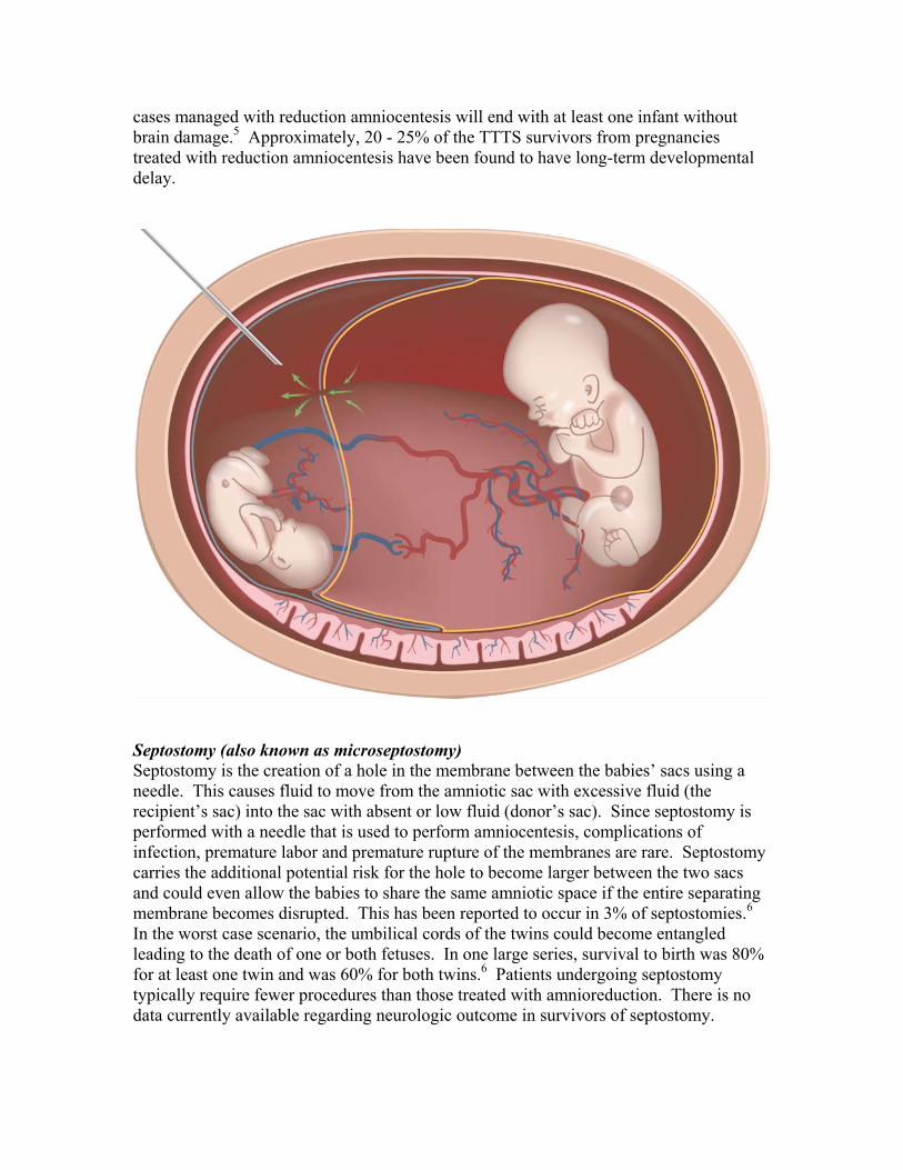
Septostomy (also known as microseptostomy) Septostomy is the creation of a hole in the membrane between the babies’ sacs using a needle. This causes fluid to move from the amniotic sac with excessive fluid (the recipient’s sac) into the sac with absent or low fluid (donor’s sac). Since septostomy is performed with a needle that is used to perform amniocentesis, complications of infection, premature labor and premature rupture of the membranes are rare. Septostomy carries the additional potential risk for the hole to become larger between the two sacs and could even allow the babies to share the same amniotic space if the entire separating membrane becomes disrupted. This has been reported to occur in 3% of septostomies.6 In the worst case scenario, the umbilical cords of the twins could become entangled leading to the death of one or both fetuses. In one large series, survival to birth was 80% for at least one twin and was 60% for both twins.6 Patients undergoing septostomy typically require fewer procedures than those treated with amnioreduction. There is no data currently available regarding neurologic outcome in survivors of septostomy.
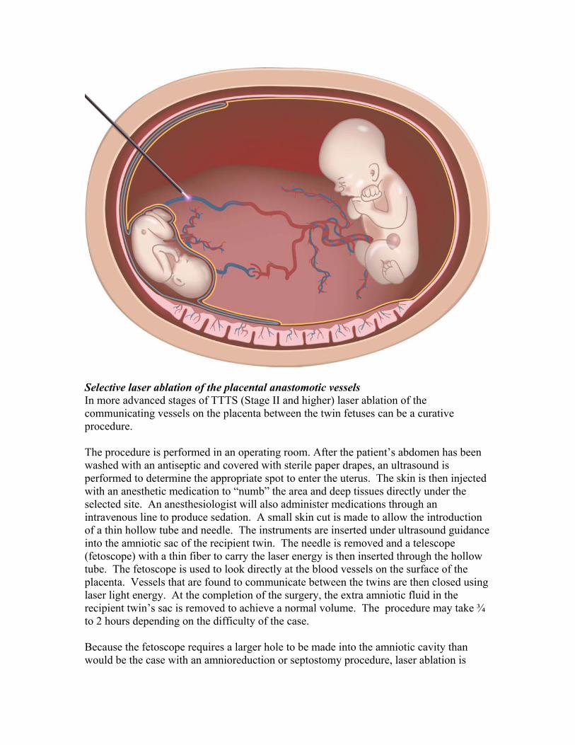
Selective laser ablation of the placental anastomotic vessels In more advanced stages of TTTS (Stage II and higher) laser ablation of the communicating vessels on the placenta between the twin fetuses can be a curative procedure. The procedure is performed in an operating room. After the patient’s abdomen has been washed with an antiseptic and covered with sterile paper drapes, an ultrasound is performed to determine the appropriate spot to enter the uterus. The skin is then injected with an anesthetic medication to “numb” the area and deep tissues directly under the selected site. An anesthesiologist will also administer medications through an intravenous line to produce sedation. A small skin cut is made to allow the introduction of a thin hollow tube and needle. The instruments are inserted under ultrasound guidance into the amniotic sac of the recipient twin. The needle is removed and a telescope (fetoscope) with a thin fiber to carry the laser energy is then inserted through the hollow tube. The fetoscope is used to look directly at the blood vessels on the surface of the placenta. Vessels that are found to communicate between the twins are then closed using laser light energy. At the completion of the surgery, the extra amniotic fluid in the recipient twin’s sac is removed to achieve a normal volume. The procedure may take ¾ to 2 hours depending on the difficulty of the case. Because the fetoscope requires a larger hole to be made into the amniotic cavity than would be the case with an amnioreduction or septostomy procedure, laser ablation is
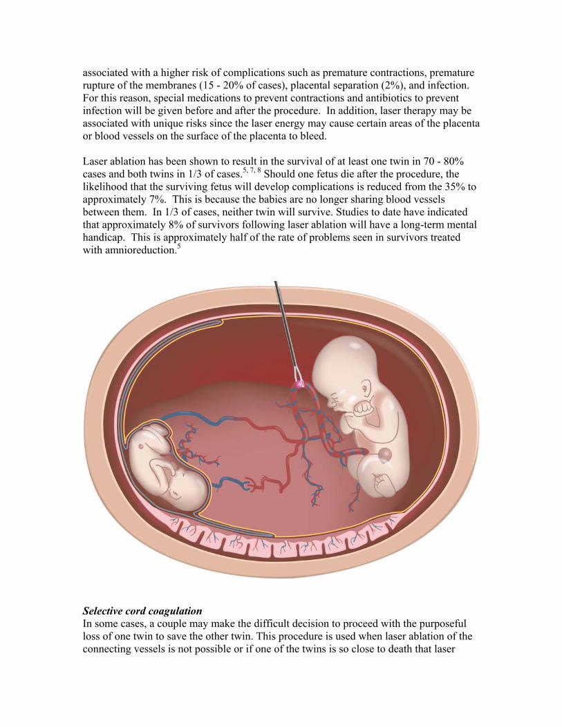
associated with a higher risk of complications such as premature contractions, premature rupture of the membranes (15 - 20% of cases), placental separation (2%), and infection. For this reason, special medications to prevent contractions and antibiotics to prevent infection will be given before and after the procedure. In addition, laser therapy may be associated with unique risks since the laser energy may cause certain areas of the placenta or blood vessels on the surface of the placenta to bleed. Laser ablation has been shown to result in the survival of at least one twin in 70 - 80% cases and both twins in 1/3 of cases.5, 7, 8 Should one fetus die after the procedure, the likelihood that the surviving fetus will develop complications is reduced from the 35% to approximately 7%. This is because the babies are no longer sharing blood vessels between them. In 1/3 of cases, neither twin will survive. Studies to date have indicated that approximately 8% of survivors following laser ablation will have a long-term mental handicap. This is approximately half of the rate of problems seen in survivors treated with amnioreduction.5
Selective cord coagulation In some cases, a couple may make the difficult decision to proceed with the purposeful loss of one twin to save the other twin. This procedure is used when laser ablation of the connecting vessels is not possible or if one of the twins is so close to death that laser
ablation would likely not be successful. By stopping the flow in the cord of the dying twin, the other twin can be protected from the consequences of its sibling’s death. The procedure is performed through the use of a special forceps that is placed into the amniotic sac of the recipient twin while watching with ultrasound. The umbilical cord is then grasped and electrical current is applied to burn (coagulate) the blood vessels in the cord so that the blood flow will stop to this fetus. The communication between the fetuses is definitively ended, however, this eliminates the chance of survival for one of the twins. Complications of this procedure include premature delivery and premature rupture of the membranes. Rupture of the membranes has been reported to occur in about 20% of cases. Survival of the one remaining fetus can be expected in 85% of cases.
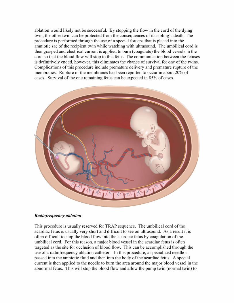
Radiofrequency ablation This procedure is usually reserved for TRAP sequence. The umbilical cord of the acardiac fetus is usually very short and difficult to see on ultrasound. As a result it is often difficult to stop the blood flow into the acardiac fetus by coagulation of the umbilical cord. For this reason, a major blood vessel in the acardiac fetus is often targeted as the site for occlusion of blood flow. This can be accomplished through the use of a radiofrequency ablation catheter. In this procedure, a specialized needle is passed into the amniotic fluid and then into the body of the acardiac fetus. A special current is then applied to the needle to burn the area around the major blood vessel in the abnormal fetus. This will stop the blood flow and allow the pump twin (normal twin) to

no longer have to send blood to the acardiac twin. Complications of infection, premature contractions and premature rupture of the membranes can occur as in any needle procedure. In one series, the risk for premature rupture of the membranes was 8%.9 In this same series, the chance for a successful livebirth for the pump twin was 90%. What do I do after my physician has made a referral for me to be seen? You will be scheduled for an ultrasound evaluation with one of our Maternal-Fetal Medicine specialists at the University of North Carolina School of Medicine. This may be scheduled in the main ultrasound unit at UNC Women’s Hospital or it may be scheduled at our satellite ultrasound office at Rex Hospital in Raleigh. The physician will discuss all findings and will review the treatment options, surgical procedures, prognosis, and recommended follow-up care. We will be able to answer your questions and concerns at this time. Next, you will meet with or have a phone conversation with our Fetal Therapy Coordinator. She will answer any further questions that you are your partner may have. In addition, she will assist you any special needs including overnight accommodations. You will receive a folder that contains information you will need for surgery and additional information that you will find helpful. We will ask that you come to the Labor & Delivery unit of the UNC Women’s Hospital (fourth floor) for your pre-operative consultation with an anesthesiologist. You will also have blood samples drawn at that time. A surgery consent form will be given to you to review. It explains the surgery in terms you can understand. You will also be given several consent forms for collection of data for an ongoing study of research to help us better understand the treatment of twin-twin transfusion syndrome. You will then be discharged home or back to your hotel room. How do I prepare for surgery? The night before surgery, you will not be allowed to eat or drink for a defined amount of time (usually 6 - 8 hours). This is to prevent the risk of vomiting during surgery. In medical terms, this is known as "NPO" (nothing by mouth). We will give you a time to come UNC Women’s Hospital to Labor & Delivery on the fourth floor on the day of surgery. Your family may come with you but will be asked to wait for you in one of our labor rooms during the surgery. An intravenous line (IV) will be inserted by needle stick to give you fluids and medications during surgery. An ultrasound will be performed prior to going to the operating room to confirm that both twins are alive. One of the specially trained nurses that will be assisting in surgery and an anesthesiology resident will accompany you to surgery. What can I expect during surgery? You will be allowed to walk into the operating room where you will be asked to move on to the operating table. You will be covered with a warm blanket and a pillow will be placed under your knees to keep you comfortable during surgery. You may be rolled to
your left side to keep your uterus from causing your blood pressure to fall. A belt will be placed across your legs to prevent you from sliding off the operating room table. Your abdomen (belly) will be cleaned with an iodine solution (let your nurse know if you are allergic to iodine). Then you will be covered with sterile paper drapes. The top of the drapes will be attached to a pole so that you do not need to watch the procedure. Medication will be given through your IV to relax you. Surgery is performed under local anesthesia, meaning you are awake but relaxed and your abdomen is numbed where the instrument is inserted. An anesthesiologist will stay with you throughout the procedure. You will be given additional medication for discomfort as needed. On rare occasions, general anesthesia, meaning you are put to sleep, may be used. During surgery, one or two small incisions, approximately 1/10 of an inch long will be made on the abdomen. These incision(s) are small. You will have short pieces of specialized tape (steri-strips) placed on your skin to close this incision at the end of the procedure. What can I expect after surgery? Following surgery, you will be taken to the recovery room in Labor & Delivery or a labor room where you and your fetuses will be closely monitored. Your abdomen will be a little tender or sore once the local anesthetic wears off. You may be given medications after surgery to relax the uterus and stop any contractions. The pain and discomfort after surgery is usually minimal. If needed, pain relief medicine is available. Your husband or other support person may remain with you in your room. Following surgery, you may have food as tolerated. You will be admitted to the hospital for an overnight stay. That night, activity is restricted to bathroom privileges only, but this depends upon your specific condition. You will undergo an ultrasound the day after surgery to determine how the babies are doing. What can I expect after I am discharged home? You will then be discharged home to the care of your primary obstetrician and/or your referring Maternal-Fetal Medicine specialist. Your instructions will include bed rest with bathroom privileges for 7 days after the surgery, with a gradual increase in activity. We will also ask that you get a thermometer and take your temperature 3 times per day. You should notify your primary obstetrician for any increase above 100.4° F of an oral temperature. The site of the surgery can get wet in a shower within 24 hours of the procedure. You can remove the steristrips over the incision yourself by one week after the surgery. After 4 weeks you can resume normal activity based on your pregnancy condition and the comfort level of your primary obstetrician. Weekly ultrasounds are recommended for the next month. After that time, if all is going well, ultrasounds are performed as directed by your doctor. Although you are returning home, we will continue to follow your pregnancy closely through our care coordinator. Please make arrangements with your doctor to forward your ultrasound reports and any other pertinent information to us. We also ask that you inform your obstetrician and labor nurse that we would like to have your placenta sent back to us after you delivery. This information is

useful to further our knowledge and will assist in the future treatment of patients with TTTS. Social services and pastoral care are available for all our patients and their families. If you would like to see them at any time, you need only to request it and they can be contacted. We are sensitive to the psychological, social, and spiritual needs of our families. We will provide any support that is necessary. Please contact us if you have any questions, concerns, or special requests. For our out of town patients, we realize that traveling may be difficult or stressful and want you to know that we will do everything we can to accommodate your special needs and schedule.
References
1. Quintero RA, Morales WJ, Allen MH, Bornick PW, Johnson PK, Kruger M.
Staging of twin-twin transfusion syndrome. J Perinatol 1999; 19:550-5. 2. Taylor MJ, Govender L, Jolly M, Wee L, Fisk NM. Validation of the Quintero
staging system for twin-twin transfusion syndrome. Obstet Gynecol 2002; 100:1257-65.
3. Dickinson JE, Evans SF. The progression of disease stage in twin-twin transfusion syndrome. J Matern Fetal Neonatal Med 2004; 16:95-101.
4. Mari G, Roberts A, Detti L, et al. Perinatal morbidity and mortality rates in severe twin-twin transfusion syndrome: results of the International Amnioreduction Registry. Am J Obstet Gynecol 2001; 185:708-15.
5. Senat MV, Deprest J, Boulvain M, Paupe A, Winer N, Ville Y. Endoscopic laser surgery versus serial amnioreduction for severe twin-to-twin transfusion syndrome. N Engl J Med 2004; 351:136-44.
6. Moise KJ, Jr., Dorman K, Lamvu G, et al. A randomized trial of amnioreduction versus septostomy in the treatment of twin-twin transfusion syndrome. Am J Obstet Gynecol 2005; 193:701-7.
7. Hecher K, Plath H, Bregenzer T, Hansmann M, Hackeloer BJ. Endoscopic laser surgery versus serial amniocenteses in the treatment of severe twin-twin transfusion syndrome. Am J Obstet Gynecol 1999; 180:717-24.
8. Quintero RA, Dickinson JE, Morales WJ, et al. Stage-based treatment of twin-twin transfusion syndrome. Am J Obstet Gynecol 2003; 188:1333-40.
9. Lee H, Wagner A, Bobert B, et al. Radiofrequency ablation for TRAP sequence. Am J Obstet Gynecol 2005; 191:S18.