tutorial - qiagen...
TRANSCRIPT

Sample to Insight
Tutorial
Whole Metagenome Functional Analysis (beta)November 21, 2017
QIAGEN Aarhus Silkeborgvej 2 Prismet 8000 Aarhus C DenmarkTelephone: +45 70 22 32 44 [email protected]

Tutorial
2
Whole Metagenome Functional Analysis (beta)This tutorial will take you through the different tools available in CLC Microbial Genomics Moduleavailable for CLC Genomics Workbench and Biomedical Genomics Workbench to perform a wholemetagenome functional analysis pipeline.
Introduction The main goal of the tutorial is to demonstrate the assembly of metagenomesderived from two different groups of samples and the subsequent investigation of functionaldifferences. It serves as a template for performing a comparative investigation into the functionalcomposition and diversity of microbial communities. The tools provide a way of looking at differentsamples in aggregate views and to drill down into differentiating functional categories that resultfrom the comparative analysis.
Prerequisites For this tutorial, you will need either CLC Genomics Workbench or BiomedicalGenomics Workbench with MetaGeneMark plugin and CLC Microbial Genomics Module installed.
Overview In this tutorial we will go through a suite of useful components in pipelines foranalyzing whole-metagenome NGS data from microbial communities.
• First, we will import "raw" NGS sequencing data into the workbench and prepare thesamples for analysis.
• We will then assemble the reads using De Novo Assemble Metagenome into contigs.
• With MetaGeneMark, we will identify genes and coding DNA sequences (CDS) on thecontigs.
• Subsequently, functional annotation of the CDS with Gene Ontology (GO) terms and Pfamdomains will be performed with Annotate CDS with Pfam Domains.
• Based on the annotations, we will construct a Gene Ontology profile using the BuildFunctional Profile tool for measuring functional diversity.
• We will also create a multi-sample abundance table using Merge Abundance Tables.
• Finally, we will set up the data for additional statistical analyses and visualisations.

Tutorial
3
Downloading and importing the data For this tutorial we will make use of the mock datasetgenerated by Lindgreen et al., 2016. The dataset contains four samples, divided in two groups (Aand B). Group A is enriched in bacteria that perform photosynthesis and nitrogen fixation, whilegroup B is enriched in pathogenic bacteria. The goal of the analysis in this tutorial is to find thosefunctional differences from whole-metagenome sequencing data.
For sake of speed, the dataset used for this tutorial is a small subset of reads that is related tothe following functional categories:
• Photosynthesis, identified by the GO id 0015979, which defines it as "the synthesis byorganisms of organic chemical compounds, especially carbohydrates, from carbon dioxide(CO2) using energy obtained from light rather than from the oxidation of chemical compounds."
• Nitrogen Fixation, identified by the GO id 0009399, which defines it as "the process inwhich nitrogen is taken from its relatively inert molecular form (N2) in the atmosphere andconverted into nitrogen compounds useful for other chemical processes, such as ammonia,nitrate and nitrogen dioxide."
• Pathogenesis, identified by the GO ids 0009405 and 0009403, which are defined as "theset of specific processes that generate the ability of an organism to cause disease in another"and "the chemical reactions and pathways resulting in the formation of toxin, a poisonouscompound (typically a protein) that is produced by cells or organisms and that can causedisease when introduced into the body or tissues of an organism," respectively.
The tutorial data consists of the following files:
• Sequence data: 4 pairs of fastq files (two for group A and two for group B). The files containsimulated paired-end sequencing reads.
• Metadata: "Group metadata.xls". A spreadsheet containing metadata information aboutthe samples and the group they belong to.
• Pfam database: "Pfam-A v29 - Tutorial subset.clc". A small subset of Pfam v29 [Batemanet al., 2004,Finn et al., 2016] containing only the Pfam domains relevant to this tutorial.
• GO database: "GO database - Tutorial subset.clc". A small subset of the GO (GeneOntology) database [Ashburner et al., 2000, The Gene Ontology Consortium, 2015] andPfam2GO mappings (which allow to infer GO terms from Pfam domains). This databasecontains only terms relevant for this tutorial.
Please note that this tutorial contains only a small subset of the Pfam and GO databases, whichshould only be used for this tutorial. For applying the functional analysis pipeline to real datasets,please download the complete databases: the complete versions of the Pfam and GO databasescan be easily obtained using the Download Pfam Database and Download GO Database tools,respectively. If you are not sure where to find these tools in the toolbox, use the Launch button( ) .
Now that the prerequisites have been described, it is time to start importing the input data.
1. Download the sample data from our website: http://resources.qiagenbioinformatics.com/testdata/functional_tutorial.zip and unzip it.

Tutorial
4
2. Start your CLC Workbench and create a folder for storing input data and results, named forexample Functional analysis.
3. Go to Import | Illumina to import the 8 sequence files (ending with "fastq") (figure 1).
Figure 1: Import paired-end reads for the four samples.
• The import type under Options is set to Paired reads.
• Paired-end (forward-reverse) is selected.
• Discard read names, Discard quality scores and MiSeq de-multiplexing are notchecked.
• Set the minimum distance to 450 and the Maximum distance to 600.
4. Click on the button labeled Next and select the location where you want to store theimported sequences. You can check that you have now 4 files labeled as "paired"
5. Import the metadata by clicking Import | Import Metadata on top of the Navigation Area.
6. A wizard opens (figure 2). Select the spreadsheet Group metadata.xls in the first field. Thecontent fills the Medatata preview table at the bottom of the dialog. Click Next.
Figure 2: Import the metadata.

Tutorial
5
7. Now select the four reads imported earlier. Check Partial for the Data association previewtable to fill in and thereby indicate that the association was successful (figure 3). ClickNext.
Figure 3: The metadata and the reads are now associated.
8. Save the metadata in the folder created earlier. You can rename the metadata table "Groupmetadata" instead of the default name "Samples".
9. Import the Pfam and GO databases by dragging the ‘‘GO database - Tutorial subset.clc( )’’ and ‘‘Pfam-A v29 - Tutorial subset.clc ( )’’ files into your destination folder in yourworkbench, or by using the Standard Import button on top of the Navigation Area.
All of the data needed to get started is now imported and you should have the objects depictedin figure 4. You are now ready to begin the analysis.
Figure 4: All files are now imported.

Tutorial
6
-dup5Assembling and annotating references
In this section, we will assemble the reads into contigs and annotate them with functionalinformation.
1. Create a new folder, for example Assemblies, to store the results. We are now ready toassemble the reads into contigs using the De Novo Assemble Metagenome tool:
Microbial Genomics Module ( ) | Metagenomics ( ) | Functional Analysis ( ) |De Novo Assemble Metagenome ( )
2. Since we are processing four samples, select the Batch option. Next, select the reads asinput (figure 5) and click Next.
Figure 5: Batch reads for de novo metagenome assembly.
3. The next window gives an overview of the batch units. If you do not see this window, itmeans you forgot to check the batch option at the previous step.
4. In the "De novo options" dialog, make sure Minimum contig length is set to 200, choosethe Longer contigs execution mode and make sure the Perform scaffolding checkbox isnot checked (figure 6). Click Next.
Figure 6: Select parameters for de novo metagenome assembly.
5. Choose to save the results in the Assemblies folder.
Once the reads have been assembled, we need to functionally annotate the contigs. Beforeannotation with functional information, we need to identify coding regions in the contigs. Wetherefore run the MetaGeneMark tool to identify genes and coding DNA sequences (CDS).
Tutorial
7
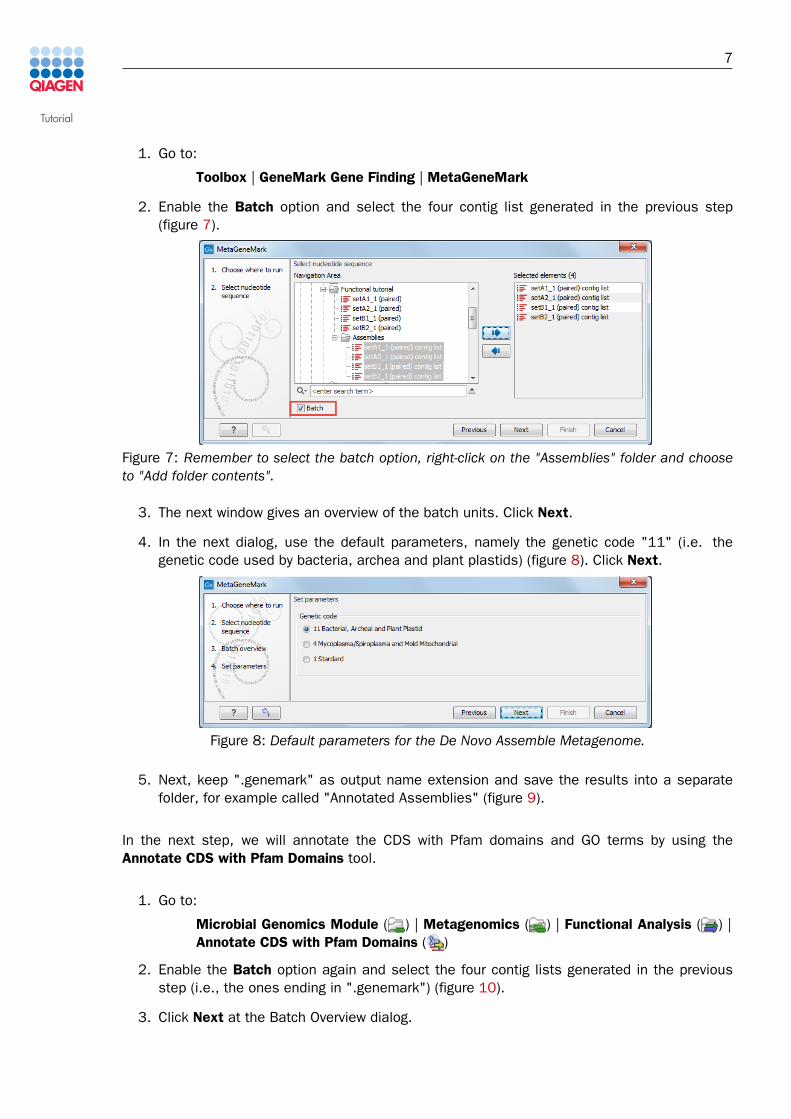
1. Go to:
Toolbox | GeneMark Gene Finding | MetaGeneMark
2. Enable the Batch option and select the four contig list generated in the previous step(figure 7).
Figure 7: Remember to select the batch option, right-click on the "Assemblies" folder and chooseto "Add folder contents".
3. The next window gives an overview of the batch units. Click Next.
4. In the next dialog, use the default parameters, namely the genetic code "11" (i.e. thegenetic code used by bacteria, archea and plant plastids) (figure 8). Click Next.
Figure 8: Default parameters for the De Novo Assemble Metagenome.
5. Next, keep ".genemark" as output name extension and save the results into a separatefolder, for example called "Annotated Assemblies" (figure 9).
In the next step, we will annotate the CDS with Pfam domains and GO terms by using theAnnotate CDS with Pfam Domains tool.
1. Go to:
Microbial Genomics Module ( ) | Metagenomics ( ) | Functional Analysis ( ) |Annotate CDS with Pfam Domains ( )
2. Enable the Batch option again and select the four contig lists generated in the previousstep (i.e., the ones ending in ".genemark") (figure 10).
3. Click Next at the Batch Overview dialog.

Tutorial
8
Figure 9: Default parameters for the MetaGeneMark tool.
Figure 10: Remember to select the batch option, right-click on the "Annotated Assemblies" folderand choose to "Add folder contents".
4. Use the ‘‘Pfam-A v29 - Tutorial subset.clc ( )’’ as pfam database and ‘‘GO database -Tutorial subset.clc ( )’’ as GO database, as shown in figure 11. Make sure the geneticcode is set to "11 Bacterial, Archeal and Plant Plastid" and that "Use profile’s gatheringcutoffs" and "Remove overlapping matches from the same clan" are checked. You cankeep "Complete GO basic" as GO subset.
Figure 11: Parameters for Annotate CDS with Pfam Domains.
5. Choose where you will save the reports and click Finish.

Tutorial
9
The tool will add annotations to the existing contigs and create a report for each batch unit.You can check that Pfam annotations have been added by opening ‘‘set_A1_1(paired) contiglist.genemark ( )’’. To see Pfam annotations, you may have to open the Annotation Type tabon the right panel and click on Pfam domain. In the Find tab, select "Annotation" and type in"Pfam" to find all Pfam annotations on each contig. If you hover over a Pfam annotation, you willbe able to see the name of the Pfam domain, its description and the score of the match. Whenthe Pfam domain can be matched to a GO term, a GO annotation will also be present, as shownin figure 12.
Figure 12: Contig_4 in set_A1 contains a Colicin V production protein domain, which is related totoxin biosynthesis.
Tutorial
10
-dup5Building functional profiles
The metagenome assemblies are now annotated. We now want to re-map the original reads toestimate the abundance of functional categories in the samples.
First, map the reads to the annotated assemblies. In this case, we will not batch the execution,but we will need to run the read mapping four times against the 4 different references.
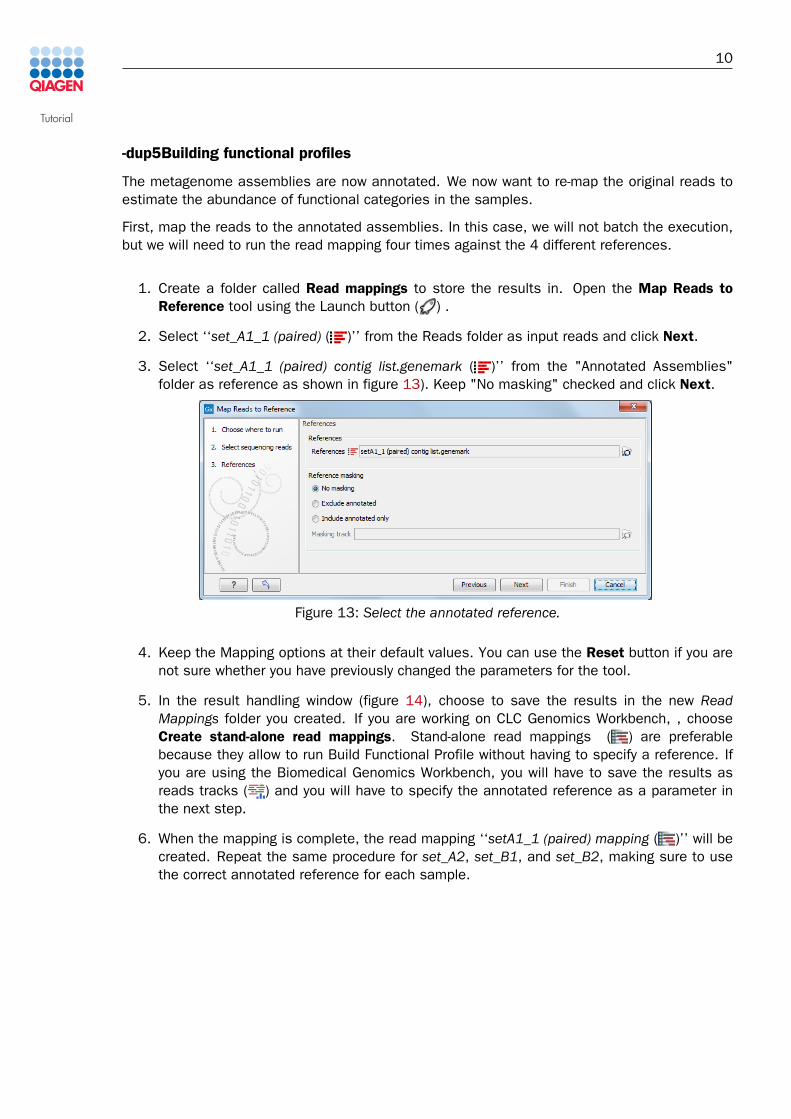
1. Create a folder called Read mappings to store the results in. Open the Map Reads toReference tool using the Launch button ( ) .
2. Select ‘‘set_A1_1 (paired) ( )’’ from the Reads folder as input reads and click Next.
3. Select ‘‘set_A1_1 (paired) contig list.genemark ( )’’ from the "Annotated Assemblies"folder as reference as shown in figure 13). Keep "No masking" checked and click Next.
Figure 13: Select the annotated reference.
4. Keep the Mapping options at their default values. You can use the Reset button if you arenot sure whether you have previously changed the parameters for the tool.
5. In the result handling window (figure 14), choose to save the results in the new ReadMappings folder you created. If you are working on CLC Genomics Workbench, , chooseCreate stand-alone read mappings. Stand-alone read mappings ( ) are preferablebecause they allow to run Build Functional Profile without having to specify a reference. Ifyou are using the Biomedical Genomics Workbench, you will have to save the results asreads tracks ( ) and you will have to specify the annotated reference as a parameter inthe next step.
6. When the mapping is complete, the read mapping ‘‘setA1_1 (paired) mapping ( )’’ will becreated. Repeat the same procedure for set_A2, set_B1, and set_B2, making sure to usethe correct annotated reference for each sample.

Tutorial
11
Figure 14: Save as stand-alone read mappings in CLC Genomics Workbench.
Next, we build the GO functional profile for each sample using the Build Functional Profile tool.
1. Create a folder called Functional profiles to store results and go to:
Microbial Genomics Module ( ) | Metagenomics ( ) | Functional Analysis ( ) |Build Functional Profile ( )
2. On Biomedical Genomics Workbench you cannot perform this step in batch, so select justone mapping and click Next. Working with CLC Genomics Workbench, enable the Batchoption and select the four read mappings (figure 15). Click Next twice to pass the Batchoverview window.
Figure 15: Select the four read mappings and analyze them in batch.
3. If you are running Biomedical Genomics Workbench, specify the annotated reference (e.g.‘‘set_A1_1 (paired) contig list.genemark ( )’’ for set_A1) in the Reference parameter, andrepeat the analysis with the appropriate reference for each read mapping. If you are workingwith CLC Genomics Workbench, you do not need to specify a reference in the "Parameters"dialog.
In both cases, use ‘‘GO database - Tutorial subset.clc ( )’’ as GO database, as shown infigure 16.
Figure 16: Parameters for Build Functional Profile.

Tutorial
12
4. Finally, choose Create GO functional profile only and save to the "Functional profiles"folder (figure 17).
Figure 17: Create only a GO functional profile.
You have now built a functional profile for each sample (figure 18).
Figure 18: GO functional profiles have been created.
We now want to merge them using the Merge Abundance Tables tools.
1. Create a folder to store the results (e.g. Statistical analyses) and go to:
Microbial Genomics Module ( ) | Metagenomics ( ) | Abundance Analysis ( ) |Merge Abundance Tables ( )
2. Select the four GO profiles as input. In this case, we do not batch the analysis, as the toolsshould be run only once using all the four functional profiles as input (figure 19).
Figure 19: Merge the GO functional profiles.
3. Save the merged profile in the "Statistical analyses" folder.

Tutorial
13
The tool will create an abundance table called ‘‘merged ( )’’ containing functional abundancesfor the four samples. You can now open the table to explore the results of the functional analysis(figure 20). Observe the functional abundance values for the four GO terms. As expected,abundance values for "pathogenesis" and "toxins" are higher in group B, whereas group A isenriched in "photosynthesis" and "nitrogen fixation".
Figure 20: Result of the GO functional analysis.
-dup5Performing statistical analyses
A heat map and dendrogram help assessing similarity between samples.
1. Open the Microbial Genomics Module ( ) | Metagenomics ( ) | Abundance Analysis( ) | Create Heat Map for Abundance Table ( )
and choose the "merged" table as input.
2. Leave the parameters as set by default, i.e., the distance to Euclidean and clusters toComplete linkage. Click Next.
3. In the next wizard window, do not set any particular filter by selecting the option "Nofiltering" and click Next.
4. Save the result in the Statistical analyses folder.
Display the heat map by double-clicking on it in the Navigation Area (figure 21).
As we would have expected from the description of the data-set by Lindgreen et al., 2016 inthe beginning of this tutorial, the normalized values for toxin biosynthesis and pathogenesis areover-expressed in group B, while the normalized values for photosynthesis and nitrogen fixationare enriched in group A. Furthermore, the samples from each group cluster together, as shownin the dendrogram at the bottom of the figure.
It is also possible to use as additional statistical analyses the Differential Abundance Analysistool, although the interest of a Venn diagram is quite limited when the data set is only made oftwo distinctive groups as it is for this tutorial.
Although the results are hardly surprising, it is always re-assuring and good scientific practice tofirst apply a method to a problem with a known solution in order to verify everything works outexactly as expected before moving on to harder problems. Well done! At this point, we’d like topoint out again that it is important to download the full versions of the Pfam and GO databases(by using the tools provided in the workbench) prior to using the functional annotation pipelinefor a complete functional analysis of your own datasets.

Tutorial
14
Figure 21: Heat map from the abundance table.

Tutorial
Bibliography
[Ashburner et al., 2000] Ashburner, M., Ball, C. A., Blake, J. A., Botstein, D., Butler, H., Cherry,J. M., Davis, A. P., Dolinski, K., Dwight, S. S., Eppig, J. T., Harris, M. A., Hill, D. P., Issel-Tarver,L., Kasarskis, A., Lewis, S., Matese, J. C., Richardson, J. E., Ringwald, M., Rubin, G. M., andSherlock, G. (2000). Gene ontology: tool for the unification of biology. Nat Genet, 25(1):25--29.
[Bateman et al., 2004] Bateman, A., Coin, L., Durbin, R., Finn, R. D., Hollich, V., Griffiths-Jones,S., Khanna, A., Marshall, M., Moxon, S., Sonnhammer, E. L. L., Studholme, D. J., Yeats, C.,and Eddy, S. R. (2004). The Pfam protein families database. Nucleic Acids Res, 32(Databaseissue):D138--D141.
[Finn et al., 2016] Finn, R. D., Coggill, P., Eberhardt, R. Y., Eddy, S. R., Mistry, J., Mitchell,A. L., Potter, S. C., Punta, M., Qureshi, M., Sangrador-Vegas, A., Salazar, G. A., Tate, J., andBateman, A. (2016). The Pfam protein families database: towards a more sustainable future.Nucleic Acids Research, 44(D1):D279--D285.
[Lindgreen et al., 2016] Lindgreen, S., Adair, K. L., and Gardner, P. P. (2016). An evaluation ofthe accuracy and speed of metagenome analysis tools. Scientific Reports, 6:19233--.
[The Gene Ontology Consortium, 2015] The Gene Ontology Consortium (2015). Gene ontologyconsortium: going forward. Nucleic Acids Research, 43(D1):D1049--D1056.
15