tumor-specific lysis of human renal cell carcinomas by...
TRANSCRIPT
(CANCER RESEARCH 53, 4020-4025. September l, 1993J
Tumor-specific Lysis of Human Renal Cell Carcinomas by Tumor-infiltrating
Lymphocytes: Modulation of Recognition through Retroviral Transductionof TYimorCellswith Interleukin 2 Complementary DNA andExogenous a Interferon Treatment1
Dolores J. Sehende!2 and Bernd Gansbacher
Institut fürImmunologie. Ludwig-Maiirnilians-Universilüt München,8000 Munich 2, Germany ¡D../. .V./, cimi Sloun-Kcrtering Institute for Cancer Research. M'w York, New York
10021 ¡B.G.I
ABSTRACT
Two cytotoxic effector cell populations were isolated from a patient withrenal cell carcinoma. The tumor-infiltrating lymphocytes comprised apopulation of highly specific, major histocompatibility complex-restricted,cytotoxic T lymphocytes (CTL). An autologous non-major histocompatibility complex-restricted lymphokine-activated killer (LAK) cell popula
tion was generated by culturing the peripheral blood lymphocytes withhigh doses of recombinant interleukin 2 (rIL-2). The capacity of these twoeffector cell types to lyse cytokine-modulated autologous tumor cells wascompared /;/ vitro. A complementary DNA for rIL-2 was introduced into
the tumor cells by retroviral transduction, and tumor cells secreting lowdoses of rIL-2 were isolated. The CTL recognition of these tumor cells was
enhanced, compared to unmodified tumor cells, whereas LAK cell recognition was unchanged or slightly reduced. Pretreatment of tumor cellswith exogenous a interferon led to an up-regulation of some major histo
compatibility complex class I molecules and to slightly better recognitionby the CTL; little effect on LAK cell recognition was observed. CTL werefound to be 50-150-fold more effective than LAK cells in lysing autologoustumor cell lines or clones modulated with both rIL-2 and a interferon. Theassessment of a patient's cytotoxic immune capacity directed against ge
netically modified autologous tumor cells in vitro provides important insight for cytokine-mediated gene therapy of cancer.
INTRODUCTION
Patients with metastatic RCC1 have a 5-year survival rate of <5%.
The failure of chemotherapy or radiotherapy to influence the course ofdisease has left treatments which enhance the host immune responseagainst tumor as the most promising form of therapy for metastaticRCC to date. The prevalence of rich lymphocytic cellular infiltrates intumors in situ (1) and the occurrence of spontaneous regression ofmetastatic lesions in a number of well documented cases (2) providedthe first suggestions that immunological responses might be directedagainst RCC. Initial studies using cell-derived interferon revealed an
improved clinical status in some patients with RCC. The availabilityof recombinant cytokines has allowed more extensive clinical trials toassess their roles in the treatment of various forms of cancer. Bothrecombinant IFN-a and rIL-2 produced partial or complete responses
in some RCC patients with metastatic disease (3, 4). Recent trials have
Received 3/15/93; accepted 6/21/93.The costs of publication of this article were defrayed in part by the payment of"page
charges. This article must therefore be hereby marked advertisement in accordance with18 U.S.C. Section 1734 solely to indicate this fact.
1This manuscript is second in a series. This work was supported by grants from the
Bundesministerium fürForschung und Technologie (BEO/21 0319527A), the SchultzFoundation, and the NIH (PO1-CA59350). B. G. is a F. M. Kirby Clinical ResearchFellow.
2 To whom requests for reprints should be addressed, at Institute of Immunology.University of Munich, Goethestrasse 31, D-S(KK)Munich 2. Germany.
3 The abbreviations used are: RCC, renal cell carcinoma; CTL, cytotoxic T lymphocytes; LAK, lymphokine-activated killer; MHC, major histocompatibility complex; PBL.peripheral blood lymphocytes; rIL-2, recombinant interleukin 2; IFN-a, a interferon; TIL.tumor-infiltrating lymphocytes; cDNA, complementary DNA: FCS, fetal calf serum; E:Tratio, effector to target ratio; IL-2, interleukin 2; mAb, monoclonal antibody; LU. lytic
units; NKC, normal kidney cells.
evaluated the role of transfer of activated lymphocytes in conjunctionwith rIL-2. Two types of activated cells have been evaluated in RCC
patients: LAK cells, derived from PBL through culture with highdoses of rIL-2, and TIL, isolated from primary or metastatic lesionsand expanded in vitro with rIL-2 (5). TIL seem to be more effective
than LAK cells in achieving partial or complete remission. Animalstudies have shown that protective host antitumor responses can beelicited by injecting tumor cells which have been genetically modifiedto express various cytokine genes (6). In one case long-lasting pro
tection against tumor challenge was achieved (7). Phase I trials usingrIL-2-modified tumor cells for vaccination of patients with melanoma
or RCC have been initiated (8). It is hoped that this form of treatmentwill support the immune system while alleviating the deleterious sideeffects of systemic rIL-2 therapy. Like patients with RCC, some
patients with melanoma respond to various forms of immunotherapy.In patients with melanoma, TIL have been found to be more effectivegenerally than LAK cells in achieving partial or complete remissions(3, 9, 10). It is thought that larger numbers of specific CTL present inthe TIL populations may account for this difference (10). Whereasboth quantitative and qualitative differences (i.e., presence of MHC-restricted versus non-MHC-restricted effector cells) between TIL and
LAK cells of melanoma patients have been repeatedly detected invitro, such distinctions have not been readily observed in cells ofpatients with RCC. Therefore, it has been questioned whether specificCTL play an important role in tumor regression in patients with RCCwho receive immunotherapy. On the other hand, newer studies demonstrate that RCC-specific CTL do exist (11, 12)4; thus, it is important
to understand how cytokines influence the capacity of the CTL torecognize and destroy autologous tumor cells, compared to non-MHC-restricted LAK cells.
We have recently characterized a tumor-specific. MHC-restrictedCD8+ CTL line that was established from TIL of a patient with RCCwho had no apparent metastatic disease.4 For comparison, a LAK cell
line was generated by culturing PBL from the same patient in mediumcontaining a high concentration of rIL-2. In addition, an autologous
RCC line was established from the primary tumor. To analyze theinfluence of rIL-2 on the responses of autologous TIL and LAK cells,we used retroviral transduction to introduce a human rIL-2 cDNA intothe tumor cell line (13). IFN-a was provided exogenously to cultures
of the tumor cells. The effect of each cytokine alone or in combinationon MHC expression and susceptibility to lysis by autologous TIL andLAK cells was studied. It was found that modulation of the tumorcells by these cytokines had different effects on the two effector cellpopulations.
4 D. J. Schendel, B. Gansbacher, R. Oberneder. M. Kricgmair. A. Hofstetter, G. Ri-ethmüller,and O. G. Segurado. Tumor-specific lysis of human renal cell carcinomas bytumor-infiltrating lymphocytes. 1. HLA-A2 restricted recognition of autologous and allo-
geneic tumor cells, J. Immunol., in press.
4020
on March 18, 2019. © 1993 American Association for Cancer Research.cancerres.aacrjournals.org Downloaded from
CTL RECOGNITION OF CYTOKINE-MODIFIED RENAL CARCINOMA CELLS
MATERIALS AND METHODS
Establishment of RCC and NKC Cell Lines. Patient 26 was diagnosed ashaving renal clear cell carcinoma staged as TI, G2; the tumor was 3 cm in
diameter. Freshly resected primary tumor material was enzymatically digestedfor 4 h at 37°Cin RPMI 1640 culture medium containing 0.1% collegenase,
0.0()2ir deoxyribonuclease, and 0.001% hyaluronidase (Sigma Chemical Co.,Munich. Germany). Following separation by density gradient centrifugation(Ficoll-Paque; LKB-Pharmacia. Freiburg. Germany), cells from the interfacewere cultured in a 75-mm culture flask in RPMI 1640 medium containing 10%PCS, 2.5 u.g/m! amphotericin B (GIBCO-BRL. Freiburg, Germany), 19r min
imum essential medium nonessential amino acids (GIBCO), 9 fig/ml insulin(Sigma), 10 (xg/ml transferrin (Sigma), and 10 ng/ml cholera toxin (C3012;Sigma). A rapidly growing tumor line designated as RCC-26 was maintained
in continuous culture. The same procedure was used to establish a short termline of NKC cultured from tissue obtained at the time of surgery from a sitedistant from the tumor. These cells were frozen at early passages, and afterthawing they could be maintained for approximately eight passages. In addition, PBL obtained at the time of surgery were used for generation of LAKcells.
Establishment of TIL and LAK Cultures. A second aliquot of the primary tumor was further subdivided into six individual pieces of 5-mm diameter, which were cultured separately in a 24-well plate in TIL medium con
sisting of RPMI 1640 medium with 5% human serum, 10% FCS, 100 units/mlpenicillin. 100 fig/ml streptomycin, 20-30 units/ml rIL-2, and 15% conditioned medium obtained from phytohemagglutinin-activated lymphocytes (14).The TIL emerging from these cultures were restimulated with RCC-26 tumorcells every 7-10 days. Tumor cells (10,000 cells/well) were distributed in24-well plates and allowed to expand until they formed a 75% confluent
monolayer. The plates were irradiated (100 Gy) and TIL were added to thetumor monolayers in medium containing no rIL-2; after 24 h exogenous rIL-2(30 units/ml) was added. This was also necessary when the IL-2-transducedcells were used as a source of antigen-presenting cells, because the tumor
monolayer was substantially damaged by the TIL after 1 day and completelydisappeared after 3 days of cocultivation. Expanding TIL were fed twiceweekly with TIL medium and split 1:1 as necessary. PBL from patient 26 wereused to establish a LAK cell line, hereafter referred to as LAK-26 cells; PBLwere suspended at 10h cells/ml in RPMI 1640 medium with supplements, 15%
FCS, and 1000 units/ml rIL-2. These cells were tested for cytotoxic activity 10
days later and cryopreserved for further studies.Exogenous Treatment of Tumor Cells with IFN-a. Kinetic and dose-
response studies revealed that incubation of the tumor cells with 500 units/mlIFN-a (Referon A3; kindly provided by Hoffmann-LaRoche, Grenzach-
Wyhlen, Switzerland) for a period of 96 h led to maximal levels of MHC classI surface expression, as determined by indirect immunofluorescence staining(data not shown). This dosage was used for all subsequent studies. Tumor celllines were analyzed for total MHC class I surface expression using a monoclonal antibody (mAb Al.4) (United Biomedicai, Inc., Lake Success, NY)which recognizes an epitope present on all HLA-A, -B. and -C molecules.HLA-A2 expression was determined using ascites (diluted 1:100) of the
MA2.1 hybridoma, obtained from the American Type Culture Collection(Rockville, MD). Ascites of the mAb 4E (diluted 1:1000) was kindly providedby S-Y. Yang (Sloan-Kettering Institute for Cancer Research, New York, NY);this reagent reacts with HLA-B and -C molecules (15). The mAb IC6E11(purified immunoglobulin) produced in this laboratory detects HLA-Cw7 molecules on RCC-26 cells.5 A fluorescein isothiocyanate-coupled goat anti-
mouse immunoglobulin (F261; Dakopatts) was used as the second reagent.Cytoflurometric analysis was made using a FACscan instrument (Becton Dickinson. Mountain View, CA).
Cell-mediated Lysis. Cytotoxicity was measured in a standard 4-h chro-mium-51 release assay (16) using a panel of target cells which includedautologous normal kidney cells (NKC-26), autologous RCC cells (RCC-26),RCC-26 cells transduced with the retroviral vector containing the rIL-2 cDNA(RCC-26-IL-2), and one clone derived from the IL-2-secreting line IL-2B2.RCC-26 cells were also transduced with control vector containing no IL-2cDNA (RCC-26-VC). In addition, natural killer cell-sensitive and LAK cell-sensitive cell lines (Daudi and K562) were used as specificity controls. Spon-
s C. Reinhardt and D. J. Schendel. unpublished observations
taneous release was determined by incubating target cells alone in completemedium. Total release was determined by directly counting an aliquot of
labeled cells. A percentage of Cytotoxicity value was calculated using theformula: % lysis = (experimental cpm - spontaneous cpm/total cpm - spon
taneous cpm) X 100. All percentages were determined using triplicate measurements with single E:T ratios or duplicate measurements with three step(¡(rationsof effector cells. The SD of the replicates were <10%. One LU was
defined as the number of effector cells required to cause 20% lysis of theautologous tumor cells and was determined from regression analyses of thepercentage of lysis obtained with varying E:T ratios for those target cells forwhich a significant regression coefficient (r > 0. 9) was obtained.
RESULTS
Establishment of the Renal Cell Carcinoma Line and RetroviralTransduction with rIL-2 cDNA. Freshly resected tumor tissue frompatient 26 was enzymatically dispersed and single-cell suspensions
were cultured in RPMI 1640 medium with 10% FCS containinginsulin, transferrin, and cholera toxin, which suppressed overgrowthof contaminating fibroblasts and appeared to help a larger number ofepithelial cells reattach to the surface of culture flasks after trypsiniza-
tion at each cell passage. After 20 passages all tumor lines werecultured further in RPMI 1640 medium containing antibiotics and5-10% FCS. To demonstrate that tumor cell cultures were composed
of epithelial cells, cytospin preparations were shown to stain positively with monoclonal antibodies specific for cytokeratin-18 and
vimentin, which are cytoskeleton components characteristic of renalcell carcinomas (17). In addition, the cells were shown to bind amonoclonal antibody reacting with G250, a determinant that is foundon >95% of renal cell carcinomas but is not expressed by adult orfetal kidney tissue (18). At an early stage of in vitro culture (i.e., thesixth passage) the parental line showed homogeneous cytokeratinstaining and was proliferating rapidly enough that it could be transduced with retrovirus to introduce the human cDNA encoding IL-2
(13). Uncloned tumor cells were selected as recipient cells for tworeasons. First, the transductants were to be used as antigen-presenting
cells for expansion of TIL; thus, they needed to express a heterogeneity which is characteristic of primary tumors in situ so as not tocause undue selection of effector cells. Second, such lines wouldrepresent most closely the mixed populations of tumor cells that areenvisioned for use in future autologous tumor vaccines.
The uncloned. cytokine-modified tumor line (RCC-26-IL-2) wasfound to produce approximately 20 units/ml/106 cells/48 h IL-2 when
compared with a commercial rIL-2 preparation (Proleukin; Cetus,
Emeryville, CA) by using a bioassay (14). The untransduced parentaltumor line (RCC-26) and the vector control line (RCC-26-VC) did notsecrete detectable levels of IL-2. A number of clones of the cytokine-modified tumor line were also characterized; clone IL-2B2 was selected for further evaluation because it secreted levels of IL-2 comparable to those of the uncloned line (i.e., 30 units/ml/10h cells/48 h),showed stable secretion over several months of continuous culture /';;
vitro, and did not reveal diminished levels of IL-2 secretion for 3
weeks following irradiation (100 Gy) (data not shown). The levels ofIL-2 secretion by the line and the clone were in the range expected forN2-based vectors, which have been used to introduce IL-2 into a
number of melanoma and renal cell carcinoma lines for use in genetherapy (7, 8). Although it has not been possible to adapt RCC-26 cellsto growth in vivo in nude mice, the parental line and the cytokine-
modified cells have been maintained in continuous culture for >80passages.
The parental, vector control, and cytokine-modified lines and clone
were analyzed for expression of a number of surface molecules byflow cytometry. HLA class I expression was monitored using severallocus- and allele-specific monoclonal antibodies (see "Materials and
4021
on March 18, 2019. © 1993 American Association for Cancer Research.cancerres.aacrjournals.org Downloaded from
CTL RECOGNITION OF CYTOKINE-MODIFIED RENAL CARCINOMA CELLS
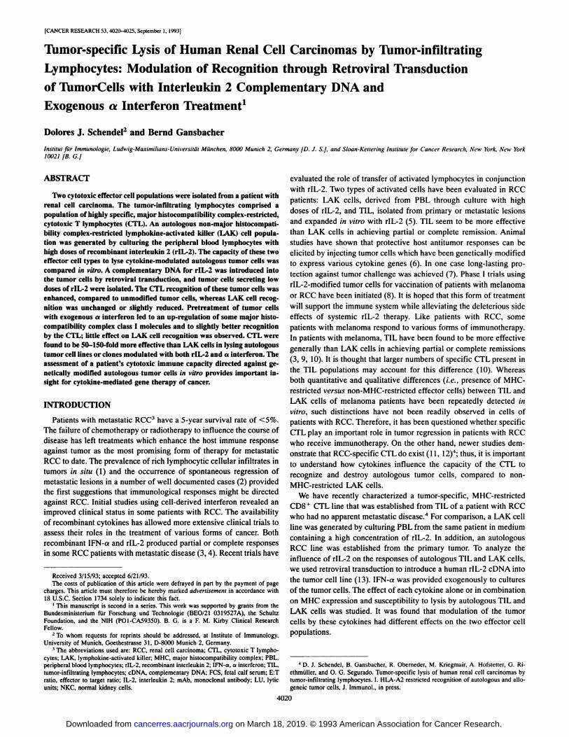
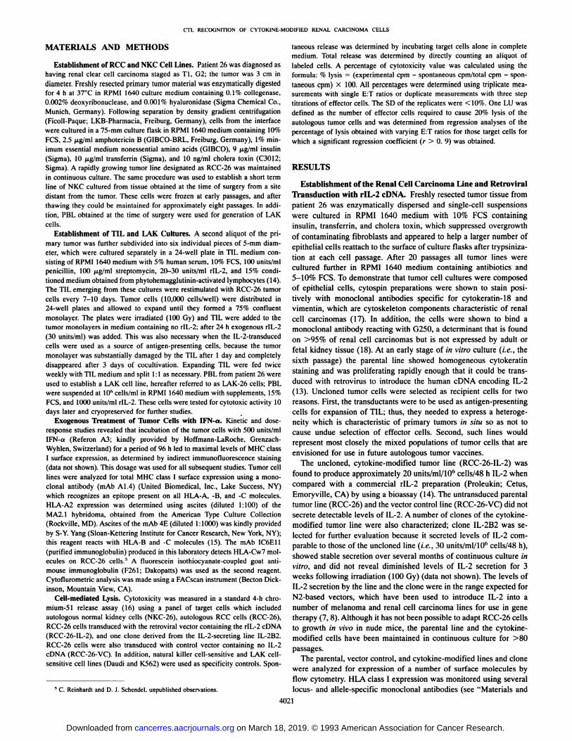
Fig. 1. Comparison of TIL-26 and LAK-26 cellcytotoxic activity directed against different targetcells, a, results with TIL-26 cells; b, results withLAK-26 cells. NKC-26 (•)cells represent a shortterm line of autologous normal kidney cells,RCC-26 (•)is the autologous tumor cell line, RCC-26-IL-2 (O) is the tumor line transduced with thehuman rlL-2 cDNA. and IL-2B2 (ED) is a clone
derived from this line. Daudi (A) and K562 ( * ) arecontrol target cells for non-MHC-restricted effectorcells. Dala represent the percenlage of specific lysismeasured at various E:T cell ratios and were calculated according to the formula given in "Materialsand Methods."
50
45
40
35
30
25
20
15
10
5
0
-51:1 2:1
E:T4:1 40:1
Methods"); in addition, staining for the adhesion molecules CD54
(intercellular adhesion molecules-1) and CD58 (lymphocyte-functionassociated molecules-3) was studied. No significant differences in
percentages of positive cells and levels of surface expression betweenunmodified and transduced cells were found with any of these markers(data not shown).
Comparison of Specificity of TIL and LAK Cells. The lympho-cytic infiltrate was isolated from mini-cultures of RCC-26 using a lowdose of rIL-2. Studies of TIL populations from a number of patients
revealed that these conditions were most suitable for isolation ofcytotoxic effector cells showing restricted patterns of specificity.4-6
Continued proliferation of TIL-26 cells was dependent upon regularrestimulation with autologous tumor cells; RCC-26, RCC-26-IL-2,and IL-2B2 tumor cells could all be used but more extensive growthover longer periods of time was achieved using the cytokine-modifiedcells, even when exogenous rIL-2 was provided to all cultures. TheTIL-26 population was composed predominantly of CD3+CD8+
(96%) cells. In comparison, the autologous LAK-26 cell population,which was generated from the patient's PBL using a high concentration of rIL-2, contained a mixture of CD3 + (59%) and CD3~ (41%)
cells.When tested for cytotoxicity against a group of target cells the
TIL-26 cells were found to be very potent, as evidenced by the small
numbers of effector cells required for detection of significant cytotoxicity (Fig. la). Additional experiments using higher effector totarget cell ratios revealed that plateaus were reached at ratios of morethan 8:1; therefore, quantitative comparisons were made using ratiosthat yielded significant linear regression values (16). Visual inspectionof cultures of TIL-26 cells with the parental and cytokine-modifiedtumor cells showed that the TIL-26 cells were capable of eliminatingall tumor cells during a 24-48-h period of cocultivation; thus, the lysis
of only a fraction of the tumor cells depicted in Fig. la is probably afunction of the incubation time of the chromium release assay, whichwas limited by the rate of spontaneous release of radioactive labelfrom the target cells.
The TIL were highly specific; they efficiently lysed cells of theautologous tumor (RCC-26) but not normal kidney cells (NKC-26) orthe natural killer cell-sensitive or LAK cell-sensitive target cells
' D. J. Schendel, unpublished observations.
(K562 and Daudi). Based on phenotype and specificity these cellsappeared to be tumor-specific CTL. Additional immunogenetic andblocking studies using T cell receptor- and MHC-specific mAb substantiated this conclusion.4 In contrast, the LAK-26 cells showed a
broad pattern of cytotoxicity, associated with non-MHC-restricted
effector cells, and lysed all of the target cells (Fig. l/>). They were alsoless potent, since substantially more LAK-26 cells were required toachieve levels of lysis equivalent to those seen with TIL-26 cells.Based on results of five experiments the TIL-26 cells were found to be3-6-fold more potent than LAK-26 cells in lysing RCC-26 cells
(Table 1 and data not shown).Influence of Endogenous 11-2 Expression by RCC-26 Cells on
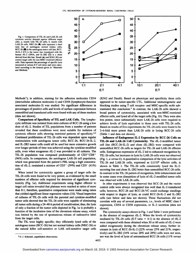
TIL-26 and LAK-26 Cell Cytotoxicity. The rIL-2-modified tumorcell line (RCC-26-IL-2) and clone (IL-2B2) were compared withunmodified RCC-26 cells as targets for TIL-26 and LAK-26 effectorcells. Endogenous expression of rIL-2 led to enhanced recognition byTIL-26 cells, but increases in lysis by LAK-26 cells were not observed
(Fig. 1, a versus b). A quantitative comparison of the lytic activities ofTIL-26 and LAK-26 cells, expressed as LU/10" effector cells, is
shown in Table 1. The TIL-26 cells consistently lysed the IL-2-secreting line and clone IL-2B2 better than unmodified RCC-26 cells.In contrast to the TIL-26 pattern of recognition, little enhancement andin some cases even dimunition of lysis of rIL-2-modified tumor cellswas observed with LAK-26 cells.
In other experiments it was observed that RCC-26 and the vectorcontrol cells were always recognized less well than IL-2-transducedcells; however, RCC-26 and RCC-26-VC could exchange standingswith respect to degree of lysis, as could RCC-26-IL-2 and IL-2B2
cells (see below and data not shown). These fluctuations did notcorrelate with any of several parameters, i.e., levels of MHC class Iexpression, CD54 or CD58 expression, or IL-2 secretion (data not
shown).The cytotoxicity assays shown in Fig. 1 and Table 1 were performed
in the absence of exogenous rIL-2. When the levels of cytotoxicitymediated by TIL-26 cells (E:T ratio = 4:1) in the absence of rIL-2were compared with those obtained when 30 or 60 units of rIL-2 were
added exogenously during the chromium release assay, further increases in lysis of RCC-26-IL-2 (22% versus 25% and 21%, respectively) and IL-2B2 (34% versus 28% and 28%) cells were not seen,nor was the level of lysis of untransduced RCC-26 cells (11% versus
4022
on March 18, 2019. © 1993 American Association for Cancer Research.cancerres.aacrjournals.org Downloaded from
CTL RECOGNITION OF CYTOKINE-MODIFIED RENAL CARCINOMA CELLS
Table 1 Lytic activity of TIL and LAK cells for aulologous lL-2-modifit'd target cells
Lytic activity (LU/10" cells)
NKC-26 cells RCC-26 cells
" This represents a clone derived from the RCC-26-IL-2 line.h NC, not calculated because the r coefficient was <0.9.' Mean LU/IO6 effector cells for the two experiments.
DC/AD/R/IL-2 vector
TILExperiment1
Experiment 2Mean'LAKExperiment
1Experiment 2MeanNo
veclor, NKC-26targetNCh
NCNC1.2
4.0 X IO--
0.6No
vector, RCC-26target2.5
X IO21.1 X IO21.8 XIO277.422.4
49.9RCC-26-IL-2
target1.2X
IO37.9 X IO21.0 XIO322.337.4
29.9IL-2B2
target-5.9
X IO28.1 X IO27.0 XIO232.4
84.058.2
10% and 8%) increased. Addition of neutralizing antibody specific forIL-2 (50 jug/ml) during the chromium release assay led to equalinhibition of TIL-26 cell lysis of IL-2B2 target cells in the absence
(38% inhibition) and in the presence (38% inhibition) of exogenousrIL-2. This antibody did not lead to any inhibition of IL-2B2 cell lysisby the LAK-26 cells.
Modulation of Tumor Cell Lysis by IFN-a. The influence ofIFN-a on tumor cell recognition by TIL-26 and LAK-26 cells was
studied by incubating the tumor lines in medium containing exogenous cytokine and testing them as target cells in cytotoxicity assays.Following preincubation with IFN-a for 96 h, RCC-26 cells werebetter recognized by TIL-26 cells, but diminished recognition wasobserved with LAK-26 cells (Table 2). Similar distinctions werefound with the IL-2-transduced cells pretreated with IFN-a and withRCC-26 cells transduced with the control vector, showing that this
effect was not altered by introduction into the tumor cells of theretroviral construct itself. Regardless of the status of the tumor cellswith respect to rIL-2, IFN-a had a potentiating effect on TIL-26 cellrecognition but not on the LAK-26 cell recognition.
One well known effect of IFN-a is its ability to up-regulate MHC
class I expression, which in turn could lead to better expression of thetarget ligands seen by TIL-26 cells. Immunogenetic studies revealedthat the target antigens recognized by TIL-26 cells involve RCC-specific peptides that are presented by HLA-A2 molecules and possibly by HLA-Cw7 molecules.4 Therefore, the influence of IFN-a
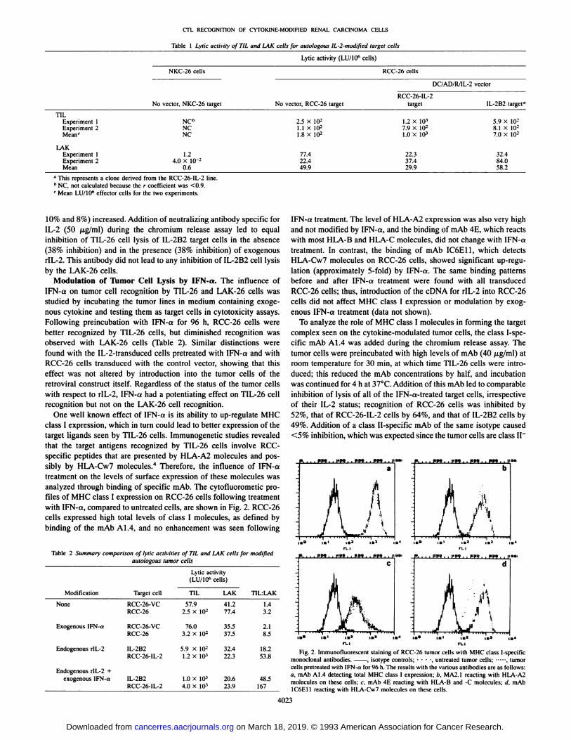
treatment on the levels of surface expression of these molecules wasanalyzed through binding of specific mAb. The cytofluorometic profiles of MHC class I expression on RCC-26 cells following treatmentwith IFN-a, compared to untreated cells, are shown in Fig. 2. RCC-26
cells expressed high total levels of class I molecules, as defined bybinding of the mAb Al.4, and no enhancement was seen following
Table 2 Summary comparison of lytic activities of TIL and 1AK cells for modifiedautologous tumor cells
Lytic activity(LU/10" cells)
ModificationNoneExogenous
IFN-aEndogenous
rlL-2Endogenous
rlL-2 +exogenous IFN-aTarget
cellRCC-26-
VCRCC-26RCC-26-VC
RCC-26IL-2B2
RCC-26-IL-2IL-2B2
RCC-26-1L-2TIL57.92.5
Xlu276.0
3.2 XIO25.9
x W-1.2 XIO31.0
XIO34.0X 10»LAK41.277.435.5
37.532.422.320.6
23.9TILiLAK1.43.22.1
8.518.2
53.848.5
167
IFN-a treatment. The level of HLA-A2 expression was also very highand not modified by IFN-a, and the binding of mAb 4E, which reactswith most HLA-B and HLA-C molecules, did not change with IFN-a
treatment. In contrast, the binding of mAb IC6E11, which detectsHLA-Cw7 molecules on RCC-26 cells, showed significant up-regu-lation (approximately 5-fold) by IFN-a. The same binding patternsbefore and after IFN-a treatment were found with all transducedRCC-26 cells; thus, introduction of the cDNA for rIL-2 into RCC-26
cells did not affect MHC class I expression or modulation by exogenous IFN-a treatment (data not shown).
To analyze the role of MHC class I molecules in forming the targetcomplex seen on the cytokine-modulated tumor cells, the class I-spe-
cific mAb Al.4 was added during the chromium release assay. Thetumor cells were preincubated with high levels of mAb (40 jug/ml) atroom temperature for 30 min, at which time TIL-26 cells were intro
duced; this reduced the mAb concentrations by half, and incubationwas continued for 4 h at 37°C.Addition of this mAb led to comparable
inhibition of lysis of all of the IFN-a-treated target cells, irrespectiveof their IL-2 status; recognition of RCC-26 cells was inhibited by52%, that of RCC-26-IL-2 cells by 64%, and that of IL-2B2 cells by49%. Addition of a class II-specific mAb of the same isotype caused<5% inhibition, which was expected since the tumor cells are class II"
P. ... RM : . 1*99 : . EBB . , ?BB . , I PBB . , l*Ba , . EBB , . EBB , , [IBB.
Fig. 2. Immunofluorescent staining of RCC-26 tumor cells with MHC class 1-specificmonoclonal antibodies. . isotype controls; •¿�•¿�•¿�-, untreated tumor cells: , tumorcells pretreated with IFN-a for 96 h. The results with the various antibodies are as follows:a, mAb A 1.4 detecting total MHC class I expression; b, MA2.1 reacting with HLA-A2molecules on these cells; c, mAb 4E reacting with HLA-B and -C molecules; d. mAb1C6E11 reacting with HLA-Cw7 molecules on these cells.
4023
on March 18, 2019. © 1993 American Association for Cancer Research.cancerres.aacrjournals.org Downloaded from
CTL RECOGNITION OF CYTOK1NE-MODIFIED RENAL CARCINOMA CELLS
and the CTL are CD8 +. The failure to achieve higher levels of secreting IL-2. The untransduced tumor cells did not express IL-2
inhibition with mAb Al.4 seems to reflect high affinity in the interaction of the CTL and the tumor cells, as evidenced by the low E:Tratio that was sufficient to yield significant lysis and the failure toinhibit the CTL with a mAb specific for the CDS molecule.4
Combined rIL-2 and IFN-a Modulation of RCC-26 Cells. The
individual and combinatorial effects of tumor cell modulation byendogenously produced rIL-2 and exogenous IFN-a on TIL-26 andLAK-26 cell lytic potential are summarized in Table 2. In all casesTIL-26 cells were more potent than LAK-26 cells in lysing untreatedand cytokine-modified target cells. Treatment of RCC-26 cells and thevector control line (RCC-26-VC) with IFN-a led to a small enhancement of recognition by TIL-26 cells but not by LAK-26 cells. Largeincreases were seen in the capacity of TIL-26 cells to lyse rIL-2-modified tumor cells, whereas this was not found with LAK-26 cells.The combined modulation with endogenous rIL-2 and exogenousIFN-a led to even better recognition by TIL-26 cells but did not havea positive influence on LAK-26 cell recognition. The LU activities
measured against target cells modulated by both cytokines, as compared to their individual effects, revealed a synergistic enhancementwhich resulted in approximately 50-150-fold greater TIL-26 cell cy-totoxicity, compared to LAK-26 cell-mediated cytotoxicity, directed
against autologous tumor cells.
DISCUSSION
The availability of both MHC-restricted CTL and non-MHC-re-
stricted LAK cells, as well as an autologous tumor line, from onepatient allowed the influence of cytokine modulation on these distincteffector populations to be compared. Clearly, rIL-2 was essential for
generating and maintaining both effector cell populations in vitro.When PBL of patient 26 were directly tested for cytotoxicity, no lysisof autologous tumor cells was detected, even by using rIL-2-modifiedtumor cells as target cells. The in vitro cultivation of PBL with rIL-2
led to development of a LAK effector population that did not requireexposure to tumor cells to show cytotoxic potential. In contrast,growth of TIL-26 cells, containing the MHC-restricted CTL, was
dependent upon regular reexposure to tumor cells; these CTL alsodied within a matter of days when rIL-2 was removed from the culturemedium.6 Both effector populations were capable of lysing autolo
gous RCC-26 cells in the 4-h chromium release assay in the absenceof exogenous rIL-2. However, when they were confronted with tumorcells which expressed endogenous rIL-2, only the TIL cells gained
potency in their lytic activity. This effect was provided by very lowdoses of rlL-2, since recognition of various tumor cell clones secretingbetween 5 and 30 units/ml rIL-2 was equal. Further enhancement oflysis was not observed with the addition of 30-60 units of exogenousrIL-2 during the cytotoxicity assay, nor were RCC-26 cells betterlysed in the presence of exogenous rIL-2. Apparently, intimate delivery of a small amount of IL-2 to the CTL at their point of contact with
the tumor cells improved their cytotoxic performance.Neither addition of exogenous rIL-2 during the cytotoxicity assay
nor endogenous rIL-2 secretion by the tumor cells influenced thepotency of LAK-26 cells. This is most likely explained by the fact thatthese cells were exposed continuously to very high levels of rIL-2 up
to the point at which they were harvested for functional studies and,therefore, they were refractory to further rIL-2 modulation. Theircytotoxic activity was also not inhibited by antibody specific for IL-2.
Since exogenous IL-2 could not enhance TIL-26 cell recognition ofRCC-26 cells and the neutralizing antibody directed against IL-2 only
partially inhibited the CTL response, it must be considered that effectsother than direct delivery of IL-2 to the CTL following T cell receptor-
ligand binding may lead to better recognition of the tumor cells
receptors, and IL-2 receptors could not be detected in the IL-2 trans-ductants.6 Since the levels of surface expression of MHC class I,
CD54, and CD58 molecules were the same in untransduced andIL-2-transduced tumor cells, alterations in these major surface mole
cules, which govern the interactions of effector cells and target cells,seemed not to be involved. It could be speculated that endogenousIL-2 expression led to altered susceptibility of the tumor cells to lysis;
if so, this effect is specifically related to cytolytic mechanisms used byCTL, since lysis by LAK-26 cells was not enhanced. The receptorsand ligands used by non-MHC-restricted cells to recognize tumor cells
are poorly understood (19); therefore, determination of the role ofIL-2 in their regulation must await future developments.
The effect of IFN-a therapy for patients with RCC has been ascribed partially to its ability to up-regulate MHC class I expression onepithelial cells. An up-regulation of MHC class I expression may leadto better presentation of tumor-peptide MHC complexes, which form
the target ligands recognized by specific CTL (20). On the other hand,a reverse correlation has been reported between the level of MHCexpression and sensitivity to lysis by some non-MHC-restricted effector cells (19, 21). Since RCC-26 tumor cells expressed very highlevels of HLA-A and -B molecules, little enhancement in their expression was achieved with IFN-a, whereas HLA-C molecules wereup-regulated about 5-fold. Indirect evidence suggests that some of theCTL in the TIL-26 population may recognize tumor-associated pep-tides restricted by HLA-Cw7.4 Determination of how much the HLA-
Cw7 increase accounts for improved recognition by the TIL-26 cellsrequires further evaluation. It is interesting to note that HLA-C molecules have also been reported to present tumor-associated peptides tomelanoma-specific CTL (22).
It is clear that combined cytokine modulation worked to the advantage of the CTL present in the TIL-26 population. Animal studies havedemonstrated that TIL are 100-fold more effective than peripheral
LAK cells in eliminating tumor cells in vivo; this effect has beenpostulated to be due to the presence of highly specific, MHC-restricted CTL in the TIL population (23). Modification of RCC-26 cellswith both rIL-2 and IFN-a revealed that these TIL were also approximately 50-150-fold more effective than LAK cells in eliminating the
autologous tumor cell line and clone in vitro. It will be important todetermine whether the observations made with patient 26 can begeneralized to other patients; if so, it would appear that those patientswho have generated a tumor-specific CTL response may benefit mostfrom combined therapy with IFN-a and rIL-2. Since the side effectsof rIL-2 therapy are highly problematic, it is hoped that vaccinesutilizing cytokine-modulated tumor cells can be substituted for sys
temic application and that they will have effects on immune responsesin vivo similar to those demonstrated in tumor models in animals. It isrewarding to find that IL-2-transduced tumor cells which secreted lowlevels of rIL-2 were very effective in potentiating the responses ofthese RCC-specific MHC-restricted CTL in vitro. If CTL can beactivated in a similar fashion following an encounter with cytokine-
modified tumor cells in vivo, they may be more effective in combatingresidual metastatic disease.
ACKNOWLEDGMENTS
The authors thank Barbara Maget, Kim Seebart, and Kathy Cronin forexcellent technical assistance and Steve Seebart for help in the preparation ofthis manuscript. We thank Dr. Gert Riethmüller for enthusiastic support of
these studies and Dr. Ralph Oberneder and other members of the GroßhadernUrology Clinic, University of Munich, for providing patient material. Dr.
Karen S. Zier is gratefully acknowledged for many helpful discussions.
4024
on March 18, 2019. © 1993 American Association for Cancer Research.cancerres.aacrjournals.org Downloaded from
CTL RECOGNITION OF CYTOKINL-MODIFIED RENAL CARCINOMA CELLS
REFERENCES
1. Vose. B. M., and Muore. M. Human tumor-infiltrating lymphocytes: a marker of hostresponse. Semin. Hematol.. 22: 27-35, 1985.
2. De Riese. W.. Allhoff. E.. Kirchner. H.. Stief. C. G.. Atzpodicn. J.. Maschek. H., andJonas. U. Complete spontaneous regression in metastatic renal cell carcinoma: anupdate and review. World J. Urol., 9: 184-191, 1991.
3. Oettgen. H. F. Cytokines in clinical cancer therapy. Curr. Opin. Immunol.. 3: 699-
705, 1991.4. Allhoff. E. P.. Liedke. S.. Kirchner. H.. Atzpodien. J.. De Riese, W., Stief, C. G., and
Jonas. U. Current clinical relevance of immunotherapy in metastatic renal cell cancer.World J. Urol.. 9: 228-231, 1991.
5. Rosenberg, S. A.. Lotze, M. T., and Mule, J. J. NIH conference: new approaches tothe ininiunotherapy of cancer using interleukin-2. Ann. Intern. Med., 108: 853-864,
1988.6. Blankenstein, T.. Rowley, D. A., and Schreiher. H. Cytokines and cancer: experimen
tal systems. Curr. Opin. Immunol., .ÃŽ:694-698, 1991.
7. Gansbachcr, B.. Zier, K., Daniels, B.. Cronin. K.. Bannerji. R., and Gilboa, E.Intcrleukin 2 gene transfer into tumor cells abrogates tumorigenicity and inducesprotective immunity. J. Exp. Med.. ¡72: 1217-1224, 1990.
8. Gansbacher. B.. Motzer. R., Houghton, A.. Bander, N.. Minasian, L., and Reuter, V.A pilot study of immunization with interleukin-2-secreting allogeneic, HLA-A2-matched renal cell carcinomas. Hum. Gene Then. 3: 691-703, 1992.
9. Knuth. A.. Wölfe!.T.. and Meyer zum Büschenfelde. K-H. Cellular and humoralimmune responses against cancer: implications for cancer vaccines. Curr. Opin.Immunol.. 3: 659-664, 1991.
10. Parmiani. G. An explanation of the variable clinical response to intc,rleukin-2 andLAK cells. Immunol. Today. 11: 113-115. 1990.
11. Koo. A. S., Tso. C-L., Shimabukuro. T.. Peyret, C.. deKernion. J. B.. and Belldegrun.A. Autologous tumor-specific cytotoxicity of tumor-infiltrating lymphocytes derivedfrom human renal cell carcinoma. J. Immunother.. 10: 347-354, 1991.
12. Finke, J. H.. Rayman. P.. Edinger, M., Tubbs, R. R.. Stanley. J.. Klein. E., andBukowski. R. Characterization of a human renal cell carcinoma specific cytotoxic
CD8 + T cell line. J. Immunother.. //: 1-11, 1992.
13. Gasll, G., Finstad, C. L., Guarini, A., Bosl, G., Gilboa, E.. Bander. N. H., andGansbacher, B. Retrovtral vector-mediated lymphokine gene transfer into humanrenal cancer cells. Cancer Res., 52: 6229-6236. 1992.
14. Schendel, D. J., and Wank. R. Production of human T cell growth factor. Hum.Immunol., 2: 325-332, 1981.
15. Trapani, J. A.. Mizuno. S.. Kang, S. H.. Yang. S. Y.. and Dupont, B. Molecularmapping of a new public HLA class I epitope shared by all HLA-B and HLA-Cantigens and defined by a monoclonal antibody. Immunogcnetics, 29: 25-32, 1989.
16. Schendel. D. J., Wank. R.. and Dupont. B. Standardization of the human in vitrocell-mediated lympholysis technique. Tissue Antigens. 13: 112-120. 1979.
17. Pitz. S.. Moll, R., Störkel.S., and Thoenes, W. Expression of intermediate filamentproteins in subtypes of renal cell carcinomas and in renal oncocytomas. Distinction oftwo classes of renal cell tumors. Lab. Invest., 56: 642-653, 1987.
18. Oosterwuk, E., Ruitter. D. J., Hoedemaeker, P. J., Pauwels, E. K. J.. Jonas, U.,Zwartenduk, J.. and Warnaar. S. O. Monoclonal antibody G 250 recognizes a determinant present in renal-cell carcinoma and absent from normal kidney. Int. J. Cancer,38: 489-494, 1986.
19. Yokoyama, W. M., and Seaman, W. E. The Ly-49 and NKR-P1 gene families encoding lectin-like receptors on natural killer cells: the NK gene complex. Annu. Rev.Immunol., //: 613-635, 1993.
20. Van der Bruggen. P.. Travrsari. C., Chômez, P., Lurquin, C., De Plaen, E., Van denEynde. B., Knuth, A., and Boon, T. A gene encoding an antigen recognized bycytolytic T lymphocytes on a human melanoma. Science (Washington DC), 254:1643-1647, 1991.
21. Janeway, C. A., Jr. To thine own self be true. Curr. Biol.. /: 239-241, 1991.
22. Horn. S. S.. Topalian. S. L.. Simonis. T., Mancini, M., and Rosenberg. S. A. Commonexpression of melanoma tumor-associated antigens recognized by human tumor infiltrating lymphocytes: analysis by human lymphocyte antigen restriction. J. Immunother.. 10: 153-164. 1991.
23. Rosenberg. S. A.. Spiess. P.. and Liifreniere, R. A new approach to the adoptiveimmunotherapy of cancer with tumor-infiltrating lymphocytes. Science (WashingtonDC), 233: 1318-1321, 1986.
4025
on March 18, 2019. © 1993 American Association for Cancer Research.cancerres.aacrjournals.org Downloaded from
1993;53:4020-4025. Cancer Res Dolores J. Schendel and Bernd Gansbacher
Interferon Treatmentα2 Complementary DNA and Exogenous through Retroviral Transduction of TumorCells with InterleukinTumor-infiltrating Lymphocytes: Modulation of Recognition Tumor-specific Lysis of Human Renal Cell Carcinomas by
Updated version
http://cancerres.aacrjournals.org/content/53/17/4020
Access the most recent version of this article at:
E-mail alerts related to this article or journal.Sign up to receive free email-alerts
Subscriptions
Reprints and
To order reprints of this article or to subscribe to the journal, contact the AACR Publications
Permissions
Rightslink site. Click on "Request Permissions" which will take you to the Copyright Clearance Center's (CCC)
.http://cancerres.aacrjournals.org/content/53/17/4020To request permission to re-use all or part of this article, use this link
on March 18, 2019. © 1993 American Association for Cancer Research.cancerres.aacrjournals.org Downloaded from