tuberculosis among patient with unilateral pleural ...eprints.usm.my/46874/1/dr. nor haslina abdul...
TRANSCRIPT

TUBERCULOSIS AMONG PATIENT WITH
UNILATERAL PLEURAL EFFUSION IN KELANTAN:
PREVALENCE AND ASSOCIATED FACTORS
DR. NOR HASLINA BINTI ABDUL RAHMAN
DISSERTATION SUBMITTED IN PARTIAL FULFILLMENT
OF THE REQUIREMENT FOR THE DEGREE OF MASTER
OF
INTERNAL MEDICINE
UNIVERSITI SAINS MALAYSIA
2018

ii
ACKNOWLEDGEMENTS
I would like to thank all my supervisors Dr Alwi Besari, Dr Haji Mat Zuki Mat Jaeb and
Dr Sudzilla Nordin for their advices, ideas and supervision on completing this
dissertation. And not to forget to Dr Najib Majdi Yaacob from the Biostatistic Department
for the advice on the statistical aspect and all the staffs who were involved in collecting
the specimens and data.
To my husband En Fairol Hisyam, my beloved children Muhammad Syakir Imaam and
Hasya Nur Aafiya, my family and friends for the full support during this years. Thank
you very much
Dr Nor Haslina Abdul Rahman

iii
TABLE OF CONTENTS
PAGE
Acknowledgement ii
Table of Contents iii
List of Tables vi
List of Figures viii
Abbreviation ix
Abstract in Bahasa Melayu x
Abstract in English
xii
CHAPTER 1 INTRODUCTION
1.1 Study Background 1
1.2 Study Rationale 4
1.3 Overview of Tuberculosis 5
1.3.1 Definition 5
1.3.2 Epidemiology 6
1.3.3 Pathogenesis 10
1.3.4 Causes of pleural effusion 14
1.3.5 Clinical manifestation 15

iv
1.3.6 Diagnostic approach of TB pleural effusion 16
1.3.6.1 Pleural fluid analyses 16
1.3.6.2 Chest Imaging
20
1.3.6.3 Thoracoscopy and pleural biopsy 22
1.3.7 Management 25
CHAPTER 2 OBJECTIVE
2.1 Research questions 26
2.2 General Objective 27
2.3 Specific Objective
27
2.4 Research Hypothesis 27
CHAPTER 2 METHODOLOGY
3.1 Study design 28
3.2 Study setting and population 28
3.3 Inclusion criteria 28
3.4 Exclusion criteria 28
3.5 Data collection 29
3.6 Definition of operational terms 30
3.7 Study flow chart 32
3.8 Sample size calculation 33

v
3.9 Statistical analysis 36
CHAPTER 4 RESULT
4.1 Descriptive analyses of overall study participants
37
4.2 Descriptive analyses of study participants based on outcome
(Non-TB versus TB)
47
4.3 Univariable analyses to determine factor associated with
Tuberculosis pleural effusion (simple logistic regression)
55
4.4 Multivariable analyses to determine factor associated with
Tuberculosis pleural effusion (Multiple logistic regression)
63
CHAPTER 5 DISCUSSION
5.1 The proportion of TB pleura among patients in unilateral
pleural effusion
66
5.2 Factors associated with TB pleura 68
CHAPTER 6 CONCLUSION
6. Conclusion 74
CHAPTER 7 STUDY LIMITATION
7. Study Limitation 75
CHAPTER 8 STUDY RECOMMENDATION
8. Study Recommendation 76
BIBLIOGRAPHY 78
APPENDICES
Appendix 1 Data Collection Sheet
Appendix 2 Ethic Approval
Appendix 3 NMRR Approval
85
87
90

vi
LIST OF TABLES
Table 4.1.1
Descriptive analysis of demographic data and clinical
presentation based on overall participant
Table 4.1.2 Descriptive analysis of pleural analysis based on overall
participant
Table 4.1.3 Descriptive analysis of culture and sensitivity based on overall
participant
Table 4.1.4 Descriptive analysis of cytology and biopsy based on overall
participant
Table 4.1.5 Descriptive analysis of diagnosis based on overall participant
Table 4.1.6 Descriptive analysis of thoracoscopy characteristics based on
overall participant
Table 4.2.1 Descriptive analysis of demographic data and clinical
presentation based on outcome (Non-TB versus TB)
Table 4.2.2 Descriptive analysis of pleural analysis based on outcome
(Non-TB versus TB)
Table 4.2.3 Descriptive analysis of culture and sensitivity based on
outcome (Non-TB versus TB)
Table 4.2.4 Descriptive analysis of cytology and biopsy based on outcome
(Non-TB versus TB)
Table 4.2.5 Descriptive analysis of thoracoscopy features based on
outcome (Non-TB versus TB)
Table 4.3.1 Univariate analysis of association between demographic data
and clinical presentation with TB pleural effusion (simple
logistic regression)

vii
Table 4.3.2 Univariate analysis of association between pleural analysis with
TB pleural effusion (simple logistic regression)
Table 4.3.3 Univariate analysis of association between culture and
sensitivity with TB pleural effusion (simple logistic regression)
Table 4.3.4 Univariate analysis of association between thoracoscopy
characteristic with TB pleural effusion (simple logistic
regression)
Table 4.4.1 Multivariable analysis of factor associated with TB pleural
effusion (Multiple logistic regression)

viii
LIST OF FIGURES
Figure 1 Cases of Tuberculosis in Kelantan since 2000 to 2017
Figure 2 Prevalence of Pulmonary TB smear positive, smear negative and
Extra-Pulmonary Tuberculosis cases in Kelantan since 2010 to
2017
Figure 3 Prevalence of Extra-Pulmonary TB cases in Kelantan in 2017
Figure 4 Pathogenesis of TB pleural effusion
Figure 5 Causes of unilateral pleural effusion
Figure 6 Imaging in TB pleural effusion
Figure 7 Thoracoscopy characteristics in TB pleural effusion
ix
ABBREVIATION
ADA Adenosine Deaminase
AFB Acid Fast Bacilli
BTS British Thoracic Society
CKD Chronic Kidney Disease
CT Computed Tomography
HIV Human Immunodeficiency Virus
HRPZ Hospital Raja Perempuan Zainab II
IGRA Interferon Gamma Release Assay
IQR Interquartile range
MT Medical Thoracoscopy
MTB Mycobcterium Tuberculosis
PCR Polymerase Chain Reaction
SD Standard Deviation
TB Tuberculosis
WHO World Health Organization

x
ABSTRAK
Pengenalan
Efusi pleura Tuberkulosis merupakan penyakit yang diketahui umum dalam kalangan
pesakit yang mempunyai efusi pleura sebelah. Dengan peningkatan prevalen kes
tuberkulosis luar pulmonari di Malaysia khususnya di negeri Kelantan, didapati
kebanyakan kes efusi pleura Tuberkulosis masih tidak dikesan dalam kalangan populasi
ini. Ini disebabkan oleh kurangnya pengalaman dalam kalangan pegawai perubatan untuk
mengenalpasti faktor risiko berkaitan dengan efusi pleura Tuberkulosis. Oleh itu,
matlamat kajian ini adalah untuk mengenalpasti jumlah perkadaran efusi pleura
Tuberkulosis dan faktor-faktor yang berkaitan dengannya.
Kaedah
Ini adalah kajian rektrospektif yang melibatkan seramai 376 orang pesakit yang
mempunyai sebelah lapisan efusi pleura di paru-paru yang masih tidak dikesan menjalani
rawatan torakoskopi bermula dari 1 Januari 2011 sehingga 31 Disember 2016 yang telah
dikenalpasti daripada buku rekod pendaftaran di Hospital Raja Perempuan Zainab II.
Maklumat-maklumat yang berkaitan dengan sosio-demografi (umur, jantina ,bangsa,
status merokok, komorbid, persembahan klinikal), ujian makmal efusi pleura (paras
protein, LDH, gula, kultur, AFB, kultur MTB, TB PCR, Xpert Gen) dan demonstrasi luka
melalui torakoskopi di pleura paru-paru (nodular, ketulan, lekatan, plak, lesi seperti keju
dan sagu) telah diambil daripada rekod. Ujian regresi logistic berganda telah digunakan
untuk tujuan analisis statistic dan nilai p nominal ditetapkan pada 0.05.

xi
Keputusan
Perkadaran efusi pleura Tuberkulosis dalam kalangan sebelah lapisan efusi pleura yang
tidak didiagnosis adalah sebanyak 38.3%. Kemalignanan paru-paru menyumbang
diagnosa yang tertinggi sebanyak 44.4% diikuti oleh jangkitan kuman selaput paru-paru
sebanyak 10.1% dan penyakit buah pinggang kronik sebanyak 2.7%. Berdasarkan analisis
regresi logistic berganda, demostrasi lesi seperti sagu (OR 37.41 (CI: 7.81, 179.24), p
value = < 0.001) dan demostrasi lesi seperti keju (OR 30.44 (CI 12.73, 72.80), p value =
< 0.001) adalah faktor yang paling berhubungkait dengan efusi pleura Tuberkulosis.
Jangkitan kuman Tuberkulosis paru-paru (OR 9.23(CI 1.95, 43.67), p value = 0.005) dan
demam (OR 4.70 (CI 2.21, 9.97), p value = <0.001) juga berhubungkait dengan efusi
pleura Tuberkulosis. Tambahan juga, positif Kultur MTB (OR 6.80 (CI 2.16, 21.45), p
value = 0.001) dan TB PCR juga berhubungkait rapat dengan efusi pleura Tuberkulosis
(OR 8.71 (CI 2.54, 29.94), p value = 0.001). Kehadiran ketulan melalui prosedur
torakoskopi kurang berhubungkait dengan efusi pleura Tuberkulosis (OR 0.13 (CI 0.02,
0.70), p value = 0.018).
Kesimpulan
Sejarah masa lalu TB pulmonari, demam, kultur MTB yang positif dan PCR TB yang
positif dan demonstrasi lesi seperti keju dan sagu dari torakoskopi sangat berkaitan
dengan efusi pleura Tuberkulosis. Mengetahui faktor-faktor penting ini akan membantu
para doktor untuk mendiagnosis efusi pleura Tuberkulosis supaya rawatan dapat bermula
lebih awal untuk mengelakkan morbiditi dan mortalitas.

xii
ABSTRACT
Background
Tuberculosis pleural effusion is a well-known aetiology among patient with unilateral
pleural effusion. With the increasing prevalence of Extra-pulmonary TB in Malaysia and
particularly in Kelantan state, many TB pleura cases still undiagnosed among this
population. This can be due to lack of experience among the clinicians in identifying the
potential risk factors for TB pleural effusion. Hence, the aims of this study are to
determine the proportion of TB pleural effusion and the factors associated with it.
Methodology
This is a retrospective study which involved a total of 376 patients with unexplained
unilateral pleural effusion who underwent medical thoracoscopy from 1st January 2011
until 31th December 2016 was identified from registration book in Respiratory Clinic,
Hospital Raja Perempuan Zainab II. Relevant details on sociodemographic data (age,
gender, race, smoking history, comorbidity, clinical presentation), laboratory pleural fluid
analysis (protein, LDH, glucose, culture, AFB, MTB culture, TB PCR, Gene Xpert ) and
thoracoscopy findings ( nodule, mass, adhesion, plaque, sago-like lesion and cheesy-like
lesion) were collected from the medical review. Multiple logistic regression were used to
analyses the data.

xiii
Results
The proportion of TB pleura amongst unexplained unilateral pleural effusion were 38.3%.
Lung malignancy contribute the highest proportion in 44.4% followed by para pneumonic
effusion in 10.1% and chronic kidney disease in 2.7%. Based on multiple logistic
regression analyses, sago-like lesion is the strongest determinant factors associated with
TB pleura (OR 37.41 (CI: 7.81, 179.24), p value = < 0.001) and cheesy-like lesion (OR
30.44 (CI 12.73, 72.80), p value = < 0.001). The present of past history of pulmonary TB
is also associated with TB pleura (OR 9.23(CI 1.95, 43.67), p value = 0.005) and fever
(OR 4.70 (CI 2.21, 9.97), p value = <0.001). In addition to that, MTB culture is also the
strongest determinant for TB pleura with (OR 6.80 (CI 2.16, 21.45), p value 0.001) and
TB PCR with (OR 8.71 (CI 2.54, 29.94), p value 0.001). The presence of a mass from
thoracoscopy is less likely associated with TB pleura (OR 0.13 (CI 0.02, 0.70), p value
0.018).
Conclusion
Past history of Pulmonary TB, fever, positive MTB culture and positive TB PCR and
demonstration of cheesy-like and sago-like lesion from thoracoscopy are strongly
associated with TB pleura. Knowing these significant factors will help the clinicians to
diagnose TB pleura so that treatment can be start earlier to avoid morbidity and mortality.

1
CHAPTER 1
INTRODUCTION
1.1 STUDY BACKGROUND
Tuberculosis is a common and potentially serious illness. It is associated with
significant morbidity and mortality. According to the World Health Organization, the
incidence of tuberculosis (TB) are 9.6 million in 2014 and it is estimated that deaths from
TB will increase from 3 million a year to 5 million by the year 2020. Between 2002 and
2020, approximately 1 billion people will be newly infected, 200 million people will get
sick, and 36 million will die of TB if proper control measures are not instituted ("World
Health Organization. Global Tuberculosis report 2016," 2016).
It is mentioned that among all cases presenting with pleural effusion, 25% are
unable to be attributed to a specific diagnosis, even after thoracoscopy and closed pleural
biopsy (JM, 2016). As many as 50% of the patients in this undiagnosed pleural effusion
will eventually be diagnosed include TB, fungal disease, connective tissue related
pleuritis, pulmonary infarction, rib fractures, asbestos-related pleural effusion, and
nonspecific pleuritis (Boutin et al., 1981).
Several studies have comprehensively analysed the etiological causes of patient
with undiagnosed pleural effusion. A retrospective cross sectional hospital based study
conducted a total of 130 cases diagnosed with pleural effusion by chest x-ray without a
consent requirement. These patient underwent medical thoracoscopy procedure in the
first 24 hours after ultrasonography under aseptic technique. In concern with the etiology,
tuberculosis (64.6%) was the most common cause of pleural effusion followed by para
pneumonic effusion (14.6%) and malignancy (11.5%) (Srivastava, 2016).

2
Department of pulmonary medicine at Rohilkhand Medical College and Hospital
Bareilly run out of 40 patients of undiagnosed pleural effusion, majority of them were
aged between 61-70 years (28.75%) and were males (53.75%). The commonest cause of
pleural effusion is tuberculosis (36.2%) followed by para pneumonic effusion (18%) and
malignancy (13%). In tuberculosis pleura, 11 out of 14 patients presented with shortness
of breath and 12 patients presented with cough (Kumar et al., 2017). From the above
studies, they conclude that there were various presenting features for pleural effusion such
as shortness of breath, fever, chest pain and there were multiple important diseases
contribute to the diagnosis of unilateral pleural effusion such as tuberculosis, para
pneumonic effusion and malignancy.
Addition to that, a prospective study conducted at Liaquat National Hospital
Karachi, Pakistan during June 2015 to December 2016 among patients with unilateral
pleural effusion. Diagnostic pleural tapping was performed in every case and pleural
aspiration was sent for laboratory analysis. From this study, majority of cases was
exudative pleural effusion (77%) and all of them (48) patients underwent diagnostic
medical thoracoscopy. The most common cause of unilateral pleural effusion was
Tuberculosis in 26 (54%) cases. Other causes included malignancies in 10 (21%),
congestive cardiac failure in 4 (8%), chronic liver disease in 3 (6%), para pneumonic in
2 (4%) and others 3 (6%) (Abbas et al., 2017).
TB pleura is a diagnostic challenge due to its nonspecific clinical presentation,
paucibacillary nature of the effusion together with the inefficiency of conventional
laboratory methods. (Amer et al., 2016).
Complete history and physical examination should be performed, including an
evaluation of disease, employment and medication history. The most usual imaging

3
technique for identifying pleural effusion is posteroanterior chest X-ray. Thoracic
ultrasound should be easily accessible for these patients and it is also more sensitive than
X-ray and better than computed tomography (CT) for identifying septa, locating small or
encapsulated pleural effusion for puncture or biopsy or providing guidance regarding the
entry point for thoracoscopy (Havelock et al., 2010). Chest CT may be useful for
modifying the probability of identifying diagnosis in undiagnosed pleural effusion as
described from study by Zhen Wang et al where CT imaging revealed pulmonary
consolidation or infiltration in 53.5%, pulmonary atelectasis in 43.4%, mediastinal
lymphadenopathy in 41.1%, pleural thickening in 33.3% , pulmonary mass or nodules in
21.9% and pleural nodularity in 3.9% (Wang et al., 2015).
Medical thoracoscopy refers to the examination of the pleural space in a
nonintubated patients under local anaesthesia, and this procedure has been well
documented to be highly sensitive and safe for diagnosing exudative pleural effusions.
Thus medical thoracoscopy has a role for further evaluation of the pleura and to see the
specific characteristics of tuberculous pleurisy.
Safaa Amer et al conduct a study among 50 patients with unexplained unilateral
pleural effusion who underwent medical thoracoscopy at Faculty of Medicine Alexandria
University. Twenty-four (48%) of the 50 studied patient were diagnose as malignant
effusion and twenty-two were diagnosed as nonspecific fibrinous pleurisy. Three patients
(6%) showed caseating granulomas and one of the total patients showed septic pleurisy.
Nodules is a common macroscopic appearance from the thoracoscopic finding in 84%,
followed by increased vascularity in 40%, adhesions in 38% ,mass in 20% and plaques in
16% among the studied patients(Amer et al., 2016).
4
1.2 STUDY RATIONALE
Hospital Raja Perempuan Zainab (HRPZ) II is the only tertiary hospital in
Kelantan. It has respiratory clinic which provides facilities such as medical thoracoscopy
(MT) procedure, thoracic ultrasound machine, sleep study machine, lung function test
and record all the patients’ file and chest X-ray film including CT film. Since 6 years ago,
MT had been used for diagnostic evaluation among patient with undiagnosed unilateral
pleural effusion in HRPZ II.
Majority of the patients presented with unspecific clinical presentation and they
already treated with multiple course of antibiotics but they still not completely recover
very well. So, these group of patients need further assessment by chest physician to
determine the final diagnosis of the patients. Bedside ultrasound is useful to see the
effusions and to facilitate MT procedure done by chest physician. Almost all the results
revealed that they already in advanced state of malignancy either primary lung or
secondary lung metastasis as well as TB pleural effusion, unresolved empyema, or others.
Therefore, early detection by complete history, physical examination, imaging,
ultrasonography and diagnostic MT should be done earlier to avoid any delay diagnosis.
If diagnosis is correct, so the treatment can be initiate as soon as possible and patients can
have a better outcome and quality of life.
So, this study intends to determine the proportion of TB pleura among patients
presented with unexplained unilateral pleural effusion who underwent MT at HRPZ II.
This study also want to determine the associated factors that attribute to the diagnosis of
TB pleura. Apart from that, we also can get additional local data regarding cases of
unexplained unilateral pleural effusion who underwent MT in HRPZ II since 2011 to
2016.

5
1.3 OVERVIEW OF TUBERCULOSIS
1.3.1 DEFINITION
Tuberculosis is one of the world’s most widespread and deadly illnesses. It is an
airborne disease caused by Mycobacterium tuberculosis. Mycobacterium tuberculosis is
carried in airborne particles, called droplet nuclei, 1-5 microns in diameter. High lipid
content of this pathogen accounts for many of its unique clinical characteristic .This
disease is usually affect the lung but also involve other organs in up to one third of cases
such as lymph nodes, liver, bones and also brain or meninges. It divides every 16 to 20
hours, an extremely slow rate compare with other bacteria which usually divide in less
than an hour (Simon et al., 1980).
MTB can withstand weak disinfectant and survive in a dry state for many weeks.
In nature the bacterium can grow only within the cell of a host organism but MTB can be
cultured in vitro (Parish and Stoker, 1999).
Since MTB has a cell wall but lacks of phospholipid outer membranes, it is
classify as a Gram positive bacterium. However, if a Gram stain is perform, MTB either
stain very weakly Gram positive or does not retain dye as a result of the high lipid and
myocolic acid content of its cell wall (Madison, 2001).
Majority of drug susceptible tuberculosis is curable if treated properly with anti-
TB medication. If untreated, the disease may be fatal within 5 years in 50-65% of cases.
This problem has been compounded by the HIV pandemic, neglect towards the disease
and international movement. About one-third of the world's population has latent TB,
which means people have been infected by TB bacteria but are not yet ill with the disease
and cannot transmit the disease. These people with latent TB can get active TB disease if
6
their immune system is low or if they are taking any treatment that can supress their
immune system.
1.3.2 EPIDEMIOLOGY
Globally, the frequency of TB pleura is highly variable and depend on the
incidence of tuberculosis in each country, for example in Spain, this entity represents a
considerable problem because it is estimated that TB pleura is affected in 23.3% among
all patients with tuberculosis (Valdés et al., 2003). TB pleura in the United States and
Brazil accounted about 4% while 20% of those in South Africa (Baumann et al., 2007).
In Korea, about 2884 new TB pleura cases are reported in 2012, which accounted for
7.3% of a total 39,545 new tuberculosis cases and 34% of all extra pulmonary tuberculosis
cases (Park et al., 2013).
Malaysia was classified as a country with an intermediate TB burden. Number of
TB cases notified in 2016 are around 24,000 and notification rate for TB about 81 cases
per 100,000 populations. Of these cases, 62% are sputum positive, 21% smear negative
and 13% extra-pulmonary TB cases. The most common form of extra pulmonary
tuberculosis seen in Malaysia are TB lymphadenitis, bone, joint, spine and military TB.
Three states with high TB cases are Sabah, Selangor and Sarawak, making up a total of
almost 50% of all new cases in Malaysia ("World Health Organization. Global
Tuberculosis report 2016," 2016).
One hundred seventy four articles related to tuberculosis are found in a search
through a database published in Malaysia between the years 2000 to 2013. About 10 to
11% of tuberculosis at a tertiary level chest clinic were classified as extra pulmonary
tuberculosis and 14% of pulmonary tuberculosis also had extra pulmonary involvement

7
(Jamaluddin et al.). TB pleura is the most common cause of exudative pleural effusion
about 44%, followed by malignancy in 30%. Conversely, in a smaller study the most
common cause for pleural effusion is malignancy in 34%, followed by tuberculosis 23%
and para pneumonic effusions in 19% (Liam et al., 2000).
In Kelantan, cases of Tuberculosis that had been notified show gradually
increasing in trend from 2000 to 2017. The rate of notification in 2017 are 73.3 per
100000 population have Tuberculosis including case with Extra-pulmonary Tuberculosis
(Figure 1).
Among those patient who has been diagnosed with tuberculosis, pulmonary TB
smear positive still the most commonest cases that has been notified since 2010 to 2017
and shows decreasing in trend for the past one year from 62.1% down to 59.8% as
compared to those who has pulmonary TB smear negative, which shows gradually
increasing in trend from 22.3% to 25%. Extra pulmonary TB only contribute a minor
population and gradually static during this period probably maybe due to lack of
notification or undiagnosed cases (Figure 2).
In 2017, TB lymph nodes is the most common among other cases of Extra-
Pulmonary TB about 65% followed by TB spine (36%) , TB meningitis (18%) and the
rest are rare cases of Extra-Pulmonary TB example gut, miliary, pericardium, ovary and
breast. About 30% of TB pleura cases has been notified in 2017 (Figure 3).
Tuberculosis is the largest killer among communicable diseases in 15 to 49 age
group, when humans are most productive and it’s always high in the elderly with other
co morbid diseases like diabetes, smoking and kidney diseases. According to WHO
Global TB report, it is estimated that up to 10.4 million Tuberculosis cases reported
globally in which 5.9 million (56%) are among men, 3.5 million (34%) among women

8
and 1.0 million (10%) among children. In 2016 there are 1,696 TB deaths reported
(excluding TB/HIV mortality), giving rise to 5.56 TB deaths per 100,000 populations.
In the WHO south-East Asia Region as estimated 4.74 million cases of TB are
reported and about 784,000 people died of it. This TB death rate is the highest among all
infectious diseases, including dengue, HIV and malaria. People living with HIV
accounted for 1.2 million (11%) of all new TB cases. Deaths from TB among HIV-
positive people are officially classified as deaths caused by HIV/AIDS in the International
classification of diseases. Deaths from TB among HIV-positive people accounted for 37%
of deaths classified as caused by HIV/AIDS in 2016 ("World Health Organization. Global
Tuberculosis report 2016," 2016).
Figure 1 Cases of Tuberculosis in Kelantan since 2000 to 2017 in per
100000 population. Adapted from Jabatan Kesihatan Negeri Kelantan.

9
Figure 2 Prevalence of PTB smear positive, smear negative and Extra- Pulmonary TB in
Kelantan since 2010 to 2017. Adapted from Jabatan Kesihatan Negeri Kelantan.
Figure 3 Total of Extra-Pulmonary TB cases in Kelantan in 2017. Adapted from Jabatan
Kesihatan Negeri Kelantan.
56.8 53.2 55.4 55.3 53.7 58.8 62.1 59.8
24.8 30.2 29.2 27.9 31 27.5 22.3 25
14.3 14.2 15.2 16.3 15.3 13.7 14.4 14.9
0
20
40
60
80
100
120
2010 2011 2012 2013 2014 2015 2016 2017
Pe
ratu
s
Peratus kes TB mengikut status smear
PTB Pos PTB Neg TB Ekstrapulm
10
1.3.3 PATHOGENESIS
Transmission occurs when a person inhales droplet containing Mycobacterium
tuberculosis and traverse through the mouth or nasal passages, upper respiratory and
bronchi to reach the lungs. Infectious droplet nuclei are generated when person who have
pulmonary of laryngeal TB disease. These tiny particles can remain suspended in the air
for several hours and is transmitted through the air not by surface contact. Although the
majority of inhaled bacilli are trapped in the upper airways and expelled by ciliated
mucosal cells, only a fraction (usually < 10%) reach the alveoli.
In the alveoli, macrophages that have not yet activated phagocytose the
mycobacterium bacilli and adhesion of mycobacteria to macrophages results largely from
binding of the bacterial cell wall to a variety of macrophage cell surface molecules,
including complement receptors, the mannose receptor, the immunoglobulin GFcƴ
receptor, and type A scavenger receptors.
Phagocytosis is enhanced by complement activation leading to opsonisation of bacilli
with C3 activation products such as C3b and C3bi. Binding of certain receptors, such as
the mannose receptor, regulates post phagocytic events such as phagosome-lysosome
fusion and inflammatory cytokine production.
After the phagosome forms, the survival of M. tuberculosis within it seems to depend
on reduced of acidification due to lack of assembly of a complete vesicular proton-
adenosine triphosphatase. The M. tuberculosis phagosome has been found to inhibit the
production of phosphatidylinositol 3- phosphate (PI3P). Normally, PI3P earmarks
phagosomes for membrane sorting and maturation, including phagolysosome formation,
which would destroy the bacteria.

11
Bacterial factors have also been found to block the host defense of autophagy, in
which the cell sequesters the phagosome in a double-membrane vesicle (auto phagosome)
that is destined to fuse the lysosomes. If the bacilli are successful in arresting phagosome
maturation, then replication begins and the macrophage eventually ruptures and release
its bacillary contents. Other uninfected phagocytic cells are then recruited to continue the
infection cycle by ingesting dying macrophages and their bacillary content, thus in turn
becoming infected themselves and expanding the infection (Longo and Harrison,
2012)(Longo and Harrison, 2012)(Longo and Harrison, 2012).
Pleural effusion is an abnormal collection of fluid in the pleural space which normally
contains 0.1-0.2 ml/kg body weight of fluid. The fluid accumulation is due to increased
pleural membrane permeability and pulmonary capillary pressure or decreased negative
intra pleural or oncotic pressure and obstruction of lymphatic flow (Miserocchi, 1997).
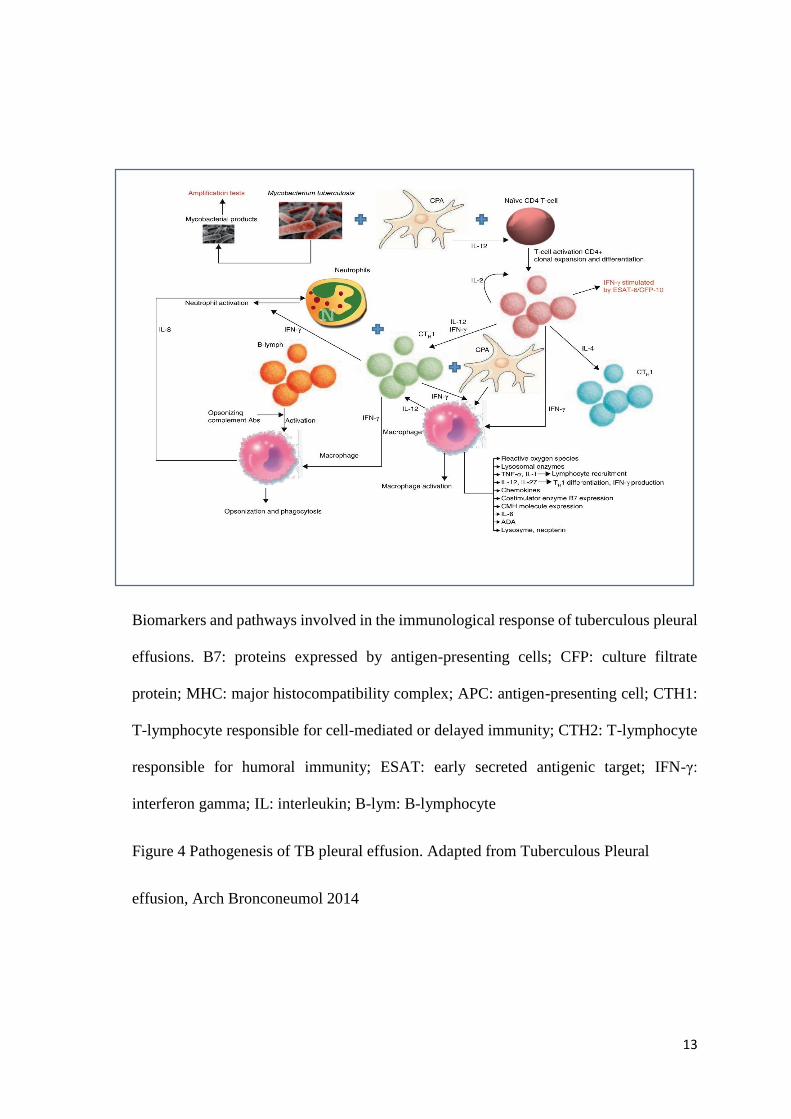
However, in TB pleura, rupture of a sub pleural caseous focus in the lung into the
pleura spaces is thought to be the initial event in the pathogenesis of primary TB pleural
effusion. Mycobacterial antigens interact with CD4+ T-lymphocytes, leading to a delayed
hypersensitivity reaction in which different cytokines stimulate macrophage anti
mycobacterial activity (Figure 4). It is mediated by T-helper type 1(Th1) cells that
activate macrophages to switch on mechanisms responsible for the killing of
mycobacteria. A strong Th1-like immunity (interferon [IFN]-ƴ dominant) is essential for
the containment of M tuberculosis, while these protective effects are antagonized by T-
helper type 2 cytokines, primarily interleukin (IL)-4. The predominance of Th1 immunity
in TB pleural effusions is demonstrated by the significantly higher levels of IFN-ƴ in
pleural fluid compared to peripheral blood of the same patient (Sharma et al., 2002)

12
In industrialized countries, it is though that more pleural effusion are due to
reactivation of TB rather than follow a primary infection 6 to 12 weeks previously. This
hypothesis is based on the observation by Steal et al that they could demonstrate a caseous
focus in the lung contiguous to the diseased pleura in 12 of 15 patients with TB pleural
effusion. The three other patient in this study had lung parenchymal disease although they
did not have caseous foci adjacent to the pleura. In addition, the lymphocytic pleurisy
obstructs the lymphatic in the parietal pleura, which leads to decreased pleural fluid
clearance from the parietal spaces. The pleural effusion results from the combination of
the increased pleura fluid formation and the decreased pleural fluid removal (Light,
2007).
13
Biomarkers and pathways involved in the immunological response of tuberculous pleural
effusions. B7: proteins expressed by antigen-presenting cells; CFP: culture filtrate
protein; MHC: major histocompatibility complex; APC: antigen-presenting cell; CTH1:
T-lymphocyte responsible for cell-mediated or delayed immunity; CTH2: T-lymphocyte
responsible for humoral immunity; ESAT: early secreted antigenic target; IFN-γ:
interferon gamma; IL: interleukin; B-lym: B-lymphocyte
Figure 4 Pathogenesis of TB pleural effusion. Adapted from Tuberculous Pleural
effusion, Arch Bronconeumol 2014

14
1.3.4 CAUSES OF PLEURAL EFFUSION
Causes of pleural effusion are classified into transudate and exudative pleural effusion.
Causes of transudate pleural effusion
Very common causes
Left ventricular failure
Liver cirrhosis
Hypoalbuminemia
Peritoneal dialysis
Less common causes
Hypothyroidism
Nephrotic syndrome
Mitral stenosis
Pulmonary embolism
Rare causes
Constrictive pericarditis
Superior vena cava obstruction
Ovarian hyperstimulation
Meigs’ syndrome
Causes of exudative pleural effusions
Very common causes
Malignancy
Parapneumonic effusions
Tuberculosis
Less Common causes
Pulmonary infarction
Rheumatoid arthritis
Autoimmune disease
Benign asbestos effusion
Pancreatitis
Post myocardial infarction syndrome
Rare causes
Yellow nail syndrome
Drugs
Fungal infections
Figure 5 Adapted from BTS guideline for the investigation of a unilateral pleural
effusion in adults, Thorax, 2013
15
1.3.5 CLINICAL MANIFESTATION
TB pleural effusion usually presents as an acute illness which is different from
pulmonary TB. Approximately one third of patients being symptomatic for < 1 week and
two third for < 1 month. Berger et al explain the most common presenting symptoms in
TB pleura are pleuritic chest pain (75%) and non-productive cough (70%). Other
symptoms included fever, night sweat, weight loss, malaise, and shortness of breath
varying in severity according to the size of effusion. Patient with TB pleural effusion tend
to be younger and immuno competent than patient with pulmonary tuberculosis (JM,
2016). However, Epstein and colleagues demonstrate a rise in the median age (56 years)
at presentation of TB pleural effusions with 19% of patients having reactivation disease
(Sharma and Mohan, 2004).
On physical examination, the patients appear cachexia, malnourished and
chronically ill. Sometimes they had generalised palpable lymphadenopathy in extra
pulmonary tuberculosis. On lung findings, they were present of collapse consolidation at
the apical area or pleural effusion in pleural tuberculosis. In immune compromised
patients like HIV, they can present with sign of other opportunistic infections such as oral
candidiasis, multiple skin lesions and meningeal signs positive.

16
1.3.6 DIGNOSTIC APPROACH OF TB PLEURAL EFFUSION
The definitive diagnosis of TB pleura depends on the demonstration of MTB in
sputum, pleural fluid, or pleural biopsy specimens. Supportive evidence includes
demonstration of classical TB granulomas in the pleura and elevated adenosine
deaminase (ADA) and IFN-ƴ levels in the pleural fluid.
The tuberculin skin test is being utilised less and less in patients suspected of
having TB pleural effusion. This is primarily because a negative test does not rule out the
diagnosis of TB pleural effusion. In a series of 254 patients from Spain, only 66.5% of
the patients had a positive skin test while in another case series from Hong Kong, more
than half the patients tested have a negative skin test (Valdés et al., 2003). If initial
negative tuberculin skin test in patient who suspected TB pleural effusion are repeated
after 8 weeks development of symptoms, the skin test will be almost always be positive.
However, if the patient is markedly immunosuppressed with HIV infection or is severely
malnourished, the skin test may remain negative.
1.3.6.1 PLEURAL FLUID ANALYSES
The initial step in assessing a pleural effusion is to ascertain whether it is a
transudate or exudate. A diagnostic pleural fluid sample should be gathered with a fine
bore (21G) needle and a 50 ml syringe. The sample should be placed in both sterile vials
and blood culture bottles and analyzed for protein, lactate dehydrogenase (LDH), pH,
Gram stain, AFB stain, cytology and microbiological culture. After performing pleural
effusion, the appearance and odor of the pleural fluid should be noted. The appearance
can be divided into serous, hemorrhagic, frankly bloody, or purulent. pH should be
performed in all non-purulent effusion.
17
A pleural fluid pH of < 7.2 represents a substantial accumulation of hydrogen ions
as normal pleural pH is about 7.6 because of bicarbonate accumulation in the pleural
cavity. In an infected effusion a pH <7.2 indicates the need for tube drainage. The pleural
protein should be measured to differentiate between a transudate and exudative pleural
effusion. Criteria known as Light’s criteria define the exudative or transudate effusion.
An exudative effusion will have a ratio of pleural fluid protein to serum protein greater
than 0.5, a ratio of pleural fluid lactate dehydrogenase greater than 0.6 or a pleural fluid
lactate dehydrogenase greater than two thirds the upper limit of normal serum lactate
dehydrogenase (Light, 2002)
Pleural lymphocytosis is common in malignancy and tuberculosis. A pleural fluid
glucose level of less than 3.3 mmol/L is found in exudative pleural effusions secondary
to empyema, rheumatoid disease, lupus, tuberculosis, malignancy or esophageal rupture.
Malignant effusion can be diagnosed by pleural fluid cytology alone in only 60% of cases.
In Malaysia and many other countries, sputum smear microscopy is still used to
diagnose TB. Trained laboratory technicians look at sputum samples under a microscope
to see if TB bacteria are present. Although inexpensive, AFB microscopy has relatively
low sensitivity (40-60%) in culture confirmed cases of pulmonary tuberculosis. Most
modern laboratories processing large numbers of diagnostic specimen use auramine –
rhodamine staining and fluorescence microscopy which more sensitive than the Ziehl
Neelsan method. Microscopy detects only half the number of TB cases and it depends on
the quality of the sputum produced by the patients. Up to 40% of active TB cases can be
missed if the sputum is used alone to diagnose TB pleura.
Pleural fluid adenosine deaminase (ADA) test is an easy and inexpensive methods
for establishing the diagnosis of TB pleural effusion. ADA is a predominant T-

18
lymphocyte enzyme, catalyzes the conversion of adenosine and deoxyadenosine to
inosine and deoxyinosine. A recent meta-analysis of 63 studies including 2796 patients
with TB pleura and 5297 with non-TB pleura reported that the sensitivity and specificity
of ADA in the diagnosis of pleural TB were 92% and 90%, respectively. ADA levels in
pleural fluid are also elevated in HIV patients even with very low CD4 cell counts. The
most widely accepted cut-off value for pleural fluid ADA is 40 U/L. The higher the level,
the greater the chance of the patient having TB while the lower level the lesser the chance
of the patient having TB (Chan et al., 1991). Unfortunately, ADA level is not available
in Malaysia.
Although interferon-gamma release assays (IGRAs) were primarily designed to
detect latent tuberculosis, it is expected that it might also contribute to the diagnosis of
TB pleural effusion. Based on the evidence so far, the IGRAs are not recommended to
make a diagnosis of TB pleural effusion. In a meta-analysis from a previous study , the
sensitivity and specificity for pleural IGRAs in diagnosing TB pleural effusion were 75%
and 82% respectively (Zhou et al., 2011).
Several test systems based on amplification of mycobacterial nucleic acid have
some available in the past few years. These test are most useful for the rapid confirmation
of TB in persons with AFB - negative pulmonary and extrapulmonary TB. One system
that permits rapid diagnosis of TB with high specificity and sensitivity is the fully
automated, real time nucleic acid amplification technology known as the Xpert MTB/RIF
assay. Xpert MTB/RIF can simultaneously detect TB and rifampicin resistance in < 2 h
and has minimal biosafety and training requirement. This test has a sensitivity of 98%
among AFB-positive cases and 70% among AFB-negative specimens. The WHO
recommends its se worldwide as the initial diagnostic test in adults and children presumed
to have MDR-TB or HIV-associated TB. Xpert MTB/RIF should be the initial test applied

19
to CSF from patients in whom TB meningitis is suspected as well as replacement test for
non-pulmonary specimens obtained by gastric lavage, fine needle aspiration, or pleural
or other biopsies from patients in whom extra pulmonary TB is suspected (Longo and
Harrison, 2012).
Definitive diagnosis depends on the isolation and identification of MTB from a
clinical specimen or the identification of specific DNA sequences in a nucleic acid
amplification test. Specimen may be inoculated onto egg or agar based medium (e.g
Lowenstein Jensen or Middlebrook medium). Because most species of mycobacteria
including MTB grow slowly by 4-8 weeks may be required before growth is detected. In
modern, well equipped laboratories, liquid culture for isolation and species identification
by molecular methods or high pressure liquid chromatography of mycolic acids has
replaced isolation on solid media and identification by biochemical tests (Yasinskyi and
Solodovnik, 2016).
A widely used technology is the mycobacterial growth indicator tube which uses
a fluorescent compound sensitive to the presence of oxygen dissolved in the liquid
medium. The appearance of fluorescence detected by fluorometric technology indicates
active growth of mycobacteria. A low cost rapid immune chromatographic lateral flow
assay based on detection of MTP64 antigen may also be used for species identification of
the MTB complex in culture isolates (Shinnick and Good, 1995).
Current methods utilised for routine drug susceptibility testing include the use of
commercial liquid medium and the proportion methods on conventional agar medium.
Line probe assay is used to detect rifampicin and isoniazid resistance in smear positive
sputum specimen or cultures isolates from smear positive and negative specimens. Line
probe assay in the routine TB diagnostic algorithm is its rapid turnaround time which has

20
a direct impact on patient management and ultimately the transmission of TB (Morgan et
al., 2005).
Polymerase chain reaction (PCR) is also new method which based on
amplification of mycobacterial DNA fragments. TB pleural is a paucibacillary disease,
so the sensitivity can be improved by TB PCR and the advantages of TB PCR include
rapid diagnosis and high specificity and sensitivity. This can be approved by several study
who evaluate the efficacy of PCR in the diagnosis of TB pleura. Based on these previous
studies, it shown that a sensitivity ranging from 20 to 90% and specificity from 78 to
100% (Villegas et al., 2000). TB PCR findings are positive in 100% of culture-positive
TB pleural fluids and in only 30 to 60% of culture-negative pleural fluids. Causes of false
positive results include DNA contamination or presence of nonviable organisms. Pleural
biopsy for PCR has 90% sensitivity and 100% specificity, as the overall accuracy being
similar to biopsy culture (Hasaneen et al., 2003).
1.3.6.2 CHEST IMAGING
Plain radiography such as PA and lateral view should be performed in the
assessment of suspected pleural effusion. The PA chest radiograph is abnormal in the
presence of about 200ml pleural fluid. However, only 50ml of pleural fluid can produce
detectable posterior costophrenic angle blunting on a lateral chest radiograph. Lateral
decubitus films are occasionally useful as free fluid gravities to the most dependent part
of the chest wall, differentiating between pleural thickening and free fluid (Blackmore et
al., 1996)
Ultrasound is more accurate than plain chest radiography for estimating pleural
fluid volume and aids thoracocentesis. After unsuccessful thoracocentesis or in a

21
loculated pleural effusion, ultrasound guided aspiration yields fluid in 97% of cases.
Ultrasound is also useful in demonstrating fibrinous location and readily differentiates
between pleural fluid and pleural thickening. Ultrasound also has the added advantages
of often being portable, allowing imaging at the bedside with the patient sitting or in the
recumbent position.
CT scanning has been shown to be superior to plain radiograph in the
differentiation of pleural from parenchymal disease. CT scan can usually differentiate
between benign and malignant pleural thickening. It is particularly helpful in the
assessment and management of loculated pleural effusion. Loculated effusions on CT
scans tend to have lenticular shape with smooth margins and relatively homogeneous
attenuation.

22
Figure 6 The representative radiographic finding of loculated tuberculous pleural
effusion. (A) Chest plain X-ray shows no shifting of pleural fluid on decubitus film, as
compared with chest posteroanterior view. (B) Thoracic real-time sonography shows
complex septated pleural effusion. (C) Chest computed tomography shows loculated
pleural fluid accumulated in nondependent portion
Adapted from Loculated Tuberculous Pleural effusion, Tuberculosis and Respiratory
Diseases, 2017
1.3.6.3 THORACOSCOPY AND PLEURAL BIOPSY
Thoracoscopy should be considered when less invasive test have failed to give a
diagnosis especially in undiagnosed pleural effusion. In addition to obtaining a tissue
diagnosis, several litres of fluid can be removed during the procedure and the opportunity
is also provided for talc pleurodesis. Thoracoscopy may therefore be therapeutic as well
as diagnostic. Pleural tissue can also be obtained during thoracoscopy but thoracoscopy
is usually not necessary to make the diagnosis of tuberculous pleural effusion.
Thoracoscopy is sometimes indicated when the diagnosis is confusion. A recent
meta-analysis revealed that the overall sensitivity 91% and specificity 100%, positive
likelihood ratio 4.92 and negative likelihood ratio 0.08 respectively; the area under curve

23
are 0.93 (Agarwal et al., 2013) Complication of this procedure appear to be few. The most
serious, but rare is severe hemorrhage caused by blood vessel trauma.
Pleural biopsy is the most common way to make the diagnosis of tuberculous
pleural effusion since 50 years ago by using a blind needle biopsy of the pleura. The
presence of granuloma in the parietal pleura suggest tuberculous pleural effusion but
caseous necrosis and AFB need not be demonstrated. Although other disorders including
fungal diseases, sarcoidosis, rheumatoid pleuritis may produce granulomatous pleuritis,
more than 95% of patient with granulomatous pleuritis have TB. The biopsy specimen
should be send for AFB and cultured for M. tuberculosis even if no granuloma presence
on the biopsy (Meldau et al., 2014)
Sugiyama et al classified tuberculous pleural effusion into 4 stages: Stage 1 where
the parietal pleura is swollen, reddened and show tiny white nodules; Stage II where the
redness and swelling become more extensive and military white nodules extending
diffusely and coalescing together; Stage III white fibrin deposits extend over the pleura
in a cord or a membrane-like fashion; Stage IV (chronic stage) the fibrin deposits become
fibrous. Part of the pleural effusion becomes encapsulated with a fibrin net, and the
parietal pleura become white, thickened, firm, and difficult to biopsy (Sugiyama et al.,
2001)
An image guided cutting needle biopsy is indicated in patient who has focal area
abnormality from contrast enhanced thoracic CT scan because it has a higher yield than
blind needle pleural biopsy in the diagnosis of malignancy. This technique also useful in
patient who are unsuitable for thoracoscopy (Maskell and Butland, 2003). Adam et al
reported that in a recent prospective study among 33 patients with a pleural effusion with
pleural thickening demonstrated on contrast enhanced CT scanning. These patients

24
underwent percutaneous image pleural biopsy and correct histological diagnosis was
made in 21 out of 24 patients, including 13 of 14 patients with mesothelioma (sensitivity
88%, specificity 100%).
Figure 7 Thoracoscopic images taken from patients with tuberculous pleurisy showing
A) tuberculous nodules with irregular distribution on parietal pleura; B) tuberculous
nodules on parietal (upper) and visceral (bottom) pleura; C) multiple military tuberculous
nodules on parietal pleura; D) diffuse parietal pleura nodules and pleural adhesions; E)
parietal pleural hyperemia with necrosis of the pleural node; F) parietal pleural hyperemia
with white pleural plaques.
Adapted from Diagnostic value and safety of medical thoracoscopy in tuberculous pleural
effusion. Respiratory Medicine, 2015.