tryptophan within basic peptide sequences triggers ...paris-en-resonance.fr/pdf/2013/bechara.pdf ·...
TRANSCRIPT

The FASEB Journal • Research Communication
Tryptophan within basic peptide sequences triggersglycosaminoglycan-dependent endocytosis
Chérine Bechara,*,†,‡ Manjula Pallerla,*,†,‡ Yefim Zaltsman,*,†,‡ Fabienne Burlina,*,†,‡
Isabel D. Alves,§ Olivier Lequin,*,†,‡ and Sandrine Sagan*,†,‡,1
*Université Pierre et Marie Curie (UPMC), Université Paris 6, Unité Mixte de Recherche (UMR)7203, †Centre National de la Recherche Scientifique (CNRS), UMR 7203, and ‡Ecole NormaleSupérieure (ENS), UMR 7203, Laboratoire des BioMolécules (LBM), Paris, France; and §Chimie etBiologie Des Membranes et Des Nanoobjets (CBMN), UMR 5248, CNRS, Pessac, France
ABSTRACT Deciphering the structural requirementsand mechanisms for internalization of cell-penetratingpeptides (CPPs) is required to improve their deliveryefficiency. Herein, a unique role of tryptophan (Trp)residues in the interaction and structuring of cationicCPP sequences with glycosaminoglycans (GAGs) hasbeen characterized, in relation with cell internalization.Using isothermal titration calorimetry, circular dichro-ism, NMR, mass spectrometry, and phase-contrast mi-croscopy, we compared the interaction of 7 basic CPPswith 5 classes of GAGs. We found that the affinity ofCPPs for GAGs increases linearly with the number ofTrp residues, from 30 nM for a penetratin analog with1 Trp residue to 1.5 nM for a penetratin analog with 6Trp residues for heparin (HI); peptides with Trpresidues adopt a predominantly �-strand structure incomplex with HI and form large, stable �-sheet aggre-gates with GAGs; and in the absence of any cytotoxicityeffect, the quantity of peptide internalized into CHOcells increased 2 times with 1 Trp residue, 10 times with2 Trp residues, and 20 times with 3 Trp residues,compared with �6 peptides with no Trp residues.Therefore, Trp residues represent molecular determi-nants in basic peptide sequences not only for directmembrane translocation but also for efficient endocy-tosis through GAGs.—Bechara, C., Pallerla, M., Zalts-man, Y., Burlina, F., Alves, I. D., Lequin, O., Sagan S.Tryptophan within basic peptide sequences triggersglycosaminoglycan-dependent endocytosis. FASEB J. 27,000–000 (2013). www.fasebj.org
Key Words: aggregation � � strand � � sheet � � helix �membrane translocation
Cell-penetrating peptides (CPPs) are short basicpeptides (8–30 aa) that can enter cells. CPPs use a
combination of multiple mechanisms for cellular entry,covering both direct translocation across the plasmamembrane and endocytic processes (1, 2). For directtranslocation, it is postulated that CPPs interact directlywith the lipid bilayer, whereas endocytic processes caninvolve other membrane partners, such as proteogly-cans. However, the molecular basis that controls theseinternalization processes is still not fully elucidated.Although there are few examples of pure hydrophobicCPP sequences, basic amino acids in the peptide se-quence are generally required for interaction with thecell surface and entry (3). Thus, as a prerequisite stepfor peptide entry, electrostatic interactions should takeplace between the positively charged peptides andnegatively charged molecules at the cell surface. Theseelectrostatic interactions involve, in particular, plasmamembrane phospholipids for membrane translocationand glycoconjugates, such as sialic acids (SAs) andglycosaminoglycans (GAGs), for endocytosis.
SAs are typically found at the terminal ends ofcarbohydrate chains on the cell surface. Because oftheir negative charge, SAs are involved in the bindingof positively charged molecules that can be furthertransported into cells. SAs contribute in various biolog-ical and pathological phenomena, including intercellu-lar adhesion, signaling, and microbial attachment (4,5). For instance, removal of SAs present at the surfaceof neuronal cells strongly reduced the uptake of thehomeodomain sequence (60 aa) of antennapedia pro-tein (6). However, this was not the case for the home-odomain-derived cell-penetrating peptide penetratin,which instead was trapped by SAs at the cell surface (7).As for GAGs, they have important roles in endocytosisand cellular signaling. They are, for example, involvedin the binding of proteins to their receptors, an inter-action that leads to the internalization of the complexes
1 Correspondence: Université Pierre et Marie Curie, Labo-ratoire des Biomolécules, UMR 7203 CNRS, ENS, Case cour-rier 182, 4 place Jussieu, 75252 Paris cedex 05, France. E-mail:[email protected]
doi: 10.1096/fj.12-216176This article includes supplemental data. Please visit http://
www.fasebj.org to obtain this information.
Abbreviations: CD, circular dichroism; CPP, cell-penetrat-ing peptide; CS, chondroitin sulfate; DLS, dynamic lightscattering; FCS, fetal calf serum; GAG, glycosaminoglycan;HI, heparin; HS, heparan sulfate; ITC, isothermal titrationcalorimetry; MALDI, matrix-assisted laser desorption/ioniza-tion; MS, mass spectrometry; SA, sialic acid; TOF, time offlight; Trp, tryptophan; WT, wild-type
10892-6638/13/0027-0001 © FASEB
The FASEB Journal article fj.12-216176. Published online October 15, 2012.
(8–11). GAGs are also implicated in the internalizationof CPPs through an endocytic process (12–15). The cellsurface displays different kinds of GAGs that are at-tached to core proteins, including heparan sulfate(HS) and chondroitin sulfate (CS), but few studies haveassessed the role of these different GAGs in the inter-action with CPPs in vitro and in cell internalization.Interactions of TAT, penetratin, oligoarginine, andother well-studied CPPs with HS and the structurallyrelated heparin (HI) mainly revealed tight binding,leading to the formation of aggregates thought to bethe result of numerous HI chains noncovalentlycrossed-linked by CPPs (16). Clusters formed withpenetratin are more stable than those formed with TATpeptide (17). Penetratin preferentially binds CS, andthe removal of endogenous CS reduces the internaliza-tion of penetratin peptides (18), whereas there is aspecific HS proteoglycan epitope necessary for endo-
cytic stimulation and to which TAT peptide is able tobind (19).
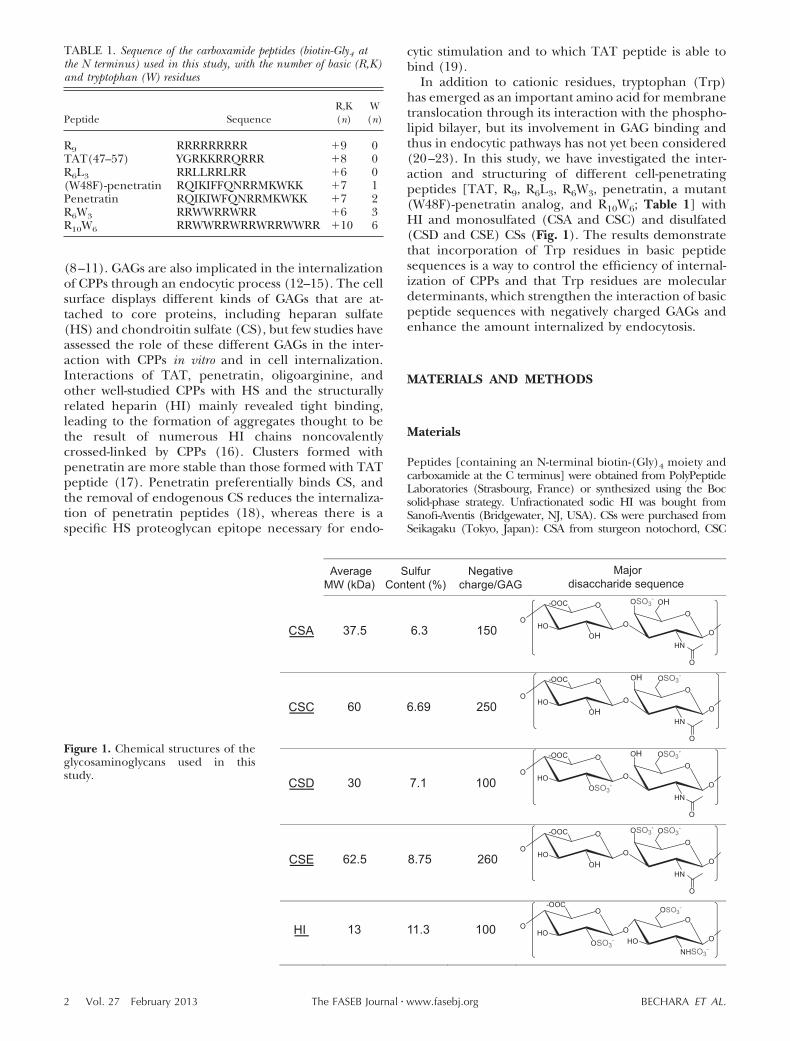
In addition to cationic residues, tryptophan (Trp)has emerged as an important amino acid for membranetranslocation through its interaction with the phospho-lipid bilayer, but its involvement in GAG binding andthus in endocytic pathways has not yet been considered(20–23). In this study, we have investigated the inter-action and structuring of different cell-penetratingpeptides [TAT, R9, R6L3, R6W3, penetratin, a mutant(W48F)-penetratin analog, and R10W6; Table 1] withHI and monosulfated (CSA and CSC) and disulfated(CSD and CSE) CSs (Fig. 1). The results demonstratethat incorporation of Trp residues in basic peptidesequences is a way to control the efficiency of internal-ization of CPPs and that Trp residues are moleculardeterminants, which strengthen the interaction of basicpeptide sequences with negatively charged GAGs andenhance the amount internalized by endocytosis.
MATERIALS AND METHODS
Materials
Peptides [containing an N-terminal biotin-(Gly)4 moiety andcarboxamide at the C terminus] were obtained from PolyPeptideLaboratories (Strasbourg, France) or synthesized using the Bocsolid-phase strategy. Unfractionated sodic HI was bought fromSanofi-Aventis (Bridgewater, NJ, USA). CSs were purchased fromSeikagaku (Tokyo, Japan): CSA from sturgeon notochord, CSC
TABLE 1. Sequence of the carboxamide peptides (biotin-Gly4 atthe N terminus) used in this study, with the number of basic (R,K)and tryptophan (W) residues
Peptide SequenceR,K(n)
W(n)
R9 RRRRRRRRR �9 0TAT(47–57) YGRKKRRQRRR �8 0R6L3 RRLLRRLRR �6 0(W48F)-penetratin RQIKIFFQNRRMKWKK �7 1Penetratin RQIKIWFQNRRMKWKK �7 2R6W3 RRWWRRWRR �6 3R10W6 RRWWRRWRRWRRWWRR �10 6
CSA 37.5 6.3 150
O
HO
-OOC
OHO
O
HN
O
OHOSO3-
O
O
CSC 60 6.69 250
O
HO
-OOC
OHO
O
HN
O
OSO3-OH
O
O
CSD 30 7.1 100
O
HO
-OOC
OSO3-
OO
HN
O
OSO3-OH
O
O
CSE 62.5 8.75 260
O
HO
-OOC
OHO
O
HN
O
OSO3-OSO3
-
O
O
HI 13 11.3 100 O
HO
-OOC
OSO3-
OO
NHSO3-O
O
HO
OSO3-
AverageMW (kDa)
SulfurContent (%)
Negativecharge/GAG
Majordisaccharide sequence
Figure 1. Chemical structures of theglycosaminoglycans used in thisstudy.
2 Vol. 27 February 2013 BECHARA ET AL.The FASEB Journal � www.fasebj.org
and CSD from shark cartilage, and CSE from squid cartilage.Tetramethylrhodamine isothiocyanate (TRITC)-streptavidin andavidin were purchased from Zymed Laboratories (Invitrogen, Carls-bad, CA, USA). Phalloidin-FITC, SA, and all other reagents werefrom Sigma-Aldrich (St. Louis, MO, USA).
Cell culture
Wild-type (WT) CHO-K1 cells, xylose transferase- or GAG-deficient CHO-pgsA745 (GAGneg) cells, and SA-deficientCHO-lec2 (SAneg) cells were obtained from American TypeCulture Collection (Rockville, MD, USA). All cell lines weregrown in Dulbecco’s modified Eagle’s medium (DMEM)supplemented with 10% fetal calf serum (FCS), penicillin(100 IU/ml), streptomycin (100 IU/ml), and amphotericin B(1 �g/ml) in a humidified atmosphere containing 5% CO2 at37°C.
Measure of cellular uptake and quantificationof membrane-bound peptide
Cellular uptake was quantified by matrix-assisted laser desorp-tion/ionization (MALDI)-time of flight (TOF) mass spec-trometry (MS) as described previously (24, 25). In thisprotocol, the peptides bear a tag on the N terminus com-posed of 4 glycine residues together with a biotin moiety forpurification purposes (1H-peptide). This enables us to obtainan internal standard for absolute quantification by MS, hav-ing the same sequence as the one to quantify, except that itbears 4 deuterated glycine residues (2H-peptide). Adherentand confluent cells (106 cells, seeded the day before in 12-wellplates) were incubated with the cell-penetrating 1H-peptide(1, 3, 5, 7, and 10 �M) in culture medium (without FCS) for75 min at 4 or 37°C. After washings, trypsin (37°C) or pronase(4°C) was added to degrade the remaining extracellular andmembrane-bound peptide and to detach cells (12). The cellswere then lysed and boiled in a solution containing a con-trolled and relevant quantity of the 2H-peptide. The lysate wasincubated with streptavidin-coated magnetic beads to extractthe peptides. For membrane-bound peptide quantification,no protease was added, and the cells were directly lysed afterwashings. The peptides were eluted from the streptavidin-coated magnetic beads with �-cyano-4-hydroxycinnamic acidmatrix and spotted on the MALDI plate. The samples wereanalyzed by MALDI-TOF MS (positive ion reflector mode) ona Voyager-DE Pro mass spectrometer (Applied Biosystems,Foster City, CA, USA). For each experiment, we used dupli-cate or triplicate wells, and the experiments were repeatedindependently �3 times.
Isothermal titration calorimetry (ITC)
ITC experiments were performed on a nano-ITC calorimeter(TA Instruments, New Castle, DE, USA). Titrations wereperformed by injecting 10-�l aliquots of polysaccharides (HI,CSA, CSC, CSD, and CSE) into the calorimeter cell contain-ing the peptide solution, with 5-min injection intervals. Peptidesand polysaccharide solutions were used at different concen-trations according to the peptide sequence and the GAGspecies (varying between 50 and 200 �M for the peptides, 10and 30 �M for CSs, and 70 and 120 �M for HI). Theexperiments were performed at 37°C in 50 mM Tris-Cl, 2 mMEDTA, and 150 mM NaCl, pH 7.5 (26, 27). Control experi-ments were performed by injection of buffer into peptidesolution and injection of GAGs into buffer. Data analysis wasperformed with NanoAnalyze software provided by TA Instru-ments. Experiments were done �3 times.
Circular dichroism (CD)
The CD spectra of the peptides were recorded using a J-815spectrophotometer (Jasco, Tokyo, Japan). The spectra wereacquired in a 1-mm pathlength at 20°C in a quartz opticalcell, from 190 to 270 nm with a 0.2-nm step. Five scans wereaccumulated and averaged after subtraction of the baselinefor an appropriate blank sample. The spectra were acquiredfor peptide concentrations varying from 10 to 50 �M (corre-sponding to peptide totally bound to HI or peptide in excesswith respect to HI, according to the stoichiometry of bindingobtained by ITC) in 10 mM sodium phosphate buffer (pH7.4) or in the presence of HI. CD measurements werereported as mean residual ellipticity (�; deg · cm2 · dmol�1 ·n�1). Secondary structure content was estimated from CDspectra using the deconvolution program CDFriend (28).The experimental error is estimated to be �5% (28).
NMR
Experiments were performed on a Avance III spectrometerequipped with a TCI 1H/13C/15N cryoprobe (Bruker, New-ark, DE, USA) and operating at a 1H frequency of 500 MHz.Peptide samples were prepared at a concentration of 0.1 mMin 3-mm NMR tubes (Norell, Inc., Landisville, NJ, USA)containing 170 �l of PBS at pH 5.5, 10% D2O, and 0.1 mMsodium 2,2-dimethyl-2-silapentane-5-sulfonate (Isotec, St.Louis, MO, USA) for chemical shift calibration; pH is classi-cally decreased to 5.5 to observe the amide protons ofpeptides on NMR spectra. Peptide samples were titrated withincreasing concentrations of HI tetrasaccharide DP4 (Dextra,Reading, UK) from a 1 mM stock solution. NaCl was added inthe samples using a 5 M stock solution. One-dimensionalNMR experiments were recorded at 25°C using a WatergateW5 pulse sequence for solvent suppression (29). Protonresonances were assigned from the analysis of two-dimen-sional total correlation spectroscopy and nuclear Overhausereffect spectroscopy experiments, as described previously (20).
RESULTS
GAG/CPP binding affinity can be controlled by thenumber of Trp residues in peptide sequences
We first examined the capacity of penetratin, (W48F)-penetratin mutant, R9, TAT, R6W3, R6L3, and R10W6,peptides (Table 1) to bind carbohydrates in vitro. Weanalyzed the interaction of CPPs with HI and 4 differ-ent classes of CSs (the most prevalent GAG on the cellsurface): monosulfated CSA and CSC and disulfatedCSD and CSE (Fig. 1). The interaction of these pep-tides with the different polysaccharides in solution wasanalyzed by ITC experiments (Table 2). The ionicstrength experimental conditions ensure that the neg-ative charges along the polymer chains were fullyscreened by the positive ion salts. The presence ofEDTA, a divalent cation chelating agent, did not signif-icantly affect the binding parameters between peptidesand GAGs. At the saturation conditions, the dissocia-tion constants were in the nanomolar to submicromo-lar range for all peptides. The affinity ranges for allpeptides were mostly in order according to the sulfatecontent of those carbohydrates (Fig. 1); that is, HI, CSE
3CATIONIC CELL-PENETRATING PEPTIDE INTERNALIZATION
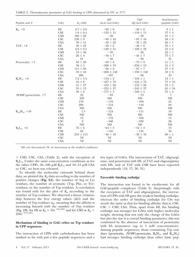
� CSD, CSC, CSA (Table 2), with the exception ofR6L3. Under the same concentration conditions as forthe other CPPs, 50–100 �M R6L3 and 10–15 �M CSAor CSC, no heat was released.
To identify the molecular rationale behind thosedata, we plotted the Kd data according to the number ofpositive charges (Fig. 2A), the number of Arg or Lysresidues, the number of aromatic (Trp, Phe, or Tyr)residues, or the number of Trp residues. A correlationwas found only for the plot of Kd according to thenumber of Trp residues. We observed a linear relation-ship between the free energy values (G) and thenumber of Trp residues nW, meaning that the affinity isincreasing linearly with the number of Trp residues(Fig. 2B), for HI as Kd 55e�0.6(nW) and for CSD as Kd 250e�0.5(nW).
Mechanism of binding to GAG relies on Trp residuesin CPP sequences
The interaction of CPPs with carbohydrates has beenstudied so far with just a few peptide sequences and a
few types of GAGs. The interactions of TAT, oligoargi-nine, and penetratin with HI, of TAT and oligoargininewith HS, and of TAT with CSB have been reportedindependently (16, 17, 30, 31).
Favorable binding enthalpy
The interaction was found to be exothermic for allGAG-peptide complexes (Table 2). Surprisingly, withthe exception of TAT and oligoarginine, the interac-tion of CPPs with HI gave the weakest binding enthalpy,whereas the order of binding enthalpy for CSs wasnearly the same as that for binding affinity; that is, CSE,CSC � CSD, CSA. Thus, apart from HI, the bindingenthalpy was stronger for GAGs with higher molecularweight, showing that not only the charge of the GAGsbut also the size is a crucial binding parameter; this wasconfirmed by the absence of interaction of penetratinwith SA monomers (up to 5 mM concentration).Among peptide sequences, those containing Trp resi-dues [penetratin, (W48F)-penetratin, R6W3, and R10W6]had stronger binding enthalpy than other basic pep-
TABLE 2. Thermodynamic parameters of GAG binding to CPPs determined by ITC at 37°C
Peptide and Z GAG Kd (nM)H°
(kcal/mol GAG)TS°
(kJ/mol GAG)Stoichiometry
(peptide/GAG)
R9, �9 HI 8.7 � 2.6 �60 � 6 �48 � 6 8 � 1CSE 1.0 � 0.4 �123 � 15 �110 � 15 37 � 4CSD 180 � 28 �48 �38 21 � 5CSC 230 � 9 �96 � 44 �87 � 44 48 � 8CSA 39 � 15 �42 � 22 �32 � 22 26 � 1
TAT, �8 HI 36 � 22 �58 � 6 �46 � 6 10 � 1CSE 8.8 � 8.5 �120 � 31 �108 � 32 43 � 6CSD 53 � 36 �39 �28 22 � 1CSC 26 � 4 �56 � 7 �45 � 7 73 � 7CSA 91 �40 �30 31
Penetratin, �7 HI 93 � 20 �83 � 9 �73 � 9 11 � 1CSE 36 � 5 �268 � 53 �258 � 53 41 � 3CSD 115 � 69 �96 � 6 �87 � 7 27 � 8CSC 59 � 44 �200 � 148 �190 � 148 58 � 9CSA 280 �107 �98 43
R6W3, �6 HI 7.2 � 5.4 �116 � 2 �104 � 1 12 � 1CSE 16 � 6 �427 � 72 �416 � 72 75 � 16CSD 51 � 27 �163 � 20 �152 � 20 33 � 2CSC 35 � 13 �253 � 37 �243 � 37 65 � 18CSA 38 � 6 �171 � 5 �160 � 5 35 � 2
(W48F)-penetratin, �7 HI 22 �83 �73 7CSE ND ND ND NDCSD 170 �110 �100 25CSC 290 �154 �144 84CSA ND ND ND ND
R10W6, �10 HI 1.5 �124 �111 10CSE ND ND ND NDCSD 12 �172 �160 14CSC 8 �340 �328 28CSA ND ND ND ND
R6L3, �6 HI 76 � 6 �64 � 3 �54 � 3 13CSE 62 �145 �135 58CSD 210 � 115 �46 � 10 �36 � 10 60 � 1CSC NI NI NI NICSA NI NI NI NI
ND, not determined; NI, no interaction in the studied conditions.
4 Vol. 27 February 2013 BECHARA ET AL.The FASEB Journal � www.fasebj.org

tides (R9, TAT, and R6L3), irrespective of the type ofGAG. In this manner, it was found that the bindingenthalpy did not vary significantly with the number ofpositive charge (Fig. 2C), but became stronger with anincreased number of Trp residues (Fig. 2D): the moreTrp residues present in the peptide, the more favorablewas the binding enthalpy. The binding enthalpy in-creased from 2- to 4.5-fold with 1 and 6 Trp residues,respectively, compared with peptides that do not con-tain Trp residues (R9, Tat, and R6L3). The improve-ment in binding enthalpy observed for peptides con-taining Trp, was reflected in better affinity (Fig. 2C),because the enthalpy gain dominated unfavorable en-tropy (Table 2 and Fig. 2D).
Unfavorable binding entropy
As for binding enthalpy, the unfavorable binding en-tropy was not dependent on the number of positivecharges, because the R9, TAT, and R6L3 interactiongave similar entropy values (Fig. 2E). However, unfa-vorable entropy increased linearly with increasing num-bers of Trp residues in the peptide sequence (Fig. 2F).Thus, greater unfavorable conformational entropy(more conformational penalties or fewer degrees offreedom) arose from the binding to GAGs of peptides
that contain hydrophobic Trp residues, compared withother basic CPPs.
Stoichiometry of GAG/CPP complexes attainselectric neutrality
The differences in binding enthalpy and entropy weremore significant with CSC and CSE than with the otherpolysaccharides. This observation demonstrated thatthe degree of sulfation and size of the polysaccharidewere crucial parameters, which determined the bindingenergy of CPPs. On the basis of the molecular weightand sulfate content of the CS and HI polysaccharides,the total negative charge per molecule was calculated(Fig. 1). Taking into account the net positive charge ofeach peptide and the stoichiometry of their binding toGAGs (Table 2), the interaction led to CPP-GAG neu-tralized charge complexes with all polysaccharides(Supplemental Table S1). However, the interaction ofCPPs with GAGs not only occurred through ion pairsbut also the peptide sequence, the length, and theposition or degree of sulfation in the polysaccharidewere determinant binding factors. In fact, the bindingaffinity increased as a function of sulfate content andchain length of GAGs (Fig. 2A). Notably, no interactionof penetratin with SA alone (up to 5 mM) could bedetected.
Trp-rich CPPs adopt predominantly a �-strandconformation in complex with GAGs
Although usually unstructured in solution, CPPs tendto adopt a well-defined secondary structure when bind-ing to lipid membranes (32). As for the interaction withpolysaccharides, it has been shown that TAT peptidedoes not undergo conformational changes in the pres-ence of HS (30). On the other hand, different studiesdemonstrated that HI binding peptides adopt an �-he-lical structure when bound to HI (and other polyelec-trolytes; refs. 33, 34). Here, we used CD spectroscopyand NMR to determine the secondary structure ofcationic peptides in the presence of carbohydrates.Based on the stoichiometry of binding calculated in theITC experiments, we tested different peptide/HI ratiosto have the peptide either saturated or not saturatedwith GAGs (Table 3).
Except for R6W3 and R10W6, all the peptides had arandom coil structure in buffer solution. R6W3, on theother hand, had partial helical content that was previ-ously characterized, with a minimum shifted to 202 nmand another shouldering at 220 nm. This peculiarsignature was ascribed to the presence of Trp residues(20). On the other hand, R10W6 could accommodate�-helical and �-strand conformations in solution. Con-formational differences among peptides were unveiledwhen bound in complex with HI (Table 3). R9, TAT,and R6L3 were partially structured into an � helix(Supplemental Fig. S1). On the other hand, R6W3,R10W6, penetratin, and (W48F)-penetratin could adopta �-strand structure principally (minimum at 213 nm
Figure 2. A, C, E) Binding affinity (A), binding enthalpy (C)and binding entropy (E) according to the number of positivecharges (number of Arg/Lys) in the CPPs for the GAGsinvestigated. B, D, F) Plots of the binding affinity (B), thebinding enthalpy (D), and the binding entropy (F) accordingto the number of Trp residues in peptide sequences; regres-sion coefficients (R2) were all between 0.85 and 0.99.
5CATIONIC CELL-PENETRATING PEPTIDE INTERNALIZATION

and maximum at 193 nm; Supplemental Fig. S1). Thus,peptides with Trp residues could adopt predominantlya higher-order �-strand conformation in interactionwith HI, better than that of the other basic peptides,which remained unstructured or adopted an �-helicalconformation. It should be noted that on addition ofthe peptides to HI, a fine precipitate formed in thesolution. This might be due to the aggregation phe-
nomena that take place with CPP-HI binding, throughformation of �-sheet aggregates (13, 16, 17). Lightscattering due to the particulate nature of the solutionmight have interfered with the CD experiment, but,nonetheless, the increase in ordered structure contentcame at the expense of other secondary structuralfeatures.
The interaction of CPPs with GAGs was furtherexamined by 1H NMR spectroscopy. Taken as modelpeptides, R6L3 and R6W3 were titrated with increasingconcentrations of HI-derived oligosaccharides. Thetitrations with HI tetrasaccharide DP4 (Fig. 3) showvery different behaviors for R6L3 and R6W3 peptides. Inthe case of R6L3 (Fig. 3A), the amide resonancesundergo gradual chemical shift perturbations on titra-tion, indicating an interaction occurring on a fastexchange regimen on the NMR time scale. No signifi-cant broadening is observed, suggesting that the inter-action does not involve the formation of high-molecu-lar-weight species. The amide protons of residues 3–9exhibit an average chemical shift variation of 0.17 ppmthat can be ascribed to direct interaction with heparintetrasaccharide or peptide folding. Notably, the H�protons also show an average upfield shift of 0.07 ppm,which supports a helical folding of the R6L3 peptide oncomplex formation. In contrast, the proton resonancesof R6W3 peptide show a gradual decrease in intensitywithout any changes in chemical shifts or linewidths asthe DP4 concentration is increased, whereas no peptideresonances can be observed beyond a 1:1 peptide/DP4ratio (Fig. 3B). The addition of 1 M NaCl in the sampleleads to peptide resonances reappearing, with chemicalshifts similar to that of the peptide in the absence ofDP4 ligand (Fig. 3C). This observation demonstratesthat an NMR-invisible peptide-GAG complex is formedduring the titration, which can be dissociated at high
TABLE 3. Secondary structure content of the CPPs in phosphatebuffer (pH 7.4) or in the presence of HI at saturating peptideconditions
CPP�-Helix
(%)Randomcoil (%)
Type IIhelix(%)
�-Sheet(%)
PenetratinBuffer 1 57 42 0HI 3 69 0 28
(W48F)-penetratinBuffer 0 58 42 0HI 31 51 5 13
R10W6Buffer 21 65 0 14HI 8 55 0 37
R6W3Buffer 18 71 11 0HI 25 48 0 27
R6L3Buffer 0 77 23 0HI 47 35 0 18
R9Buffer 0 83 17 0HI 18 70 12 0
TATBuffer 0 92 8 0HI 13 78 0 9
Experimental error is estimated to be �5% (28).
Figure 3. NMR titration of R6L3 and R6W3peptides with HI tetrasaccharide DP4. A)Amide region of the 1-dimensional 1HNMR spectra of R6L3 as a function ofadded DP4 (peptide/DP4 ratios of 1:0 to1:5) B, C) Amide region of the 1-dimen-sional 1H NMR spectra of R6W3 as afunction of added DP4 (peptide/DP4ratios of 1:0 to 1:1) in the presence of150 mM NaCl (B) or 1 M NaCl (C).
6 Vol. 27 February 2013 BECHARA ET AL.The FASEB Journal � www.fasebj.org

ionic strength. From the titration, it can be calculatedthat the interaction involves a binding of 0.7 peptideper disaccharide unit and leads to electrostatic neutral-ity of the complex. Therefore, as opposed to R6L3, theinteraction of R6W3 with HI saccharides leads to thehigh-molecular-weight aggregates consistent with �-sheetformation, which are too large to be observed bysolution-state NMR.
Trp-rich peptides form stable and large aggregates incomplex with GAGs
Because precipitates were observed in saturated pep-tide-GAG complexes, we further explored this possibil-ity by turbidity and differential light-scattering mea-sures and phase-contrast microscopy. It was previouslyreported that conformational transition of penetratinfrom an � helix to a � strand could be associated withaggregation on lipid binding (35, 36).
Turbidity measurements of saturated GAG-CPP ITCsamples showed that there were no significant or ratio-nal differences in absorbance among peptides andGAGs (not shown). Obviously, all peptides inducedsupramolecular assemblies of GAGs in solution, withthe exception of R6L3 with CSA and CSC, as expectedfrom the absence of interaction found between thepeptide and those carbohydrate molecules in ITCexperiments. Thus, in principle, all peptide-GAG com-plexes could form aggregated molecular species. Dy-namic light scattering (DLS) measurements did nothelp to further decipher the mechanism because thesupramolecular assemblies between GAGs and peptideswere too polydispersed. In addition, accurate DLSmeasurement requires particles to be randomly diffus-ing. This places the upper size limit of DLS analysis asthe point at which sedimentation of the particle dom-inates diffusion, and the upper size limit was obviouslyreached in the case of the CPP-GAG complexes. Phase-contrast microscopy imaging was thus used to observethe number and size of aggregates formed between HI,monosulfated CSD, disulfated CSC, and CPPs (Supple-mental Fig. S2). Trp-rich peptides systematically in-duced the formation of numerous, large, and stableGAG aggregates (as can be seen in Supplemental Fig.S2). The situation was quite different for the otherbasic peptides (R9, TAT, and R6L3), for which few or noaggregates were observed, with the exception of HI. Inthis latter case, TAT and R6L3 caused the formation ofmany small aggregates, whereas for other peptides,including R9, larger aggregates were stably formed.This observation highlights the fact that HI behavesdifferently from the other GAGs and is not representa-tive of the interactions of all types of GAGs with CPPs.The higher density of negative charges in HI might bethe reason for such differences in the aggregationbehavior of this carbohydrate compared with that ofothers.
Thus, these results demonstrated that stable aggre-gates have been formed between the Trp-rich peptidesand the different types of GAGs. On the other hand, for
peptides that do not contain Trp residues, either theaggregates were formed but transiently so that theycould not be observed, or they were simply not inducedduring peptide-GAG interaction.
Trp-rich peptides internalize efficiently through cellsurface GAG clustering
We further explored the role of cell surface carbohy-drates in the interaction of Trp-rich peptides withplasma membrane. Plasma membrane glycoconjugatesform a pool of negative charges on the cell surface thatbind the positively charged CPPs, thus facilitating theirinteraction with the cell (10). We used 3 CHO cell linesthat express all types of GAGs (WT), no CS and HS(GAGneg), or no SAs (SAneg).
Membrane translocation and endocytosis dependingon GAG clustering are observed for CPPs
The internalization of cell-penetrating peptides occursat both 37 and 4°C. It is generally assumed that alltranslocation and endocytic processes are active at37°C, whereas endocytic pathways are inhibited at 4°C.Therefore, the amount of internalized peptide viadirect membrane translocation pathways can be esti-mated from the comparison of internalization at 37 and4°C. This amount might be underestimated becausethe gel-phase state of the lipid membrane at 4°C mighthinder the translocation process. Nevertheless, wechose to modify temperature to distinguish betweentranslocation and endocytic pathways rather than useendocytosis inhibitors because these inhibitors havenegative side effects in internalization studies (37).
At low micromolar extracellular peptide concentra-tions and at 37°C, the amount of internalized peptidewas similar in all three cell types for both penetratinand R6W3 peptides. This result was also observed pre-viously with a penetratin analog and was related tomembrane translocation pathways (12). When the pep-tide concentration is increased, a significantly higheruptake in SAneg and WT cells than in GAGneg cells wasmeasured. This finding supports previous results show-ing that a minimum amount of bound CPP is necessaryto trigger cooperative GAG clustering and internaliza-tion (12, 13, 16, 17).
Notably, the internalization was significantly moreefficient in SAneg cells than in WT cells (�5 �Mextracellular peptide concentration). Thus, at the cellsurface, SAs are inert CPP binders that trap CPPs andprevent them from interacting with other membranepartners, such as GAGs and plasma membrane lipids in-volved in internalization processes, as described previously(7). Internalization was also visualized by confocal micros-copy, at 37°C in the three cell lines. Because it was notpossible to observe any differences in peptide quantityamong cell lines with this technique, experiments are shownfor penetratin only (Supplemental Fig. S4).
At 4°C, internalization occurred with similar amountsirrespective of the cell surface carbohydrate composition
7CATIONIC CELL-PENETRATING PEPTIDE INTERNALIZATION

and the extracellular peptide concentration (Fig. 4A, Bfor R6W3 and penetratin intracellular amounts, respec-tively, and Supplemental Fig. S4 for intracellular imag-ing of penetratin). As for 37°C, one should note thatR6W3 was internalized more efficiently than penetratin.
Finally, these data indicate that these peptides entervia direct translocation at low extracellular concentra-tions and that both GAG-dependent endocytosis anddirect translocation occur at higher concentrations, asdescribed previously for a penetratin analog (12).
Efficiency of internalization can be controlled by Trpresidues within CPP sequences
To further evaluate the effect of CPP sequences oninternalization efficiency, the amount of peptide inter-nalized at a saturating (10 �M) extracellular peptideconcentration was plotted for each peptide, taking intoaccount the number of positive charges and the num-ber of aromatic (F, Y, W) or Arg (R) or Trp (W)residues (Fig. 4C). The internalization data were ob-tained with WT and GAGneg cells at 37 and 4°C. Thehighest efficiency of internalization was obtained for R9
and R6W3, which enter cells in similar amounts. Thisresult shows that basic residues in CPPs can be partiallyreplaced by tryptophans, maintaining the same effi-ciency of uptake as was also observed with pseudopep-tide carriers (21). For the other peptide sequences, it isclear that in the two cell lines, regardless of thetemperature, the amounts of internalized peptides cor-related with the number of Trp residues (and not thetotal number of aromatic residues; Fig. 4C). The inter-nalization curve fits showed decreased efficiency ofinternalization in the absence of GAG or at 4°C com-pared with that f wild-type cells at 37°C. However, thecurve fits were shifted parallel lines (Fig. 4C), anobservation demonstrating that crucial steps in themechanism of internalization are not influenced bytemperature.
DISCUSSION
Our data show that introducing Trp residues into theCPP sequence can enhance the entry efficiency of CPPsas recently reported (38). The role of Trp in efficientinternalization of peptides is observed not only fordirect translocation, as already described for penetratin(39–41), but also for endocytosis through GAGs, al-though these peptides are not cytotoxic (42–44). Posi-tive charges in the sequence of CPPs are necessary tointeract with negatively charged GAGs and phospholip-ids, but the additional presence of one or more Trpresidues governs the binding properties of the pep-tides.
In addition, our results show that Trp-rich peptidescan adopt a �-strand structure in contact with GAGs,which lead to higher-order complexes and probablyform �-sheet stable and large aggregates. These resultswere consistent with the thermodynamic data. A strongbinding enthalpy contributed in a major way to thefavorable free energy change that was measured for allCPPs. Thus, strong ion pairs, hydrogen bonds, and vander Waals contacts dominate the unfavorable enthalpyassociated with the desolvation of polar groups in GAGsand peptides. However, Trp-rich peptides had strongerbinding enthalpy and higher entropy loss comparedwith other basic sequences, which can be explained bythe fact that hydrogen bonds of �-strands are consid-ered slightly stronger than those found in �-helices.With regard to entropy of binding, two major factorscontribute to it: desolvation and conformational en-tropy changes. The desolvation entropy should befavorable because it originates from the release of watermolecules on interaction between the peptide andGAGs. Favorable desolvation entropy should arise fromthe binding energy of hydrophobic groups. However,the conformational entropy change should be unfavor-able, because the binding process involves the loss ofconformational degrees of freedom for both the pep-tide and GAGs. Thus, the stronger hydrogen bonds inthe Trp-containing peptide-GAG interaction lead to
Figure 4. A, B) Quantification of the internalized peptides R6W3 (A) and penetratin (B) in 106 WT, GAGneg, and SAneg cells at37 and 4°C. Error bars were obtained from n � 4 independent experiments. C) Plots of the amount of internalized peptides(logarithmic scale) in WT and GAGneg cells at 37 and 4°C, according to the number of positively charged amino acids [n(�)],the number of total aromatic residues [n(W,Y,F)], the number of Trp residues [n(W)], and the number of Arg residues[n(Arg)].
8 Vol. 27 February 2013 BECHARA ET AL.The FASEB Journal � www.fasebj.org

strong unfavorable conformational entropy that shoulddominate the favorable desolvation entropy. Notably,Ye et al. (45) reported in live cells that the secondarystructure of penetratin (used at high concentration, 20�M) was found to be mainly random coil and � strandin the cytoplasm and � sheet in the nucleus.
With regard to the molecular mechanisms of CPP-GAG interactions, the first binding step should involveion-pair formation between all basic peptides andGAGs. Arg and Trp residues could contact the sugarunits either by electrostatic and bidentate hydrogenbond interactions with the sulfates or by hydrophobicinteractions to the sugar rings, respectively. Trp resi-dues may also bind to sulfate groups of GAGs (46)through �-anion interactions (47–49). In addition, it iswell-known that Trp and Arg side chains from twodifferent peptide � strands can also interact through�-cation noncovalent bonds and evoke self-assembly ofpeptides, which should lead to the formation of �-sheetaggregates in complex with GAGs. Finally, �-cationinteractions between Trp and Arg residues can tune thepKa of the guanidinium side chain, increasing theabundance of the protonated form of arginyl residues.Together these interactions increase both the bindingenthalpy and the unfavorable binding entropy of pep-tide-GAG complexes.
In the absence of Trp, Arg, more so than Lys,mediated the binding properties of CPPs, which betterfit �-helical conformational structures. This was notsurprising because Arg is a better HI binder than Lys(46, 50) through the formation of higher-energy biden-tate hydrogen bondings. These peptide helices do notform stable and large aggregates as do Trp-rich pep-tides. However, with a minimum number of 9 Argresidues, a level of internalization similar to that of apeptide containing 6 Arg and 3 Trp residues wasrecovered, suggesting that �-helical peptides with ahigher density of positive charges could also transientlybut efficiently aggregate GAGs and internalize (17).Thus, Trp-rich basic peptides are more prone thanother CPPs to form stable clusters of GAGs, when GAGclustering is a key step for subsequent endocytosis (10,51, 52). In the case studied herein, this property is notrelated to the primary or secondary amphipathic struc-ture of the peptides, because R6L3 and R6W3, withsimilar amphipathic profiles, behaved differently interms of interaction with GAGs and internalization.However, the position of Trp within the peptide se-quence can be an important issue (38). In the absenceof Trp residues, the number of Arg residues more sothan the number of positive charges, regulates theefficient internalization of CPPs into cells. In thismanner, a minimum number of 6 Arg residues seemsrequisite for entry of conventional peptide sequencesinto cells, as reported previously (53, 54). Thus, ourdata highlight the importance of Trp and Arg residuesin the binding, structuring, and internalization of someCPP sequences through GAG-dependent endocytosisbut also through translocation processes. However, this
result does not exclude the possibility that other aminoacids may be important for GAG interaction (55).
Finally, the fact that the amounts of internalizedpeptides are linearly proportional to the number of Trpresidues in a CPP sequence, regardless of the cellsurface (the absence or the presence of GAGs) andtemperature (37 or 4°C) strongly suggests a shared firststep in the mechanism for both internalization path-ways: interaction with GAGs for endocytosis and withphospholipids for membrane perturbation/transloca-tion. It was previously reported that penetratin canadopt a �-strand conformation in complex with acidic(negatively charged) phospholipids and that thecharge density is a crucial parameter in the transloca-tion process (35, 36, 56, 57). It is also shown herein thatthe negative charge alone is not sufficient to allow forCPP binding, because no interaction of penetratin withSA alone could be detected. These results are consis-tent with those from Ziegler and Seelig (16), whoshowed that the cationic charge alone is not sufficientto provide binding to GAGs. Indeed, these researchersreported a tight binding of nona-arginine but not ofthe isolated arginine amino acid for HI, althoughstudied at identical arginine monomer concentrations(1.2 mM for nona-arginine and 10 mM for Arg; ref. 16).This result demonstrates that interactions at low nano-molar concentrations require a minimum charge den-sity to fulfill the polyelectrolyte theory, as alreadypointed out (58).
Thus, we propose, as a first step for internalization ofcationic CPP, clustering of negatively charged mole-cules, either lipids or carbohydrates, at the cell surface,triggered differently by peptides containing Trp, whichfurther leads to translocation or endocytosis, respec-tively (Scheme 1). For the mechanism of endocytosis,an energy- and temperature-dependent process, in-vagination or ruffling of the plasma membrane,could occur, a process assisted by GTPase activity andactin dynamics (59). For membrane translocation,different types of membrane perturbation have beendescribed so far: inverted micelles (60), membranetubulat ion (61), transient pore-formation/membrane repair (62), or permeant interface be-tween segregated gel-like and more fluid membranedomains (63). A previous study has shown that theprocess of translocation or membrane crossing of apenetratin analog, was limited in time and reached aplateau in kinetics experiments (12), a result thatfavors the hypothesis of a coentry of CPP withnegatively charged glycolipids or lipids.
In summary, we found that the entry of peptidesinto cells can be rationally controlled by the presenceof Arg and Trp in the CPP sequence. The � helix and� strand, previously described as being essentialsecondary structures for interaction with HI (64), donot have the same efficiency in inducing GAG clus-tering; � strands are better inducers of stable GAGaggregates, which can be related to their betterinternalization efficiency into cells than � helices.This finding is beyond the scope of CPPs and encom-
9CATIONIC CELL-PENETRATING PEPTIDE INTERNALIZATION

passes the role of Trp residues in the interaction ofany signaling peptide or protein, with either secretedcarbohydrates or those attached to the cell surface.Notably, in prokaryotes, antimicrobial activity couldbe enhanced with Trp end-tagged antimicrobial pep-tide sequences (65). In this latter case, the Trp-tagged antimicrobial peptide sequences had in-creased potency not only to bind the negativelycharged lipopolysaccharide but also to evoke wallrupture in bacteria or a model lipid vesicle. As foreukaryotes, it has been shown that the proteinPDX-1, a protein containing an antennapedia-likeprotein transduction domain, has two Trp residuesimportant for the internalization of the protein (66,67). Conserved Trp residues are also found in home-odomains of homeobox proteins (68), which cantraffic from cell to cell (69, 70). The acidic fibroblastgrowth factor protein also contains a Trp residue ina �-strand domain that interacts with HI (71). Thus,as a general rule, interaction and signaling proper-ties of endogenous basic peptides and proteins haveto be reexamined in the light of the potential stronginteractions between Trp residues and carbohydratesat the cell surface.
Support for this research was provided the UniversitéPierre et Marie Curie (UPMC; Université Paris 6), by ANR-BLAN2010-ParaHP (postdoctoral position for M.P.), the Cen-tre National de la Recherche Scientifique (CNRS), and the
French Ministère de l’Enseignement Supérieur et de laRecherche (Ph.D. fellowship for C.B.). Y.Z. was supported bythe U.S. National Science Foundation (grant CHE-0755225,for a fellowship sponsored by the University of Florida).
REFERENCES
1. Futaki, S., Nakase, I., Tadokoro, A., Takeuchi, T., and Jones,A. T. (2007) Arginine-rich peptides and their internalizationmechanisms. Biochem. Soc. Trans. 35, 784–787
2. Alves, I. D., Jiao, C. Y., Aubry, S., Aussedat, B., Burlina, F.,Chassaing, G., and Sagan, S. (2010) Cell biology meets biophys-ics to unveil the different mechanisms of penetratin internaliza-tion in cells. Biochim. Biophys. Acta 1798, 2231–2239
3. Lindgren, M., and Langel, U. (2011) Classes and prediction ofcell-penetrating peptides. Metab. Mol. Biol. 683, 3–19
4. Jones, L. S., Yazzie, B., and Middaugh, C. R. (2004) Polyanionsand the proteome. Mol. Cell. Proteomics 3, 746–769
5. Varki, A. (2007) Glycan-based interactions involving vertebratesialic-acid-recognizing proteins. Nature 446, 1023–1029
6. Joliot, A. H., Triller, A., Volovitch, M., Pernelle, C., and Prochi-antz, A. (1991) �-2,8-Polysialic acid is the neuronal surfacereceptor of antennapedia homeobox peptide. New Biol. 3,1121–1134
7. Alves, I. D., Bechara, C., Walrant, A., Zaltsman, Y., Jiao, C. Y.,and Sagan, S. (2011) Relationships between membrane binding,affinity and cell internalization efficacy of a cell-penetratingpeptide: penetratin as a case study. PloS One 6, e24096
8. Zimmermann, P., and David, G. (1999) The syndecans, tunersof transmembrane signaling. FASEB J. 13(Suppl), S91�S100
9. Sasisekharan, R., and Venkataraman, G. (2000) Heparin andheparan sulfate: biosynthesis, structure and function. Curr.Opin. Chem. Biol. 4, 626–631
Scheme 1. Mechanisms of internalization (translocation and endocytosis) of CPPs. Unstructured in solution, positivelycharged CPPs interact with negatively charged polymers (GAGs and phospholipids) at the cell surface. The interactionbetween CPPs and negatively charged polymers leads to a �-strand conformation of Trp-containing peptides and to an�-helical structure for other basic peptide sequences. Interactions with CPPs trigger the clustering of the negativelycharged molecules, which form stable aggregated �-sheet complexes with Trp-containing peptides and might lead totransient aggregates for basic �-helical peptides. Then, membrane ruffling or invagination of the plasma membrane evokesendocytosis of CPP/GAG complexes, whereas membrane destabilization/perturbation of the lipid membrane leads todirect translocation of the peptides into cells.
10 Vol. 27 February 2013 BECHARA ET AL.The FASEB Journal � www.fasebj.org

10. Belting, M., Mani, K., Jonsson, M., Cheng, F., Sandgren, S.,Jonsson, S., Ding, K., Delcros, J. G., and Fransson, L. A. (2003)Glypican-1 is a vehicle for polyamine uptake in mammaliancells: a pivotal role for nitrosothiol-derived nitric oxide. J. Biol.Chem. 278, 47181–47189
11. Belting, M. (2003) Heparan sulfate proteoglycan as a plasmamembrane carrier. Trends Biochem. Sci. 28, 145–151
12. Jiao, C. Y., Delaroche, D., Burlina, F., Alves, I. D., Chassaing, G.,and Sagan, S. (2009) Translocation and endocytosis for cell-penetrating peptide internalization. J. Biol. Chem. 284, 33957–33965
13. Ziegler, A., and Seelig, J. (2011) Contributions of glycosamino-glycan binding and clustering to the biological uptake of thenonamphipathic cell-penetrating peptide WR9. Biochemistry 50,4650–4664
14. Ram, N., Aroui, S., Jaumain, E., Bichraoui, H., Mabrouk, K.,Ronjat, M., Lortat-Jacob, H., and De Waard, M. (2008) Directpeptide interaction with surface glycosaminoglycans contributesto the cell penetration of maurocalcine. J. Biol. Chem. 283,24274–24284
15. Letoha, T., Keller-Pinter, A., Kusz, E., Kolozsi, C., Bozso, Z.,Toth, G., Vizler, C., Olah, Z., and Szilak, L. (2010) Cell-penetrating peptide exploited syndecans. Biochim. Biophys. Acta1798, 2258–2265
16. Ziegler, A., and Seelig, J. (2008) Binding and clustering ofglycosaminoglycans: a common property of mono- and multiva-lent cell-penetrating compounds. Biophys. J. 94, 2142–2149
17. Rullo, A., Qian, J., and Nitz, M. (2011) Peptide-glycosaminogly-can cluster formation involving cell penetrating peptides. Biopo-lymers 95, 722–731
18. Yang, H., Liu, S., Cai, H., Wan, L., Li, S., Li, Y., Cheng, J., andLu, X. (2010) Chondroitin sulfate as a molecular portal thatpreferentially mediates the apoptotic killing of tumor cells bypenetratin-directed mitochondria-disrupting peptides. J. Biol.Chem. 285, 25666–25676
19. Wittrup, A., Zhang, S. H., ten Dam, G. B., van Kuppevelt, T. H.,Bengtson, P., Johansson, M., Welch, J., Morgelin, M., andBelting, M. (2009) ScFv antibody-induced translocation of cell-surface heparan sulfate proteoglycan to endocytic vesicles: evi-dence for heparan sulfate epitope specificity and role of bothsyndecan and glypican. J. Biol. Chem. 284, 32959–32967
20. Walrant, A., Correia, I., Jiao, C. Y., Lequin, O., Bent, E. H.,Goasdoue, N., Lacombe, C., Chassaing, G., Sagan, S., and Alves,I. D. (2011) Different membrane behaviour and cellular uptakeof three basic arginine-rich peptides. Biochim. Biophys. Acta 1808,382–393
21. Aussedat, B., Sagan, S., Chassaing, G., Bolbach, G., and Burlina,F. (2006) Quantification of the efficiency of cargo delivery bypeptidic and pseudo-peptidic Trojan carriers using MALDI-TOFmass spectrometry. Biochim. Biophys. Acta 1758, 375–383
22. Delaroche, D., Aussedat, B., Aubry, S., Chassaing, G., Burlina, F.,Clodic, G., Bolbach, G., Lavielle, S., and Sagan, S. (2007)Tracking a new cell-penetrating (W/R) nonapeptide, throughan enzyme-stable mass spectrometry reporter tag. Anal. Chem.79, 1932–1938
23. Derossi, D., Chassaing, G., and Prochiantz, A. (1998) Trojanpeptides: the penetratin system for intracellular delivery. TrendsCell Biol. 8, 84–87
24. Burlina, F., Sagan, S., Bolbach, G., and Chassaing, G. (2005)Quantification of the cellular uptake of cell-penetrating pep-tides by MALDI-TOF mass spectrometry. Angew. Chem. Int. Ed.44, 4244–4247
25. Burlina, F., Sagan, S., Bolbach, G., and Chassaing, G. (2006) Adirect approach to quantification of the cellular uptake ofcell-penetrating peptides using MALDI-TOF mass spectrometry.Nat. Protoc. 1, 200–205
26. Binder, H., and Lindblom, G. (2003) Charge-dependent trans-location of the Trojan peptide penetratin across lipid mem-branes. Biophys. J. 85, 982–995
27. Epand, R. M., Epand, R. F., Arnusch, C. J., Papahadjopoulos-Sternberg, B., Wang, G., and Shai, Y. (2010) Lipid clustering bythree homologous arginine-rich antimicrobial peptides is insen-sitive to amino acid arrangement and induced secondary struc-ture. Biochim. Biophys. Acta 1798, 1272–1280
28. Jean-Francois, F., Khemtemourian, L., Odaert, B., Castano, S.,Grelard, A., Manigand, C., Bathany, K., Metz-Boutigue, M. H.,and Dufourc, E. J. (2007) Variability in secondary structure of
the antimicrobial peptide Cateslytin in powder, solution, DPCmicelles and at the air-water interface. Eur. Biophys. J. 36,1019–1027
29. Liu, M., Mao, X. A., Ye, C., Huang, H., Nicholson, J. K., andLindon, J. C. (1998) Improved WATERGATE pulse sequencesfor solvent suppression in NMR spectroscopy. J. Magn. Reson.132, 125–129
30. Ziegler, A., and Seelig, J. (2004) Interaction of the proteintransduction domain of HIV-1 TAT with heparan sulfate: bind-ing mechanism and thermodynamic parameters. Biophys. J. 86,254–263
31. Goncalves, E., Kitas, E., and Seelig, J. (2005) Binding of oli-goarginine to membrane lipids and heparan sulfate: structuraland thermodynamic characterization of a cell-penetrating pep-tide. Biochemistry 44, 2692–2702
32. Eiriksdottir, E., Konate, K., Langel, U., Divita, G., and Deshayes,S. (2010) Secondary structure of cell-penetrating peptides con-trols membrane interaction and insertion. Biochim. Biophys. Acta1798, 1119–1128
33. Mulloy, B., Crane, D. T., Drake, A. F., and Davies, D. B. (1996)The interaction between heparin and polylysine: a circulardichroism and molecular modelling study. Braz. J. Med. Biol. Res.29, 721–729
34. Jayaraman, G., Wu, C. W., Liu, Y. J., Chien, K. Y., Fang, J. C., andLyu, P. C. (2000) Binding of a de novo designed peptide tospecific glycosaminoglycans. FEBS Lett. 482, 154–158
35. Magzoub, M., Eriksson, L. E., and Graslund, A. (2003) Compar-ison of the interaction, positioning, structure induction andmembrane perturbation of cell-penetrating peptides and non-translocating variants with phospholipid vesicles. Biophys. Chem.103, 271–288
36. Persson, D., Thoren, P. E., and Norden, B. (2001) Penetratin-induced aggregation and subsequent dissociation of negativelycharged phospholipid vesicles. FEBS Lett. 505, 307–312
37. Vercauteren, D., Vandenbroucke, R. E., Jones, A. T., Rejman, J.,Demeester, J., De Smedt, S. C., Sanders, N. N., and Braeckmans,K. (2010) The use of inhibitors to study endocytic pathways ofgene carriers: optimization and pitfalls. Mol. Ther. 18, 561–569
38. Rydberg, H. A., Matson, M., Amand, H. L., Esbjorner, E. K., andNorden, B. (2012) Effects of tryptophan content and backbonespacing on the uptake efficiency of cell-penetrating peptides.Biochemistry 51, 5531–5539
39. Christiaens, B., Symoens, S., Verheyden, S., Engelborghs, Y.,Joliot, A., Prochiantz, A., Vandekerckhove, J., Rosseneu, M., andVanloo, B. (2002) Tryptophan fluorescence study of the inter-action of penetratin peptides with model membranes. Eur. J.Biochem. 269, 2918–2926
40. Lindberg, M., Biverstahl, H., Graslund, A., and Maler, L. (2003)Structure and positioning comparison of two variants of pen-etratin in two different membrane mimicking systems by NMR.Eur. J. Biochem. 270, 3055–3063
41. Christiaens, B., Grooten, J., Reusens, M., Joliot, A., Goethals, M.,Vandekerckhove, J., Prochiantz, A., and Rosseneu, M. (2004)Membrane interaction and cellular internalization of penetratinpeptides. Eur. J. Biochem. 271, 1187–1197
42. Aubry, S., Burlina, F., Dupont, E., Delaroche, D., Joliot, A.,Lavielle, S., Chassaing, G., and Sagan, S. (2009) Cell-surfacethiols affect cell entry of disulfide-conjugated peptides. FASEB J.23, 2956–2967
43. Delaroche, D., Cantrelle, F. X., Subra, F., Van Heijenoort, C.,Guittet, E., Jiao, C. Y., Blanchoin, L., Chassaing, G., Lavielle, S.,Auclair, C., and Sagan, S. (2010) Cell-penetrating peptides withintracellular actin-remodeling activity in malignant fibroblasts.J. Biol. Chem. 285, 7712–7721
44. Lamaziere, A., Burlina, F., Wolf, C., Chassaing, G., Trugnan, G.,and Ayala-Sanmartin, J. (2007) Non-metabolic membrane tubu-lation and permeability induced by bioactive peptides. PloS One2, e201
45. Ye, J., Fox, S. A., Cudic, M., Rezler, E. M., Lauer, J. L., Fields,G. B., and Terentis, A. C. (2010) Determination of penetratinsecondary structure in live cells with Raman microscopy. J. Am.Chem. Soc. 132, 980–988
46. Mascotti, D. P., and Lohman, T. M. (1995) Thermodynamics ofcharged oligopeptide-heparin interactions. Biochemistry 34,2908–2915
11CATIONIC CELL-PENETRATING PEPTIDE INTERNALIZATION

47. Demeshko, S., Dechert, S., and Meyer, F. (2004) Anion-piinteractions in a carousel copper(II)-triazine complex. J. Am.Chem. Soc. 126, 4508–4509
48. Maeda, H., Osuka, A., and Furuta, H. (2004) Anion bindingproperties of N-confused porphyrins at the peripheral nitrogen.J. Incl. Phenom. Macrocycl. Chem. 49, 33–36
49. Ito, K., Olsen, S. L., Qiu, W., Deeley, R. G., and Cole, S. P.(2001) Mutation of a single conserved tryptophan in multidrugresistance protein 1 (MRP1/ABCC1) results in loss of drugresistance and selective loss of organic anion transport. J. Biol.Chem. 276, 15616–15624
50. Fromm, J. R., Hileman, R. E., Caldwell, E. E., Weiler, J. M., andLinhardt, R. J. (1995) Differences in the interaction of heparinwith arginine and lysine and the importance of these basicamino acids in the binding of heparin to acidic fibroblastgrowth factor. Arch. Biochem. Biophys. 323, 279–287
51. Poon, G. M., and Gariepy, J. (2007) Cell-surface proteoglycansas molecular portals for cationic peptide and polymer entry intocells. Biochem. Soc. Trans. 35, 788–793
52. Sarrazin, S., Wilson, B., Sly, W. S., Tor, Y., and Esko, J. D. (2010)Guanidinylated neomycin mediates heparan sulfate-dependenttransport of active enzymes to lysosomes. Mol. Ther. 18, 1268–1274
53. Wender, P. A., Mitchell, D. J., Pattabiraman, K., Pelkey, E. T.,Steinman, L., and Rothbard, J. B. (2000) The design, synthesis,and evaluation of molecules that enable or enhance cellularuptake: peptoid molecular transporters. Proc. Natl. Acad. Sci.U. S. A. 97, 13003–13008
54. Rothbard, J. B., Garlington, S., Lin, Q., Kirschberg, T., Kreider,E., McGrane, P. L., Wender, P. A., and Khavari, P. A. (2000)Conjugation of arginine oligomers to cyclosporin A facilitatestopical delivery and inhibition of inflammation. Nat. Med. 6,1253–1257
55. Caldwell, E. E., Nadkarni, V. D., Fromm, J. R., Linhardt, R. J.,and Weiler, J. M. (1996) Importance of specific amino acids inprotein binding sites for heparin and heparan sulfate. Int. J.Biochem. Cell Biol. 28, 203–216
56. Magzoub, M., Eriksson, L. E., and Graslund, A. (2002) Confor-mational states of the cell-penetrating peptide penetratin wheninteracting with phospholipid vesicles: effects of surface chargeand peptide concentration. Biochim. Biophys. Acta 1563, 53–63
57. Bellet-Amalric, E., Blaudez, D., Desbat, B., Graner, F., Gauthier,F., and Renault, A. (2000) Interaction of the third helix ofAntennapedia homeodomain and a phospholipid monolayer,studied by ellipsometry and PM-IRRAS at the air-water interface.Biochim. Biophys. Acta 1467, 131–143
58. Mitchell, D. J., Kim, D. T., Steinman, L., Fathman, C. G., andRothbard, J. B. (2000) Polyarginine enters cells more efficientlythan other polycationic homopolymers. J. Pept. Res. 56, 318–325
59. Gerbal-Chaloin, S., Gondeau, C., Aldrian-Herrada, G., Heitz, F.,Gauthier-Rouviere, C., and Divita, G. (2007) First step of the
cell-penetrating peptide mechanism involves Rac1 GTPase-de-pendent actin-network remodelling. Biol. Cell 99, 223–238
60. Derossi, D., Calvet, S., Trembleau, A., Brunissen, A., Chassaing,G., and Prochiantz, A. (1996) Cell internalization of the thirdhelix of the Antennapedia homeodomain is receptor-indepen-dent. J. Biol. Chem. 271, 18188–18193
61. Lamaziere, A., Chassaing, G., Trugnan, G., and Ayala-Sanmar-tin, J. (2009) Tubular structures in heterogeneous membranesinduced by the cell penetrating peptide penetratin. Commun.Integr. Biol. 2, 223–224
62. Palm-Apergi, C., Lorents, A., Padari, K., Pooga, M., and Hall-brink, M. (2009) The membrane repair response masks mem-brane disturbances caused by cell-penetrating peptide uptake.FASEB J. 23, 214–223
63. Verdurmen, W. P., Thanos, M., Ruttekolk, I. R., Gulbins, E., andBrock, R. (2010) Cationic cell-penetrating peptides induceceramide formation via acid sphingomyelinase: implications foruptake. J. Control. Release 147, 171–179
64. Margalit, H., Fischer, N., and Ben-Sasson, S. A. (1993) Compar-ative analysis of structurally defined heparin binding sequencesreveals a distinct spatial distribution of basic residues. J. Biol.Chem. 268, 19228–19231
65. Pasupuleti, M., Chalupka, A., Morgelin, M., Schmidtchen, A.,and Malmsten, M. (2009) Tryptophan end-tagging of antimicro-bial peptides for increased potency against Pseudomonasaeruginosa. Biochim. Biophys. Acta 1790, 800–808
66. Noguchi, H., Kaneto, H., Weir, G. C., and Bonner-Weir, S.(2003) PDX-1 protein containing its own antennapedia-likeprotein transduction domain can transduce pancreatic duct andislet cells. Diabetes 52, 1732–1737
67. Ueda, M., Matsumoto, S., Hayashi, S., Kobayashi, N., andNoguchi, H. (2008) Cell surface heparan sulfate proteoglycansmediate the internalization of PDX-1 protein. Cell Transplant.17, 91–97
68. Merabet, S., Hudry, B., Saadaoui, M., and Graba, Y. (2009)Classification of sequence signatures: a guide to Hox proteinfunction. Bioessays 31, 500–511
69. Le Roux, I., Joliot, A. H., Bloch-Gallego, E., Prochiantz, A., andVolovitch, M. (1993) Neurotrophic activity of the Antennapediahomeodomain depends on its specific DNA-binding properties.Proc. Natl. Acad. Sci. U. S. A. 90, 9120–9124
70. Balayssac, S., Burlina, F., Convert, O., Bolbach, G., Chassaing,G., and Lequin, O. (2006) Comparison of penetratin and otherhomeodomain-derived cell-penetrating peptides: interaction ina membrane-mimicking environment and cellular uptake effi-ciency. Biochemistry 45, 1408–1420
71. Blaber, M., DiSalvo, J., and Thomas, K. A. (1996) X-ray crystalstructure of human acidic fibroblast growth factor. Biochemistry35, 2086–2094
Received for publication July 26, 2012.Accepted for publication October 1, 2012.
12 Vol. 27 February 2013 BECHARA ET AL.The FASEB Journal � www.fasebj.org