trypanosome reh1 is an rna helicase involved with the 3 5 ... · anchor duplexes just downstream of...
TRANSCRIPT
Trypanosome REH1 is an RNA helicase involved withthe 3′–5′ polarity of multiple gRNA-guided uridineinsertion/deletion RNA editingFeng Lia,1, Jeremy Herreraa, Sharleen Zhoub, Dmitri A. Maslovc, and Larry Simpsona,1
aDepartment of Microbiology, Immunology and Molecular Genetics, David Geffen School of Medicine at University of California, Los Angeles, CA 90095;bHoward Hughes Medical Institute Mass Spectrometry Laboratory, University of California, Berkeley, CA 94720; and cDepartment of Biology,University of California, Riverside, CA 92521
Edited by Joan A. Steitz, Howard Hughes Medical Institute, New Haven, CT, and approved January 4, 2011 (received for review September 22, 2010)
Uridine insertion/deletion RNA editing in kinetoplastid mitochon-dria corrects encoded frameshifts in mRNAs. The genetic informa-tion for editing resides in small guide RNAs (gRNAs), which formanchor duplexes just downstream of an editing site and mediateediting within a single editing “block.” Many mRNAs requiremultiple gRNAs; the observed overall 3′ to 5′ polarity of editing isdetermined by the formation of upstreammRNA anchors by down-stream editing. Hel61, a mitochondrial DEAD-box protein, was pre-viously shown to be involved in RNA editing, but the functionalrole was not clear. Here we report that down-regulation of Hel61[renamed REH1 (RNA editing helicase 1)] expression in Trypanoso-ma brucei selectively affects editing mediated by two or moreoverlapping gRNAs but has no effect on editing within a singleblock. Down-regulation produces an increased abundance of thegRNA/edited mRNA duplex for the first editing block of the A6mRNA. Recombinant REH1 has an ATP-dependent double strandRNA unwinding activity in vitro with a model gRNA-mRNA duplex.These data indicate that REH1 is involved in gRNA displacementeither directly by unwinding the gRNA/edited mRNA duplex orindirectly, to allow the 5′ adjacent upstream gRNA to form ananchor duplex with the edited mRNA to initiate another blockof editing. Purified tagged REH1 is associated with the RNA editingcore complex by RNA linkers and a colocalization of REH1, REL1,and two kinetoplast ribosomal proteins with the kinetoplast DNAwas observed by immunofluorescence, suggesting that editing,transcription, and translation may be functionally linked.
Leishmania ∣ RNAi ∣ tandem affinity purification ∣ streptavidin bindingand protein A purification
Uridine insertion/deletion RNA editing (1, 2) in the mitochon-dria of kinetoplastid protists is an essential process that re-
sults in the correction of encoded frameshifts thereby renderingthe mRNAs translatable. The reaction involves the interaction of“preedited” mRNAs transcribed from maxicircle kinetoplastDNA with small gRNAs that act as templates for the preciseinsertion and deletion of uridylyl residues at multiple sites (3).The editing reaction is mediated by a holoenzyme or “editosome”(4). The core component of the editosome is the RECC (RNAediting core complex) (5) that contains approximately 15–20polypeptides (2, 6–9). Eight RECC proteins have been so farfunctionally characterized. The remaining proteins contain a vari-ety of motifs including zinc fingers, single-strand binding (SSB),and OB folds, and the roles of these proteins in the editing reac-tion are not known, although several are required for structuralstability of the RECC. Low-resolution cryoEM 3D structuresfor RECC particles from Trypanosoma brucei and Leishmaniatarentolae have been published (7, 10), but these do not havesufficient resolution to shed light on the precise mechanismsinvolved in the editing reactions.
Several multiprotein complexes have been described thatinteract with the RECC via RNA in substoichiometric amounts.These include the MRP1/2 complex (11, 12) and the guide RNA
binding complex (GRBC)/mitochondrial RNA binding (MRB1)complex (13–16). Heterogeneous-sized high molecular weightcomplexes (L* or RECC*) consisting of the RECC togetherwith several RNA-linked complexes have been visualized byelectrophoresis in blue native gels (4). These break down withRNase treatment, giving rise to the 1-MDa RECC, and we havesuggested that they may represent the holoenzyme (4).
The initial editing event occurs when a gRNA forms an RNAduplex with a complementary mRNA sequence just downstreamof the editing site (3). A single gRNA encodes the informationfor several adjacent editing sites; this constitutes an “editingblock.” RNA editing has a 3′ to 5′ polarity within a single block(3) due to the fact that the gRNA first forms a duplex anchorjust downstream of an editing site. Insertion and deletion of uresidues occurs at the first gRNA/mRNAmismatch. This extendsthe mRNA-gRNA duplex in a 5′ direction and editing is thenreinitiated at the next upstream editing site. However, “mise-dited” sequences occur that are due in some cases to the hybri-dization of the incorrect gRNA and in other cases apparently tostochastic errors in the editing mechanism. Alternative editinghas been identified in several mRNAs that contain multiplegRNA-mediated editing domains (17). This mechanism wouldgreatly increase the repertoire of proteins, but confirmation ofthis phenomenon and its generality remain to be examined.
The editing of most mRNAs is mediated by multiple overlap-ping gRNAs. Editing utilizing the adjacent upstream gRNAcannot proceed until the first block is completely edited becausethe gRNA can only form an anchor duplex with edited sequenceto initiate the second editing block. This is responsible for theobserved overall 3′ to 5′ polarity (18), but the mechanism of dis-placement of adjacent gRNAs is unknown. An RNA helicasehas been suggested to be involved in this process by displacingthe initial gRNA, thereby allowing the adjacent upstream gRNAto form an anchor duplex with the edited sequence (19). Twotrypanosome mitochondrial DExD/H-box proteins have beenidentified (14, 16, 20, 21) and shown to be involved in RNAediting. RNA editing helicase 2 or REH2 is a component ofGRBC or MRB1 complexes and is involved in gRNA biogenesis(13, 14, 16). Hel61, another DEAD-box protein, was shown to beinvolved in RNA editing by a gene disruption experiment inT. brucei, which produced a slow growth phenotype and affectedediting efficiency, and ectopic reexpression of Hel61 rescued apartially restored editing phenotype (21). However, gene disrup-tion had no effect on either an experimentally observed mito-
Author contributions: F.L., D.A.M., and L.S. designed research; F.L., J.H., S.Z., and D.A.M.performed research; F.L. and L.S. analyzed data; and F.L., D.A.M., and L.S. wrote the paper.
The authors declare no conflict of interest.
This article is a PNAS Direct Submission.1To whom correspondence may be addressed. E-mail: [email protected] or [email protected].
This article contains supporting information online at www.pnas.org/lookup/suppl/doi:10.1073/pnas.1014152108/-/DCSupplemental.
3542–3547 ∣ PNAS ∣ March 1, 2011 ∣ vol. 108 ∣ no. 9 www.pnas.org/cgi/doi/10.1073/pnas.1014152108
chondrial RNA unwinding activity (20) or on full cycle in vitroediting reactions (21). Furthermore, the observed RNA unwind-ing activity did not cosediment with Hel61 (21). The functionalrole of Hel61 and the mechanism of gRNA displacement werenot clear.
In this paper we present evidence that Hel61 is involved in thedisplacement of gRNAs to allow the 5′ adjacent gRNA to forman anchor duplex with the edited sequence. We also show thatrecombinant REH1 has ATP-dependent duplex RNA unwindingactivity. We have therefore relabeled Hel61 with the functionalname, REH1 (RNA editing helicase 1).
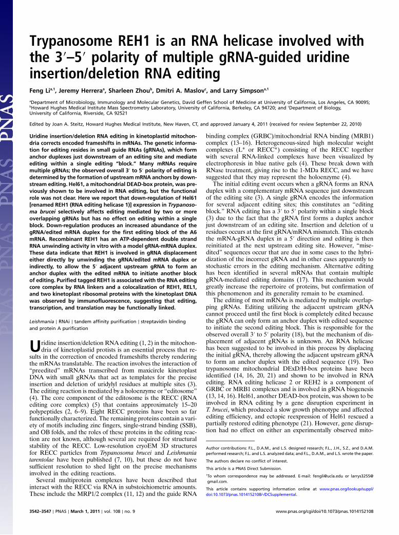
ResultsEffect of Down-Regulation of REH1 Expression in T. brucei on RelativeAbundance of mRNAs Edited in Block 1 Versus Those Edited in Two orMore Blocks. Down-regulation of expression of REH1 by condi-tional RNAi in T. brucei procyclic cells produced a slow growthphenotype (Fig. 1A). An 80% decrease in the abundance ofREH1 mRNA by day 3 was demonstrated by RT-PCR andreal-time RT-PCR (Fig. 1 B and D) and REH1 protein was notdetectable by Western blot analysis (Fig. 1B). Repression ofREH1 expression showed no effect on the stability or length ofthe gRNA 3′ oligo U tails (Fig. 1C), as was reported for the try-panosome REH2 mitochondrial RNA helicase (14, 16)
Real-time RT-PCR was also used to quantitate the effect ofREH1 down-regulation on the relative abundance of severalpreedited and mature edited mRNAs (Fig. 1D and Table S1).The abundances of edited CR3 and A6mRNAs were significantlyreduced with down-regulation of REH1, but the effects on editedmRNAs for Cyb, ND7, CO3, and ND9 were small and probablynot significant because a similar decrease was observed for theCO2 edited mRNA, which is mediated by a single in cis gRNAand does not require an overlapping gRNA. Interestingly, theabundances of preedited mRNAs for CO2 and ND9 were in-creased 30–40%, raising the possibility that REH1 has an effecton RNA turnover of some mRNAs. Two never-edited RNAs,ND4 and COI, were examined as controls: Neither showed asignificant change in abundance. The changes in the abundancesof the A6, CR3, Cyb, ND7, and CO3 preedited mRNAs werenot statistically significant.
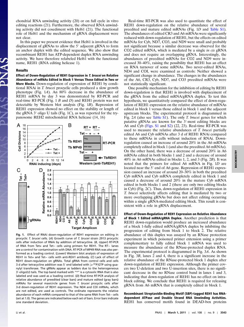
One possible mechanism for the inhibition of editing by REH1down-regulation is that REH1 is involved with displacement ofthe gRNA from the edited mRNA/gRNA duplex. To test thishypothesis, we quantitatively compared the effect of down-regu-lation of REH1 expression on the relative abundance of mRNAsedited in block 1 versus those edited in block 1 and the adjacentupstream blocks. The experimental protocol is diagrammed inFig. 2A (also see Table S1). The only T. brucei genes for whichputative gRNAs are known for the 3′-most editing blocks areA6 and Cyb (Figs. S1 and S2) (22, 23). Real-time RT-PCR wasused to measure the relative abundances of T. brucei partiallyedited A6 and Cyb mRNAs after 3 d of REH1 RNAi comparedto these mRNAs in cells without induction of RNAi. Down-regulation caused an increase of around 20% in the A6 mRNAscompletely edited in block 1 (and also the preedited A6 mRNAs).On the other hand, there was a decrease of around 20% in A6mRNAs edited in both blocks 1 and 2 and a decrease of around40% in A6 mRNAs edited in blocks 1, 2, and 3 (Fig. 2B). It wasnoted that the primers for edited A6 mRNA in Fig. 1D arelocated near the 5′ end of A6 gene. Repression of REH1 expres-sion caused an increase of around 20–30% in both the preeditedCyb mRNA and Cyb mRNA completely edited in block 1 andcaused a decrease of around 20% in the mature Cyb mRNAedited in both blocks 1 and 2 (there are only two editing blocksin Cyb) (Fig. 2C). Thus, down-regulation of REH1 expression inT. brucei selectively affects editing that is mediated by two ormore overlapping gRNAs but does not affect editing occurringwithin a single gRNA-mediated editing block. This result is con-sistent with a role in gRNA displacement.
Effect of Down-Regulation of REH1 Expression on Relative Abundanceof Block 1 Edited mRNA/gRNA Duplex. Another prediction is thatREH1 down-regulation would produce an increased abundanceof a block 1-fully edited mRNA/gRNA duplex by inhibiting theprogression of editing from block 1 to block 2. The relativeabundance of this duplex was assayed by an RNase protectionexperiment in which poisoned primer extension using a primercomplementary to fully edited block 1 mRNA was used tomeasure the abundance of the RNase-protected duplex RNA.The experimental protocol is diagrammed in Fig. 3A. As shownin Fig. 3B, lanes 2 and 4, there is a significant increase in therelative abundance of the RNase-protected block 1 duplex afterdown-regulation of REH1 expression. Although the primer cov-ers two U-deletion and two U-insertion sites, there is no signifi-cant decrease in the no RNase control band in lanes 1 and 3,indicating that down-regulation of REH1 has no effect on intra-block editing. We conclude that REH1 is required for releasinggRNA from A6 mRNA that is completely edited in block 1.
Recombinant Streptavidin-Binding Motif (SBP)-tagged REH1 has RNA-dependent ATPase and Double Strand RNA Unwinding Activities.REH1 has conserved motifs found in DEAD-box proteins
A
C D
B
Fig. 1. Effect of RNAi down-regulation of REH1 expression on editing inprocyclic T. brucei cells. (A) Growth curve of T. brucei strain 2913 procycliccells after induction of RNAi by addition of tetracycline. (B, Upper) RT-PCRof RNA from Tet+ and Tet− cells using primers for REH1. The RT− lanesare a control for contamination with DNA. RT-PCR of MP49 RNAwas also per-formed as a loading control. (Lower) Western blot analysis of expression ofREH1 in Tet+ and Tet− cells with anti-REH1 antibody. (C) Lack of effect ofREH1 down-regulation on gRNAs. Total gRNA from control cells and cells3 d after tetracycline addition was 5′ end labeled with ½α-32P�GTP using gua-nylyl transferase. The gRNAs appear as ladders due to the heterogeneous3′-oligo(U) tails. The top band marked with “*” is a cytosolic RNA that is alsolabeled and was used as a loading control. (D) Real-time RT-PCR analysis ofrelative abundance of preedited (clear bars) and mature edited (gray bars)mRNAs for several maxicircle genes from T. brucei procyclic cells after3-d down-regulation of REH1 expression. The ND4 and COI mRNAs, whichare not edited, are used as controls. The ordinate represents the relativeabundance of each mRNA compared to that of the same RNA from Tet− cells(set at 1.0). The genes are indicated below each set of bars. Error bars indicateone standard deviation.
Li et al. PNAS ∣ March 1, 2011 ∣ vol. 108 ∣ no. 9 ∣ 3543
BIOCH
EMISTR
Y

(Fig. 4A and Fig. S3), but there is no evidence in the literaturefor REH1 having RNA helicase activity. To address this question,we expressed tagged Leishmania major (Lm) REH1 (Fig. 4B) inSf9 cells and analyzed the activity. A modified tandem affinitypurification (TAP) tag (24), the streptavidin binding and proteinA purification (SAP) tag (Fig. 4B), was used in which the calmo-dulin binding peptide motif was substituted with a high affinitystreptavidin binding peptide (SBP) motif. The rREH1 waspurified to homogeneity by IgG-Sepharose binding, MonoS ion-exchange chromatography, and streptavidin affinity chromato-
graphy (Fig. 4C, lane 1). The expressed band was confirmedto be SBP-tagged REH1 by Western analysis using anti-REH1or HRP-conjugated streptavidin (SA-HRP) (Fig. 4C, lanes 2, 3).His6x-tagged REH1 was also expressed in Escherichia coli andpurified to homogeneity for antibody generation.
The purified SBP-tagged recombinant REH1 protein showeda robust poly U–stimulated ATPase activity. This activity was de-stroyed by RNase treatment (Fig. 4D). As a control for ATPasecontamination from the Sf9 cells, SAP-tagged RET2 RECCprotein was expressed and purified using the same procedure.The tagged RET2 did not show ATPase activity (Fig. 4D). TheREH1 ATPase activity requires ATP or dATP (Fig. S4 D and E).The optimal pH is around 8.3, the Km for the reaction is 620 μMATP, and the Kcat is 82.3 min−1 (Fig. S4 A–C).
Recombinant REH1 showed an ATP-dependent double strandRNA unwinding activity using a partially edited model gRNA/edited mRNA duplex (Fig. 4E and Table S2). The reaction re-quired ATP and showed a protein dose response (Fig. 4 E and F).We also tested the unwinding of shorter RNA duplexes with 5′or 3′ overhangs or no overhang (Fig. 4 F–H). The latter wereunwound more efficiently probably due to their lower stability.It has been shown that helicase activity in vitro shows an inverserelationship between duplex stability and unwinding (25). As acontrol, a mutation of K143A in motif I, which is the conservedATP binding domain of REH1, was introduced and the mutantprotein lost ATPase and unwinding activities (Fig. S5). Thesedata indicate that REH1 is an ATP-dependent RNA helicase.The REH1 helicase apparently differs from most other DEAD-box RNA helicases that exhibit very poor in vitro unwindingactivity and require a 5′ or 3′ single-stranded RNA region forunwinding activity (25).
REH1, REL1, and the Kinetoplast Ribosome Proteins, L3 and S17,Show Concentration in the Kinetoplast Region of the Mitochondrion.Immunolocalization of SAP-tagged REH1 within the singlemitochondrion of transfected L. tarentolae cells was performedusing an antibody against the C-terminal FLAG epitope (Fig. 5A).This antibody does not have cross-reactivity with any endogenousprotein (Fig. S6D). The cells were stained with MitoTrackerRed to visualize the single mitochondrion and costained withDAPI to detect the nuclear and kinetoplast DNA, and then trea-ted with anti-FLAG antibody and secondary anti-IgG antibodyconjugated with Alexa Fluor 488 to visualize SAP-tagged REH1.The REH1 protein colocalized with the MitoTracker mitochon-drial image. Interestingly, in addition to a dispersed localizationthroughout the tubular mitochondrion there is an apparentconcentration of REH1 protein in the kinetoplast DNA-contain-ing region. Lower resolution images of entire fields that showthat the selected fields in Fig. 5A are representative are shownin Fig. S6A. As a control, cells were analyzed for the localizationof glutamate dehydrogenase, a soluble mitochondrial proteinnot involved in RNA editing (Fig. S6C). These cells showed asomewhat punctate distribution of immunofluorescence through-out the mitochondrion without the level of concentration in thekinetoplast region observed in the REH1 immunofluorescence,indicating that the observed kinetoplast concentration of REH1is probably not artifactual (Fig. S6).
The localizations of TAP-tagged REL1 RNA ligase, a RECCcore component, and two TAP-tagged kinetoplast ribosomeproteins (Fig. S6E), S17 and L3, were also analyzed by indirectimmunofluorescence. REL1 and the two ribosome proteinsshowed a similar kinetoplast concentration as the SAP-taggedREH1 (Fig. 5 B–D and Fig. S6B).
REH1 Is Associated with the RECC by RNA Linkers. To investigatepossible interactions between REH1 and the RECC, SAP-taggedLm REH1 with a mitochondrial target signal was expressed inL. tarentolae. The tagged REH1 was found only in the whole cell
A B
Fig. 3. Relative abundance of the T. brucei gRNA I/edited A6 block 1 mRNAduplex after RNAi down-regulation of REH1 expression. (A) Diagram of assay.The abundance is assayed by poisoned primer extension of RNase-protectedA6 mRNA. (B) Poisoned primer extension assay of RNase-resistant A6 block 1duplex RNAwith and without down-regulation of REH1 expression. Note thepresence of a +4 labeled band only in the +Tet, +RNase lane. (Lower) Diagramof the extension reaction.
block 1block 2block 3
block 1 + block 2block 1 + block 2 + block 3
block 1
A
1.4
1.6
1.8
gRNA IgRNA II
gRNA III
P1P2P3
P4
1.2
1.4
1.6
1.8 CBA6 Cyb
Rel
ativ
e m
RN
A A
bun
dan
ce
0.0
0.2
0.4
0.6
0.8
1.0
1.2
Rel
ativ
e m
RN
A A
bun
dan
ce
0.0
0.2
0.4
0.6
0.8
1.0
1.2
Target Amplicon
preed
ited
block
1
block
s 1+2
Target Amplicon
preed
ited
block
1
block
s 1+2
block
s 1+2
+3
Fig. 2. Effect of repression of REH1 T. brucei on relative abundances of A6and Cyb mRNAs edited in block 1 versus mRNAs edited in both blocks 1 and 2and 3. (A) Diagram of real-time RT-PCR analysis. Primer sets are indicatedwith arrows. See Table S1 for sequences. (B) Relative abundance of A6mRNAsin T. brucei procyclic cells induced for down-regulation of expression of REH1by RNAi. The abundance of these mRNAs from untreated cells is set at 1.0.Clear bar, preedited mRNA; gray bars, partially edited mRNAs. (C) Relativeabundance of Cyb mRNAs in T. brucei procyclic cells induced for down-regulation of expression of REH1 by RNAi. See B for details. The differencebetween the effect of RNAi on the abundance of the A6 edited mRNA andthe results in this experiment are due to the use of an upstream primer nearthe 5′ end of the mRNA in Fig. 1D instead of the primer used in this assay,which is close to the 3′ end.
3544 ∣ www.pnas.org/cgi/doi/10.1073/pnas.1014152108 Li et al.

and mitochondrial fractions and not in the cytosol fraction(Fig. S7A). Cell lysate was allowed to bind to IgG-Sepharose,and the bound material was released by digestion with tobaccoetch virus (TEV) protease. Half of the eluted material was trea-ted with RNase A prior to fractionation on a glycerol gradient.Gradient fractions were subjected to blue native gel electrophor-
esis and the blots probed with anti-REH1 antibody (Fig. 6A,Upper).
Autoadenylation of REL1/2 with ½α-32P�ATP (26) was alsoused to detect REL1 as a marker for the RECC (Fig. 6A, Lower).The gradient fractions were run in SDS gels that were probedwith anti-REH1 antibody and with anti-MRP1/2 antibody(Fig. 6B). MRP1/2 are proteins that are known to associate withthe RECC via RNA linkers (11).
The tagged REH1 sedimented as free oligomeric protein inthe 5–10S region. A portion of the REH1 sedimented in the20–25S region migrating in the blue native gel at ∼1 MDa(Fig. 6 A and B, Left) and a substantial amount sedimented inthe >25S region, migrating in the blue native gel at >1 MDa(Fig. 6A, Left). The high molecular weight material was sensitiveto RNase as were the REH1-containing RECC materials in frac-tions 10–13 (Fig. 6A, Right). This can also be seen in SDS gels ofthe same gradient fractions (Fig. 6B). The RNase-sensitive highmolecular weight material is reminiscent of the previously re-ported RECC* particles that bound substoichiometric amountsof several other editing complexes (4). This evidence suggeststhat a portion of the tagged REH1 protein is associated withthe RECC* particles via RNA linkers.
Additional evidence for an association of REH1 with theRECC was obtained by TAP purification, performed with orwithout RNase pretreatment (Fig. 6C). The REL1/REL2internal RECC proteins and the MRP1/2 proteins were detectedby Western analysis in the pull-down prior to RNase treatmentbut not after such treatment (Fig. 6C, Right). The coprecipitatedREL1 and REH1 proteins were in substoichiometric amountsbecause they could not be visualized in the SDS gel by Syprostaining (Fig. 6C, Left). The three visible Sypro-stained bandsin the REH1 TAP pull-down were identified by mass spectrome-try as the SPB-tagged REH1 (II), a 10-kDa REH1 C-terminalbreakdown product (III), and a 70-kD band (I) containing twocontaminating proteins: TEV protease and a cytosolic DEAD-box protein (Fig. 6C and Table S3). The presence of this cytosolichelicase in the REH1-TAP pull-down should be investigatedfurther.
A
B
C
D
Fig. 5. Intracellular localization in late log phase L. tarentolae of SAP-tagged Lm REH1, TAP-tagged REL1, and the S17 and L3 TAP-tagged kineto-plast ribosome proteins. (A) Representative cells expressing SAP-taggedREH1. Cells were stained with mouse anti-FLAG antibody + Alexa Fluor 488conjugated F(ab) fragment (α-FLAG, green) to visualize the SAP-taggedREH1. DAPI was used to visualize the nucleus and kinetoplast, andMitoTrack-er CMXRos was used to visualize the entire mitochondrion. A merge of theFLAG and DAPI panels is shown as MergI and a merge of the FLAG andMitoTracker panels is shown as MergII. N: nucleus, K: kinetoplast, M: mito-chondrion. (B) Representative cell expressing TAP-tagged REL1. See legendin A for details. Images of entire fields are shown in Fig. S6. (C) A cell expres-sing TAP-tagged mitochondrial ribosomal protein S17. (D) A cell expressingTAP-tagged mitochondrial ribosomal protein L3.
A
D
G H
B
C
E F
Fig. 4. Purification, RNA-dependent ATPase activity, and ATP-dependent double strand RNA unwinding activity of recombinant SBP-tagged REH1. (A) Dia-gram showing the conserved motifs of REH1. Open boxes represent the conserved helicase motifs with the indicated the amino acid sequences. The numberof amino acids separating the motifs is shown. (B) Diagram of the SAP-tagged Lm REH1. SBP, streptavidin-binding peptide; PrA, protein (A). (C) Purification ofSBP-tagged rREH1 expressed in sf9 insect cells using the Baculovirus expression system (Invitrogen). SDS gels stained with Sypro. Blots probed with SA-HRP oranti-REH1 antibody. (D) RNA-dependent ATPase activity of purified SBP-tagged REH1. The ATPase reactions were performed without added RNA, with addedpoly U, or with added poly U plus 0.05 mg∕mL RNase. (E) Model gRNA/mRNA substrate with a 46-bp duplex and a 49-nt 3′ overhang. The 32P-labeled end isindicated by * in the diagram. The unwinding reaction required ATP and showed a dose response with the amount of protein. (F) Model gRNA/mRNAsubstrate with a 15-bp duplex and a 24-nt 3′ overhang; 5 nM rREH1. (G) 22-bp duplex with 15-nt 5′ overhang; 5 nM rREH1. (H) 15-bp blunt end duplex;5 nM rREH1.
Li et al. PNAS ∣ March 1, 2011 ∣ vol. 108 ∣ no. 9 ∣ 3545
BIOCH
EMISTR
Y

DiscussionREH1 is essential as shown by the growth phenotype caused bydown-regulation of REH1 expression. The partial growth inhibi-tion could be due to incomplete down-regulation, a slow turnoverof the protein or an enzyme redundancy. Down-regulation alsocauses a decrease in abundance of edited mRNAs for the sixgenes assayed, with editing of the A6 and CR3 mRNAs beingthe most affected. Possible roles of the REH1 helicase in RNAediting are (i) to directly assist in the processive editing reactionitself within a single gRNA-mediated block as editing proceedsfrom site to site or (ii) to be involved in gRNA displacementbetween adjacent editing blocks. Model 1 is ruled out by the ob-served lack of effect of REH1 down-regulation on intrablockediting. Model 2 is supported by the significant decrease inthe relative abundance of mRNAs edited in two or more adjacentblocks by REH1 down-regulation as compared to those editedin a single block, and by the accumulation of the gRNA/block 1edited A6 mRNA duplex. These data suggest that REH1 is in-volved with the progression of editing from one gRNA-mediatedediting block to the next adjacent upstream block.
The mechanism of this involvement, however, is not clear. Thefact that recombinant REH1 protein has gRNA/mRNA duplexunwinding activity in vitro may suggest an in vivo role in directlyunwinding the mRNA/gRNA duplex formed by the editing ofall the sites mediated by a single gRNA and liberating an editedmRNA single strand available for hybridization of the adjacentgRNA. However, in the case of REH1, the evidence does notdistinguish between a direct effect on unwinding of the gRNA/mRNA duplex or an indirect effect, such as described foreIF4AIII, a core component of the exon junction complex, andsome putative RNA helicases that are involved in snoRNA re-lease from preribosomes (27–31). Another potential functionof REH1 could be to unwind cis elements within the preeditedmRNA. A dominant-negative mutation of REH1 and ectopicreexpression of wild-type REH1 might indeed help to explore therole of REH1 in vivo. In fact, the evidence that the largest effectof REH1 down-regulation on editing is on the pan-edited A6and CR3 mRNAs suggests that the in vivo activity of REH1 issubstrate-specific and perhaps is regulated by transient bindingof cofactors. Along this line, no detectable in vitro unwindingactivity had been observed with T. brucei gradient fractions thatshowed a peak of Hel61 (REH1) by blot analysis (21) nor in theLm REH1 SAP pull-down (Fig. S7B). These data suggest thepossibility of regulation of helicase activity in vivo.
There have been conflicting reports on the association ofREH1 with the RECC. Tb REH1 was detected by mass spectro-metry analysis in an MP63-immunoprecipitated sample and alsoin a RECC preparation isolated by ion-exchange chromatography(32–34). However, REH1 was not detected in REN1-, REN2-, orREN3-TAP pull-downs from T. brucei (32, 35), nor in REL1-TAPand MP44-TAP pull-downs from L. tarentolae (7, 11). These re-sults could be explained by our finding that REH1 is associatedwith the RECC by RNA linkers, as are several other editing-associated complexes (6, 11), and that this linkage is easily dis-rupted during the isolation.
Immunofluorescence of REH1 showed a concentration in thekinetoplast DNA (kDNA) region of the mitochondrion. Inter-estingly, the REL1 RNA ligase, a component of the RECC,and the S17 and L3 kinetoplast ribosome proteins also exhibiteda concentration in the kDNA region of the mitochondrion, sug-gesting the intriguing possibility of a physical linkage of kDNAtranscription, translation, and editing pathways, but this remainsto be investigated. Our localization results differ somewhat fromseveral previous studies that showed that editing proteins aredistributed throughout the mitochondrion with no apparent con-centration (36, 37) and that the RNA-linked editing proteins,GAP1 and GAP2, are localized in discrete particles throughoutthe mitochondrion (14). These differences could be species-dependent or could be due to technical details.
More work is required to fully understand the precise role ofthe REH1 helicase in RNA editing, but it is clear from the resultsin this paper that REH1 is involved in the 3′ to 5′ polarity of edit-ing in a multi-gRNA-mediated editing domain.
Materials and MethodsSI Text contains details of plasmid construction, real-time PCR, poisonedprimer extension, purification of SAP-tagged Lm REH1, ATPase activity assay,immunofluorescent localization of REH1, REL1, S17, L3, and glutamate dehy-drogenase, mitochondrial importation of L3-TAP and S17–TAP proteins, andsedimentation analysis of kinetoplast ribosomal complexes. The SI Text tablescontain sequences of DNA primers and RNA oligonucleotides used in theassays, and sequences of peptides derived from protein bands in Fig. 3C
ACKNOWLEDGMENTS. We thank Bob Nelson for the original construction ofthe SAP vector in our laboratory, Kent Hill for use of a fluorescence micro-scope, Stephen Smale and Jian Xu for the use of a Real-Time PCR apparatus,and Agda Simpson and Kestrel Rogers for generation of antibodies. Theantibody against Tb REH1 was provided by Ulrich Göringer (Technical Univer-sity of Darmstadt, Darmstadt, Germany) and the 2913 strain of T. bruceiwas provided by George Cross (Rockfeller University, New York). We thankDavid King for mass spectrometry of gel bands to identify proteins. This workwas supported by National Institutes of Health Grant AI09102 (to L.S.).
A C
B
Fig. 6. RNA-dependent association of REH1 with the RECC. (A) Clarified lysate from L. tarentolae cells expressing SAP-tagged REH1 was bound to IgG-Sepharose and the SBP-tagged REH1 released with TEV protease. Half was treated with RNase A (0.1 mg∕mL). After glycerol gradient sedimentation, fractionswere run in blue native gels, which were blotted and probed with anti-REH1. Aliquots of fractions were also autoadenylated with α½32P�ATP to label the REL1RECC protein prior to blue native gel analysis. These gels were dried and exposed to a phosphoimager screen. (B) Gradient fractions from A were run in SDSgels, which were blotted and probed with anti-REH1 or anti-MRP1/2. (C) The TEV-released material in A, with or without RNase treatment, was bound tostreptavidin–Sepharose and the SBP-tagged REH1 released with 2 mM biotin. The SBP-tagged REH1 was run in SDS gels, which were autoadenylated andstained with Sypro (Invitrogen), or blotted and probed with anti-REH1 or anti-MRP1/2. Note that the labeled REL1/REL2 and theMRP1/2 proteins were detectedonly prior to RNase treatment. The three major stained bands were subjected to mass spectrometry (data shown in Table S3).
3546 ∣ www.pnas.org/cgi/doi/10.1073/pnas.1014152108 Li et al.

1. Simpson L, Sbicego S, Aphasizhev R (2003) Uridine insertion/deletion RNA editing intrypanosome mitochondria: A complex business. RNA 9:265–276.
2. Stuart KD, Schnaufer A, Ernst NL, Panigrahi AK (2005) Complex management: RNAediting in trypanosomes. Trends Biochem Sci 30:97–105.
3. Blum B, Bakalara N, Simpson L (1990) A model for RNA editing in kinetoplastidmitochondria: “Guide” RNA molecules transcribed from maxicircle DNA providethe edited information. Cell 60:189–198.
4. Osato D, et al. (2009) Uridine insertion/deletion RNA editing in trypanosomatidmitochondria: In search of the editosome. RNA 15:1338–1344.
5. Simpson L, Aphasizhev R, Lukes J, Cruz-Reyes J (2010) Guide to the nomenclature ofkinetoplastid RNA editing: A proposal. Protist 161:2–6.
6. Aphasizhev R, et al. (2003) Isolation of a U-insertion/deletion editing complex fromLeishmania tarentolae mitochondria. EMBO J 22:913–924.
7. Li F, et al. (2009) Structure of the core editing complex (L-complex) involved in uridineinsertion/deletion RNA editing in trypanosomatid mitochondria. Proc Natl Acad SciUSA 106:12306–12310.
8. Simpson L, Aphasizhev R, Gao G, Kang X (2004) Mitochondrial proteins and complexesin Leishmania and Trypanosoma involved in U-insertion/deletion RNA editing. RNA10:159–170.
9. Worthey EA, Schnaufer A, Mian IS, Stuart K, Salavati R (2003) Comparative analysis ofeditosome proteins in trypanosomatids. Nucleic Acids Res 31:6392–6408.
10. Golas MM, et al. (2009) Snapshots of the RNA editing machine in trypanosomescaptured at different assembly stages in vivo. EMBO J 28:766–778.
11. Aphasizhev R, Aphasizheva I, Nelson RE, Simpson L (2003) A 100-kD complex oftwo RNA-binding proteins from mitochondria of Leishmania tarentolae catalyzesRNA annealing and interacts with several RNA editing components. RNA 9:62–76.
12. SchumacherMA, Karamooz E, Zikova A, Trantirek L, Lukes J (2006) Crystal structures ofT. brucei MRP1/MRP2 guide-RNA binding complex reveal RNA matchmaking mechan-ism. Cell 126:701–711.
13. Weng J, et al. (2008) Guide RNA-binding complex frommitochondria of trypanosoma-tids. Mol Cell 32:198–209.
14. Hashimi H, Čičová Z, Novotná L, Wen YZ, Lukeš J (2009) Kinetoplastid guide RNAbiogenesis is dependent on subunits of the mitochondrial RNA binding complex 1and mitochondrial RNA polymerase. RNA 15:588–599.
15. Acestor N, Panigrahi AK, Carnes J, Zikova A, Stuart KD (2009) The MRB1 complexfunctions in kinetoplastid RNA processing. RNA 15:277–286.
16. Hernandez A, et al. (2010) REH2 RNA helicase in kinetoplastid mitochondria:Ribonucleoprotein complexes and essential motifs for unwinding and guide RNA(gRNA) binding. J Biol Chem 285:1220–1228.
17. Ochsenreiter T, Hajduk SL (2006) Alternative editing of cytochrome c oxidase III mRNAin trypanosome mitochondria generates protein diversity. EMBO Rep 7:1128–1133.
18. Feagin JE, Abraham J, Stuart K (1988) Extensive editing of the cytochrome c oxidase IIItranscript in Trypanosoma brucei. Cell 53:413–422.
19. Maslov DA, Simpson L (1992) The polarity of editing within a multiple gRNA-mediateddomain is due to formation of anchors for upstream gRNAs by downstream editing.Cell 70:459–467.
20. Missel A, Goringer HU (1994) Trypanosoma brucei mitochondria contain RNA helicaseactivity. Nucleic Acids Res 22:4050–4056.
21. Missel A, Souza AE, Norskau G, Goringer HU (1997) Disruption of a gene encoding anovel mitochondrial DEAD-box protein in Trypanosoma brucei affects edited mRNAs.Mol Cell Biol 17:4895–4903.
22. Hong M, Simpson L (2003) Genomic organization of Trypanosoma brucei kinetoplastDNA minicircles. Protist 154:265–279.
23. Ochsenreiter T, Cipriano M, Hajduk SL (2007) KISS: The kinetoplastid RNA editingsequence search tool. RNA 13:1–4.
24. Puig O, et al. (2001) The tandem affinity purification (TAP) method: A general proce-dure of protein complex purification. Methods 24:218–229.
25. Cordin O, Banroques J, Tanner NK, Linder P (2006) The DEAD-box protein family ofRNA helicases. Gene 367:17–37.
26. Peris M, et al. (1994) Characterization of two classes of ribonucleoprotein complexespossibly involved in RNA editing from Leishmania tarentolae mitochondria. EMBO J13:1664–1672.
27. Andersen CB, et al. (2006) Structure of the exon junction core complex with a trappedDEAD-box ATPase bound to RNA. Science 313(5795):1968–1972.
28. Bono F, Ebert J, Lorentzen E, Conti E (2006) The crystal structure of the exon junctioncomplex reveals how it maintains a stable grip on mRNA. Cell 126:713–725.
29. Kos M, Tollervey D (2005) The putative RNA helicase Dbp4p is required for release ofthe U14 snoRNA from preribosomes in saccharomyces cerevisiae. Mol Cell 20:53–64.
30. Liang XH, Fournier MJ (2006) The helicase Has1p is required for snoRNA release frompre-rRNA. Mol Cell Biol 26:7437–7450.
31. Bohnsack MT, Kos M, Tollervey D (2008) Quantitative analysis of snoRNA associationwith pre-ribosomes and release of snR30 by Rok1 helicase. EMBO Rep 9(12):1230–1236.
32. Panigrahi AK, et al. (2006) Compositionally and functionally distinct editosomes inTrypanosoma brucei. RNA 12:1038–1049.
33. Panigrahi AK, Allen TE, Stuart K, Haynes PA, Gygi SP (2003) Mass spectrometricanalysis of the editosome and other multiprotein complexes in Trypanosoma brucei.J Am Soc Mass Spectr 14:728–735.
34. Stuart K, Panigrahi AK, Schnaufer A (2004) Identification and characterization oftrypanosome RNA-editing complex components. Methods Mol Biol 265:273–291.
35. Carnes J, Trotter JR, Peltan A, Fleck M, Stuart K (2008) RNA editing in Trypanosomabrucei requires three different editosomes. Mol Cell Biol 28:122–130.
36. Oberholzer M, Morand S, Kunz S, Seebeck T (2006) A vector series for rapidPCR-mediated C-terminal in situ tagging of Trypanosoma brucei genes. Mol BiochemParasitol 145:117–120.
37. Panigrahi AK, et al. (2001) Association of two novel proteins, TbMP52 and TbMP48,with the Trypanosoma brucei RNA editing complex. Mol Cell Biol 21:380–389.
Li et al. PNAS ∣ March 1, 2011 ∣ vol. 108 ∣ no. 9 ∣ 3547
BIOCH
EMISTR
Y