trophic upgrading and mobilization of wax esters in
TRANSCRIPT

Trophic upgrading and mobilization ofwax esters in microzooplanktonKeyana Roohani1, Brad A. Haubrich1,2, Kai-Lou Yue1, Nigel D’Souza3,4,Amanda Montalbano3, Tatiana Rynearson3, Susanne Menden-Deuer3
and Christopher W. Reid1
1 Science and Technology, Bryant University, Smithfield, RI, USA2 Chemistry, University of Nevada, Reno, Reno, NV, USA3 Graduate School of Oceanography, University of Rhode Island, Narragansett, RI, USA4 Marine Science Institute, University of California, Santa Barbara, Santa Barbara, CA, USA
ABSTRACTHeterotrophic protists play pivotal roles in aquatic ecosystems by transferring matterand energy, including lipids, from primary producers to higher trophic predators.Using Oxyrrhis marina as a model organism, changes to the non-saponifiable protistlipids were investigated under satiation and starvation conditions. During activefeeding on the alga Cryptomonas sp., the O. marina hexane soluble non-saponifiablefraction lipid profile reflected its food source with the observed presence of longchain mono-unsaturated fatty alcohols up to C25:1. Evidence of trophic upgrading inO. marina was observed with long chain mono-unsaturated fatty alcoholaccumulation of up to C35:1. To the best of our knowledge, this is the first evidencethat heterotrophic dinoflagellates are capable of producing ester derived alcoholsand that dinoflagellates like O. marina are capable of synthesizing fatty alcohols up toC35. Additionally, we show evidence of trophic upgrading of lipids. During a 20-dayresource deprivation, the lipid profile remained constant. During starvation, themobilization of wax esters as energy stores was observed with long chain fattyalcohols mobilized first. Changes in lipid class profile and utilization of wax esters inO. marina provides insight into the types of lipids available for energy demand,the transfer of lipids through the base of marine food webs, and the catabolicresponse induced by resource deprivation.
Subjects Biochemistry, Marine Biology, Microbiology, Aquatic and Marine ChemistryKeywords Oxyrrhis marina, Wax ester, Resource deprivation, Trophic upgrading, Catabolism,Microzooplankton
INTRODUCTIONHeterotrophic dinoflagellates are ubiquitous, important components of the pelagicprotozoan community. They are significant consumers of bacterial and phytoplanktonbiomass, and contribute to the cycling of organic matter and nutrients, serving asimportant trophic links in marine microbial food webs (Strom, 1991; Sherr & Sherr, 1994;Steinberg & Landry, 2017). Trophic interactions within complex marine food webs canstrongly influence pathways and efficiencies of material and energy transfer to higher levelconsumers (Anderson & Menden-Deuer, 2017; Mitra & Flynn, 2005; Rose et al., 2011).Heterotrophic protists, like dinoflagellates, add biochemical value during this transfer
How to cite this article Roohani K, Haubrich BA, Yue K-L, D’Souza N, Montalbano A, Rynearson T, Menden-Deuer S, Reid CW. 2019.Trophic upgrading and mobilization of wax esters in microzooplankton. PeerJ 7:e7549 DOI 10.7717/peerj.7549
Submitted 7 April 2019Accepted 25 July 2019Published 19 August 2019
Corresponding authorChristopher W. Reid,[email protected]
Academic editorIvo Feussner
Additional Information andDeclarations can be found onpage 8
DOI 10.7717/peerj.7549
Copyright2019 Roohani et al.
Distributed underCreative Commons CC-BY 4.0

through the production and chemical elaboration of compounds (Klein Breteler et al.,1999). Thus changes to the diet of heterotrophic dinoflagellates (i.e., through starvation)can alter the biomass and cellular composition of herbivores. Although heterotrophicprotists add biochemical value during trophic transfer, little is known about how cellularcomposition changes in response to food availability. These changes in biomass can impacthigher trophic levels through changes in cellular composition. It has been shown thatheterotrophic dinoflagellates such as Oxyhrris marina, Gyrodinium dominans, andG. spirale can survive long periods (>10 days) without algal prey. For example, starvationof O. marina for up to 3 weeks resulted in a reduction in cell volume of 17–57% with somecells deformed and transparent (Menden-Deuer et al., 2005). It has been puzzling howa single celled heterotrophic organism can sustain survival in the absence of substantiveorganic matter, particularly over such extended periods.
Lipids are important energy stores that can be used in times of resource deprivation.Many of the studies on lipids of dinoflagellates fed on algal prey have focused on fatty acidand sterol composition (Klein Breteler et al., 1999; Veloza, Chu & Tang, 2006; Parket al., 2016). These studies have suggested that the fatty acid composition ofO. marinamaynot be dependent on its prey and have highlighted this organism’s ability to upgrade lipidsacquired from its diet. A subclass of the neutral lipids, wax esters, have traditionallyonly been found in marine animal phyla (Sargent, Gatten & McIntosh, 1977; Bauermeister &Sargent, 1979), but some examples have been reported in zooplankton species.Wax esters havebeen observed in the chlorophyte Chlorella kessleri (Sargent, Gatten & Henderson, 1981),the cryptomonad Chroomonas salina (Antia Naval et al., 1974; Henderson & Sargent, 1989),and the euglenoid Euglena gracilis (Furuhashi et al., 2015). In Chroomonas salina ester derivedalcohols are almost exclusively saturated with the most predominant species C13 and C15
while in E. gracilis ester alcohol moieties of up to C22 have been observed.The mechanism of trophic upgrading by heterotrophic protists may bridge the gap and
deliver essential nutrients between higher trophic levels (Klein Breteler et al., 1999; Veloza,Chu & Tang, 2006). Given the importance of heterotrophic dinoflagellates in marinefood webs by providing essential nutrients to higher trophic levels, an understanding of thechanges to the lipid profile under varying availability of prey can provide insight into thenutritional quality available to higher trophic levels. Here, we report the changes to thenon-saponifiable fraction (NSF) lipid composition of the representative dinoflagellateO. marina, as our model organisms during active feeding and in response to long termstarvation. O. marina is a free living, cosmopolitan, and phagotrophic alveolate that feedson a variety of algae and bacteria (Landry et al., 2000) and has been recognized for itsunique starvation ability lasting for several months (Menden-Deuer et al., 2005; Calbetet al., 2013).
MATERIALS AND METHODSMaterialsLong chain fatty alcohol standards were obtained from Millipore Sigma (Burlington,MA, USA). High pressure liquid chromatography (HPLC) lipid standards included aphospholipid mixture and mono-, di-, and tri-acylglycerol mixtures (Millipore Sigma,
Roohani et al. (2019), PeerJ, DOI 10.7717/peerj.7549 2/12

Burlington, MA, USA). Nile Red was purchased from Invitrogen (Carlsbad, CA, USA). Allsolvents used were of HPLC or spectroscopic grade. All HPLC mobile phases were filteredthrough a 0.22 μm membrane prior to use.
Cell cultureNon-axenic cultures of the cryptophyte alga Cryptomonas sp. were maintained in triplicatetwo-L, transparent polycarbonate (PC) bottles to serve as prey under culture conditionsthat included a 12:12 h light-dark cycle at 14 �C, salinity of approximately 30 practicalsalinity units (PSU), and a light intensity of 70–80 µmol photons · m−2 · s−1. The culturemedium was prepared from sterile autoclaved 0.2 µm filtered seawater amended withnutrients following the f/2 medium without silica recipe of Guillard (1975). The seawaterwas collected at high tide from Narragansett Bay, Rhode Island, USA.
Non-axenic, clonal cultures of the heterotrophic dinoflagellate O. marina (CCMP3375;Om), were established via single-cell isolation and grown in two-L transparent PC bottleson a 12 h:12 h light–dark cycle at 14.5 �C, salinity of approximately 30 psu, and alight intensity of 8–15 µmol photons · m−2 · s−1. Grazers were maintained in exponentialgrowth phase by feeding them once a week with Cryptomonas sp. prey and diluted withautoclaved filtered seawater.
Estimating cell abundance, size, and biomassGrazer and phytoplankton prey abundance and cell size were monitored using aMultisizer TM 3 Coulter counter (version 3.53; Beckman Coulter, Indianapolis, IN, USA).The Coulter counter provided a more rapid and reliable sampling approach thanmicroscopy and allowed convenient monitoring of the cultures over the course of theexperiments (Kim & Menden-Deuer, 2013). Grazer and phytoplankton prey were easilydistinguishable on the Coulter counter based on their respective size distributions. Grazervolume was determined using the equivalent spherical diameter measurements from theCoulter counter, and converted to carbon biomass (pg C. cell−1) using previouslyestablished conversion equations (Menden-Deuer & Lessard, 2000).
Starvation and re-feeding experiments were set-up using established methods(Anderson & Menden-Deuer, 2017). Briefly, grazers fed with prey were transferred totriplicate four-L bottles and starved for 1–3 weeks until a reduction in predator abundanceor cell size was detected, which indicated a negative impact of algal prey deprivation andmarked the initiation of grazer starvation (Anderson & Menden-Deuer, 2017). Grazerswere starved for 20 days, received a fresh pulse of phytoplankton prey after starvation, andwere monitored for 3–7 days after re-feeding. Samples were taken at regular intervals(0, 8, 10, 15, 18, 20 days) during the starvation for measurements of grazer abundance andcell size. Grazer and prey abundances obtained using the coulter counter and flowcytometer were verified by light microscopy.
Lipid isolationFilter membranes containing Cryptomonas sp. or O. marina cells were extracted using theBligh-Dyer procedure (Bligh & Dyer, 1959). Briefly, 105 to 107 cells adhered to membrane
Roohani et al. (2019), PeerJ, DOI 10.7717/peerj.7549 3/12
were suspended in five mL of 1:2 (v/v) CHCl3:MeOH and sonicated for 5 min at65% amplitude, (10 s on, 20 s off). Lipids were extracted with the addition of three mL of2:1 MeOH:CHCl3 and mixed by vortexing. The sample was then converted to a two phaseBligh-Dyer by the addition of one mL CHCl3 and 1.8 mL dH2O. The resulting biphasicsamples were centrifuged (4,000 rpm, 2 min) and the bottom layer was recovered andtransferred to a clean sample vial. The organic layer was dried under a stream on N2.Samples were then analyzed by HPLC. Portions of the lipid extracts were subjected tosaponification (methanolic KOH) (Haubrich et al., 2015) for analysis of neutral lipids andanalyzed by gas chromatography-mass spectrometry (GCMS) without furtherderivatization. Negative controls of solvent extracted filter membranes were incorporatedinto the experiment to control for contaminants arising from the filter membranes.
Lipid staining and flow cytometrySamples were stained with Nile Red as described by De la Jara et al. (2003). The optimalfluorescence of Nile Red can be highly selective and variable based on dye concentrationand cell type stained (Rumin et al., 2015), and given the novelty of Nile Red stainingwith marine heterotrophic dinoflagellates, we followed an optimization protocol. The effectof several parameters on dye permeation and fluorescence were tested such as final dyeconcentration (0.5–5 µg mL−1), incubation time (5–30 min), solvents (e.g., DMSO andacetone), and temperature (Rumin et al., 2015). An optimum Nile Red concentration oftwo µg mL−1 dissolved in acetone was determined based on fluorescence profiles (via flowcytometry), and thus represented the dye concentration used in starvation experiments.Triplicate five mL samples were spiked with Nile Red, gently vortexed, and incubatedfor 10 min at room temperature in the dark to ensure dye permeation while avoidingquenching effects. Nile Red samples were then fixed using glutaraldehyde (0.5% finalconc. v/v), flash frozen in liquid N2, and stored at −80 �C until flow cytometry analysis(measured within 1–2 months).
Triplicate samples were analyzed using flow cytometry (BD-Influx flow cytometer,Becton Dickinson Instruments) with an excitation wavelength of 488 nm. A minimum of200 cells were counted for each sample. Populations of cells were identified based onfluorescence vs forward and side scatter. Chlorophyll autofluorescence was determinedusing a 692/40 nm filter and differentiated autotrophic prey from grazer lipid fluorescence.Lipid content, measured as fluorescence intensity per cell, was estimated from thefluorescence emission of NR-stained cells using 580/30 nm (neutral lipid), and 610/20 nm(polar lipid) filters (Alonzo &Mayzaud, 1999; De la Jara et al., 2003). Non-stained sampleswere used to control for NR autofluorescence; fluorescence of unstained cells wasconsistently less than 10% of stained samples. Lipid content for each grazer species wasmeasured as a function of fluorescence, and expressed as relative fluorescence units, ratherthan as equivalent lipid concentrations.
Non-saponifiable lipid extractsLipid extracts were treated with 10:10:80 (v/w/v) of dH2O:KOH:MeOH and refluxed for30 min (Haubrich et al., 2015). After cooling to room temperature, water and hexanes were
Roohani et al. (2019), PeerJ, DOI 10.7717/peerj.7549 4/12

added. Saponified lipids were extracted three times with hexanes and pooled. Thecombined hexane extracts were dried over anhydrous Na2SO4 and evaporated to drynessunder a stream of N2. Prior to analysis samples were dissolved in equal volumes of CHCl3.
Chromatographic analysisRP-HPLCChromatographic separation was performed on a Prominence uHPLC system (Shimadzu,Columbia, MD, USA) equipped with a COSMOSIL 5 μ C18-MS II (4.6 × 150 mm) column(Nacalai Tesque Inc., Kyoto, Japan). Lipid classes were separated employing a binarygradient system described by Knittelfelder et al. (2014) with detection at 205 nm (Guarrasiet al., 2010). Mobile phase A consisted of water:methanol (1:1 v/v), mobile phase B was2-propanol. Both solvents contained phosphoric acid (eight μM) and formic acid(0.1% v/v). A linear gradient with initial conditions starting at 45% mobile phase B wasincreased to 90% B over 30 min. Mobile phase B was then increased to 100% over 2 minand was held at 100% for 10 min. The column was re-equilibrated for 15 min betweeninjections. Retention time regions for polar, and neutral lipids were established usingcommercial lipid standards. Data was analyzed using LabSolutions (Shimadzu, Columbia,MD, USA) and statistical analysis (ANOVA) performed using GraphPad Prism version7.0. All samples were run in biological triplicate and technical duplicate.
GCMSNSF lipid samples were analyzed GCMS with an Agilent 7890A gas chromatographequipped with a 5975C electron impact mass spectrometer set to 70 eV using a RestekRtx-5 column (30 m × 25 μm diameter). The GC flow rate of He was set at 1.2 mL/min,injector port set to 250 �C, and the initial temperature set at 170 �C, held for 1 min,then increased at 20 �C/min to a final temperature of 280 �C (Haubrich et al., 2015).Chromatograms were processed using ChemStation (Agilent, Santa Clara, CA, USA) andanalyte identification of the resulting chromatograms was performed via interrogationof resulting electron impact mass spectra with the NIST database and manual analysis.Octadecanol (RT = 5.001 min) was the standard for determination of relative retentiontime. All samples were run in biological triplicate and technical duplicate.
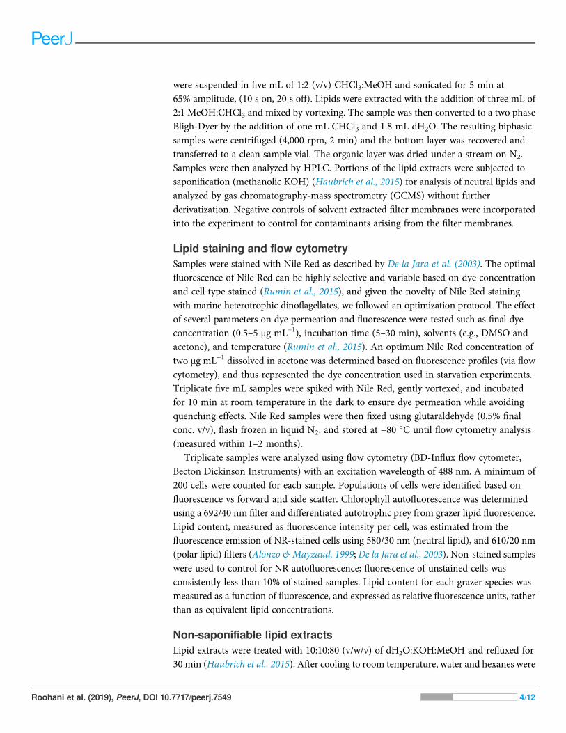
RESULTS AND DISCUSSIONO. marina lipid class profileThe lipidome of protist predators can change rapidly in response to environmentalconditions. Nile Red lipid staining of O. marina demonstrated significant changes in theconcentration of neutral and polar lipids in satiated and starved cells (Fig. 1A). A linearrelationship (r2 ¼ 0.9044) in the decrease in polar and neutral lipids during starvationwas observed via flow cytometry (Fig. 1A). The apparent differences in total polar andneutral lipids after 7 and 15 days respectively are non-significant (p > 0.05). The change inpolar and neutral lipid concentration during starvation was further investigated byRP-HPLC. While total polar and neutral lipids decreased during starvation the relativeabundance of the subclasses of these lipids showed a consistent ratio of polar to neutral
Roohani et al. (2019), PeerJ, DOI 10.7717/peerj.7549 5/12
lipids of 92.1 ± 3.2:3.0 ± 2.2:5.7 ± 2.6 (PL:MAG/DAG:TAG) was maintained (Fig. 1B).The lipid class composition of O. marina is comparable to what has been previously observedin dinoflagellates and phytoplankton (Harvey et al., 1988; Bourdier & Amblard, 1989;Yoon et al., 2017). Nutrient deprived O. marina have been shown to decrease cell volume by17–57% (Anderson & Menden-Deuer, 2017) and increase expression of genes involved inthe degradation of lipids (Rubin et al., 2019) suggesting a homeostatic requirement forO. marina to maintain relative amounts of each lipid class as cell volume decreases andstress-induced catabolism progresses.
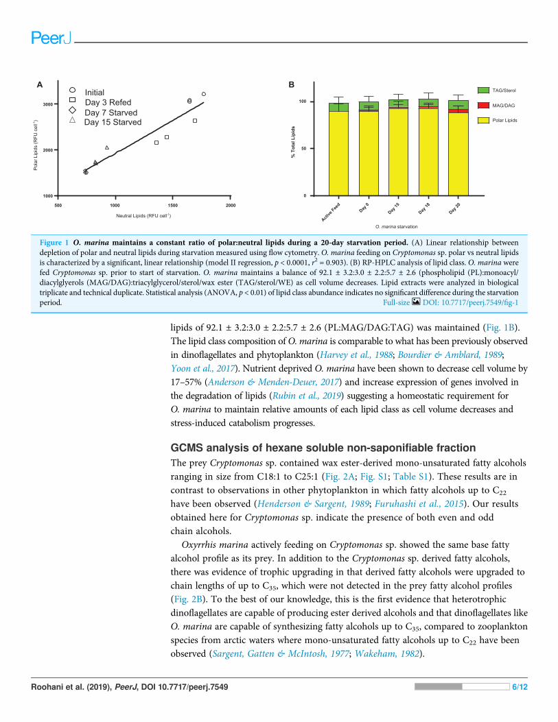
GCMS analysis of hexane soluble non-saponifiable fractionThe prey Cryptomonas sp. contained wax ester-derived mono-unsaturated fatty alcoholsranging in size from C18:1 to C25:1 (Fig. 2A; Fig. S1; Table S1). These results are incontrast to observations in other phytoplankton in which fatty alcohols up to C22
have been observed (Henderson & Sargent, 1989; Furuhashi et al., 2015). Our resultsobtained here for Cryptomonas sp. indicate the presence of both even and oddchain alcohols.
Oxyrrhis marina actively feeding on Cryptomonas sp. showed the same base fattyalcohol profile as its prey. In addition to the Cryptomonas sp. derived fatty alcohols,there was evidence of trophic upgrading in that derived fatty alcohols were upgraded tochain lengths of up to C35, which were not detected in the prey fatty alcohol profiles(Fig. 2B). To the best of our knowledge, this is the first evidence that heterotrophicdinoflagellates are capable of producing ester derived alcohols and that dinoflagellates likeO. marina are capable of synthesizing fatty alcohols up to C35, compared to zooplanktonspecies from arctic waters where mono-unsaturated fatty alcohols up to C22 have beenobserved (Sargent, Gatten & McIntosh, 1977; Wakeham, 1982).
A B
% T
otal
Lip
ids
Active
Feed
Day 0
Day 15
Day 18
Day 20
0
50
100
Polar Lipids
MAG/DAG
TAG/Sterol
O. marina starvation
500 1000 1500 2000
1000
2000
3000
Neutral Lipids (RFU cell-1)
Pol
ar L
ipid
s (R
FU c
ell-1
) Day 15 StarvedDay 7 StarvedDay 3 RefedInitial
Figure 1 O. marina maintains a constant ratio of polar:neutral lipids during a 20-day starvation period. (A) Linear relationship betweendepletion of polar and neutral lipids during starvation measured using flow cytometry. O. marina feeding on Cryptomonas sp. polar vs neutral lipidsis characterized by a significant, linear relationship (model II regression, p < 0.0001, r2 = 0.903). (B) RP-HPLC analysis of lipid class. O. marina werefed Cryptomonas sp. prior to start of starvation. O. marina maintains a balance of 92.1 ± 3.2:3.0 ± 2.2:5.7 ± 2.6 (phospholipid (PL):monoacyl/diacylglyerols (MAG/DAG):triacylglycerol/sterol/wax ester (TAG/sterol/WE) as cell volume decreases. Lipid extracts were analyzed in biologicaltriplicate and technical duplicate. Statistical analysis (ANOVA, p < 0.01) of lipid class abundance indicates no significant difference during the starvationperiod. Full-size DOI: 10.7717/peerj.7549/fig-1
Roohani et al. (2019), PeerJ, DOI 10.7717/peerj.7549 6/12

A
B
C
D
1:811:12
1:221:32
1:421:52
1:621:72
1:53
0 . 0
0 . 2
0 . 4
0 . 6
18
: 1 1:12 22
: 1 1:321:42 2
5: 1
26
: 1 1:721:53
0 . 0
0 . 2
0 . 4
0 . 6
18: 1 1:12 22
: 1 1:321:42 2
5: 1
26
: 1 1:721:53
0 . 0
0 . 2
0 . 4
0 . 6
F a t t y a l c o h o l s
Res
pons
e ce
ll-1
18
: 1 1:12 22: 1 1:32
1:42 25: 1
26
: 1 1:721:53
0 . 0
0 . 2
0 . 4
0 . 6
Figure 2 Changes in hexane soluble NSF lipid extracts during active feeding and prolongedstarvation of O. marina. (A) Prey Cryptomonas sp., (B) O. marina during active feeding on Crypto-monas sp., (C) O. marina after 15 days starvation, (D) O. marina after 18 days starvation. Day zero ofstarvation commenced when prey were not detectable by Coulter Counter and microscopy. Evidence oftrophic upgrading of observed fatty alcohols in actively feeding O. marina. During a 20-day starvation,O. marina mobilized wax esters as an energy source. Full-size DOI: 10.7717/peerj.7549/fig-2
Roohani et al. (2019), PeerJ, DOI 10.7717/peerj.7549 7/12

Over a 20 day starvation period, O. marina appeared to mobilize the fatty alcohols asenergy reserves with longer chain fatty alcohols utilized first (Figs. 2C and 2D). After20 days near complete depletion of fatty alcohols was observed, consistent withobservations of increased expression of genes involved in lipid degradation in starvedO. marina (Rubin et al., 2019).
Wax ester production in dinoflagellates has been suggested to be involved in buoyancyregulation and as a deposit of an energy rich food reserve during periods of low preyabundance (Sargent, Gatten & Henderson, 1981). Wax esters and TAGs are commonlyfound in lipid bodies within dinoflagellates. These compounds have been the focus ofinvestigations during nitrogen stress (Dagenais Bellefeuille et al., 2014) and in coral-dinoflagellate symbiont relationships (Chen et al., 2012). This analysis of changes in lipidclass profile and utilization of wax esters in O. marina provides insight into the catabolicresponse induced by general resource deprivation. We have provided evidence thatduring starvation in O. marina that wax esters are mobilized as energy stores while theratio of polar:non-polar lipids remain constant as cell volume decreases. These dataprovide information on the changes in lipid content, in particular the NSF in O. marinaduring prolonged resource deprivation.
CONCLUSIONSHere, we evaluated the lipid profile of a marine herbivorous zooplankton during starvationand contrasted this with the NSF lipid profile of its phytoplankton prey. We foundevidence both of direct trophic transfer of lipids from the algal source, as well as trophicupgrade of neutral lipids. While diet deprivation did not seem to affect ratios of polarto neutral lipids in starved O. marina, starvation was accompanied by a time-dependentdepletion of longer-chain fatty alcohols from energy stores. In addition, the presence ofremarkably long fatty alcohols was noted in saponified lipids of a heterotrophicdinoflagellate. Characterization and quantification of catabolic responses to resource stressin marine herbivores provides opportunities to use lipids as biomarkers for energy demandand assessment of energy status in marine microbial food webs and improve coastalecosystem models.
ACKNOWLEDGEMENTSWe would like to thank Krystyna Kula for technical assistance.
ADDITIONAL INFORMATION AND DECLARATIONS
FundingThis research was supported by the Rhode Island Science and Technology AdvisoryCouncil (to Susanne Menden-Deuer, Tatiana Rynearson, Christopher W. Reid) andNASA grant 80NSSC17K0716 (to Susanne Menden-Deuer, Tatiana Rynearson) as partof the Export Processes in the Global Ocean from Remote Sensing (EXPORTS) fieldcampaign. This study was conducted using infrastructure supported by NSF EPSCoRresearch infrastructure improvement awards EPS-1004057 and OIA-1655221. There was
Roohani et al. (2019), PeerJ, DOI 10.7717/peerj.7549 8/12

no additional external funding received for this study. The funders had no role in studydesign, data collection and analysis, decision to publish, or preparation of themanuscript.
Grant DisclosuresThe following grant information was disclosed by the authors:Rhode Island Science and Technology Advisory Council.NASA: 80NSSC17K0716.Export Processes in the Global Ocean from Remote Sensing (EXPORTS) field campaign.NSF EPSCoR research infrastructure improvement awards: EPS-1004057 andOIA-1655221.
Competing InterestsSusanne Menden-Deuer is an Academic Editor for PeerJ.
Author Contributions� Keyana Roohani performed the experiments, analyzed the data, authored or revieweddrafts of the paper, approved the final draft.
� Brad A. Haubrich conceived and designed the experiments, performed the experiments,analyzed the data, prepared figures and/or tables, authored or reviewed drafts of thepaper, approved the final draft.
� Kai-Lou Yue performed the experiments, approved the final draft, prepared lipidextracts, saponified lipid samples, prepared and ran samples on GCMS.
� Nigel D’Souza performed the experiments, approved the final draft.� Amanda Montalbano performed the experiments, analyzed the data, authored orreviewed drafts of the paper, approved the final draft.
� Tatiana Rynearson conceived and designed the experiments, analyzed the data, authoredor reviewed drafts of the paper, approved the final draft.
� Susanne Menden-Deuer conceived and designed the experiments, analyzed the data,contributed reagents/materials/analysis tools, prepared figures and/or tables, authoredor reviewed drafts of the paper, approved the final draft.
� Christopher W. Reid conceived and designed the experiments, analyzed the data,contributed reagents/materials/analysis tools, prepared figures and/or tables, authoredor reviewed drafts of the paper, approved the final draft.
Data AvailabilityThe following information was supplied regarding data availability:
Raw data in the format of GCMS and HPLC chromatograms are provided as .netCDFfiles in the Supplemental Materials.
Supplemental InformationSupplemental information for this article can be found online at http://dx.doi.org/10.7717/peerj.7549#supplemental-information.
Roohani et al. (2019), PeerJ, DOI 10.7717/peerj.7549 9/12

REFERENCESAlonzo F, Mayzaud P. 1999. Spectrofluorometric quantification of neutral and polar
lipids in zooplankton using Nile red. Marine Chemistry 67(3–4):289–301DOI 10.1016/S0304-4203(99)00075-4.
Anderson SR, Menden-Deuer S. 2017. Growth, grazing, and starvation survival in threeheterotrophic dinoflagellate species. Journal of Eukaryotic Microbiology 64(2):213–225DOI 10.1111/jeu.12353.
Antia Naval J, Lee Richard F, Nevenzel Judd C, Cheng Joseph Y. 1974.Wax ester production bythe marine Cryptomonad Chroomonas salina grown photoheterotrophically on glycerol.Journal of Protozoology 21(5):768–771 DOI 10.1111/j.1550-7408.1974.tb03749.x.
Bauermeister A, Sargent JR. 1979. Wax esters: major metabolites in the marine environment.Trends in Biochemical Sciences 4(9):209–211 DOI 10.1016/0968-0004(79)90082-3.
Bligh EG, Dyer WJ. 1959. A rapid method of total lipid extraction and purification.Canadian Journal of Biochemistry and Physiology 37(1):911–917 DOI 10.1139/y59-099.
Bourdier GG, Amblard CA. 1989. Lipids in Acanthodiaptomus denticomis during starvationand fed on three different algae. Journal of Plankton Research 11(6):1201–1212DOI 10.1093/plankt/11.6.1201.
Calbet A, Isari S, Martínez RA, Saiz E, Garrido S, Peters J, Borrat RM, Alcaraz M. 2013.Adaptations to feast and famine in different strains of the marine heterotrophic dinoflagellatesGyrodinium dominans and Oxyrrhis marina. Marine Ecology Progress Series 483:67–84DOI 10.3354/meps10291.
Chen W-NU, Kang H-J, Weis VM, Mayfield AB, Jiang P-L, Fang L-S, Chen C-S. 2012. Dielrhythmicity of lipid-body formation in a coral-Symbiodinium endosymbiosis. Coral Reefs31(2):521–534 DOI 10.1007/s00338-011-0868-6.
Dagenais Bellefeuille S, Dorion S, Rivoal J, Morse D. 2014. The dinoflagellate Lingulodiniumpolyedrum responds to N depletion by a polarized deposition of starch and lipid bodies.PLOS ONE 9(11):e111067 DOI 10.1371/journal.pone.0111067.
De la Jara A, Mendoza H, Martel A, Molina C, Nordstron L, De la Rosa V, Diaz R. 2003. Flowcytometric determination of lipid content in a marine dinoflagellate, Crypthecodinium cohnii.Journal of Applied Phycology 15(5):433–438 DOI 10.1023/A:1026007902078.
Furuhashi T, Ogawa T, Nakai R, Nakazawa M, Okazawa A, Padermschoke A, Nishio K,Hirai MY, Arita M, Ohta D. 2015. Wax ester and lipophilic compound profiling of Euglenagracilis by gas chromatography-mass spectrometry: toward understanding of wax esterfermentation under hypoxia. Metabolomics 11(1):175–183 DOI 10.1007/s11306-014-0687-1.
Guarrasi V, Mangione MR, Sanfratello V, Martorana V, Bulone D. 2010. Quantification ofunderivatized fatty acids from vegetable oils by HPLC with UV detection. Journal ofChromatographic Science 48(8):663–668 DOI 10.1093/chromsci/48.8.663.
Guillard RRL. 1975. Culture of phytoplankton for feeding marine invertebrates. In: Smith WL,Chanley MH, eds. Culture of Marine Invertebrate Animals: Proceedings—1st Conference onCulture of Marine Invertebrate Animals Greenport. Boston: Springer, 29–60.
Harvey HR, Bradshaw SA, O’Hara SCM, Eglinton G, Corner EDS. 1988. Lipid composition ofthe marine dinoflagellate Scrippsiella trochoidea. Phytochemistry 27(6):1723–1729DOI 10.1016/0031-9422(88)80432-1.
Haubrich BA, Singha UK, Miller MB, Nes CR, Anyatonwu H, Lecordier L, Patkar P, Leaver DJ,Villalta F, Vanhollebeke B, Chaudhuri M, Nes WD. 2015.Discovery of an ergosterol-signalingfactor that regulates Trypanosoma brucei growth. Journal of Lipid Research 56(2):331–341DOI 10.1194/jlr.M054643.
Roohani et al. (2019), PeerJ, DOI 10.7717/peerj.7549 10/12

Henderson RJ, Sargent JR. 1989. Lipid composition and biosynthesis in ageing cultures of themarine cryptomonad, Chroomonas salina. Phytochemistry 28(5):1355–1361DOI 10.1016/S0031-9422(00)97745-8.
Kim H, Menden-Deuer S. 2013. Reliability of rapid, semi-automated assessment of planktonabundance, biomass, and growth rate estimates: Coulter counter versus light microscopemeasurements. Limnology and Oceanography: Methods 11(7):382–393DOI 10.4319/lom.2013.11.382.
Klein Breteler WCM, Schogt N, Baas M, Schouten S, Kraay GW. 1999. Trophic upgrading offood quality by protozoans enhancing copepod growth: role of essential lipids. Marine Biology135(1):191–198 DOI 10.1007/s002270050616.
Knittelfelder OL, Weberhofer BP, Eichmann TO, Kohlwein SD, Rechberger GN. 2014.A versatile ultra-high performance LC-MS method for lipid profiling. Journal ofChromatography B 951–952:119–128 DOI 10.1016/j.jchromb.2014.01.011.
Landry MR, Constantinou J, Latasa M, Brown SL, Bidigare RR, Ondrusek ME. 2000. Biologicalresponse to iron fertilization in the eastern equatorial Pacific (IronEx II). III. Dynamics ofphytoplankton growth and microzooplankton grazing. Marine Ecology Progress Series201:57–72 DOI 10.3354/meps201057.
Menden-Deuer S, Lessard EJ. 2000. Carbon to volume relationships for dinoflagellates, diatoms,and other protist plankton. Limnology and Oceanography 45(3):569–579DOI 10.4319/lo.2000.45.3.0569.
Menden-Deuer S, Lessard EJ, Satterberg J, Grünbaum D. 2005. Growth rates and starvationsurvival of three species of the pallium-feeding, thecate dinoflagellate genus Protoperidinium.Aquatic Microbial Ecology 41:145–152 DOI 10.3354/ame041145.
Mitra A, Flynn KJ. 2005. Predator–prey interactions: is ‘ecological stoichiometry’sufficient when good food goes bad? Journal of Plankton Research 27(5):393–399DOI 10.1093/plankt/fbi022.
Park J, Jeong HJ, Yoon EY, Moon SJ. 2016. Easy and rapid quantification of lipid contentsof marine dinoflagellates using the sulpho-phospho-vanillin method. ALGAE 31(4):391–401DOI 10.4490/algae.2016.31.12.7.
Rose KA, Allen JI, Artioli Y, Barange M, Blackford J, Carlotti F, Cropp R, Daewel U,Edwards K, Flynn K, Hill SL, HilleRisLambers R, Huse G, Mackinson S, Megrey B, Moll A,Rivkin R, Salihoglu B, Schrum C, Shannon L, Shin Y-J, Smith SL, Smith C, Solidoro C,St. John M, Zhou M. 2011. End-to-end models for the analysis of marine ecosystems:challenges, issues, and next steps. Marine and Coastal Fisheries 2(1):115–130DOI 10.1577/C09-059.1.
Rubin ET, Cheng S, Montalbano AL, Menden-Deuer S, Rynearson TA. 2019. Transcriptomicresponse to feeding and starvation in a herbivorous dinoflagellate. Frontiers in Marine Science6:246 DOI 10.3389/fmars.2019.00246.
Rumin J, Bonnefond H, Saint-Jean B, Rouxel C, Sciandra A, Bernard O, Cadoret J-P, Bougaran G.2015. The use of fluorescent Nile red and BODIPY for lipid measurement in microalgae.Biotechnology for Biofuels 8(1):42 DOI 10.1186/s13068-015-0220-4.
Sargent JR, Gatten RR, Henderson RJ. 1981. Marine wax esters. Pure and Applied Chemistry53(4):867–871 DOI 10.1351/pac198153040867.
Sargent JR, Gatten RR, McIntosh RJ. 1977. Wax esters in the marine environment—theiroccurrence, formation, transformation and ultimate fates. Marine Chemistry 5(4–6):573–584DOI 10.1016/0304-4203(77)90043-3.
Roohani et al. (2019), PeerJ, DOI 10.7717/peerj.7549 11/12

Sherr EB, Sherr BF. 1994. Bacterivory and herbivory: key roles of phagotrophic protists in pelagicfood webs. Microbial Ecology 28(2):223–235 DOI 10.1007/BF00166812.
Steinberg DK, Landry MR. 2017. Zooplankton and the ocean carbon cycle. Annual Review ofMarine Science 9(1):413–444 DOI 10.1146/annurev-marine-010814-015924.
Strom SL. 1991. Growth and grazing rates of the herbivorous dinoflagellate Gymnodinium sp.from the open subarctic Pacific Ocean. Marine Ecology Progress Series 78:103–113DOI 10.3354/meps078103.
Veloza AJ, Chu F-LE, Tang KW. 2006. Trophic modification of essential fatty acids byheterotrophic protists and its effects on the fatty acid composition of the copepod Acartia tonsa.Marine Biology 148(4):779–788 DOI 10.1007/s00227-005-0123-1.
Wakeham SG. 1982. Organic matter from a sediment trap experiment in the equatorial northAtlantic: wax esters, steryl esters, triacylglycerols and alkyldiacylglycerols. Geochimica etCosmochimica Acta 46(11):2239–2257 DOI 10.1016/0016-7037(82)90198-3.
Yoon EY, Park J, Jeong HJ, Rho J-R. 2017. Fatty acid composition and docosahexaenoic acid(DHA) content of the heterotrophic dinoflagellateOxyrrhis marina fed on dried yeast: comparedwith algal prey. ALGAE 32(1):67–74 DOI 10.4490/algae.2017.32.3.5.
Roohani et al. (2019), PeerJ, DOI 10.7717/peerj.7549 12/12