treatment with calcimimetics in kidney transplantation
TRANSCRIPT

Available online at www.sciencedirect.com
Transplantation Reviews 24 (2010) 79–88www.elsevier.com/locate/trre
Treatment with calcimimetics in kidney transplantationEnrique Morales⁎, Eduardo Gutierrez, Amado Andres
Nephrology Department, Hospital 12 de Octubre, Madrid, Spain
Abstract
Graft and patient survival in renal transplantation has increased with better immune suppression treatment, leading to the appearance ofnew complications such as posttransplant bone disease. After renal transplantation and the recovery of renal function, mineral metabolismdisorders secondary to renal failure could be expected to normalize. However, both immediately after transplantation and later, and even withgood renal graft function, we see bone disorders associated to renal osteodystrophy, a high incidence of osteopenia, persistenthyperparathyroidism, hypercalcemia, hypophosphoremia, and less commonly, aseptic bone necrosis. The causes potentially responsible forthese disorders have basically been identified as different degrees of renal insufficiency in the graft, persistent posttransplant secondaryhyperparathyroidism, and negative impact of immunosuppression treatment, particularly corticosteroids. The most important factor in theevolution of metabolic and bone disorders after renal transplantation, however, is pretransplant bone status. Special attention should be paidto other osteoarticular complications such as loss of bone mass and fractures, leading to significant morbidity. In the therapeutic approach tothese patients, as well as encouraging physical exercise and advice about diet or other habits, the use of drugs such as calcium and vitamin Dsupplements, bisphosphonates, and more recently, calcimimetics have made significant improvements in the prevention and treatment ofbone-mineral metabolism. It has been shown that calcimimetic agents can control the parathyroid hormone, reduce episodes ofhypercalcemia, and improve hypophosphatemia. Their properties have to be assessed in broader studies to establish the basis for theirwidespread use among renal transplant recipients.© 2010 Elsevier Inc. All rights reserved.
1. Introduction
Most of the bone and mineral metabolism disorderscaused by uremia are corrected after renal transplantation.The factors that can have a negative impact on bone andmineral metabolism after transplantation are as follows:persistent hyperparathyroidism, adverse effects of immuno-suppressants, and persistence of different degrees of renalfailure, acidosis, and the use of other drugs [1-3]. Secondaryhyperparathyroidism frequently persists after renal trans-plantation and is associated with hypercalcemia and negativeeffects on bone. More information has become available inthe last years about the effect of different treatments for bonemineral metabolism disorders [4,5]. Cinacalcet is a calcimi-metic agent approved for treatment of patients with primaryand secondary hyperparathyroidism. Several studies haveshown that treatment with cinacalcet normalized serumcalcium concentration and lowering the blood levels ofparathyroid hormone (PTH) in almost all patients with
⁎ Corresponding author.E-mail address: [email protected] (E. Morales).
0955-470X/$ – see front matter © 2010 Elsevier Inc. All rights reserved.doi:10.1016/j.trre.2010.01.001
persistent hypercalcemic hyperparathyroidism within a fewweeks. Other studies reported that cinacalcet also increasedphosphorus levels in such patients [6,7]. Control of persistenthyperparathyroidism and calcium and phosphorus levelsleads to a positive effect on bones in these transplantrecipients. Indeed, several studies have shown that treatmentwith calcimimetics is effective for bone mass density [8].The use of these drugs, however, has led to a series ofquestions, such as: When should treatment start in transplantpatients? When should it be suspended? What dose shouldbe used? Should it be associated to other drugs? Does thedrug have other pleiotropic effects? This review aims toprovide information that could constitute a rationale for thetherapeutic use of calcimimetics in persistent hyperparathy-roidism in kidney transplant recipients, attempting to answersome of these questions.
1.1. Risk factors associated to bone complications inrenal transplantation
The pathogenics of transplant bone disease are multifac-torial. There are therefore many risk factors to be considered

80 E. Morales et al. / Transplantation Reviews 24 (2010) 79–88
in transplant recipients, both before and after the graft isperformed. Before transplantation, the patient's history ofosteodystrophy has to be assessed. If it is due to secondaryhyperparathyroidism, osteomalacia, or adynamic bone diseaseshould be considered. Coco et al [4], in a study assessing theefficacy of bisphosphonates in renal transplantation, foundthat 50% of the bone biopsies performed immediately afterthe renal transplant showed high bone turnover, 30% showedadynamic bone disease, and 20% a combination of the two.Before transplantation, it is important to identify the etiologyof the renal condition and the treatment received (steroids,immunosuppressants, antiepileptic agents, and others). It isparticularly interesting to discover whether the patient hasreceived vitamin D, vitamin D analogs, or calcimimetics.Dialysis duration and modality are also factors to beconsidered. Lastly, there are other risk factors to beconsidered such as immobilization, malnutrition, history offractures, and others (see Table 1) [2,9].
Table 1Risk factors associated to bone complications in renal transplant
Pretransplant risk factorsHistory of renal osteodystrophySecondary hyperparathyroidismOsteomalaciaMixed bone diseaseAdynamic bone diseaseAluminum toxicity
Drug treatmentVitamin D and calciumPrevious steroid treatmentImmunosuppressive drugsAnticonvulsant therapy
Other factorsSmokingType of dialysis (duration, modality)ImmobilizationMalnutritionImpaired gonadal statusHistory of fracturesHistory of musculoskeletal symptoms
Posttransplant risk factorsIncreasing ageDiabetes mellitusTransplant function (kidney function)Immunosuppressive therapySteroid-induced osteoporosis
Persistent hyperparathyroidism (3° HPT)Elevated PTHHypercalcemia and hypophosphatemia
Vitamin D statusLow levels of 25-OH-vitamin D3
Reduced formation of 1,25 (OH)2-vitamin D3
Vitamin deficiency may aggravate secondary hyperparathyroidismHypercalcemiaPersistent hyperphosphatonismHypophosphatemia (elevated FGF-23)
HypomagnesemiaMetabolic acidosisLoop diureticsHypogonadism, dysregulation of sex hormonesSmoking, inactivity
1.2. Metabolic disorders after renal transplantation
Posttransplant risks to be considered include renal graftfunctionality and the immunosuppression treatment admin-istered, the degree of hyperparathyroidism, and vitamin Ddisorders. For the prevention and treatment of differentmetabolic conditions, we have to identify the impact of themain factors influencing bone disease (see Table 1) [10].
2. Persistent hyperparathyroidism
Approximately two thirds of kidney transplant recipientspresent high PTH levels immediately after surgery [11].Parathyroid hormone concentration falls by 50% in the first 2weeks, followed by a more gradual reduction. As a result,about half of all kidney transplant recipients, with normalkidney function, have normal levels after around 3 months.After the first year, however, hyperparathyroidism (HPT)resolution is incomplete in 10% to 50% of patients [12]. Up to20% of recipients can present abnormally elevated PTHlevels 15 years after transplantation [13]. The risk ofdeveloping posttransplant hyperparathyroidism, irrespectiveof the level of renal function, is increased by the time patientsunderwent dialysis [14] and the severity of their pretransplanthyperparathyroidism [3]. The primary clinical consequencesof HPT in kidney transplant recipients are as follows. (1) Itcan contribute to the genesis of acute posttransplant tubularnecrosis. Elevated PTH values can cause an increase incytosolic calcium in proximal tube cells during reperfusion,favoring the negative effects of ischemia-reperfusion [15]. (2)With regard to bone-mineral metabolism, HPT is a veryimportant factor in greater bone mass loss and a higherincidence of osteonecrosis and hypercalcemia. After 5 yearsof posttransplant follow-up, we can find hypercalcemiaepisodes in 46% of all cases after the first year, 40% after 3years, and 24% after 5 years [11]. This hypercalcemiaetiopathogenesis is not only due to HPT but also to otherfactors, including corticoids, immobilization, tissue calcifi-cations, and hypophosphoremia. Hypercalcemia can presentin different clinical forms. Subacute hypercalcemia, whichoccurs in the first 3 months after transplantation, basicallyarises in patients with severe hyperparathyroidism whopresented calcium values of 12 to 15 mg/dL in dialysis.Another form is transient hypercalcemia that can occur in25% to 50% of transplant recipients but normally resolvesafter the first year. Finally, persistent hypercalcemia persistsfor more than 1 year after transplantation [16,17]. Hypercal-cemia is associated to posttransplant polyglobulia [18] andelevated blood pressure [19]. The most important aspect ofmaintained posttransplant hypercalcemia, however, is that itcan increase the risk of nephrolithiasis and nephrocalcinosis[20]. In this respect, hyperparathyroidism and hypercalcemiahave been related to the histologic presence of interstitialmicrocalcifications in the renal graft. They are believed to bea poor long-term prognosis factor for renal function and

81E. Morales et al. / Transplantation Reviews 24 (2010) 79–88
related to the development of chronic transplant nephropathy[21]. (3) Finally, hyperparathyroidism has an impact on thehematologic system. Elevated PTH levels have traditionallybeen seen as a uremic toxin, capable of interfering withhematopoiesis [22]. Some experimental studies have alsorelated PTH to an immunomodulator effect. The mechanismis not completely clear. Parathyroid hormone could act onlymphocyte receptors or increase interleukin-2 production,thus, favoring a higher incidence of rejection in renaltransplantations [23].
3. Hypophosphatemia
Hypophosphatemia resulting from hypophosphaturia im-mediately after transplantation is a common complication,found in 90% of all patients [24]. Although phosphorusconcentrations normalize in most patients after the first year,this is not true in hyperphosphaturia, as increased fractionalphosphate excretion can be detected after that time [25]. Thishypophosphatemia is the result of altered phosphorusresorption through the proximal tubule, which can be reducedby several factors such as hyperparathyroidism, calcitrioldeficiency, the use of corticoids, phosphatonins such asfibroblast growth factor (FGF23), hormones (insulin, thyroidhormones, glucagon), cytokines (insulin-like growth factor I,epidermal growth factor), high phosphate diet, and metabolicacidosis [9]. Phosphatonins can play a decisive role in thedevelopment of hypophosphatemia. Serum concentrations ofFGF23 are elevated immediately after renal transplantation,gradually decreasing in the following weeks [26]. Fibroblastgrowth factor-23 inhibits 1α-hydroxylase, resulting indeficient calcitriol production, which can enhance hypopho-sphatemia [27]. Hypophosphatemia is associated to anincrease in osteoblast apoptosis, reduced osteoblast activity,and reduced osteoblastogenesis factors, all of which favor thedevelopment of posttransplant osteomalacia [9].
able 2anifestations of bone disease after renal transplantation
ain bone alterationsOsteopenia/osteoporosisSteroids
Osteitis fibrosa cystica3° hyperparathyroidism
OsteomalaciaVitamin D deficiency
Adynamic bone diseaseParathyroidectomyBisphosphonatesVitamin D therapy
linical consequences of altered bone disease
4. Vitamin D disorders
Numerous studies have shown that posttransplant 1.25(OH)2D concentrations reach levels similar to or slightlybelow those of the healthy population [28]. Other studies,however, reveal that a percentage of transplant recipientshave above normal values, whereas others have abnormallylow. Graft dysfunction, hyperparathyroidism, hypophospha-temia, exposure to the sun, and immunosuppressiontreatment could be some of the factors leading to a shortageof vitamin D production [3]. Recent studies relate vitamin Ddeficiency to loss of bone mass [29].
Fracture ratesBone mineral densityBone painAvascular necrosisPredisposition for cardiovascular diseaseExtraskeletal calcifications
5. Adverse effects of immunosuppressants
The effect of calcineurin inhibitors (cyclosporine, tacro-limus) on bone metabolism continues to be uncertain. Some
experimental studies with cyclosporine show loss of bonemass, especially in trabecular bone, with increased boneremodeling and high osteocalcin levels [30]. Rapamycin canfavor hypophosphatemic osteomalacia by increasing hyper-phosphaturia. The drugs most involved in the bone-mineralmetabolism of transplant recipients, however, are steroids.Many studies have found that steroids cause loss of bonemass, osteoporosis, and bone necrosis, increasing the risk offractures [9].
5.1. Bone disorders after renal transplantation.Clinical consequences
The main clinical problems related to bone metabolismafter renal transplantation are loss of bone mass andassociated fractures, vascular bone necrosis, and bone painrelated to calcineurin inhibitors (see Table 2). There is rapidbone mass loss in the first few months after transplantation,largely due to corticoids [31]. Loss of bone mass generateshigher incidence (3%-5%) and prevalence (7%-20%) offractures than in the general population [32]. There areconcomitant factors for fracture development, such asdiabetes, menopause, or preexisting osteoporosis. Avascularbone necrosis is the most disabling complication of renaltransplantation [33]. Bone pain is relatively common andrelated to the administration of anticalcineurin inhibitors,which could be justified by an intrabone vasoconstrictoreffect [34]. All these bone disorders are closely related tovascular calcifications, one of the most important cardio-vascular risk factors in transplant recipients [35].
5.2. Prevention and treatment of renal posttransplant bonedisease. Therapeutic alternatives
Prevention and therapy should target 2 major factors inrenal transplantation patients: (1) prevention of posttrans-plant bone mass loss and (2) treatment of persistenthyperparathyroidism. For prevention purposes, calcium,
TM
M
C

Fig. 1. Calcimimetics—mechanism of action.
82 E. Morales et al. / Transplantation Reviews 24 (2010) 79–88
vitamin D, or its metabolites are recommended when notcontraindicated [10]. Bisphosphonates are the most promis-ing agents, as they increase bone density and reduce fracturerisk, although they also reduce bone remodeling and havenephrotoxic potential and should therefore be reserved forpatients with a high risk of fractures [36].
With regard to the treatment of persistent hyperparathy-roidism, although PTH levels tend to return to normal, thiscan be a very slow process, particularly in cases ofsuboptimal renal function. After renal transplantation, therecan be a high incidence rate of hypercalcemia episodes dueto the persistent hyperparathyroidism that is often accompa-nied by normal vitamin D production and extrabone calciumdeposit reabsorption [37]. This has a negative impactthroughout the body, including the renal graft. Bone diseasemanagement in renal transplant recipients is thereforeparticularly challenging because (a) preventive treatmentwith vitamin D and calcium supplements is contraindicatedin hypercalcemia, (b) bisphosphonates considerably reducebone remodeling, resulting in adynamic forms, and (c) up to70% of renal transplantations can be associated to hyper-parathyroidism with interstitial calcifications in the grafts.These microcalcifications are a poor prognosis factor for thegraft's long-term survival [21]. Early and effective treatmentof hyperparathyroidism is therefore required to prevent suchcomplications, currently in the form of parathyroidectomyand treatment with calcimimetics.
5.2.1. ParathyroidectomyParathyroidectomy is performed in 1.3% to 20% of all
renal transplant recipients [38]. In most centers, the criterionfor parathyroidectomy is symptomatic or asymptomatichypercalcemia with inappropriate PTH levels and normalrenal function more than 1 year after transplantation [39].Parathyroidectomy presents a series of intra and perioperativecomplications such as hypocalcemia and hypoparathyroidismand is associated to the risk of worsening preexistingadynamic bone disease. Parathyroid hormone plays a centralrole in bone formation, due to osteoblast stimulation, partiallycounteracting the proapoptotic effect of steroids on osteo-blasts. There is evidence of a relationship between parathy-roidectomy and renal function [40]. Up to 5 retrospectivestudies in renal transplant recipients show a reduction in renalglomerular filtration after parathyroidectomy [41-43]. Themechanism underlying this decline is not clear. On the otherhand, some, but not all, studies have found a higher graftrejection rate after parathyroidectomy. Ultimately, there is notsufficient evidence of reduced graft survival after parathy-roidectomy. Finally, parathyroidectomy does not appear to beindicated immediately after transplantation because that iswhen the negative effect of steroids on bone mass is greatest,and the PTH can protect against this effect [9].
5.2.2. Calcimimetic agentsThe use of cinacalcet has been widely studied in patients
with chronic renal failure in dialysis. The results of thesestudies constantly showed reduced PTH levels and subse-
quently found reduced risk of fracture and number ofhospital stays due to cardiovascular complications [44,45].
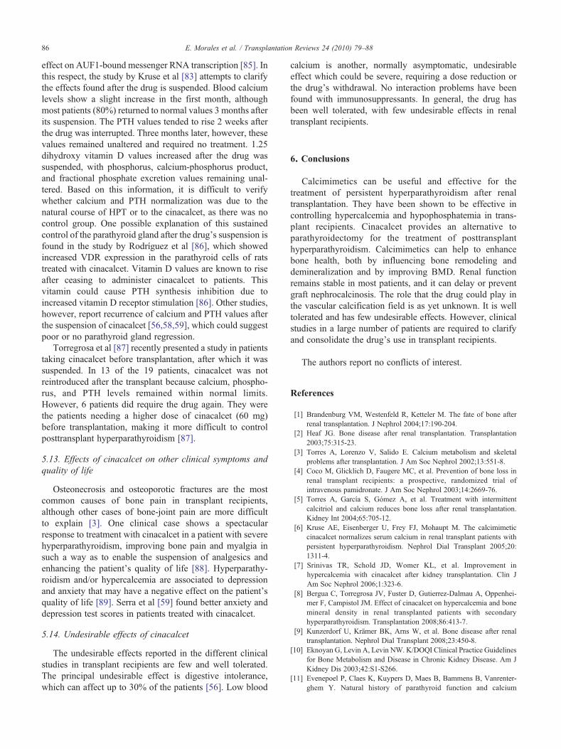
5.3. Mechanisms of action
Calcium receptor (RCa) cloning has enabled the subse-quent development of calcimimetic agents, a completely newgroup of drugs that act as allosteric regulators of the receptor(type II calcimimetics). Type II calcimimetics thereforeincrease sensitivity to extracellular Ca by inducing changesin RCa conformation, reducing PTH secretion and thusproviding a new treatment possibility for secondaryhyperparathyroidism (see Fig. 1) [46,47]. Indeed, nodularhyperplasia of the parathyroid glands is associated to thereduction of not only vitamin D (VDR) but also RCareceptors. The calcium receptor is a transmembrane receptorthat initiates a transduction signal by activating adenylatecyclase or phospholipase C after binding to calcium [48]. Ithas also been found that calcimimetics appear to delay thedevelopment of parathyroid hyperplasia, prevent the devel-opment of renal osteodystrophy, and increase bone mineraldensity, at least in experimental animals [49,50]. Indeed,RCa-related calcium appears to be more important thanVDRs in the control or modification of parathyroidhyperplasia. Some experimental data, however, show thatparathyroid hyperplasia precedes RCa infraregulation.Beneficial effects of calcimimetics on renal functionprogression, cardiovascular risk factors such as bloodpressure and low-density lipoprotein cholesterol and cardiacremodeling have also been found in experimental animals[51]. The first generation of type II calcimimetics, basicallyrepresented by NPS R-568, has been abandoned due to itslow bioavailability and a pharmacokinetic profile that variesfrom patient to patient [52]. Of the second generation,AMG073 or cinacalcet is the only drug in the familycurrently approved by the Food and Drug Administration.
5.4. Clinical experience in renal transplant recipients
Tertiary hyperparathyroidism is relatively commonamong renal transplant recipients. It is clinically character-ized by hypercalcemia and high PTH levels [53]. To date,

Table3
Studies
conductedwith
cinacalcet
inkidn
eytransplant
recipients
Reference
NTypeof
stud
yDuration
(mo)
Change
SCa(%
)Change
SP(%
)Change
PTH
(%)
Chang
eALP(%
)Chang
eCa
inurine(%
)ChangeP
inurine(%
)Chang
eSCr(%
)Chang
eGFR(%
)Chang
eProtu
(%)
Highestdo
seCNC(m
g)
Kruse
etal,2
005[6]
14Prospectiv
e3
↓(11)
=↓(50)
––
–↑(6)
––
30Serra
etal,2
005[55]
11Prospectiv
e2.5
↓(9)
↑(17)
↓(21)
–=
=↑(6)
↑(7)
–60
Srinivaset
al,20
06[7]
11Retrospectiv
e3-18
↓(14)
↑(14)
↓(42)
––
–↓(5)
––
30Szw
arcet
al,2
006[56]
9Prospectiv
e6
↓(11)
↑(10)
↓(13)
–=
––
=↑(100)
60Apo
stolou
etal,20
06[57]
7Retrospectiv
e3-18
↓(15)
=↓(57)
–=
==
––
30Lecaet
al,20
06[58]
10Prospectiv
e6
↓(13)
↑(18)
↓(40)
↓(44)
––
==
–60
Serra
etal,2
007[59]
12Prospectiv
e6
↓(12)
↑(6)
↓(30)
↑(20)
↓(20)
↓(15)
==
↓(10)
60Bergu
aet
al,2
007[60]
13Prospectiv
e6
↓(12)
↑(14)
↓(32)
↑(15)
==
=–
–60
El-Amm
etal,20
07[61]
18Retrospectiv
e6
↓(8)
↑(13)
↓(42)
↓(29)
––
↑(11)
↓(14)
–18
0Bergu
aet
al,2
008[8]
9Prospectiv
e12
↓(14)
↑(6)
↓(40)
=↓(14)
↓(7)
↓(21)
↑(6)
–45
Borchhardtet
al,20
08[62]
32Prospectiv
e1.5
↓(10)
↑(12)
=–
↑(37)
↑(19)
=–
–30
Góm
ezet
al,2
009[63]
48Retrospectiv
e12
↓(12)
↑(13)
↓(39)
––
–=
=–
180
Toroet
al,20
09[64]
27Prospectiv
e6
↓(8)
↑(7)
↓(19)
–↑(29)
↓(16)
↑(9)
––
45López
etal,2
009[65]
29Prospectiv
e12
↓(12)
↑(28)
↓(18)
=–
––
↑(8)
–12
0Morales
etal,20
09[66]
51Retrospectiv
e18
↓(11)
↑(15)
↓(28)
↓(25)
==
==
=90
Protu
indicatesproteinuria;
N,numberof
patients;SCa,serum
calcium;SP,
serum
phosphorus;ALP,alkalin
ephosphatase;SCr,serum
creatin
ine;
GFR,g
lomerular
filtrationrate;CNC,cinacalcet.
83E. Morales et al. / Transplantation Reviews 24 (2010) 79–88
parathyroidectomy is the treatment of choice in thesepatients, particularly with persistent hypercalcemia a yearafter transplantation (see Table 3).
5.5. Effects of cinacalcet on calcium-phosphorusmetabolism
Hyperparathyroidism can be found in 17% to 50% of renaltransplant recipients a year after transplantation. Hypercal-cemia is the most harmful effect of this hyperparathyroidism.It is responsible for graft loss and a mortality risk factor forthese patients [54]. In our review of different clinical trials onthe use of cinacalcet in transplant recipients [6-8,55-66], theinclusion criteria were heterogeneous with regard to baselinecalcium, ranging from 10.2 to 11 mg/dL, and PTH values,ranging from 65 to 150 pg/mL. We first observe an 8% to15% reduction in calcium values. These values normalized inthe first month of treatment, and similar results were found inpatients with higher calcium figures (Ca N 11 mg/dL) [66].
Phosphorus values increased in most studies by 6% to28%. Kruse et al [6] were the first group to find beneficialeffects of cinacalcet in a small group of renal transplantrecipients. One interesting find in this study, on which otherstudies were based, was the effect of the calcimimetic onphosphorus levels. Parathyroid hormone increases the urineexcretion of phosphate through the proximal tube's Na2+/Pco-carrier [67]. Patients with primary hyperparathyroidismtreated with cinacalcet show up to a 10% increase inphosphorus levels [68], whereas dialysis patients show an8% reduction (approximately) [44]. Green et al [25] showthat uremic phosphatonin clearance plays a fundamental rolein final phosphate values.
5.6. Effects of cinacalcet on PTH and vitamin D
The reduction in PTH values in the different studiesanalyzed was 20% to 57%, although the dose, the time used,and the association of vitamin D varied. In this respect, thestudy by Gómez et al [63] shows PTH reduction in patientswithout hypercalcemia. This enabled the association ofvitamin D in these patients and prevented undesirable effectssuch as hypocalcemia.
With regard to 1.25 dihydroxyvitamin D3 or 25 hydro-xyvitamin D, there are few studies including these variations.Szwarc et al [56] find a small increase in 25-OH vitamin D3
values (17.1 ± 6 to 25.4 ± 6.1 ng/mL; P b .02), but novariations in 1,25 OH2 vitamin D3. On the other hand,Bergua et al [8] report a reduction in 1.25 (OH)2 vitamin D(53 ± 18 to 32 ± 9 pg/mL; P b .01) during follow-up, with nochange in 25 OH vitamin D. It is therefore difficult toestablish a conclusion regarding vitamin D.
5.7. Effects of cinacalcet on alkaline phosphatase
With regard to the effect of cinacalcet on alkalinephosphatase, the results are somewhat contradictory. Becauseof the increase in bone alkaline phosphatase after transplan-tation, which would show increased osteoblast activity,

84 E. Morales et al. / Transplantation Reviews 24 (2010) 79–88
cinacalcet could inhibit bone formation and normalize thetrophic effects of PTH on bone [69]. Leca et al [58] foundreduced alkaline phosphatase values (not of the bonefraction), although they suggest that, if there are notransaminase changes, it could be bone alkaline phosphatase.Our study also confirms a reduction in alkaline phosphatasevalues [66]. Serra et al [59], however, find an increase inalkaline phosphatase and bone alkaline phosphatase at the endof the study. This study's findings are related to other similardiscoveries in patients with primary HPT, where cinacalcetcan also increase the levels of bone markers such as alkalinephosphatase and the deoxypyridinoline-creatinine ratioremains unaltered, compared with placebo [68]. One possibleexplanation would be that the cyclic fluctuations in PTHvalues after introducing cinacalcet could stimulate boneformation [68].
5.8. Effects of cinacalcet on calcium and phosphorusexcretion in urine
More than 90% of calcium is reabsorbed through theproximal tubule and the loop of Henle through paracellularmechanisms [59]. It is transported by a transepithelialgradient generated by a Na-K-2Cl cocarrier in the ascendingloop. This gradient depends on the sensitive calciumreceptor. Calcium elimination adjustment occurs in the distalconvoluted tubule and collector tubes. This process iscontrolled by PTH, calcitriol, calcitonin, and other factors[62]. Calcium flows through a TRPV5 calcium channel, itsactivity being influenced by the PTH. Calbindin-D28kcarries calcium through the cell and sodium/calciumbasolateral exchanger (NCX1), eliminating it from the cell.This entire mechanism is regulated by PTH [70]. Theadministration of cinacalcet reduces systemic PTH valuesand, therefore, the effect of the TRPV5 epithelial channeland calcium-carrying proteins. The ultimate effect ofcalcimimetics is reduced calcium reabsorption. Locally,calcimimetics reduce PTH-dependent tubular calcium reab-sorption through calcium-sensitive receptors. Cinacalcet can
Table 4Evolution of different parameters in our patients according to GFR
Pre-CIN(FG b 60), n = 29
Post-CIN(FG b 60), n = 29
Ca (mg/dL) 11.1 ± 0.37 9.9 ± 0.58P (mg/dL) 2.6 ± 0.6 3.2 ± 0.57ALP (U/L) 172.8 ± 60.7 139.4 ± 59.8Ca/24 h in urine (mg/24 h) 130.7 ± 75.1 143.8 ± 77.2P/24 h in urine (mg/24 h) 906.5 ± 395 909.8 ± 294.9TRP (%) 63.2 ± 16.2 70.1 ± 8.8TmP/GF (mg/dL) 1.7 ± 0.7 2.2 ± 0.5iPTH (pg/mL) 270.2 ± 247.1 209 ± 198.5SCr (mg/dL) 1.55 ± 0.46 1.50 ± 0.46GFR (mL/min) 47.1 ± 11.9 49.5 ± 14Proteinuria (g/24 h) 0.46 ± 0.58 0.34 ± 0.29
GFR indicates glomerular filtration rate; CIN, cinacalcet; TRP, tubular reabsorptglomerular filtration rate; iPTH, parathyroid hormone.
therefore increase calcium excretion through 2 mechanisms;PTH reduction leads to less tubular calcium reabsorption andtubular calcium receptor sensitivity increases [71]. Moststudies do not find significant changes in the final calciumbalance in urine relative to baseline (see Table 3) eventhough cinacalcet could reduce tubular calcium reabsorption,leading to hypercalciuria, nephrolithiasis, and osteopenia[72]. Szwarc et al [56] monitored fractional calciumexcretion, finding an increase 4 hours after the administra-tion of cinacalcet that was not observed at the end of thestudy. That the calcium excretion fraction does not increaseover time could be explained by a reduction in the tubularcalcium load when serum calcium levels diminish, counter-acting the calciuric effect of calcimimetics and reduced PTHvalues [56]. Parathyroid hormone reduces phosphorusreabsorption by internalization of the 2a phosphorus-sodiumtubular carrier and a reduction in the phosphorus-sodiumcarrier transcription gene [73]. The recently describedFGF23 reduces phosphorus reabsorption through this tubularcarrier, inhibiting renal 1-α hydroxylase [74]. The FGF23levels are elevated in patients with primary and secondaryHPT and contribute to the development of posttransplanthypophosphatemia. Serra et al [75] conducted an interestingstudy in which they observed the effects of cinacalcet onphosphatemia in transplant recipients. The administration ofcinacalcet reduces PTH values more than FGF23 levels.They found that phosphorus excretion decreased as PTHvalues fell, whereas FGF23 values remained elevated. Theseclinical findings suggest that PTH has a greater impact thanFGF23 on phosphorus reabsorption in renal transplantrecipients [75]. When we classified patients according toglomerular filtration in our study, the group with GF lessthan 60 presented increased phosphate reabsorption relativeto the group with a higher GF value, suggesting that theeffect on phosphorus is greater in patients with a lowerglomerular filtration (GF) (see Table 4) [66]. Studies arerequired, however, to identify the pathogenic mechanisms ofcalciuria and phosphaturia and the role played by cinacalcet.
P Pre-CIN(FG N 60), n = 22
Post-CIN(FG N 60), n = 22
P
b.001 11.17 ± 0.44 9.91 ± 0.48 b.001b.001 2.59 ± 0.45 2.81 ± 0.5 .054b.001 165.1 ± 54.7 108.6 ± 32.8 b.001.474 189.5 ± 145.3 192.4 ± 112.9 .930.925 1050.5 ± 509.4 1259 ± 669.7 .102.038 70.2 ± 12 66.5 ± 21 .297.001 1.86 ± 0.6 1.9 ± 0.8 .543
b.001 290.3 ± 142.6 181.4 ± 112.8 .003.147 1.07 ± 0.16 1.1 ± 0.23 .442.047 73.8 ± 8.49 72.6 ± 13.2 .637.401 0.34 ± 0.38 0.36 ± 0.55 .446
ion of phosphate; TmP/GF, tubular maximum for phosphate corrected for

85E. Morales et al. / Transplantation Reviews 24 (2010) 79–88
5.9. Effects of cinacalcet on bone mass
Persistent hyperparathyroidism has a negative effect onbone mineral density (BMD) [3]. Experimental studies withrats have shown increased BMD in cortical bone after theadministration of a calcimimetic agent [76]. Preliminarystudies report that cinacalcet can increase BMD [77] andreduce fracture risk in patients with secondary hyperpara-thyroidism [78]. We found a clinical case of a transplantrecipient whose BMD values showed spectacular improve-ment after the introduction of cinacalcet [79]. The possibleexplanations of this effect are greater control of HPT,increased osteoblastic activity, and reduced bone turnover. Ingeneral, studies of the correlation of biochemical boneformation or resorption markers with bone histomorphome-try in renal transplant recipients have been disappointing[80]. The study by El-Amm et al [61] finds a drop in alkalinephosphatase and urine N-telopeptide values, probably relatedto slower bone turnover. Cinacalcet may slow bone turnover.Bergua et al [8] find better BMD in most patients. Althoughthe mechanism behind this effect is unknown, it could berelated to better PTH control, cyclic PTH changes due to theadministration of cinacalcet, or the direct effect of cinacalceton bone. We recently found another study in whichcinacalcet reduces bone mass loss in the femoral neck butnot in the lumbar area, suggesting that it has a greater effecton cortical than on trabecular bone [63].
5.10. Effects of cinacalcet on renal function andimmunosuppression
Results are also contradictory in this area. There arestudies [6,55,61,64] in which the patients presented aslight decline in renal function, whereas others [7,8,65]show a slight improvement at the end of follow-up or[57-60,62,63,66] no difference in creatinine levels orglomerular filtration. One case in the literature reports anacute decline in renal function secondary to hypercalciuriaafter the administration of cinacalcet, which remitted after thedrug was suspended [81]. The renal graft biopsy showed thepresence of intratubular calcium deposits. The differentmechanisms suggested to explain this case, as well ascalcimimetic agents, would be tissue calcium reabsorption,increased calcitriol synthesis by the graft, increasing calciumresorption in the intestine, and severe HPT. Calcineurinicagents and corticoids also increase the calcium load.Calcineurin inhibitors can increase calcium excretion byregulating a calcium-binding protein (calbindin D28K) in thedistal tubule [82]. Administration of the calcimimetic wouldincrease calcium excretion by enhancing the sensitivity of thecalcium sensitive receptors of the ascending loop and couldinhibit tubular transport of calcium [80]. Kruse et al [83]found an interesting factor related to the renal function oftheir patients. It had deteriorated during follow-up (51.1 ± 4.2baseline to 47.9 ± 4.2 mL/min per 1.73 m2 at the end offollow-up). After the drug was suspended, kidney functionrecovered (52.4 ± 5.9 mL/min per 1.73 m2) after 3 months.
This was confirmed by a decrease in cystatin C values. Thereversibility of these kidney function disorders suggestshemodynamic changes associated to calcium and PTH valuesrather than structural renal graft changes. El-Amm et al [61]found that their patients presented slight deterioration of renalfunction in spite of unaltered immunosuppression medica-tion. In this study, 13 of 18 patients presented biopsies duringfollow-up, finding no signs of acute rejection, so immuno-logic involvement in renal function deterioration seemsunlikely. The PTH elevation could possibly be a nonimmu-nologic risk factor in chronic allograft nephropathy. We wereable to study the effect of cinacalcet according to renalfunction (FGR N or b 60 mL/min per 1.73 m2) and found asimilar effect on calcium-phosphorus metabolism para-meters, with no changes in renal function in either of the 2groups (see Table 4).
Proteinuria information is missing in most studies so it isdifficult to assess. Our study found no change in proteinuriavalues during follow-up [66].
Finally, there were no acute rejection episodes or changesin the immunosuppression treatment dosage during follow-up. Cinacalcet is known to inhibit cytochrome P450 2D6(CYP2D6) and is metabolized by CYP3A4, CYP2D6, andCYP1A2 [6], but no interactions have been found withimmunosuppression treatment. This provides a safety marginfor the use of cinacalcet together with immunosuppressants.
5.11. What dose should be used? How often a day?
This is another controversial aspect of the drug. In moststudies, the initial dose is 30 mg once a day. The highest dosewas 180 mg/d [61,63], according to the patient's require-ments, with no additional undesirable effects. In the nextsection, we will discuss how the dose could primarily dependon the severity of the patient's pretransplant hyperparathy-roidism. Apostolou et al [57] present an interestingconsideration concerning the number of doses. There arestudies analyzing the pharmacokinetics of cinacalcet inpatients with primary hyperparathyroidism, finding a rapidbut transient decrease in PTH [68]. This could lead us toconsider that transplant recipients with normal renal functionshould receive 2 doses per day to improve the outcome. Inthis respect, some studies use 2 doses per day when the totaldaily dose is greater than 30 mg [61,65]. It is difficult toestablish a conclusion with this information, and furtherstudies are required.
5.12. When should the calcimimetic be suspended?
In different clinical studies, cinacalcet has reduced bloodcalcium and PTH values in renal transplant recipients. Weknow little, however, about what happens to phosphorous-calcium metabolism parameters when the drug is withdrawnand its effect on the parathyroid glands. The mechanism ofaction of calcimimetics includes reduced PTH values andparathyroid hyperplasia [84]. However, another mechanismhas recently been described, in which these drugs have an
86 E. Morales et al. / Transplantation Reviews 24 (2010) 79–88
effect on AUF1-bound messenger RNA transcription [85]. Inthis respect, the study by Kruse et al [83] attempts to clarifythe effects found after the drug is suspended. Blood calciumlevels show a slight increase in the first month, althoughmost patients (80%) returned to normal values 3 months afterits suspension. The PTH values tended to rise 2 weeks afterthe drug was interrupted. Three months later, however, thesevalues remained unaltered and required no treatment. 1.25dihydroxy vitamin D values increased after the drug wassuspended, with phosphorus, calcium-phosphorus product,and fractional phosphate excretion values remaining unal-tered. Based on this information, it is difficult to verifywhether calcium and PTH normalization was due to thenatural course of HPT or to the cinacalcet, as there was nocontrol group. One possible explanation of this sustainedcontrol of the parathyroid gland after the drug's suspension isfound in the study by Rodríguez et al [86], which showedincreased VDR expression in the parathyroid cells of ratstreated with cinacalcet. Vitamin D values are known to riseafter ceasing to administer cinacalcet to patients. Thisvitamin could cause PTH synthesis inhibition due toincreased vitamin D receptor stimulation [86]. Other studies,however, report recurrence of calcium and PTH values afterthe suspension of cinacalcet [56,58,59], which could suggestpoor or no parathyroid gland regression.
Torregrosa et al [87] recently presented a study in patientstaking cinacalcet before transplantation, after which it wassuspended. In 13 of the 19 patients, cinacalcet was notreintroduced after the transplant because calcium, phospho-rus, and PTH levels remained within normal limits.However, 6 patients did require the drug again. They werethe patients needing a higher dose of cinacalcet (60 mg)before transplantation, making it more difficult to controlposttransplant hyperparathyroidism [87].
5.13. Effects of cinacalcet on other clinical symptoms andquality of life
Osteonecrosis and osteoporotic fractures are the mostcommon causes of bone pain in transplant recipients,although other cases of bone-joint pain are more difficultto explain [3]. One clinical case shows a spectacularresponse to treatment with cinacalcet in a patient with severehyperparathyroidism, improving bone pain and myalgia insuch a way as to enable the suspension of analgesics andenhancing the patient's quality of life [88]. Hyperparathy-roidism and/or hypercalcemia are associated to depressionand anxiety that may have a negative effect on the patient'squality of life [89]. Serra et al [59] found better anxiety anddepression test scores in patients treated with cinacalcet.
5.14. Undesirable effects of cinacalcet
The undesirable effects reported in the different clinicalstudies in transplant recipients are few and well tolerated.The principal undesirable effect is digestive intolerance,which can affect up to 30% of the patients [56]. Low blood
calcium is another, normally asymptomatic, undesirableeffect which could be severe, requiring a dose reduction orthe drug's withdrawal. No interaction problems have beenfound with immunosuppressants. In general, the drug hasbeen well tolerated, with few undesirable effects in renaltransplant recipients.
6. Conclusions
Calcimimetics can be useful and effective for thetreatment of persistent hyperparathyroidism after renaltransplantation. They have been shown to be effective incontrolling hypercalcemia and hypophosphatemia in trans-plant recipients. Cinacalcet provides an alternative toparathyroidectomy for the treatment of posttransplanthyperparathyroidism. Calcimimetics can help to enhancebone health, both by influencing bone remodeling anddemineralization and by improving BMD. Renal functionremains stable in most patients, and it can delay or preventgraft nephrocalcinosis. The role that the drug could play inthe vascular calcification field is as yet unknown. It is welltolerated and has few undesirable effects. However, clinicalstudies in a large number of patients are required to clarifyand consolidate the drug's use in transplant recipients.
The authors report no conflicts of interest.
References
[1] Brandenburg VM, Westenfeld R, Ketteler M. The fate of bone afterrenal transplantation. J Nephrol 2004;17:190-204.
[2] Heaf JG. Bone disease after renal transplantation. Transplantation2003;75:315-23.
[3] Torres A, Lorenzo V, Salido E. Calcium metabolism and skeletalproblems after transplantation. J Am Soc Nephrol 2002;13:551-8.
[4] Coco M, Glicklich D, Faugere MC, et al. Prevention of bone loss inrenal transplant recipients: a prospective, randomized trial ofintravenous pamidronate. J Am Soc Nephrol 2003;14:2669-76.
[5] Torres A, García S, Gómez A, et al. Treatment with intermittentcalcitriol and calcium reduces bone loss after renal transplantation.Kidney Int 2004;65:705-12.
[6] Kruse AE, Eisenberger U, Frey FJ, Mohaupt M. The calcimimeticcinacalcet normalizes serum calcium in renal transplant patients withpersistent hyperparathyroidism. Nephrol Dial Transplant 2005;20:1311-4.
[7] Srinivas TR, Schold JD, Womer KL, et al. Improvement inhypercalcemia with cinacalcet after kidney transplantation. Clin JAm Soc Nephrol 2006;1:323-6.
[8] Bergua C, Torregrosa JV, Fuster D, Gutierrez-Dalmau A, Oppenhei-mer F, Campistol JM. Effect of cinacalcet on hypercalcemia and bonemineral density in renal transplanted patients with secondaryhyperparathyroidism. Transplantation 2008;86:413-7.
[9] Kunzerdorf U, Krämer BK, Arns W, et al. Bone disease after renaltransplantation. Nephrol Dial Transplant 2008;23:450-8.
[10] Eknoyan G, Levin A, Levin NW. K/DOQI Clinical Practice Guidelinesfor Bone Metabolism and Disease in Chronic Kidney Disease. Am JKidney Dis 2003;42:S1-S266.
[11] Evenepoel P, Claes K, Kuypers D, Maes B, Bammens B, Vanrenter-ghem Y. Natural history of parathyroid function and calcium

87E. Morales et al. / Transplantation Reviews 24 (2010) 79–88
metabolism after kidney transplantation: a single-centre study. NephrolDial Transplant 2004;19:1281-7.
[12] Dumoulin G, Hory B, Nguyen NU, et al. No trend toward aspontaneous improvement of hyperparathyroidism and high boneturnover in normocalcemic long-term renal transplant recipients. Am JKidney Dis 1997;29:746-53.
[13] Heaf J, Tvedegaard E, Kanstrup IL, Fogh-Andersen N. Bone loss afterrenal transplantation: role of hyperparathyroidism, acidosis, cyclo-sporine and systemic disease. Clin Transplant 2000;14:457-63.
[14] Messa P, Sindici C, Cannella G, et al. Persistent secondaryhyperparathyroidism after renal transplantation. Kidney Int 1998;13:436-42.
[15] Torregrosa JV, Campistol JM, Fenollosa B, Montesinos M, Romar A,Martinez de Osaba MJ. Role of secondary hyperparathyroidism in thedevelopment of post-transplant acute tubular necrosis. Nephron 1996;73:67-72.
[16] Rodríguez M, Felsenfeld AJ, Llach F. Calcemic response toparathyroid hormone in renal failure: role of phosphorus and its effecton calcitriol. Kidney Int 1991;40:1055-62.
[17] Julian BA, Quarles LD, Niemann KM. Musculoskeletal complicationsafter renal transplantation: pathogenesis and treatment. Am J KidneyDis 1992;19:99-120.
[18] Kurella M, Butterly DW, Smith SR. Post transplant erythrocytosis inhypercalcemic renal transplant recipients. Am J Transpl 2003;3:873-7.
[19] Campese VM. Calcium, parathyroid hormone, and blood pressure. AmJ Hypertens 1989;2:34-44.
[20] Leapman SB, Vidne BA, Butt KM, Waterhouse K, Kountz SL.Nephrolithiasis and nephrocalcinosis after renal transplantation: a casereport an review of the literature. J Urol 1976;115:129-32.
[21] Gwinner W, Suppa S, Mengel M, et al. Early calcification of renalallografts detected by protocol biopsies: causes and clinical implica-tions. Am J Transplant 2005;5:1934-41.
[22] García-Cantón C, Palomar R, Moreno A, et al. Evolution of anemia ofchronic renal failure after the treatment of hyperparathyroidism.Nephron 1996;74:444-5.
[23] Shutz-Swirski R, Shkolnik T, Shasha SM. Parathyroid hormone andthe cellular immune system. Nephron 1995;70:21-4.
[24] Ambuhl PM, Meier D, Wolf B, Dydak U, Boesiger P, Binswanger U.Metabolic aspects of phosphate replacement therapy for hypopho-sphatemia after renal transplantation: impact on muscular phosphatecontent, mineral metabolism, and acid/base homeostasis. Am J KidneyDis 1999;34:875-83.
[25] Green J, Debby H, Lederer E, Levi M, Zajicek HK, Bick T. Evidencefor a PTH-independent humoral mechanism in post-transplanthypophosphatemia and phosphaturia. Kidney Int 2001;60:1182-96.
[26] Bhan I, Shah A, Holmes J, et al. Post-transplant hypophosphatemia:“tertiary hyper-phosphatoninism”? Kidney Int 2006;70:1486-94.
[27] Saito H, Kusano K, Kinosaki M, et al. Human fibroblast growth factor-23 mutants suppress Na+-dependent phosphate co-transport activityand 1alpha, 25-dihydroxyvitamin D3 production. J Biol Chem 2003;278:2206-11.
[28] Briner VA, Thiel G, Monier-Faugere MC, et al. Prevention ofcancellous bone loss but persistence of renal bone disease despitenormal 1,25 vitamin D levels two years after kidney transplantation.Transplantation 1995;59:1393-400.
[29] Lim WH, Coates PS, Russ GR, Coates PT. Hyperparathyroidism andvitamin D deficiency predispose to bone loss in renal transplantrecipients. Transplantation 2009;88:678-83.
[30] Epstein S. Post-transplantation bone disease: the role of immunosup-pressive agents and the skeleton. J Bone Miner Res 1996;11:1-7.
[31] Massari PU. Disorders of bone and mineral metabolism after renaltransplantation. Kidney Int 1997;52:1412-21.
[32] O'Shaughnessy EA, Dahl DC, Smith CL, Kasiske BL. Risk factors forfractures in kidney transplantation. Transplantation 2002;74:362-6.
[33] Tang G, Chan TM, Lui SL, Li FK, Lo WK, Lai KN. Risk factors foravascular bone necrosis after renal transplantation. Transplant Proc2000;32:1783-875.
[34] Goffin E, Vandeberg B, Devogelaer JP, et al. Post-renal transplantsyndrome of transient lower limb joint pain: description undertacrolimus-based immunosuppression. Clin Nephrol 2003;59:98-105.
[35] Block GA, Klassen PS, Lazarus JM, Ofsthun N, Lowrie EG, ChertowGM. Mineral metabolism, mortality, and morbidity in maintenancehemodialysis. J Am Soc Nephrol 2004;15:2208-18.
[36] EBPG Expert Group on Renal Transplantation: European Best PracticeGuidelines for renal transplantation. Section IV: Long term manage-ment of the transplant recipient. Nephrol Dial Transplant 2002;17:S43-8.
[37] Rojas E, Carlini RG, Clesca P, et al. The pathogenesis ofosteodystrophy after renal transplantation as detected by earlyalterations in bone remodeling. Kidney Int 2003;63:1915-23.
[38] Schmid T, Muller P, Spelsberg F. Parathyroidectomy after renaltransplantation: a retrospective analysis of long-term outcome. NephrolDial Transplant 1997;12:2393-6.
[39] Lewin E, Olgaard K. Parathyroidectomy vs calcimimetics for treatmentof persistent hyperparathryroidism after kidney transplantation.Nephrol Dial Transplant 2006;21:1766-9.
[40] Rostaing L, Moreau-Gaudry X, Baron E, Cisterne JM, Monroziès-Bernadet P, Durand D. Changes in blood pressure and renal functionfollowing subtotal parathyroidectomy in renal transplant patientspresenting with persistent hypercalcemic hyperparathyroidism. ClinNephrol 1997;47:248-55.
[41] Garcia A, Mazuecos A, Garcia T, Gonzalez P, Ceballos M, Rivero M.Effect of parathyroidectomy on renal graft function. Transplant Proc2005;37:1459-61.
[42] Lee PP, Schiffmann L, Offermann G, Beige J. Effects of parathyroid-ectomy on renal allograft survival. Kidney Blood Press Res 2004;27:191-6.
[43] Evenepoel P, Claes K, Kuypers D, Maes B, Vanrenterghem Y. Impactof parathyroidectomy on renal graft function, blood pressure and serumlipids in kidney transplant recipients: a single centre study. NephrolDial Transplant 2005;20:1714-20.
[44] Block GA, Martin KJ, de Francisco AL, et al. Cinacalcet for secondaryhyperparathyroidism in patients receiving hemodialysis. N Engl J Med2004;350:1516-25.
[45] Cunningham J, Danese M, Olson K, Klassen P, Chertow GM. Effectsof the calcimimetic cinacalcet HCl on cardiovascular disease, fracture,and health-related quality of life in secondary hyperparathyroidism.Kidney Int 2005;68:1793-800.
[46] Rodríguez M, Nemeth E, Martín D. The calcium-sensing receptor: akey factor in the pathogenesis of secondary hyperparathyroidism. Am JPhysiol Renal Physiol 2005;288:F253-64.
[47] Lindberg JS, Moe SM, Goodman WG, et al. The calcimimetic AMG073 reduces parathyroid hormone and calcium × phosphorus insecondary hyperparathyroidism. Kidney Int 2003;63:248-54.
[48] Coburn JW, Elangovan L, Goodman WG, Frazao JM. Calcium-sensing receptor and calcimimetic agents. Kidney Int 1999;73:S52-8.
[49] Ishii H, Wada M, Furuya Y, Nagano N, Nemeth EF, Fox J. Dailyintermittent decreases in serum levels of parathyroid hormone have ananabolic-like action on the bones of uremic rats with low-turnoverbone and osteomalacia. Bone 2000;26:175-82.
[50] Colloton M, Shatzen E, Miller G, et al. Cinacalcet HCl attenuatesparathyroid hyperplasia in a rat model of secondary hyperparathyroid-ism. Kidney Int 2005;67:467-76.
[51] Ogata H, Ritz E, Odoni G, Amann K, Orth SR. Beneficial effects ofcalcimimetics on progression of renal failure and cardiovascular riskfactors. J Am Soc Nephrol 2003;14:959-67.
[52] Ureña P, Frazão J. Calcimimetic agents: Review and perspectives.Kidney Int 2003;63:91-6.
[53] David DS, Sakai S, Brennan BL, et al. Hypercalcemia after renaltransplantation. Long-term follow-up data. N Engl J Med 1973;289:398-401.
[54] Egbuna OI, Taylor JG, Bushinsky DA, Zand MS. Elevated calciumphsophate product after renal transplantation is a risk factor for graftfailure. Clin Transplant 2007;21:558-66.

88 E. Morales et al. / Transplantation Reviews 24 (2010) 79–88
[55] Serra LA, Schwarz AA, Wick FH, Marti HP, Wüthrich RP. Successfultreatment of hypercalcemia with cinacalcet in renal transplantrecipients with persistent hyperparathyroidism. Nephrol Dial Trans-plant 2005;20:1315-9.
[56] Szwarc I, Argilés A, Garrigue V, Delmas S, Chong G, Deleuze S,Mourad G. Cinacalcet chloride is efficient and safe in renal transplantrecipients with posttransplant hyperparathyroidism. Transplantation2006;82:675-80.
[57] Apostolou T, Kollia K, Damianou L, et al. Hypercalcemia due toresistant hyperparathyroidism in renal transplant patients treated withcalcimmetic agent cinacalcet. Transplant Proc 2006;38:3514-6.
[58] Leca N, Laftavi M, Gundroo A, et al. Early and severe hyperparathy-roidism associated with hypercalcemia after renal transplant treatedwith cinacalcet. Am J Transplant 2006;6:2391-5.
[59] Serra A, Savoca R, Huber AR, et al. Effective control of persistenthyperparathyroidism with cinacalcet in renal allograft recipients.Nephrol Dial Transplant 2007;22:577-83.
[60] Bergua C, Torregrosa JV, Cofán F, Oppenheimer F. Cinacalcet for thetreatment of hypercalcemia in renal transplanted patients withsecondary hyperparathyroidism. Transplant Proc 2007;29:2254-5.
[61] El-Amm JM, Doshi MD, Singh A, et al. Preliminary experience withcinacalcet use in persistent secondary hyperparathyroidism afterkidney transplant. Transplantation 2007;28:546-9.
[62] Borchhardt KA, Heinzl H, Mayerwöger E, Hörl WH, Hass M, Sunder-Plassmann G. Cinacalcet increases calcium excretion in hyepercalce-mic hyperparathyroidism after kidney transplantation. Transplantation2008;86:919-24.
[63] Gómez G, Obrador A, Vilar A, et al. Treatment with cinacalcet ofsecondary hyperparathyroidism after renal transplantation. TransplantProc 2009;41:2139-43.
[64] Toro FJ, Bernal G, Navarro M, et al. Calcimimetics and bone mineraldensity in renal transplant patients with persistent secondaryhyperparathyroidism. Transplant Proc 2009;41:2144-7.
[65] López V, Toledo R, Sola E, et al. Treatment with cinacalcet in 29kidney transplant patients with persistent hyperparathyroidism.Transplant Proc 2009;41:2394-5.
[66] Morales E, Gutierrez E, Bengoa I, et al. Effectiveness of cinacalcet inthe treatment of hypercalcemia after renal transplantation. J Am SocNephrol 2009;20:883A (PUB253).
[67] Murer H, Hernando N, Forster L, Biber J. Molecular mechanisms inproximal tubular and small intestinal phosphate reabsortion. MolMembr Biol 2001;18:3-11.
[68] Peacok M, Bilezikian JP, Klassen PS, Guo MD, Turner SA, ShobackD. Cinacalcet hydrochloride maintains long normocalcemia in patientswith primary hyperparathyroidism. J Clin Endocrinol Metab 2005;90:135-41.
[69] Withold W, Friedrich W, Degenhardt S. Serum bone alkalinephosphatase is superior to plasma levels of bone matrix proteins forassessment of bone metabolism in patients receiving renal transplants.Clin Chim Acta 1997;261:105-15.
[70] van Abel M, Hoenderop JG, van der Kemp AW, Friedlaender MM, vanLeeuwen JP, Bindels RJ. Coordinated control of renal Ca2+ transportproteins by parathyroid hormone. Kidney Int 2005;68:1708-21.
[71] Esposito L, Rostaing L, Gennero I, Mehrenberger M, Durand D,Kamar N. Hypercalciuria induced by high dose of cinacalcet in a renal-transplant recipient. Clin Nephrol 2007;68:245-8.
[72] Misael da Silva AM, dos Reis LM, Pereira RC, et al. Bone involvementin idiopathic hypercalciuria. Clin Nephrol 2002;57:183-91.
[73] Ohkido I, Segawa H, Yanagida R, Nakamura M, Miyamoto K.Cloning, gene structure and dietary regulation of the type-IIc Na/picotransporter in the mouse kidney. Pflugers Arch 2003;446:106-15.
[74] Shimada T, Yamazaki Y, Muto T, et al. Cloning and characterizationof FGF23 as a causative factor of tumor-induced osteomalacia. ProcNatl Acad Sci U S A 2001;98:6500-5.
[75] Serra AL, Wuhrmann C, Wüthrich RP. Phosphatemic effect ofcinacalcet in kidney transplant recipients with persistent hyperpara-thyroidism. Am J Kidney Dis 2008;52:1151-7.
[76] Wada N, Nagano N, Furuya Y, Chin J, Nemeth EF, Fox J.Calcimimetic NPS R-568 prevents parathyroid hyperplasia in ratswith severe secondary hyperparathyroidism. Kidney Int 2000;57:50-8.
[77] Lien YH, Silva AL, Whittman D. Effects of cinacalcet on bone mineraldensity in patients with secondary hyperparathyroidism. Nephrol DialTransplant 2005;20:1232-7.
[78] Cunningham J, Danese M, Olson K, Klassen P, Chertow GM. Effectsof the calcimimetic cinacalcet HCl on cardiovascular disease, fracture,and health related quality of life in secondary hyperparathyroidism.Kidney Int 2005;68:1793-800.
[79] Decleire PY, Devogelaer JP, Goffin E. Cinacalcet improves bonemineral density in a renal transplant recipient with persistenthyperparathyroidism. Clin Nephrol 2008;69:231-2.
[80] Sprague SM, Josephson MA. Bone disease after kidney transplanta-tion. Semin Nephrol 2004;24:82-90.
[81] Peng LW, Logan JL, James SH, Scout KM, Lien YHH. Cinacalcetassociated graft dysfunction and nephrocalcinosis in a kidneytransplant recipient. Am J Med 2007;120:e7-9.
[82] Lee CT, Huynh VM, Lai LW, lien YH. Cyclosporine A inducedhypercalciuria in calbidin-D28K knockout and wild type-mice. KidneyInt 2002;62:2055-61.
[83] Kruse AE, Eisenberger U, Frey FJ, Mohaupt MG. Effect of cinacalcetcessation in renal transplant recipients with persistent hyperparathy-roidism. Nephrol Dial Transplant 2007;22:2362-5.
[84] Wada M, Ishii H, Furuya Y, Fox J, Nemeth EF, Nagano N. NPS R-568halts or reverses osteitis fibrosa in uremia rats. Kidney Int 1998;53:448-53.
[85] Levi R, Ben-Dov IZ, Lavi-Moshayoff V, et al. Increased parathyroidhormone gene expression in secondary hyperparathyroidism ofexperimental uremia is reserved by calcimimetics: correlation withpost-translational modification of the transacting factor AUF1. J AmSoc Nephrol 2006;17:107-12.
[86] Rodriguez ME, Almaden Y, Canadillas S, et al. The calcimimetics R568 increases vitamin D receptor expresión in rat parathyroid glands.Am J Physiol Renal 2007;292:390-5.
[87] Torregrosa JV, Bergua C, Martínez de Osaba MJ, Oppenheimer F,Campistol JM. Evolution of secondary hyperparathyroidism afterkidney transplantation in patients receiving cinacalcet on diálisis.Transplant Proc 2009;41:2396-8.
[88] Leonard N, Brown JH. Persistent and symptomatic post-transplanthyperparathyroidism: a dramatic response to cinacalcet. Nephrol DialTransplant 2006;21:1736.
[89] White RE, Pickering A, Spathis GS. Mood disorder and chronichypercalcemia. J Psychosom Res 1996;41:343-7.