treatment of melasma and the use of intense pulsed light ... · melasma is an acquired disorder of...
TRANSCRIPT
JDD
November 2012 1316 VOLUME 11 • ISSUE 11
Copyright © 2012 ORIGINAL ARTICLE
SPECIAL TOPIC
Journal of Drugs in Dermatology
Treatment of Melasma and the Use of IntensePulsed Light: A Review
Lisa Zaleski DO LCDR MC USN,a* Sabrina Fabi MD,b and Mitchel P.Goldman MDb
aDepartment of Dermatology, U.S. Naval Medical Center Okinawa, Okinawa,Japan bDermatology/Cosmetic Laser Associates, La Jolla, CA*The views expressed in this article are those of the authors and do notreflect the official policy of the Department of the Navy, Department ofDefense, or U.S. Government.
ABSTRACT
Melasma is a complex multifactorial disorder whose pathogenesis is not well understood. In addition toincreased pigmentation, increased vascularity associated with pigmentation is present. A variety of topicaltreatments targeting pigmentation are available with temporary improvement of mainly the epidermalcomponents of melasma. Intense pulsed light (IPL) is a broadband light source that can target a widerange of cutaneous structures, including deeper pigmentation and vasculature. We describe 5 cases ofpersistent facial melasma treated with the IPL and a hydroquinone-based skin care system (Obagi Nu-Derm; Obagi Medical Products, Long Beach, CA), showing improvement of facial melasma pigmentationand vascularity.
J Drugs Dermatol. 2012;11(11):1316-1230.
INTRODUCTION
Melasma is a complex multifactorial disorder whose pathogenesis is not well understood. In addition toincreased superficial and/or deep pigmentation, increased vascularity is often present. Vascular endothelialgrowth factor (VEGF) is an angiogenic factor demonstrated within melasma patches that is a likely cause ofthe increased vasculature. Interactions between melanocytes and the cutaneous vasculature may influencethe development of pigmentation. Topical treatments targeting pigmentation are available with temporaryimprovement of mainly the epidermal component of melasma.1 Intense pulsed light (IPL) is a broadband lightsource that can target a wide range of cutaneous structures, including deeper pigmentation and theincreased vasculature. With a lower side effect profile compared with other devices used to treat melasma,IPL is a good potential treatment option for dermal and mixed forms of melasma. 2 We describe 5 cases ofpersistent facial melasma treated with IPL and a hydroquinone (HQ)-based skin care system (Obagi Nu-Derm; Obagi Medical Products, Long Beach, CA), showing improvement of facial melasma pigmentation andvascularity.
To order reprints or e-prints of JDD articles please contact [email protected]
Journal of Drugs in Dermatology. All Rights Reserved. No reproduction or use of any portion of the contents ofthese materials may be made without the express written consent of JDD. If you feel you have obtained this
copy illegally, please contact JDD immediately. Licensed to [email protected].
CASE REPORTS
Case 1
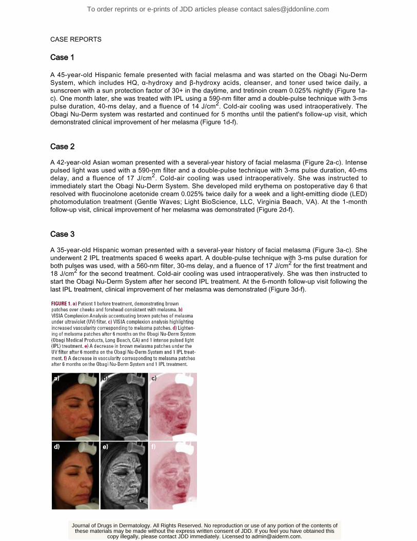
A 45-year-old Hispanic female presented with facial melasma and was started on the Obagi Nu-DermSystem, which includes HQ, α-hydroxy and β-hydroxy acids, cleanser, and toner used twice daily, asunscreen with a sun protection factor of 30+ in the daytime, and tretinoin cream 0.025% nightly (Figure 1a-c). One month later, she was treated with IPL using a 590-nm filter amd a double-pulse technique with 3-mspulse duration, 40-ms delay, and a fluence of 14 J/cm2. Cold-air cooling was used intraoperatively. TheObagi Nu-Derm system was restarted and continued for 5 months until the patient's follow-up visit, whichdemonstrated clinical improvement of her melasma (Figure 1d-f).
Case 2
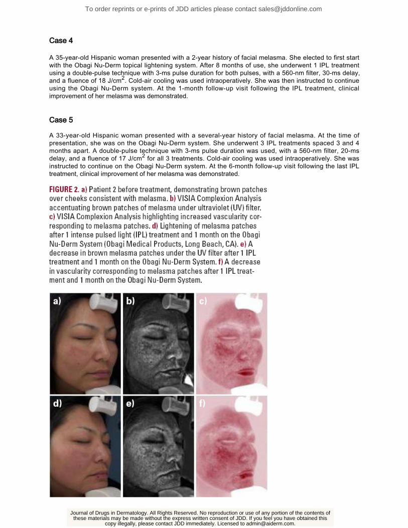
A 42-year-old Asian woman presented with a several-year history of facial melasma (Figure 2a-c). Intensepulsed light was used with a 590-nm filter and a double-pulse technique with 3-ms pulse duration, 40-msdelay, and a fluence of 17 J/cm2. Cold-air cooling was used intraoperatively. She was instructed toimmediately start the Obagi Nu-Derm System. She developed mild erythema on postoperative day 6 thatresolved with fluocinolone acetonide cream 0.025% twice daily for a week and a light-emitting diode (LED)photomodulation treatment (Gentle Waves; Light BioScience, LLC, Virginia Beach, VA). At the 1-monthfollow-up visit, clinical improvement of her melasma was demonstrated (Figure 2d-f).
Case 3
A 35-year-old Hispanic woman presented with a several-year history of facial melasma (Figure 3a-c). Sheunderwent 2 IPL treatments spaced 6 weeks apart. A double-pulse technique with 3-ms pulse duration forboth pulses was used, with a 560-nm filter, 30-ms delay, and a fluence of 17 J/cm2 for the first treatment and18 J/cm2 for the second treatment. Cold-air cooling was used intraoperatively. She was then instructed tostart the Obagi Nu-Derm System after her second IPL treatment. At the 6-month follow-up visit following thelast IPL treatment, clinical improvement of her melasma was demonstrated (Figure 3d-f).
To order reprints or e-prints of JDD articles please contact [email protected]
Journal of Drugs in Dermatology. All Rights Reserved. No reproduction or use of any portion of the contents ofthese materials may be made without the express written consent of JDD. If you feel you have obtained this
copy illegally, please contact JDD immediately. Licensed to [email protected].
Case 4
A 35-year-old Hispanic woman presented with a 2-year history of facial melasma. She elected to first startwith the Obagi Nu-Derm topical lightening system. After 8 months of use, she underwent 1 IPL treatmentusing a double-pulse technique with 3-ms pulse duration for both pulses, with a 560-nm filter, 30-ms delay,and a fluence of 18 J/cm2. Cold-air cooling was used intraoperatively. She was then instructed to continueusing the Obagi Nu-Derm system. At the 1-month follow-up visit following the IPL treatment, clinicalimprovement of her melasma was demonstrated.
Case 5
A 33-year-old Hispanic woman presented with a several-year history of facial melasma. At the time ofpresentation, she was on the Obagi Nu-Derm system. She underwent 3 IPL treatments spaced 3 and 4months apart. A double-pulse technique with 3-ms pulse duration was used, with a 560-nm filter, 20-msdelay, and a fluence of 17 J/cm2 for all 3 treatments. Cold-air cooling was used intraoperatively. She wasinstructed to continue on the Obagi Nu-Derm system. At the 6-month follow-up visit following the last IPLtreatment, clinical improvement of her melasma was demonstrated.
To order reprints or e-prints of JDD articles please contact [email protected]
Journal of Drugs in Dermatology. All Rights Reserved. No reproduction or use of any portion of the contents ofthese materials may be made without the express written consent of JDD. If you feel you have obtained this
copy illegally, please contact JDD immediately. Licensed to [email protected].

DISCUSSION
Melasma is an acquired disorder of hyperpigmented macules or patches on sun-exposed areas of the body.Risk factors include a genetic predisposition, sun exposure, stress, medications, and pregnancy. Three typesof melasma exist, with increased amounts of melanin, melanocytes, and melanosomes within the epidermis,dermis, or a mix of the two.3 A bimodal age response has been reported, with improved treatment responsein patients younger than 35 years and older than 45 years. This is thought to be due to hormonal effects andincreased dermal melasma in patients aged 35 to 45 years.4 Traditional therapies are more effective forepidermal melasma and include sunscreens, depigmenting agents, mild topical corticosteroids, tretinoin, andchemical peels.3 Intense pulsed light and various lasers have also been used, including the quality (Q)-switched ruby (694 nm), Q-switched neodymium- doped yttrium aluminum garnet (Nd:YAG; 1,064 nm), diode(840 nm), pulsed dye laser (595 nm), nonablative fractionated 1,550-nm erbium (Er)-doped laser, 2,940-nmEr:YAG with dermabrasion, and combined ultrapulsed CO2 laser with Q-switched alexandrite (755 nm).4-7
To order reprints or e-prints of JDD articles please contact [email protected]
Journal of Drugs in Dermatology. All Rights Reserved. No reproduction or use of any portion of the contents ofthese materials may be made without the express written consent of JDD. If you feel you have obtained this
copy illegally, please contact JDD immediately. Licensed to [email protected].

The pathogenesis of melasma is important when considering laser treatment. Kim et al1 reported increasedvascularity as a major finding in melasma with increased amounts of VEGF and blood vessels withinmelasma lesional skin. It has been postulated that ultraviolet (UV) radiation-induced dermal inflammationactivates fibroblasts and stem cell factors in melasma dermal skin, causing melanogenesis. The increasedvascularity could be why melasma occurs in select regions and not uniformly across the face, despite equalUV damage.
Vascular endothelial growth factor has also been shown to stimulate the release of arachidonic acid, and themetabolites of this pathway may affect melanogenesis. Steroids in triple-agent creams used to treat melasmacan also induce telangiectasias, possibly exacerbating this component of melasma.1 Bak et al8 reportedincreased nerve growth factor and neural endopeptidase in melasma lesional skin, also suggesting itsassociation in the pathogenesis of melasma.
Multiple studies have shown varied effectiveness of lasers in the treatment of melasma. The nonablative1,550-nm fractional laser has been used to treat melasma with greater patient satisfaction 3 weeks aftertreatment compared with topical triple-agent therapy of HQ 5%, tretinoin 0.05%, and triamcinolone acetonide0.1% cream. It was thought the laser brought greater satisfaction early on because of a faster initial clearanceand a possible increased effectiveness in treating dermal melasma. However, 6 months posttreatment, thepigmentation returned in both treatment groups with equal patient satisfaction rates. Side effects of the 1,550-nm laser treatment included erythema, burning sensation, edema, and pain. Kroon et al6 noted nopostinflammatory hyperpigmentation (PIH) with this treatment modality using conservative settings.
Wind et al9 described the use of the nonablative 1,550-nm fractional laser in the treatment of melasma, and 9patients (31%) developed PIH after 2 or more laser sessions. The increased PIH seen in the study is likelysecondary to more aggressive treatment settings. Skin findings and side effects associated with topical triple-agent therapy included erythema, scale, and burning. Triple-agent topical therapy is still the treatment ofchoice because of similar efficacies 6 months posttreatment.10-11
The Q-switched Nd:YAG laser has also been reported to temporarily improve melasma with commoncomplications, including hypopigmentation, melasma recurrence, and rebound hyperpigmentation. Transienterythema, transient burning, and slight edema occurred for 1 hour postprocedure. Wattanakrai et al12 founddecreased epidermal and dermal pigmentation for up to 1 year after 10 weekly treatments with the Q-switched Nd:YAG laser at subthreshold photothermolytic fluencies (<5 J/cm2). However, reboundhyperpigmentation was common, and the risk of mottled hypopigmented macules increased with greaternumber of laser sessions.12,13 Narrowband UVB has successfully been used to treat depigmentation withgood clinical results.13 The short-pulsed deep Er:YAG laser temporarily but effectively reduces epidermaltype melasma, with a recurrence upon discontinuation of treatment.14
A review of the literature suggests that laser and light source treatments can result in reboundhyperpigmentation, relapse, and darkening of melasma. It has been postulated that the laser unmasks aprevious subclinical melasma. This is thought to be secondary to stimulation of hyperactive melanocytes,which can increase melanin production and therefore pigmentation.15
Intense pulsed light is a noncoherent filtered flashlamp light source, emitting light between 515 and 1,200 nm.Filters allow for selective photothermolysis of chromophores, including melanin and hemoglobin.5,16 Since itsintroduction in 1992, there are now more than 20 different IPL devices available worldwide.17 Each IPLdevice has a unique set of wavelengths, fluences, pulse durations, epidermal temperature effects, and otherpulse parameters in addition to duration, such as unifor-
mity of the pulse delivery. Intense pulsed light has been used to treat melasma, telangiectasias, spider nevi,rosacea, lentigines, postburn hyperpigmentation, erythrosis, poikiloderma of Civatte, photoinduced skin agingand to reduce hair.2,18The IPL activates fibroblasts, resulting in the synthesis of new collagen with wrinklereduction, increased skin elasticity, contraction of larger pores, reduction of brown spots, and a decrease intelangiectasias.19 Side effects of IPL include a transient erythema and slight edema that resolve within 12hours, PIH, and desquamating microcrusts for 7 to 10 days.3 The major problem in evaluating the peer-reviewed medical literature is that each IPL device has a unique set of parameters that makes it different fromthe others. Thus, when reviewing the IPL literature, the improvement and complication profile may not be100% reproducible from one IPL device to another. Our experience, and most of the published literature, iswith the Lumenis IPL systems (Santa Clara, CA), but even within one company, the IPL systems differ based
To order reprints or e-prints of JDD articles please contact [email protected]
Journal of Drugs in Dermatology. All Rights Reserved. No reproduction or use of any portion of the contents ofthese materials may be made without the express written consent of JDD. If you feel you have obtained this
copy illegally, please contact JDD immediately. Licensed to [email protected].

on the model.
"We feel that IPL is the light source of choice for the treatment of dermal and mixed melasma because of itslower side effect profile and ability to target both melanin and hemoglobin as chromophores."
Intense pulsed light has multiple advantages over other lasers for the treatment of melasma. The longerwavelengths used with IPL allow deeper penetration for treatment of dermal melasma. The larger spot allowsfor more extensive areas of the face to be treated in a shorter time period, minimizing patient discomfort.There is also a decrease in nonhomogeneous resolution with a decrease in polka-dot treatment results withthe IPL that can be seen with smaller laser round-spot sizes. In addition, there are fewer local or systemiceffects because of the pulse delays in more advanced IPL systems, so the skin can be cooled betweenpulses.2 This decrease in photothermal injury leads to less PIH in comparison with Q-switched lasers.5
The IPL has been used in combination with the Q-switched ruby laser, with 19/25 (76%) of patients reportinggood to excellent responses.20 Side effects mainly included PIH in 12% and linear hypopigmentation in 4%.The IPL was advantageous because of the minimal preoperative preparation, easy application, limitedposttreatment care, and a lack of downtime. However, multiple treatments are often needed to obtain thedesired results, and deeper-pigmented patches tend to be less responsive. The addition of the Q-switchedruby laser allows for deeper penetration of dermal melasma but a higher risk of PIH. Repeated IPLtreatments could decrease PIH caused by the Q-switched laser. The pulse duration of IPL is in milliseconds,resulting in a greater thermal diffusion and a more generalized destruction of pigment. Quality-switchedlasers are in a nanosecond range, which selectively targets melanosomes with decreased thermaldiffusion.20
Poikiloderma of Civatte is similar to melasma, as both conditions involve hemoglobin and melanin aschromophores targeted with treatment. Goldman and Weiss reported a 50% to 75% clearance oftelangiectasias and hyperpigmentation in poikiloderma with an average of 2.8 IPL treatments. There was a5% incidence of mild pigmentary side effects. Improvement in skin texture was an added bonus with the IPLtreatments.18,21
The successful use of IPL for skin rejuvenation has been well documented in the literature.17,22-24 Noothetiet al found a 40% improvement in photoaging after a single IPL treatment.25 Feng et al found an 84.6%pigmentation improvement and an 81.25% telangiectasia improvement after 3 IPL treatments.26 However,JØrgensen et al27 found the long-pulsed dye laser to be advantageous over the IPL in photodamaged skinbecause of superior vessel clearance and less pain associated with the procedure. Both the laser and IPLhad similar efficacy with pigmentation clearance.27
Repigmentation with melasma eventually recurs, likely secondary to persistent triggering factors.5 We feelthat IPL is the light source of choice for the treatment of dermal and mixed melasma because of its lower sideeffect profile and ability to target both melanin and hemoglobin as chromophores.17,21 Targeting the vascularcomponent of melasma in addition to the pigmentation may be the key to improved results.
Our melasma patients demonstrate a strong correlation of vascularity with their melasma on the VISIAComplexion Analysis (VISIA, Fairfield, NJ). Currently, triple-agent therapy is the firstline treatment formelasma. The VISIA Complexion Analysis may be an easy method to determine which patients are the bestcandidates for concurrent IPL therapy.
DISCLOSURES
Drs. Zaleski and Fabi have no conflict of interest to declare. Mitchel P. Goldman MD is a stockholder andconsultant to Lumenis Ltd. and a consultant to Obagi Medical Products, Inc.
REFERENCES
Kim EH, Kim YC, Lee ES, Kang HY. The vascular characteristics of melasma. J Dermatol Sci.2007;46(2):111-116.
1.
Campolmi P, Bonan P, Cannarozzo G, et al. Intense pulsed light in the treatment of non-aesthetic facial2.
To order reprints or e-prints of JDD articles please contact [email protected]
Journal of Drugs in Dermatology. All Rights Reserved. No reproduction or use of any portion of the contents ofthese materials may be made without the express written consent of JDD. If you feel you have obtained this
copy illegally, please contact JDD immediately. Licensed to [email protected].

and neck vascular lesions: report of 85 cases. J Eur Acad Dermatol Venereol. 2011;25(1):68-73.Li YH, Chen JZ, Wei HC, et al. Efficacy and safety of intense pulsed light in treatment of melasma inChinese patients. Dermatol Surg. 2008;34(5):693-700.
3.
Shin JW, Lee DH, Choi SY, et al. Objective and non-invasive evaluation of photorejuvenation effectwith intense pulsed light treatment in Asian skin. J Eur Acad Dermatol Venereol. 2011;25(5):516-522.
4.
Wang CC, Hui CY, Sue YM, Wong WR, Hong HS. Intense pulsed light for the treatment of refractorymelasma in Asian persons. Dermatol Surg. 2004;30(9):1196-1200.
5.
Kroon MW, Wind BS, Beek JF, et al. Nonablative 1550-nm fractional laser therapy versus triple topicaltherapy for the treatment of melasma: a randomized controlled pilot study. J Am Acad Dermatol.2011;64(3):516-523.
6.
Rusciani A, Motta A, Fino P, Menichini G. Treatment of poikiloderma of Civatte using intense pulsedlight source: 7 years of experience. Dermatol Surg. 2008;34(3):314-319.
7.
Bak H, Lee HJ, Chang SE, Choi JH, Kim MN, Kim BJ. Increased expression of nerve growth factorreceptor and neural endopeptidase in the lesional skin of melasma. Dermatol Surg. 2009;35(8):1244-1250.
8.
Wind BS, Kroon MW, Meesters AA, et al. Non-ablative 1,550 nm fractional laser therapy versus tripletopical therapy for the treatment of melasma: a randomized controlled split-face study. Lasers SurgMed. 2010;42(7):607-612.
9.
Katz TM, Glaich AS, Goldberg LH, Firoz BF, Dai T, Friedman PM. Treatment of melasma usingfractional photothermolysis: a reports of eight cases with long-term follow-up. Dermatol Surg.2010;36(8):1273-1280.
10.
Lee HS, Won CH, Lee DH, et al. Treatment of melasma in Asian skin using a fractional 1,550-nmlaser: an open clinical study. Dermatol Surg. 2009;35(10):1499-1504.
11.
Wattanakrai P, Mornchan R, Eimpunth S. Low-fluence Q-switched neodymium-doped yttriumaluminum garnet (1,064 nm) laser for the treatment of facial melasma in Asians. Dermatol Surg.2010;36(1):76-87.
12.
Chan NP, Ho SG, Shek SY, Yeung CK, Chan HH. A case series of facial depigmentation associatedwith low fluence Q-switched 1,064 nm Nd:YAG laser for skin rejuvenation and melasma. Lasers SurgMed. 2010;42(8):712-719.
13.
Waniphakdeedecha R, Manuskiatti W, Siriphukpong S, Chen TM. Treatment of melasma usingvariable square pulse Er:YAG laser resurfacing. Dermatol Surg. 2009;35(3):475-481.
14.
Kang WH, Yoon KH, Lee ES, et al. Melasma: histopathological characteristics in 56 Korean patients.Br J Dermatol. 2002;146(2):228-237.
15.
Galeckas KJ, Collins M, Ross EV, Uebelhoer NS. Split-face treatment of facial dyschromia: pulseddye laser with a compression handpiece versus intense pulsed light. Dermatol Surg. 2008;34(5):672-680.
16.
Goldman MP, Weiss RA, Weiss MA. Intense pulsed light as a nonablative approach to photoaging.Dermatol Surg. 2005;31(9 Pt 2):1179-1187.
17.
Goldman MP, Weiss RA. Treatment of poikiloderma of Civatte on the neck with an intense pulsed lightsource. Plast Reconstr Surg. 2001;107(6):1376-1381.
18.
Li YH, Wu Y, Chen JZ, et al. Application of a new intense pulsed light device in the treatment ofphotoaging skin in Asian patients. Dermatol Surg. 2008;34(11):1459-1464.
19.
Park JM, Tsao H, Tsao S. Combined use of intense pulsed light and Qswitched ruby laser for complexdyspigmentation among Asian patients. Lasers Surg Med. 2008;40(2):128-133.
20.
To order reprints or e-prints of JDD articles please contact [email protected]
Journal of Drugs in Dermatology. All Rights Reserved. No reproduction or use of any portion of the contents ofthese materials may be made without the express written consent of JDD. If you feel you have obtained this
copy illegally, please contact JDD immediately. Licensed to [email protected].

Weiss RA, Goldman MP, Weiss MA. Treatment of poikiloderma of Civatte with an intense pulsed lightsource. Dermatol Surg. 2000;26(9):823-827.
21.
Li YH, Wu Y, Chen JZ, et al. A split-face study of intense pulsed light on photoaging skin in Chinesepopulation. Lasers Surg Med. 2010;42(2):185-191.
22.
Hedelund L, Due E, Bjerring P, Wulf HC, Haedersdal M. Skin rejuvenation using intense pulsed light:a randomized controlled split-face trial with blinded response evaluation. Arch Dermatol.2006;142(8):985-990.
23.
Negishi K, Wakamatsu S, Kushikata N, Tezuka Y, Kotani Y, Shiba K. Full-face photorejuvenation ofphotodamaged skin by intense pulsed light with integrated contact cooling: initial experiences in Asianpatients. Lasers Surg Med. 2002;30(4):298-305.
24.
Nootheti PK, Pettit KA, Yosowitz G, Goldman MP. Clinical improvement of photodamaged skin after asingle intense pulsed light treatment. Am J Cosmet Surg. 2007;24(1):15-20.
25.
Feng Y, Zhao J, Gold MH. Skin rejuvenation in Asian skin: the analysis of clinical effects and basicmechanisms of intense pulsed light. J Drugs Dermatol. 2008;7(3):273-279.
26.
JØrgensen GF, Hedelund L, Haedersdal M. Long-pulsed dye laser versus pulsed light forphotodamaged skin: a randomized split-face trial with blinded response evaluation. Lasers Surg Med.2008;40(5):293-299.
27.
Address for Correspondence
Lisa Zaleski DO LCDR MC USN
E-mail: [email protected]
To order reprints or e-prints of JDD articles please contact [email protected]
Journal of Drugs in Dermatology. All Rights Reserved. No reproduction or use of any portion of the contents ofthese materials may be made without the express written consent of JDD. If you feel you have obtained this
copy illegally, please contact JDD immediately. Licensed to [email protected].