treatment concepts - geistlich pharma · suggested treatment concept for periodontally ... 2-wall...
TRANSCRIPT

Treatment Concepts for Periodontal Regenerative Surgery

Intr
oduc
tion
3C
ontent2 Content Why periodontal regeneration?
Why periodontal regeneration? 3
Regenerative therapy: getting to the root of the problem 4
Suggested treatment concept for periodontally compromised teeth 5
Defect morphology influences outcome of regenerative therapy 6
Scientific and clinical evidence for the surgical preservative phase 7
Case 1: Dr. Frank Bröseler | Intrabony 2-wall defect: interproximal crater 8
Case 2: Dr. Diego Capri | 3-wall defect: rapid progression of lesion 9
Case 3: Prof. Dr. Michael Christgau | Extended 2-wall defect 10
Case 4: Dr. Pierpaolo Cortellini | Periodontal regenerative surgery 11
Case 5: Dr. Daniel Etienne | Treatment of infrabony 1-wall defect 12
Case 6: Prof. Dr. Markus Hürzeler | Combination defect 13
Case 7: Dr. Syed Mahnaz | Regenerative surgery 11 – perio-endo 14
Case 8: Prof. Dr. Giulio Rasperini | 2-wall defect in the non-aesthetic region 15
Case 9: Prof. Dr. Anton Sculean | Deep intrabony 2-wall defect 16
Case 10: Dr. Beat Wallkamm | 2-wall defect in the aesthetic zone 17
Case 11: Prof. Dr. Giovanni Zucchelli | 2-wall wide intrabony defect 18
References 19
Product Range for periodontal treatment 20
Helping patients affected by periodontitis to create and
maintain good oral health, function, and aesthetics is the
goal of every dentist. To accomplish this, various therapeu-
tic approaches have been developed in response to the
grades of severity of periodontitis. The role of biomaterials
in treating periodontal disease has gained in significance
and is now an integral part of many protocols. Carefully
selected biomaterials used with proven treatment proto-
cols may not only stop progression of periodontal disease,
but effectively regenerate both hard and soft tissue.1,2
TooTh preservaTion or implanT?
Teeth will last for life, unless they are affected by oral dis-
eases or service interventions. Many retained teeth there-
fore may be an indicator of positive oral health behaviour
throughout the life course. Tooth longevity is largely de-
pendent on the health status of the periodontium, the pulp
or periapical region and the extent of reconstructions.3
Multiple risks lead to a critical appraisal of the value of a
tooth. Choosing between periodontal regeneration to sup-
port tooth preservation and tooth extraction has been
called one of the most complex and debatable decisions a
dentist is confronted with in daily clinical practice.4
Assigning a questionable prognosis – where the tooth re-
quires advanced treatment to maybe preserve it –
or a hopeless prognosis, where the tooth needs to be ex-
tracted as soon as possible, is often a delicate situation.
This decision significantly impacts both treatment planing
and patient lifestyle. Accordingly, it has been argued that
periodontally compromised teeth should be treated for as
long as possible, and only being extracted when periodon-
tal and endodontic treatment is no longer possible.4, 5
Regardless of whether the tooth is preserved or extracted,
biomaterials are often required to reach the individual
therapeutic goals. Some criteria to categorise the progno-
sis of periodontally affected teeth are summarised in
Table 1.6-8
Table 1. Prognosis of periodontally affected teeth: For classification at least one of the parameters (respectively two for hopeless teeth) has to be met.6-8
Good QuesTionable hopeless
> teeth with < 50% bone loss > teeth with 50-75% bone loss or > 6–8 mm PD or> class 2 furcation or> angular defect
> teeth with > 75% bone loss or> more than 8mm PD or> Class 3 furcation or> Class 3 mobility or> teeth with at least 2 characteristics
of questionable category
The present treatment concept serves to summarise pro-
ven Guided Bone Regeneration (GBR) and Guided Tissue
Regeneration (GTR) techniques for the successful treat-
ment of common periodontal defects.
It provides scientific evidence and presents step-by-step
clinical cases, demonstrating stable favorable outcomes.
This guide is intended for the clinician and highlights reli-
able treatment options with the highest quality biomateri-
als. It aims to present techniques and tools used for oral
tissue regeneration to offer optimised therapy, leading to
greater patient long-term satisfaction.2
Trea
tmen
t Con
cept
5Regenerative Therapy
4 Regenerative therapy: getting to the root of the problem
Suggested treatment concept for periodontally compromised teeth
Good – Questionable – Hopeless … now what?
In advance of any regenerative therapy, an initial non-
surgical hygienic phase is crucial. This may include patient
education on oral hygiene, scaling and root planing, anti-
bacterial therapy, and removal of plaque retentive fac-
tors – all aimed to yield a good tissue response by elimina-
ting infection and alleviating inflammation. When these
methods fail to prevent bone loss, surgical or even rege-
nerative therapy for periodontally compromised teeth is
the recommended next-line therapy (Figure 2).9-11
In questionable cases, regenerative therapy may be fa-
vored over tooth extraction. This because extracting perio-
dontitis-affected teeth will not resolve the underlying host
response-related problems contributing to the disease.
The followinG TreaTmenT plan ouTlines a possible clinical meThodoloGY:
Moreover, periodontally compromised but treated teeth
are known to have survival rates equal to the survival rates
of implants in well-maintained patients.12
A growing amount of evidence indicates that periodontal
regeneration can result in long-term retention of teeth
originally presenting with deep pockets associated with
intra-bony defects.12-15 A randomised, long-term clinical
trial in 50 patients comparing periodontal regeneration
with extraction and prosthetic replacement of hopeless
teeth showed that regenerative therapy enabled retention
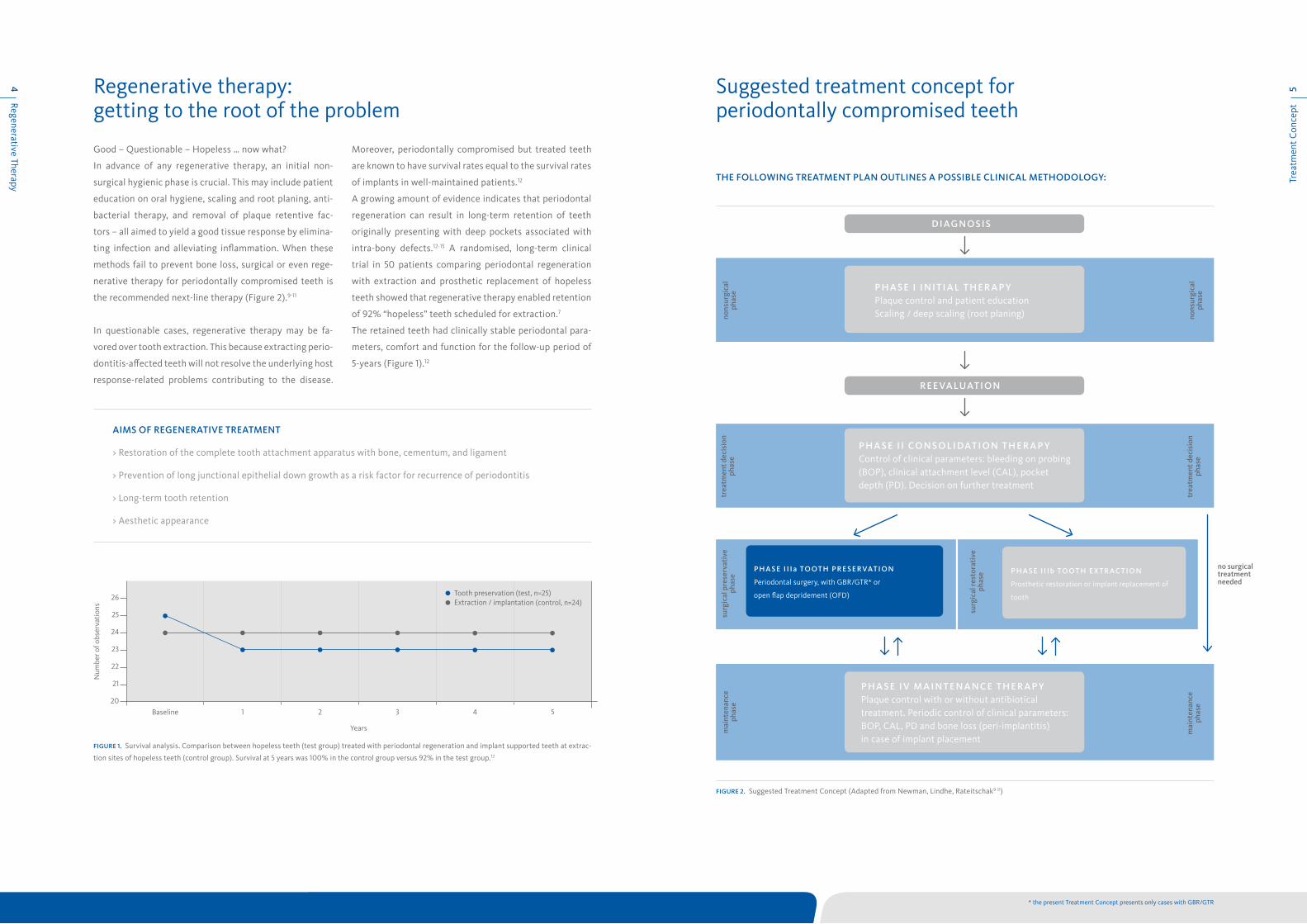
of 92% “hopeless” teeth scheduled for extraction.7
The retained teeth had clinically stable periodontal para-
meters, comfort and function for the follow-up period of
5-years (Figure 1).12
aims of reGeneraTive TreaTmenT
> Restoration of the complete tooth attachment apparatus with bone, cementum, and ligament
> Prevention of long junctional epithelial down growth as a risk factor for recurrence of periodontitis
> Long-term tooth retention
> Aesthetic appearance
fiGure 1. Survival analysis. Comparison between hopeless teeth (test group) treated with periodontal regeneration and implant supported teeth at extrac-
tion sites of hopeless teeth (control group). Survival at 5 years was 100% in the control group versus 92% in the test group.12
Years
Baseline 1 2 3 4 5
26
25
24
23
22
21
20
Num
ber o
f obs
erva
tion
s
Tooth preservation (test, n=25)Extraction / implantation (control, n=24)
fiGure 2. Suggested Treatment Concept (Adapted from Newman, Lindhe, Rateitschak9-11)
diaGnosis
reevaluaTion
p h a s e i i n i T i a l T h e r a p YPlaque control and patient educationScaling / deep scaling (root planing) no
nsur
gica
lph
ase
nons
urgi
cal
phas
e
p h a s e i i co n s o l i daT i o n T h e r a p YControl of clinical parameters: bleeding on probing (BOP), clinical attachment level (CAL), pocket depth (PD). Decision on further treatment
trea
tmen
t dec
isio
nph
ase
trea
tmen
t dec
isio
nph
ase
p h a s e i v m a i n T e n a n c e T h e r a p YPlaque control with or without antibiotical treatment. Periodic control of clinical parameters: BOP, CAL, PD and bone loss (peri-implantitis)in case of implant placement
mai
nten
ance
phas
e
mai
nten
ance
phas
e
p h a s e i i ib To oT h e x T r ac T i o n
Prosthetic restoration or implant replacement of
tooth
surg
ical
rest
orat
ive
ph
ase
no surgical treatment needed
p h a s e i i i a To oT h p r e s e rvaT i o n
Periodontal surgery, with GBR/GTR* or
open flap depridement (OFD)
surg
ical
pre
serv
ativ
e ph
ase
* the present Treatment Concept presents only cases with GBR/GTR

Scie
ntifi
c &
Clin
ical
Evi
denc
e7
Defect M
orphology6 Defect morphology influences outcome of regenerative therapy
Scientific and clinical evidence for the surgical preservative phase
There is a wide range of general factors that are known or
assumed to influence periodontal healing (e.g., age, smok-
ing, concomitant medication, postsurgical care, periodon-
tal maintenance, oral hygiene, nutrition, stress).
Furthermore, defect morphology is a key factor for the
therapy outcome.16 Each periodontal osseous lesion pre-
sents a unique anatomy. A first level of classification dif-
ferentiates between horizontal, infrabony, and furcation
defects as represented in Figure 3.17
Horizontal defects are defined when the base of the pock-
et is located coronal to the alveolar crest whereas infra-
bony defects are apical (vertical defects).
Regenerative therapy (GBR, GTR) is indicated in bony de-
fects with three, two or at least one remaining walls. To
some extend also Class II furcation defects can be treated
with GTR.18 There is evidence, that 2- and 3 wall intrabony
defects respond better to GTR therapy than 1-wall defects.
However, the deeper the infrabony defect, the more at-
tachment gain and bony fill may be expected.16 Other de-
fect characteristics influencing outcomes of regenerative
therapy are presented in Table 2:
Upon decision to preserve the tooth, the next step is to
decide for a surgical therapy: Leading treatment methods
often utilise a combination of a slowly resorbing osteocon-
ductive bone substitute and a membrane.19
Guided Tissue reGeneraTion
Some evidence shows, that Guided Tissue Regeneration
(GTR) is superior to Open Flap Debridement (OFD) for
the treatment of periodontal intrabony and furcation
defects.20-22 Overall, GTR is consistently more effective
than OFD in reducing:
> open horizontal furcation depths,
> horizontal and vertical attachment levels, and
> pocket depths for mandibular or maxillary class II
furcation defects.
With the use of Geistlich Bio-Oss® orthodontic movement
is possible in patients after GTR therapy.23,24 Moreover, re-
sorbable membranes have proven superior to non-resorb-
able membranes in generating vertical bone fill.15
GeisTlich bio-oss® (collaGen) and
GeisTlich bio-Gide® (perio)
Combined filling of periodontal defects with the graft ma-
terial Geistlich Bio-Oss® Collagen or Geistlich Bio-Oss®
followed by Geistlich Bio-Gide® membrane coverage has a
history of proven effectiveness in regenerative periodon-
tal therapy.25-31
Treatment of intra-bony defects with Geistlich Bio-Oss®
and Geistlich Bio-Gide® Perio resulted in sustained higher
clinical attachment level gain as compared to treatment
with OFD alone after 5 years (Figure 5).2
First clinical and histological results of treatment of endo-
dontic-periodontic lesion with endodontic therapy
followed by Guided Tissue Regeneration with Geistlich
Bio-Oss® and Geistlich Bio-Gide® demonstrated that the
combined approach can promote the formation of new ce-
mentum, periodontal ligament, and bone around the apex,
as well as the complete bone regeneration of the buccal
bone plate (Figure 6).19
os s e o us d e f e c T s
horizont al de fec ts
infrabony de fec ts
Intrabony Defects
1 Wal l
2 Wal ls
3 Wal ls
Combinat ions
Craters
Class I: Horizontal loss up to 3 mm
Class II: Horizontal loss > 3 mm; not total
Class III: Total loss of tissue in furcation
furc ation de fec ts
fiGure 4. Infrabony defects (modified from Papapanou et al. 2000)17
fiGure 3. Classification of periodontal osseous defects (modified from
Papapanou et al. 2000)17
posiTive influence neGaTive influence
> Deep infrabony compo-nent (> 3 mm)
> Narrow radiographic defect angle
> Deep baseline pocket depth
> Shallow infrabony component (≤ 3 mm)
> Wide radiographic defect angle
> Tooth motility
Table 2: Positive and negative defect characteristics 16
1 wall defect 2 wall defect 3 wall defect Interproximal crater
fiGure 5. The gain in clinical attachment level (CAL) and the reduction in pocket depth (PD) are significantly larger in the test group than in the control
group respectively, (p=0.01 and ≤ 0.05 respectively) both after one year and after 5 years.2
open flap debridement + Geistlich Bio-Oss® and Geistlich Bio-Gide® Perio (n=10)
open flap debridement (n=9)
p < 0.01 p < 0.01
0
1
2
3
4
5
6
7
After 1 year After 5 years
CAL gain
mm
1.8 4.0 1.4 3.7
CAL gain
p ≤ 0.05 p ≤ 0.05
0
1
2
3
4
5
6
7
After 1 year After 5 years
PD reduction
mm
4.0 5.4 3.3 4.8
PD reduction
fiGure 6. The histologic assessment demonstrates the presence of new
periodontal ligament, cementum, and bone. The newly formed woven
bone can be observed maturating into bone trabeculae completely
surrounding Geistlich Bio-Oss particles. BO=Bio-Oss; NB=new bone
L=ligament; NC=new cementum; OC=old cementum; D=dentin 19
The present Treatment Concept shows different cases that
have been appointed to a classification system combining
the remaining walls and the vertical dimension of the bony
defect (Figure 4).
BO
BO
NB
NBNC
D
200 µm
L
Cas
e 2:
Dr.
Die
go C
apri
9
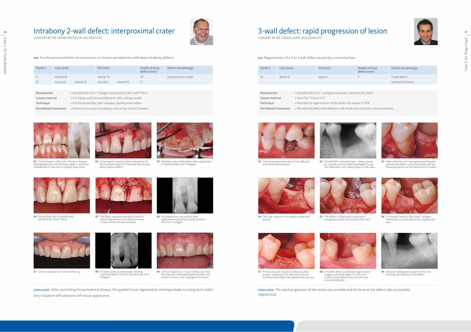
SURGERY BY DR. FRANk BRöSELER, AACHEN (DE)
aim Functional and esthetic reconstruction in chronic periodontitis with deep intrabony defects.
Tooth # cal (mm) pd (mm) depth of bony defect (mm)
defect morphology
11 mesial 10 mesial 10 10 interproximal crater
21 buccal 6 mesial 10 buccal 5 mesial 10 9
conclusion After controlling the periodontal disease, this guided tissue regeneration technique leads to a long-term stable
bony situation with pleasant soft-tissue appearance.
09 Clinical situation at 7 years follow-up; note the naturally reformed papilla between the central incisors, and no gingival recession.
06 Postoperative x-ray control after regenerative procedure using Geistlich Bio-Oss® Collagen.
05 The flap is repositioned and sutured to relieve flap tension and obtain primary closure of the interdental space.
04 The grafted site is covered with Geistlich Bio-Gide® Perio.
07 Clinical situation at 3 years follow-up 08 4.5 years post-op radiograph showing sustained defect fill from Geistlich Bio-Oss® Collagen.
01 Initial situation after anti-infective therapy. Radiographically, the intrabony defect cannot be represented in toto due to palatal bone plate.
02 Intrasurgical situation after preparation of the mucoperiostal full-thickness flap reveals deep osseous defect.
03 Palatinal view of the defect after application of Geistlich Bio-Oss® Collagen.
Intrabony 2-wall defect: interproximal crater
biomaterials > Geistlich Bio-Oss® Collagen and Geistlich Bio-Gide® Perio.
suture material > 4-0 classic and 6-0 monofilament with cutting needle
Technique > Full thickness flap, split released, papilla preservation
periodontal treatment > Patient instruction and plaque control for at least 8 weeks.
Case 1: D
r. Frank Bröseler8 3-wall defect: rapid progression of lesion
SURGERY BY DR. DIEGO CAPRI, BOLOGNA (IT)
aim Regeneration of a 2 to 3 wall defect caused by a cemental tear.
Tooth # cal (mm) pd (mm) depth of bony defect (mm)
defect morphology
35 distal 12 distal 7 5 3 wall defect
without furcation
conclusion The rapid progression of the lesion was arrested and the bone at the defect side successfully regenerated.
04 The root surface is thoroughly scaled and planed.
06 A trimmed Geistlich Bio-Gide® collagen membrane is positioned on the augmented area.
05 The defect is filled with a mixture of autogenous bone and Geistlich Bio-Oss®.
01 Clinical preoperative view of the affected area showing the lesion.
03 After reflection of a mucoperiosteal flap the periodontal defect is de-granulated and the fractured portion of the cementum is visible.
02 DIAGNOSIS: Cemental tear – likely caused by a parafunctional habit overlapped to par-tial edentulism and malocclusion in the area.
07 Primary wound closure is achieved, after proper releasing of the flap with internal mattress and single interrupted Gore sutures.
09 Intraoral radiographic aspect of the site showing the healing of the defect.
08 4 months after periodontal regenerative surgery a probing depth of 3 mm and a clinical attachment loss of 6 mm was measured distally.
biomaterials > Geistlich Bio-Oss® , autogeneous bone, Geistlich Bio-Gide®
suture material > Gore-Tex® Suture CV7
Technique > Periodontal regeneration of the defect by means of GTR
periodontal treatment > Periodontal defect debridement with hand and ultrasonic instrumentation.

Cas
e 4:
Pro
f. D
r. Pi
erpa
olo
Cor
telli
ni11
Case 3: Prof. D
r. Michael C
hristgau10 Extended 2-wall defect
SURGERY BY PROF. DR. MICHAEL CHRISTGAU, DüSSELDORF (DE)
aim Defect resolution of an extended 2-wall defect with regenerative periodontal surgery.
Tooth # cal (mm) pd (mm) depth of bony defect (mm)
defect morphology
32 mesial 14 distal 4 mesial 11 distal 2 ca. 10 2 wall defect
buccal 4 oral 4 buccal 1 oral 2
conclusion Regenerative periodontal surgery with Geistlich Bio-Oss® Collagen and Geistlich Bio-Gide® Perio results in long-
term defect resolution.
05 Coverage with a trimmed Geistlich Bio-Gide® Perio membrane without further fixation.
04 Autogeneous bone covered and defect filled completely with Geistlich Bio-Oss® Collagen.
06 Coronal flap repositioning and wound closure with horizontal mattress and single sutures.
07 Clinical and radiological situation after 6 months with clinical attachment gain of 7 mm mesial and vast defect fill.
08 Clinical and radiological situation at 12 months with clinical attachment gain of 8 mm mesial and considerable defect fill.
09 Clinical and radiological situation 6 years after surgery showing stable long-term situation.
01 Preoperative clinical and radiological situa-tion showing an inflammation-free gingiva and the bone defect.
02 Intraoperative view of the extended 2-wall defect.
03 Basal defect is filled with autogenous bone chips after debridement and root planing.
biomaterials > Geistlich Bio-Oss® Collagen, Geistlich Bio-Gide® Perio, autogenous bone
suture material > Seralene® 5-0 and 6-0
Technique > Papilla-Preservation technique, sulcular incision Regio 41–33 without vertical releasing incisions
periodontal treatment > Semipermanent adhesive tooth splinting with composite material and non-surgical periodontal therapy with additional systemic antibiotic therapy ( 3 x 400 mg metronidazol, 7 days)
Periodontal regenerative surgery SURGERY BY DR. PIERPAOLO CORTELLINI, FIRENZE (IT)
aim Resolution of deep pockets associated with deep intrabony defects and preservation of aesthetics on upper incisors.
Tooth # cal (mm) pd (mm) depth of bony defect (mm)
defect morphology
21 (22) mesial 7 (4) distal 2 (7) mesial 6 (2) distal 2 (6) max. 10 (8) 2 wall defect
buccal 4 (4) lingual 3 (4) buccal 4 (2) lingual 3 (3) without furcation
referencesCortellini P, Tonetti MS. Improved wound stability with a modified minimally invasive surgical technique in the regenerative treatment of isolated interdental intrabony defects. J Clin Periodontol 2009: 36: 157–163.
Cortellini P, Tonetti MS. Clinical and radiographic outcomes of the modified minimally invasive surgical technique with and without regenerative materials: a randomized- con-trolled trial in intra-bony defects. J Clin Peridontol 2011: 38: 365–373.
conclusion The combination of the modified minimally invasive surgical technique with Geistlich Bio-Oss® was effective in treating multiple intrabony defects associated with deep pockets in the upper incisors.
04 Buccal incision design. 06 Geistlich Bio-Oss® is positioned to fill the intrabony components of the defects. In larger and/or less contained defects, the additional use of a collagen membrane, such as Geistlich Bio-Gide®, is recommended.
05 Intraoperative probing at tooth 21. Note the absence of the interdental bone peak between teeth 11 and 21 and the severe buccal dehiscence. Geistlich Bio-Oss® was used to prevent the postoperative shrinkage of the soft tissues.
01 Preoperative probing at tooth 21 showing probing depth of 6 mm.
03 Preoperative radiograph showing the intra-bony defects mesial to tooth 21 and distal to tooth 22 .
02 Preoperative probing at tooth 22 with probing depth of 6 mm.
07 The flap is sealed over Geistlich Bio-Oss® with internal modified mattress sutures.
09 1 year radiographs showing the resolution of the intrabony components of the defects.
08 1 year clinical situation showing healthy condition and a minimal gingival recession relative to baseline.
biomaterials > Geistlich Bio-Oss®
suture material > Gore-Tex® Suture 6-0
Technique > Modified minimally invasive surgical procedure (M-MIST) with a Microblade USM 6900
periodontal treatment > Root planing was performed before surgery.
Cas
e 6:
Pro
f. D
r. M
arku
s H
ürze
ler
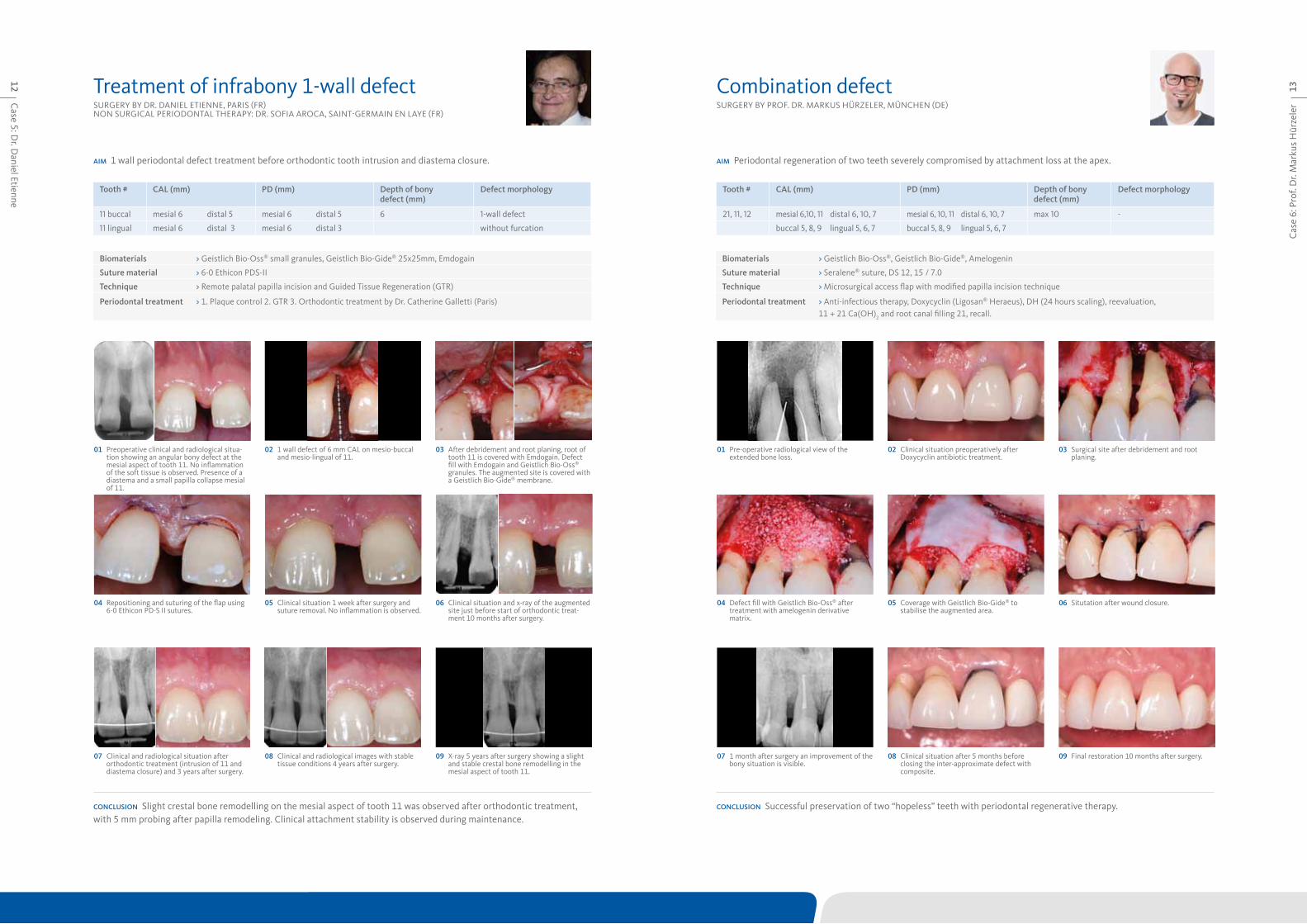
13Combination defectSURGERY BY PROF. DR. MARkUS HüRZELER, MüNCHEN (DE)
aim Periodontal regeneration of two teeth severely compromised by attachment loss at the apex.
Tooth # cal (mm) pd (mm) depth of bony defect (mm)
defect morphology
21, 11, 12 mesial 6,10, 11 distal 6, 10, 7 mesial 6, 10, 11 distal 6, 10, 7 max 10 -
buccal 5, 8, 9 lingual 5, 6, 7 buccal 5, 8, 9 lingual 5, 6, 7
conclusion Successful preservation of two “hopeless” teeth with periodontal regenerative therapy.
04 Defect fill with Geistlich Bio-Oss® after treatment with amelogenin derivative matrix.
06 Situtation after wound closure.05 Coverage with Geistlich Bio-Gide® to stabilise the augmented area.
01 Pre-operative radiological view of the extended bone loss.
03 Surgical site after debridement and root planing.
02 Clinical situation preoperatively after Doxycyclin antibiotic treatment.
07 1 month after surgery an improvement of the bony situation is visible.
09 Final restoration 10 months after surgery.08 Clinical situation after 5 months before closing the inter-approximate defect with composite.
biomaterials > Geistlich Bio-Oss®, Geistlich Bio-Gide®, Amelogenin
suture material > Seralene® suture, DS 12, 15 / 7.0
Technique > Microsurgical access flap with modified papilla incision technique
periodontal treatment > Anti-infectious therapy, Doxycyclin (Ligosan® Heraeus), DH (24 hours scaling), reevaluation, 11 + 21 Ca(OH)2 and root canal filling 21, recall.
Case 5: D
r. Daniel Etienne
12
SURGERY BY DR. DANIEL ETIENNE, PARIS (FR) NON SURGICAL PERIODONTAL THERAPY: DR. SOFIA AROCA, SAINT-GERMAIN EN LAYE (FR)
aim 1 wall periodontal defect treatment before orthodontic tooth intrusion and diastema closure.
Tooth # cal (mm) pd (mm) depth of bony defect (mm)
defect morphology
11 buccal mesial 6 distal 5 mesial 6 distal 5 6 1-wall defect
11 lingual mesial 6 distal 3 mesial 6 distal 3 without furcation
conclusion Slight crestal bone remodelling on the mesial aspect of tooth 11 was observed after orthodontic treatment, with 5 mm probing after papilla remodeling. Clinical attachment stability is observed during maintenance.
04 Repositioning and suturing of the flap using 6-0 Ethicon PD-S II sutures.
06 Clinical situation and x-ray of the augmented site just before start of orthodontic treat-ment 10 months after surgery.
05 Clinical situation 1 week after surgery and suture removal. No inflammation is observed.
01 Preoperative clinical and radiological situa-tion showing an angular bony defect at the mesial aspect of tooth 11. No inflammation of the soft tissue is observed. Presence of a diastema and a small papilla collapse mesial of 11.
03 After debridement and root planing, root of tooth 11 is covered with Emdogain. Defect fill with Emdogain and Geistlich Bio-Oss® granules. The augmented site is covered with a Geistlich Bio-Gide® membrane.
02 1 wall defect of 6 mm CAL on mesio-buccal and mesio-lingual of 11.
07 Clinical and radiological situation after orthodontic treatment (intrusion of 11 and diastema closure) and 3 years after surgery.
09 X-ray 5 years after surgery showing a slight and stable crestal bone remodelling in the mesial aspect of tooth 11.
08 Clinical and radiological images with stable tissue conditions 4 years after surgery.
biomaterials > Geistlich Bio-Oss® small granules, Geistlich Bio-Gide® 25x25mm, Emdogain
suture material > 6-0 Ethicon PDS-II
Technique > Remote palatal papilla incision and Guided Tissue Regeneration (GTR)
periodontal treatment > 1. Plaque control 2. GTR 3. Orthodontic treatment by Dr. Catherine Galletti (Paris)
Treatment of infrabony 1-wall defect
Cas
e 8:
Pro
f. D
r. G
iulio
Ras
peri
ni15
Case 7: D
r. Syed Mahnaz
14 2-wall defect in the non-aesthetic regionRegenerative surgery 11 – perio-endo SURGERY BY PROF. DR. GIULIO RASPERINI, MILAN (IT)SURGERY BY DR. SYED MAHNAZ, PERTH (AUS)
aim Periodontal regeneration to reduce probing depth by increasing bone and periodontal attachment with a minimal
gingival recession, to change the prognosis of the tooth # 46 and preserve its function.aim Retention of the central incisor and improvement of its mobility.
Tooth # cal (mm) pd (mm) depth of bony defect (mm)
defect morphology
46 mesial 14 distal 3 mesial 14 distal 3 max 10 2 wall defect
without furcation
Tooth # cal (mm) pd (mm) depth of bony defect (mm)
defect morphology
11 mesial 9 distal 5 mesial 7 distal 4 4 2 wall defect
buccal 5 lingual 5 buccal 3 lingual 3
conclusion 2 months after conclusion of presurgical, cause-related therapy, the patient reported the complete resolution of
inflammation, resulting in a decrease of the full mouth plaque and bleeding scores. 1 year after the surgery, the soft-tissue
was well preserved and represented with a sufficient width of keratinised gingiva. Radiographs after 1 year show a stable
situation with an almost complete bone fill.
conclusion Predictable treatment outcomes were achieved to help retain teeth in situations where perio-endo problems exist. Regenerative surgery offers sustainable options for treatment of advanced periodontal disease.
04 The Geistlich Bio-Oss® fills the defect and is protected by a Geistlich Bio-Gide® membrane. After flap release, the wound is closed without tension.
05 Re-evaluation at 1 year. A residual 5 mm probing depth is present with a 9 mm probing depth loss as compared to baseline measurements.
01 Baseline situation showing the 14 mm pocket depth mesial to tooth 46.
03 Elevation of a full-thickness buccal and lingual flap with papilla preservation. The 10 mm deep, 2-wall intrabony defect mesial to tooth 46 is evident after careful debridement.
02 Baseline radiograph showing the presence of an angular bony defect involving the mesial site of tooth 46.
06 Nearly complete bone fill of the angular defect at 1 year.
04 Geistlich Bio-Oss® granules in the defect. 06 Immediate post-op passive closure and coronal repositioning of the mucosa.
05 Geistlich Bio-Gide® membrane trimmed and placed in the interproximal region .
01 Non-responding residual pocket associated with a perio-endo involved tooth 11.
03 Elevation of flap with papilla preservation to access the infrabony pocket.
02 Radiograph of infrabony angular defect on tooth 11 with subsequent endodontic treatment.
07 Improved pocketing and mobility 8 months after surgery and additional composite bond-ing to improve the aesthetics.
08 Geistlich Bio-Oss® mesial of tooth 11 is well integrated after 8 months.
08 Follow up 2 years post surgery showing good bone stability and improved clinical status of this tooth.
biomaterials > Geistlich Bio-Oss®, Geistlich Bio-Gide®
suture material > Vicryl 5.0 suture materials
Technique > Endodontic treatment followed by non-surgical debridement and a modified papilla preservation technique.
periodontal treatment > Nonsurgical periodontal debridement therapy under local anaesthesia with endodontic treatment was undertaken.
biomaterials > Geistlich Bio-Oss® , Geistlich Bio-Gide®
suture material > Gore-Tex® Suture 5-0
Technique > Periodontal regeneration procedure with preservation of the interdental tissue and mesial releasing incision.
periodontal treatment > Cause related periodontal therapy, including motivation and instructions for home care; professional supra-gingival debridment and sub-gingival root planing. Re-evaluation for potential additional therapy.

Cas
e 10
: Dr.
Beat
Wal
lkam
m17
Case 9: Prof. D
r. Anton Sculean
16 Deep intrabony 2-wall defectSURGERY BY PROF. DR. ANTON SCULEAN, BERN (CH)
aim Treatment of intrabony defect with a complicated, noncontained morphology using a combination of collagen barrier
membrane and a natural bone mineral.
Tooth # cal (mm) pd (mm) depth of bony defect (mm)
defect morphology
36 distal 11 distal 11 5 2 wall, large non-contained
defect
conclusion Good appearance of soft tissue and sufficient bone fill at 1 year after regeneration of a deep non-contained bony
defect.
2-wall defect in the aesthetic zoneSURGERY BY DR. BEAT WALLkAMM, LANGENTHAL (CH)
aim Periodontal regeneration with a minimally invasive surgical technique in combination with Geistlich Bio-Oss® Collagen
and Geistlich Bio-Gide® Perio.
Tooth # cal (mm) pd (mm) depth of bony defect (mm)
defect morphology
11 mesial 11 distal 4 mesial 8 distal 3 5 2 wall defect
buccal 4 lingual 4 buccal 2 lingual 3
conclusion The minimally invasive surgical technique in combination with Geistlich Bio-Oss® Collagen and Geistlich
Bio-Gide® Perio resulted in markedly improved clinical and radiographic outcome.
04 Following removal of granulation tissue and root planing, the defect is filled with Geistlich Bio-Oss®.
06 Minimal recession of the soft tissues and attachment gain and reduced PD measured 6mm and 7 mm respectively at 1 year.
05 The grafting material and the surrounding alveolar bone are covered with a Geistlich Bio-Gide® Perio.
01 Preoperative probing indicating the presence of a deep pocket distal to the mandibular left molar.
03 Intraoperative view revealing a deep non-contained intrabony defect.
02 Preoperative radiograph demonstrating the extent of bone loss.
07 Postoperative radiograph at 1 year reveals an almost complete fill of the intrabony defect.
04 A trimmed Geistlich Bio-Gide® Perio is inserted lingually and Geistlich Bio-Oss® Collagen is applied into the defect.
06 Primary closure of the wide interdental papilla is obtained with an internal mattress suture with an external loop and two oblique hang-up mattress sutures.
05 The Geistlich Bio-Gide® Perio is folded over the augmented site and inserted under the buccal full thickness flap.
01 Tooth 11 presents with a pocket depth of 8 mm and a clinical attachment level of 11 mm with some loss of papillary tissue.
03 After elevation of a tiny buccal flap and positioning of the interdental papilla slightly to the palatal side, the defect is debrided.
02 Baseline radiograph shows the bone loss mesially to the first right incisor reaching the apical third of the root.
07 6 weeks after surgery the inter-dental soft tissues are well healed.
09 The 2 year radiograph shows a horizontal gain of 3 mm bone in the treated area.
08 Clinical situation after 2 years with a probing pocket depth of 3 mm and a clinical attach-ment level gain of 5 mm.
biomaterials > Geistlich Bio-Oss® Collagen, Geistlich Bio-Gide® Perio
suture material > Seralene® 7/0 (PVDF, Serag Wiessner)
Technique > Minimal invasive surgical technique (MIST) (Cortellini 2009)
periodontal treatment > Initial periodontal treatment (4hrs), 3-months recall
referenceCortellini P, Tonetti MS. Improved wound stability with a modified minimally invasive surgical technique in the regenerative treatment of isolated interdental intrabony defects. J Clin Periodontol 2009: 36: 157–163.
biomaterials > Geistlich Bio-Gide® Perio, Geistlich Bio-Oss®
suture material > 4-0 silk
Technique > Periodontal regeneration of a large non-contained defect through GTR with the use of grafting material.
periodontal treatment > Hygienic phase 3 months before regenerative surgery consisting of patient instruction for oral hygiene, and full-mouth scaling and root planing in conjunction with systemically administered antibiotic therapy (3 x 375 mg Amoxicillin and 3 x 250 mg Metronidazol) for one week.
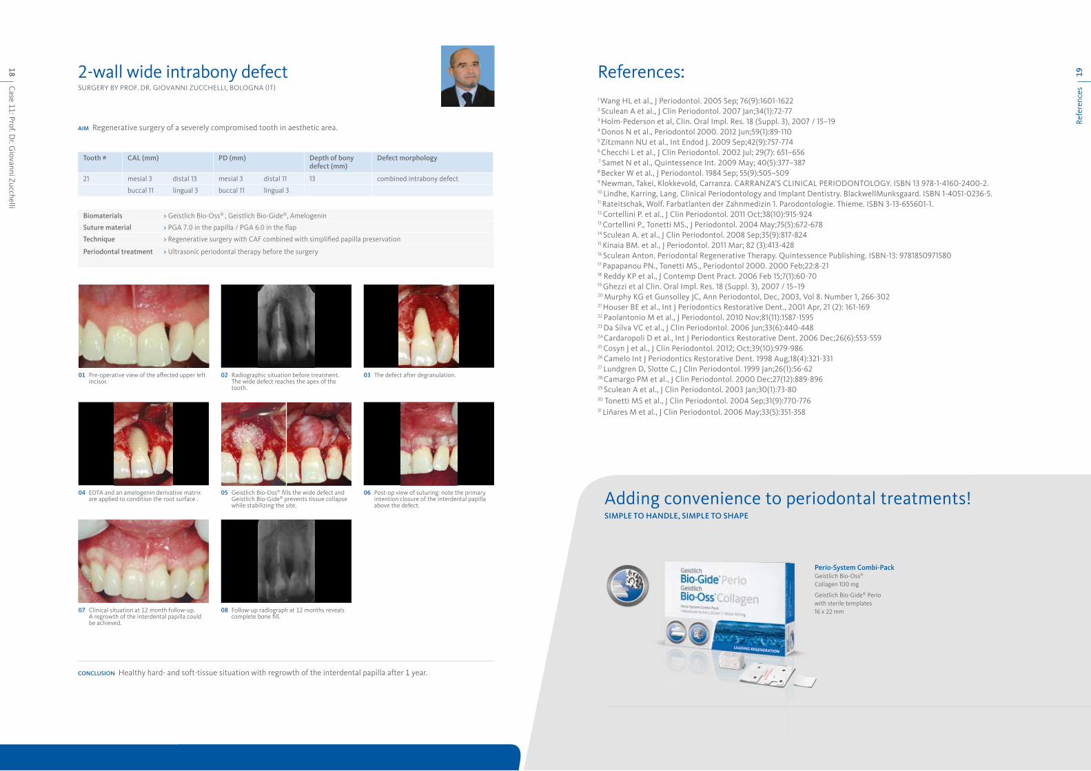
Case 11: Prof. D
r. Giovanni Zucchelli
18Re
fere
nces
192-wall wide intrabony defectSURGERY BY PROF. DR. GIOVANNI ZUCCHELLI, BOLOGNA (IT)
aim Regenerative surgery of a severely compromised tooth in aesthetic area.
Tooth # cal (mm) pd (mm) depth of bony defect (mm)
defect morphology
21 mesial 3 distal 13 mesial 3 distal 11 13 combined intrabony defect
buccal 11 lingual 3 buccal 11 lingual 3
conclusion Healthy hard- and soft-tissue situation with regrowth of the interdental papilla after 1 year.
biomaterials > Geistlich Bio-Oss® , Geistlich Bio-Gide®, Amelogenin
suture material > PGA 7.0 in the papilla / PGA 6.0 in the flap
Technique > Regenerative surgery with CAF combined with simplified papilla preservation
periodontal treatment > Ultrasonic periodontal therapy before the surgery
04 EDTA and an amelogenin derivative matrix are applied to condition the root surface .
06 Post-op view of suturing: note the primary intention closure of the interdental papilla above the defect.
05 Geistlich Bio-Oss® fills the wide defect and Geistlich Bio-Gide® prevents tissue collapse while stabilizing the site.
01 Pre-operative view of the affected upper left incisor.
03 The defect after degranulation.02 Radiographic situation before treatment. The wide defect reaches the apex of the tooth.
07 Clinical situation at 12 month follow-up. A regrowth of the interdental papilla could be achieved.
08 Follow-up radiograph at 12 months reveals complete bone fill.
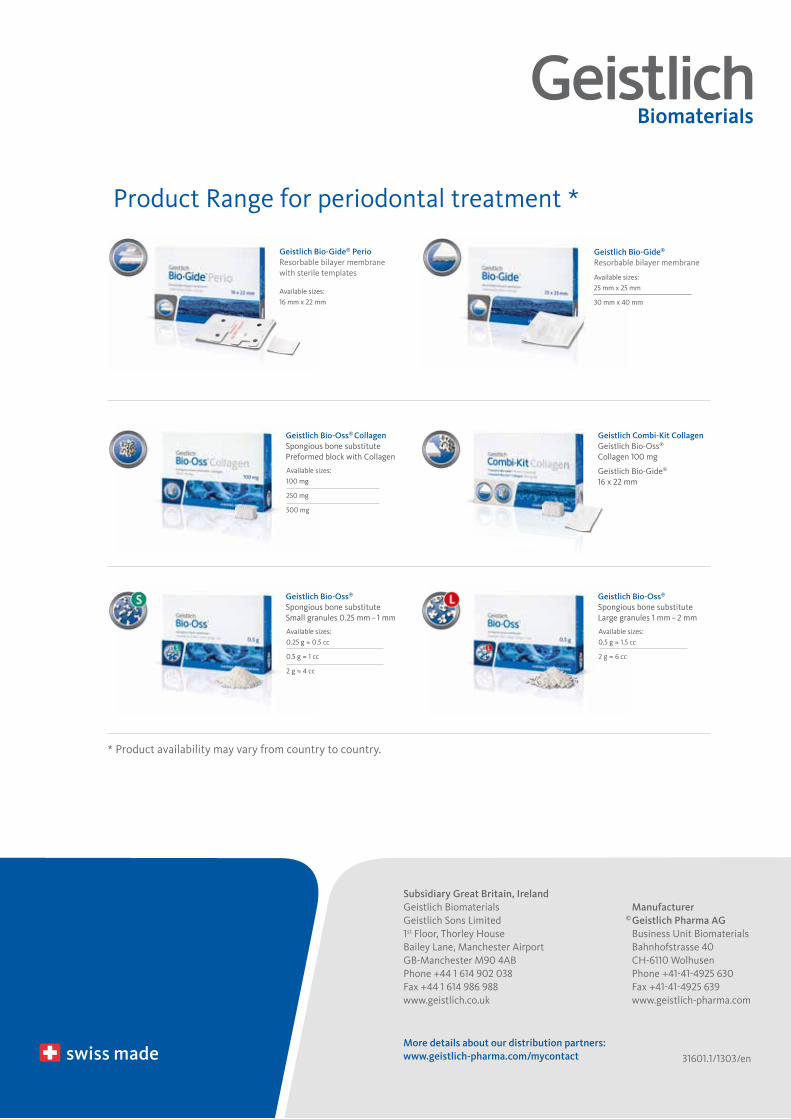
Adding convenience to periodontal treatments!simple To handle, simple To shape
perio-system combi-packGeistlich Bio-Oss® Collagen 100 mg
Geistlich Bio-Gide® Perio with sterile templates 16 x 22 mm
1 Wang HL et al., J Periodontol. 2005 Sep; 76(9):1601-16222 Sculean A et al., J Clin Periodontol. 2007 Jan;34(1):72-773 Holm-Pederson et al, Clin. Oral Impl. Res. 18 (Suppl. 3), 2007 / 15–19 4 Donos N et al., Periodontol 2000. 2012 Jun;59(1):89-1105 Zitzmann NU et al., Int Endod J. 2009 Sep;42(9):757-7746 Checchi L et al., J Clin Periodontol. 2002 Jul; 29(7): 651–656 7 Samet N et al., Quintessence Int. 2009 May; 40(5):377–3878 Becker W et al., J Periodontol. 1984 Sep; 55(9):505–5099 Newman, Takei, klokkevold, Carranza. CARRANZA’S CLINICAL PERIODONTOLOGY. ISBN 13 978-1-4160-2400-2.10 Lindhe, karring, Lang. Clinical Periodontology and Implant Dentistry. BlackwellMunksgaard. ISBN 1-4051-0236-5.11 Rateitschak, Wolf. Farbatlanten der Zahnmedizin 1. Parodontologie. Thieme. ISBN 3-13-655601-1.12 Cortellini P. et al., J Clin Periodontol. 2011 Oct;38(10):915-92413 Cortellini P., Tonetti MS., J Periodontol. 2004 May;75(5):672-67814 Sculean A. et al., J Clin Periodontol. 2008 Sep;35(9):817-82415 kinaia BM. et al., J Periodontol. 2011 Mar; 82 (3):413-42816 Sculean Anton. Periodontal Regenerative Therapy. Quintessence Publishing. ISBN-13: 978185097158017 Papapanou PN., Tonetti MS., Periodontol 2000. 2000 Feb;22:8-2118 Reddy kP et al., J Contemp Dent Pract. 2006 Feb 15;7(1):60-7019 Ghezzi et al Clin. Oral Impl. Res. 18 (Suppl. 3), 2007 / 15–1920 Murphy kG et Gunsolley JC, Ann Periodontol, Dec, 2003, Vol 8. Number 1, 266-30221 Houser BE et al., Int J Periodontics Restorative Dent., 2001 Apr, 21 (2): 161-16922 Paolantonio M et al., J Periodontol. 2010 Nov;81(11):1587-159523 Da Silva VC et al., J Clin Periodontol. 2006 Jun;33(6):440-44824 Cardaropoli D et al., Int J Periodontics Restorative Dent. 2006 Dec;26(6):553-559 25 Cosyn J et al., J Clin Periodontol. 2012; Oct;39(10):979-986 26 Camelo Int J Periodontics Restorative Dent. 1998 Aug;18(4):321-33127 Lundgren D, Slotte C, J Clin Periodontol. 1999 Jan;26(1):56-6228 Camargo PM et al., J Clin Periodontol. 2000 Dec;27(12):889-89629 Sculean A et al., J Clin Periodontol. 2003 Jan;30(1):73-8030 Tonetti MS et al., J Clin Periodontol. 2004 Sep;31(9):770-77631 Liñares M et al., J Clin Periodontol. 2006 May;33(5):351-358
References:
manufacturer© Geistlich pharma aG Business Unit Biomaterials Bahnhofstrasse 40 CH-6110 Wolhusen Phone +41-41-4925 630 Fax +41-41-4925 639 www.geistlich-pharma.com
subsidiary Great britain, irelandGeistlich BiomaterialsGeistlich Sons Limited1st Floor, Thorley HouseBailey Lane, Manchester AirportGB-Manchester M90 4ABPhone +44 1 614 902 038Fax +44 1 614 986 988www.geistlich.co.uk
more details about our distribution partners:www.geistlich-pharma.com/mycontact 31601.1/1303/en
Geistlich bio-oss®
Spongious bone substitute Small granules 0.25 mm – 1 mm
Available sizes: 0.25 g ≈ 0.5 cc
0.5 g ≈ 1 cc
2 g ≈ 4 cc
Geistlich bio-oss®
Spongious bone substitute Large granules 1 mm – 2 mm
Available sizes: 0.5 g ≈ 1.5 cc
2 g ≈ 6 cc
Geistlich bio-oss® collagen
Spongious bone substitute Preformed block with Collagen
Available sizes: 100 mg
250 mg
500 mg
Geistlich combi-Kit collagenGeistlich Bio-Oss® Collagen 100 mg
Geistlich Bio-Gide® 16 x 22 mm
* Product availability may vary from country to country.
Available sizes: 25 mm x 25 mm
30 mm x 40 mm
Geistlich bio-Gide®
Resorbable bilayer membraneGeistlich bio-Gide® perio
Resorbable bilayer membranewith sterile templates
Available sizes: 16 mm x 22 mm
Product Range for periodontal treatment *