transmembrane orientation and topology of the nadh:quinone oxidoreductase putative quinone binding...
TRANSCRIPT

Transmembrane orientation and topology of the NADH:quinoneoxidoreductase putative quinone binding subunit NuoH
Robert Roth, Cecilia Ha«gerha«ll *Department of Biochemistry, Lund University, Box 124, 22100 Lund, Sweden
Received 11 May 2000; received in revised form 13 November 2000; accepted 14 November 2000
Abstract
NADH:quinone oxidoreductase, or Complex I, is a multi-subunit membrane-bound enzyme in the respiratory chain ofmany pro- and eukaryotes. The enzyme catalyzes the oxidation of NADH and donates electrons to the quinone pool,coupled to proton translocation across the membrane, but the mechanism of energy transduction is not understood. Inbacteria the enzyme consists of 14 subunits, seven membrane spanning and seven protruding from the membrane. Thehydrophobic NuoH (NQO8, ND1, NAD1, NdhA) subunit is seemingly involved in quinone binding. A homologous,structurally and most likely functionally similar subunit is also found in F420H2 oxidoreductases and in complex membrane-bound hydrogenases. We have made theoretical analyses of NuoH and NuoH-like polypeptides and experimentally analyzedthe transmembrane topology of the NuoH subunit from Rhodobacter capsulatus by constructing and analyzing alkalinephosphatase fusion proteins. This demonstrated that the NuoH polypeptide has eight transmembrane segments, and fourhighly conserved hydrophilic sequence motifs facing the inside, bacterial cytoplasm. The N-terminal and C-terminal ends arelocated on the outside of the membrane. A topology model of NuoH based on these results is presented, and implicationsfrom the model are discussed. ß 2001 Elsevier Science B.V. All rights reserved.
Keywords: Complex I; NADH:quinone oxidoreductase; Hydrogenase; F420 ; NuoH; ND1; NQO8; NAD1; NdhA; Alkaline phospha-tase fusion; (Rhodobacter capsulatus)
1. Introduction
NADH:quinone oxidoreductase, or Complex I, isthe largest, most complex and least understood ofthe respiratory chain enzymes. The membrane-
bound, multi-subunit enzyme catalyzes the oxida-tion of NADH to NAD� and donates electronsto the quinone pool. In this reaction non-covalentlybound FMN and 6^8 iron^sulfur clusters serve asintrinsic redox components. The electron transferthrough the enzyme is coupled to proton trans-location across the membrane with a stoichiom-etry of 4H�/2e3 [1], but the mechanism of energytransduction is not understood. Mammalian Com-plex I may consists of up to 43 protein subunits,whereas bacterial Complex I generally contains14 di¡erent subunits, all with homologues in themammalian enzyme. Seven of these subunits arelocated in the promontory part of the complex,
0005-2728 / 01 / $ ^ see front matter ß 2001 Elsevier Science B.V. All rights reserved.PII: S 0 0 0 5 - 2 7 2 8 ( 0 0 ) 0 0 2 6 5 - 6
Abbreviations: PhoA, alkaline phosphatase; IPTG, isopropyl-thio-L-D-galactoside; X-phosphate, 5-bromo-4-chloro-3-indolyl-phosphate; NADH, nicotinamide adenine dinucleotide ^ reducedform; LHON, Leber's hereditary optical neuropathy
* Corresponding author. Fax: +46-46-222-4534;E-mail : [email protected]
BBABIO 45017 26-2-01 Cyaan Magenta Geel Zwart
Biochimica et Biophysica Acta 1504 (2001) 352^362www.bba-direct.com

facing the bacterial cytoplasm (or mitochondrialmatrix) whereas the remaining polypeptides(NuoA, -H, -J, -K, -L, -M and -N) are very hydro-phobic and reside in the membrane spanning partof the enzyme. The latter seven correspond tomitochondrially encoded subunits in mammalianComplex I. Interestingly, 56% of all reporteddisease causing missense mutations in structuralgenes of human mitochondria a¡ect Complex I sub-units, and of those, 40% are found in the geneencoding the ND1 subunit. (MITOMAP: A HumanMitochondrial Genome Database. Center for Mo-lecular Medicine, Emory University, Atlanta, GA,USA. http://www.gen.emory.edu/mitomap.html.)The ND1 subunit corresponds to NuoH or NQO8in bacterial Complex I, NAD1in plant mitochon-drial Complex I and NdhA in the chloroplast Com-plex I-like NADPH:plastoquinone oxidoreductase.Defect quinone interaction have been implied inComplex I a¡ected by some of the disease causingmutations, however, due to the di¤culties involvedin biochemical analyses of clinical samples these in-dications are not conclusive. However, mutationstudies of NQO8 in Paracoccus denitri¢cans Com-plex I, mimicking the human mitochondrial muta-tion ND1/3460 (commonly found associated withLeber's hereditary optical neuropathy, LHON) andalso targeting residues in the immediate vicinitydemonstrated that the mutant Complex I were in-deed defect in quinone interaction [2], suggestingthat this subunit interacts with quinone. In beefheart Complex I the ND1 subunit was photolabeledwith rotenone (one of the most commonly usedComplex I speci¢c inhibitors) [3,4], indicating thatthis subunit is involved in quinone interaction. Inthis paper we have experimentally analyzed thetransmembrane topology of the NuoH subunitfrom Rhodobacter capsulatus Complex I using thephoA gene fusion technique developed by Manoiland Beckwith [5^7]. We present a topology modelof the subunit that accommodates both NuoH andall homologous polypeptides from F420H2 oxidore-ductases and from various complex, membrane-bound hydrogenases. Apart from the listed experi-mental data indicating that NuoH contains a qui-none binding site, the evolutionary history of sub-unit provides important implications regarding itsfunction.
2. Materials and methods
2.1. Growth of bacteria
Bacteria and plasmids used are listed in Table 1.All cultures of R. capsulatus and Escherichia coliwere grown aerobically in ba¥ed E-£asks at 37³C,200 rpm, in MPYE and LB [8] medium, respectively.Bacterial growth was monitored as the optical den-sity at 600 nm in a Pharmacia Ultrospec 4000 spec-trophotometer. For solid cultures 1.5% agar wasadded to the medium. Ampicillin was used at 50Wg/ml medium where appropriate. LB-agar platescontaining IPTG (isopropyl-thio-L-D-galactoside)and X-phosphate (5-bromo-4-chloro-3-indolyl-phos-phate) at 15 Wg/ml and 40 Wg/ml, respectively, wereused to screen for clones expressing an active alkalinephosphatase.
2.2. Standard molecular biological techniques
Restriction enzymes were from Boehringer^Mann-heim or Promega, T4 DNA ligase from Promega,and used as recommended by the manufacturer.Agarose gel electrophoreses, plasmid preparation,electroporation of E. coli and preparation of electro-competent cells was done by standard methods [8].DNA fragments were puri¢ed from agarose gels withthe Gene-Clean kit (BIO101, La Jolla, CA) based onsilica particles for DNA binding. DNA sequencingwas performed at the Biomolecular Resource Fa-cility, Lund University, using FusSeq as primer (Ta-ble 1). Polymerase chain reactions (PCR) were runwith an initial denaturing step of 95³C for 3 min,followed by repetitive cycles of a denaturing step(30 s), an annealing step using di¡erent temperaturesdepending of the primer pair (30 s) and an elonga-tion step at 72³C (2 min/kbp).
2.3. Cloning of phoA and nuoH
Chromosomal DNA was prepared from E. coli asin [9], and was subsequently used as template for aPCR. The phoA gene encoding alkaline phosphatasewas ampli¢ed using the primers PhoAup and PhoA-down (Table 1; annealing temperature 61³C). In thisconstruct the codons encoding the ¢rst 21 aminoacids that constitute the N-terminal export signal se-
BBABIO 45017 26-2-01 Cyaan Magenta Geel Zwart
R. Roth, C. Ha«gerha«ll / Biochimica et Biophysica Acta 1504 (2001) 352^362 353

quence of PhoA has been omitted. The resultingPCR fragment was ligated into the SmaI^HindIIIsite of pUC18, creating the plasmid pPHOA. Anadditional codon, GGG, was added to the beginningof the ampli¢ed part of phoA gene to regenerate theblunt-end restriction site (SmaI) making it possible tocreate in-frame fusions irrespectively of the C-termi-nal sequence of the insert.
Chromosomal DNA from R. capsulatus was pre-pared using the method of Marmur, [10], with theaddition of a step precipitating polysaccharides andproteins with CTAB (N-acetyl-N,N,N-trimethyl am-moniumbromide). After treatment with lysozyme,NaCl (0.7 M) and CTAB (1% w/v) were added,and the solution was incubated at 65³C for 15 min.The DNA was used as a template for subsequent
using the primers NuoHup and NuoHdown (Table1; annealing temperature 64³C). The DNA fragmentcontaining the nuoH gene was ligated into the SacI^HindIII site of pUC18. The resulting plasmid,pNHW was used as template in the following PCRreactions subcloning parts of the nuoH gene.
2.4. Construction of fusion proteins
Fusion proteins were made by PCR ampli¢cationof chosen part of the cloned nuoH gene using theprimer Nhfup and either of the downstream primersNhf1-Nhf13 (Table 1). The fragments were ligated itin to the SacI^SmaI site of pPHOA. By using aDNA polymerase (VENT, New England Biolabs)giving blunt ends, and the upstream SacI site in
Table 1Bacteria, plasmids and primers used
Bacteria/plasmid Relevant properties/sequence Reference or source
R. capsulatus ATCC 17015 Wild type, (type strain) DSMZ, Braunschweig,Germany
E. coli XL1-Blue recA1, endA1, gyrA96, thi, hsdR17, supE44, relA1 (lac) PromegaE. coli CC118 araD139 v(ara,leu)7697 vlacX74 phoAv20 galE galK thi
rpsE rpoB argEam recA1[44]
pUC18 ampR [45]pNHW nuoH, ampR This workpPHOA phoAP, ampR This workpNHF1-13 phoAP, ampR This work
Primersa
NuoHup 5P-GCACTAGGAGCTCAGGGACAACATGGCTG-3PNuoHdown 5P-GTGACGGAAAGCTTCGGTCGGTTCGG-3PPhoAup 5P-GGGCGGACACCAGAAATGCCT-3PPhoAdown 5P-CTGCCATTAAGCTTGGTTGCTAACAGC-3PNhfup 5P-GGAATTCGAGCTCAGGGACAAC-3PNhf1 5P-GGCGCCCACCACGTTCGG-3PNhf2 5P-ATAGACCGGCTTGTCGACG-3PNhf3 5P-GTTGAAGGGCACGACGACC-3PNhf4 5P-GGGATATTTCGAGTTCGAGGC-3PNhf5 5P-GGCAGAGCGCAGCGAGC-3PNhf6 5P-CTCGACGATCGCCGAAAG-3PNhf7 5P-CGGCAGGTCAAACGGCG-3PNhf8 5P-ATATTCGACCATGAACCCCGC-3PNhf9 5P-GGGAATGGGCGAAAGCCA-3PNhf10 5P-CATCTTGGCGACCATCCAGA-3PNhf11 5P-CAGCTGGTCATAGCGGTAGCG-3PNhf12 5P-GAACACCTTCCAACCGATCCG-3PNhf13 5P-CCAGAAGCCGCCCAGAAC-3PFusSeq 5P-TTTTGCAGGTTTATCGCTA-3PaAll primers were synthesized by Life Technologies, Sweden.
BBABIO 45017 26-2-01 Cyaan Magenta Geel Zwart
R. Roth, C. Ha«gerha«ll / Biochimica et Biophysica Acta 1504 (2001) 352^362354

pPHOA, the fragments could be ligated directly inframe, with the correct orientation. The correct insertwas con¢rmed by restriction cleavage analysis usingEcoRI. Subsequently, the constructs were sequencedover the fusion point.
2.5. Expression of fusion protein
E. coli CC118 cells, containing the respective NHFplasmids, were grown for 4 h before induction withIPTG at a ¢nal concentration of 1 mM. Growth wascontinued for 2 h before the cells were harvested bycentrifugation, at 5900Ug for 15 min. For membranepreparations the cells were disrupted by passingthrough a French pressure cell at 6.9U106 Pa. Celldebris was removed by centrifugation 10 000Ug for20 min. Membranes were collected from the resultingsupernatant by centrifugation, 184 000Ug (50.2 Ti,Beckman) for 45 min and homogenized in 30 mMTris (pH 7), 10 mM KCl. Protein content was mea-sured by the BCA method (Pierce) using bovine se-rum albumin as standard.
2.6. Western blot
Sodium dodecyl sulfate^polyacrylamide gel elec-trophoresis (SDS^PAGE) was performed accordingto Schaegger and von Jagow [11], using a 10% acryl-amide/bis (29:1) gel, and a BioRad Protean II xielectrophoresis unit. Electrophoresis was run for15 h at 90 V. Membrane preparations from cells ex-pressing fusion protein (see Section 2.5) were incu-bated at room temperature in sample bu¡er contain-ing 0.2 M Tris^HCl (pH 8.8), 20% glycerol, 4% SDS,1% L-mercaptoethanol and 0.01% bromophenol bluebefore loading on the gel. After electrophoresis thegel was soaked in gel transfer bu¡er for 5 h to re-move glycerol, that hampers subsequent blotting.Transfer to PVDF membrane (Millipore) was doneaccording to the manufacturer's instructions. Anti-phoA antibodies (kindly provided by Lars Heder-stedt) were used at 17 000-times dilution. Bound anti-bodies were detected using goat-anti rabbit antibod-ies (30 000-times dilution) as secondary antibodyfollowed by a chemiluminescence assay and wasmade according to the manufacturer's instruction(Clonetech Laboratories). Biomax Light-2 ¢lm fromKodak was used to document the result.
2.7. Alkaline phosphatase activity
Alkaline phosphatase activity in cells expressingfusion protein was measured using cells inducedand harvested as in Section 2.5. The cells werewashed once and kept on ice until used, permeabi-lized and assayed as described in Manoil, [12]. p-ni-trophenyl phosphate is used as an arti¢cial substratefor PhoA in the reaction. Units of activity is calcu-lated as described in Eq. 1, where V is the volume ofcells, t is the time the permeabilized cells are incu-bated with substrate at 37³C. The optical density(OD) at 600 nm is measured before adding substrate,and at 550 nm and 420 nm after incubation withsubstrate.
Units of activity � �OD4203�1:75UOD550��U1000t �min�UOD600UV �ml�
�1�
3. Results
3.1. Theoretical predictions of transmembranetopology
Primary sequence comparisons and Kyte^Doolittleplots [13] of NuoH and NuoH-like polypeptides in-dicate that the subunit is capable of forming eighttransmembrane helical segments ([14,15], Fig. 1). Theorientation of a polypeptide in the membrane can bepredicted using the `positive inside rule' [16] thatseems to be generally applicable to proteins in alltypes of membranes, including bacterial, mitochon-drial [17] and thylakoid [18] membranes. The chargedistribution in the NuoH and homologous polypep-tides indicate that the conserved hydrophilic stretchesfaces the inside (bacterial cytoplasm, mitochondrialmatrix), except for that the polypeptide stretch be-tween helices V and VI generally contains many neg-atively charged residues, but few if any positivelycharged. This obstructs a straight forward predictionof the polypeptide orientation. Alternatively, the pre-dicted hydrophobic helix pairs could correspond tolong, single, tilted transmembrane segments, sincethe connecting loops are very small and poorly de-¢ned (Fig. 1). If this was indeed the case, the longerconserved hydrophilic segments would end up onalternating sides of the membrane. Assigning the
BBABIO 45017 26-2-01 Cyaan Magenta Geel Zwart
R. Roth, C. Ha«gerha«ll / Biochimica et Biophysica Acta 1504 (2001) 352^362 355

Fig. 1. Alignment of the primary sequence of NuoH from R. capsulatus (NuoH Rc) with the fusion points indicated by numbersabove the sequence, with PhoA-positive fusion proteins in bold typeface, and representative homologous polypeptides. NdhA Nt isfrom a chloroplast Complex I-like NADPH:plastoquinone oxidoreductase in Nicotiana tabacum. This protein deviates most dramati-cally from a typical Complex I NuoH protein from a prokaryote or eukaryote source. Two homologous proteins from F420H2 oxido-reductases (which are the archaeal version of Complex I) are shown, FpoH Mm the corresponding polypeptide from Methanosarcinamazei, where the enzyme use methanophenazine as lipid-soluble electron carrier and AF1831; the homologous subunit inF420H2 :quinone oxidoreductase from Archaeoglobus fulgidus. Primary sequences of subunits from four complex hydrogenases are alsoincluded. Of the latter polypeptides one is from a methanogenic archaea. EchB Mb: from Methanosarcina barkeri, whereas the otherthree are from bacteria. These are HycD Ec, from a formate:hydrogen lyase in E. coli, that contains one antiporter-like subunit inthe complex enzyme [36]; HyfC Ec, another E. coli formate:hydrogen lyase that contain three antiporter-like subunits in the enzyme[37] ; and ¢nally CooK Rr, from a carbon monoxide:hydrogen lyase in Rhodospirillum rubrum that contains two antiporter-like mod-ules in the enzyme, that are fused into one polypeptide [39]. The alignment was done using the software Macaw [40^42], and ¢nal ad-justments were done by hand. The transmembrane helices, indicated with roman numbers below the sequences, were predicted withKyte^Doolittle plots [13] using a window of nine residues and the secondary structure prediction algorithms included in the softwareAntheprot [43]. As mentioned in Section 1, a set of mutants in P. denitri¢cans NQO8 mimicking the LHON mutation ND1/3460showed decreased quinone reductase activities [2]. The positions of these mutations are indicated by stars.
BBABIO 45017 26-2-01 Cyaan Magenta Geel Zwart
R. Roth, C. Ha«gerha«ll / Biochimica et Biophysica Acta 1504 (2001) 352^362356

stretches between helices I and II, and between heli-ces V and VI to the outside, such an arrangementwould comply reasonably well with the `positive in-side' rule [16]. Thus, to discriminate between thesetopologies we experimentally analyzed the transmem-brane topology of the NuoH polypeptide from R.capsulatus. We used the phoA gene fusion techniquedeveloped by Manoil and Beckwith. This method hassuccessfully been applied to a very large number ofmembrane proteins, summarized in [19], many of inwhich the topology is either fully or partially con-¢rmed by other methods, cf. [20].
3.2. Construction of fusion proteins
We chose to construct a minimum number of fu-sions as described in [21], based on the theoreticalanalyses of the polypeptide. Fusion points were se-lected at the end of predicted transmembrane helices,and if present, including positively charged residuesthat could potentially act as `stop transfer' signals[16]. In the short connecting loops one fusion pointwas generally selected, except between helices VI andVII, where two di¡erent fusions points were chosen:one in the middle of the predicted loop, and one aftera conserved lysine residue. In the long, hydrophilicstretches two fusions were made, one towards eachend of the conserved motif. Finally, one fusion wasmade to the C-terminal end of the polypeptide. Thefusion proteins were named NHF for NuoH Fusionprotein and numbered consecutively 1^13. All fusionpoints in the polypeptide are indicated in Fig. 1. The
phoA gene encoding alkaline phosphatase (PhoA)was cloned from E. coli chromosomal DNA byPCR, but deleted for the N-terminal signal sequenceneeded for export of the gene product. A recognitionsite for a restriction enzyme generating blunt endswas used at the upstream end of the truncatedgene, to facilitate in frame insertion of any gene frag-ment in front of phoA. Using this approach we ob-served a low frequency of clones containing insertedNuoH fragments of apparently correct size, but con-taining an extra base pair causing a shift in the fol-lowing phoA reading frame. Such clones could, if theresulting polypeptide was close to the expected size,result in false-negative fusion proteins. Thus, all plas-mid constructs were isolated and sequenced over thefusion point.
3.3. Expression and detection of fusion proteins
Isolated membranes from E. coli CC118, express-ing the fusion proteins were subjected to Westernblot analyzes using anti-PhoA antibodies to detectthe proteins (Fig. 2). It was previously observedthat the NuoH polypeptide shows anomalous behav-ior on SDS^PAGE gels, particularly when heatedbefore loading on the gel [4]. The expressed NHF1^13 proteins showed the same tendency. We ob-tained best results by incubating the membrane sam-ples containing NuoH fusion proteins in prep. bu¡erat room temperature before loading on the gel, andusing the SDS^PAGE protocol of Scha«gger and vonJagow [11] followed by a soaking step, removing
Fig. 2. Western blot of membranes isolated from E. coli cells expressing fusion proteins NHF1^13. The lanes containing the di¡erentfusion proteins are labeled correspondingly 1^13. The last lane, labeled B, contains plain E. coli CC118 membranes. Due to the di¡er-ent amounts of detectable fusion protein in the PhoA-positive and -negative clones, di¡erent total amounts of protein were loaded onthe gel. For NHF1, 1 Wg; NHF2, 2.3 Wg; NHF3, 0.02 Wg; NHF4, 4.5 Wg; NHF5, 6 Wg; NHF6, 0.8 Wg; NHF7, 11 Wg; NHF8, 16 Wg;NHF9, 0.4 Wg; NHF10, 0.7 Wg; NHF11, 11 Wg, NHF12, 11 Wg; NHF13, 1 Wg; for lane B, 16 Wg protein was loaded. Sizes indicatedto the left are estimated from an identical SDS^PAGE gel containing a molecular mass standard that was run in parallel with the gelused for the blotting procedure.
BBABIO 45017 26-2-01 Cyaan Magenta Geel Zwart
R. Roth, C. Ha«gerha«ll / Biochimica et Biophysica Acta 1504 (2001) 352^362 357
glycerol from the gel before blotting. When PhoAfusion proteins are expressed from a constitutive pro-moter, the inactive fusion proteins containing aPhoA domain lacking disul¢de bonds tend to be rap-idly degraded and thus be more di¤cult to detect[22]. Using an inducible promoter for expression ofthe fusion proteins we had no di¤culty of detectingprotein of the expected size in all our membranes.However, the amount of protein is typically less ininactive versus active clones. In both some active andsome inactive clones we also detect smaller size pro-teolytic cleavage products of PhoA, in addition tothe full-length fusion proteins. The lac promoter isnot a tight promoter, and it is thus conceivable thatsome amount of protein is synthesized before IPTGaddition. Since the native PhoA domain is quite sta-ble and more proteolysis resistant compared to thefull-length fusion proteins, such fragments will accu-mulate in the cells. Some cross-reactivity of the anti-PhoA antibodies to other E. coli proteins is also seenat high protein concentrations (Fig. 2, lane 14).
3.4. Analyses of fusion protein activity
PhoA activity in bacteria expressing fusion proteincan be rapidly screened for on IPTG and X-phos-phate containing plates. NHF1, -2, -4, -5, -11 and -12demonstrate a PhoA-negative phenotype, with white
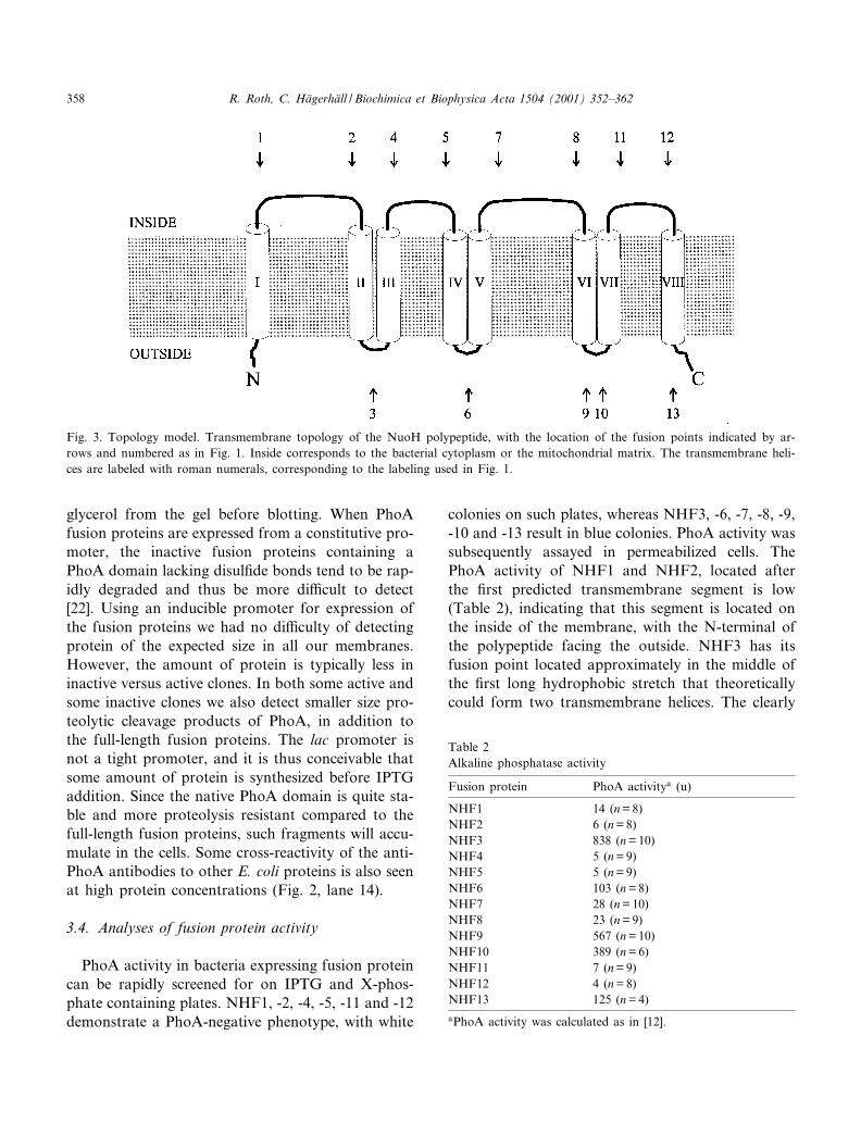
colonies on such plates, whereas NHF3, -6, -7, -8, -9,-10 and -13 result in blue colonies. PhoA activity wassubsequently assayed in permeabilized cells. ThePhoA activity of NHF1 and NHF2, located afterthe ¢rst predicted transmembrane segment is low(Table 2), indicating that this segment is located onthe inside of the membrane, with the N-terminal ofthe polypeptide facing the outside. NHF3 has itsfusion point located approximately in the middle ofthe ¢rst long hydrophobic stretch that theoreticallycould form two transmembrane helices. The clearly
Fig. 3. Topology model. Transmembrane topology of the NuoH polypeptide, with the location of the fusion points indicated by ar-rows and numbered as in Fig. 1. Inside corresponds to the bacterial cytoplasm or the mitochondrial matrix. The transmembrane heli-ces are labeled with roman numerals, corresponding to the labeling used in Fig. 1.
Table 2Alkaline phosphatase activity
Fusion protein PhoA activitya (u)
NHF1 14 (n = 8)NHF2 6 (n = 8)NHF3 838 (n = 10)NHF4 5 (n = 9)NHF5 5 (n = 9)NHF6 103 (n = 8)NHF7 28 (n = 10)NHF8 23 (n = 9)NHF9 567 (n = 10)NHF10 389 (n = 6)NHF11 7 (n = 9)NHF12 4 (n = 8)NHF13 125 (n = 4)aPhoA activity was calculated as in [12].
BBABIO 45017 26-2-01 Cyaan Magenta Geel Zwart
R. Roth, C. Ha«gerha«ll / Biochimica et Biophysica Acta 1504 (2001) 352^362358

PhoA-positive phenotype of this construct both onplates and as assayed indicates that this is indeed thecase. We did not ¢nd it meaningful to attempt toquantify the amount of fusion protein and correlateto activity. Not only are the PhoA-negative con-structs more susceptible to proteolysis but the moreprotease resistant 43 kDa native PhoA domain canalso show activity, just as the intact fusion protein.However, for NHF3 it is clear that the high activitycan also be correlated to a very large amount ofprotein (Fig. 2, lane 3). Fusion proteins NHF4 andNHF5 have fusion points located in the next, shorterhydrophilic stretch, and both have PhoA-negativephenotype, and are thus located on the inside ofthe membrane. NHF6 is located in the next long,hydrophobic stretch, which like NHF3 is locatedon the outside. Notably, in tobacco chloroplastNdhA (Fig. 1) a large segment in this region is miss-ing, suggesting that transmembrane helices IV and Vare absent in this enzyme. NHF7 and NHF8 exhibitPhoA-positive features on plates. However, whenPhoA activity is assayed in induced bacteria, thePhoA activity of both NHF7 and -8 is substantiallylower than in the other PhoA-positive clones (Table2), and only slightly higher than in the PhoA-nega-tive clones. Similar amounts of full-length protein asin the PhoA-negative NHF11 and NHF12 arepresent, but large amounts of smaller size PhoA isalso seen. We interpret the result such that the seg-ment containing the fusion points in NHF7 andNHF8 is located on the inside of the membrane,but lacks positively charged residues that could actas stop transfer signals [23]. In combination with thelack of the native downstream polypeptide in thefusion protein constructs, this causes a small fractionof PhoA domains to slip through the membrane,where it folds and become proteolysis resistant.This small activity, caused by accumulation of na-tive, folded PhoA, is apparently enough to causeblue color in the plate screening. The NHF9 proteinexhibits a high PhoA activity in combination withmoderate amounts of protein. But particularly inter-esting is NHF10, which is also clearly PhoA-positive,in spite of having the fusion point after the conservedlysine (Fig. 1). This demonstrate that the region be-tween helix VI and VII, which consists of a longerloop particularly in fungi mitochondria ND1, resideson the outside of the membrane. Finally, as men-
tioned NHF11 and -12 are PhoA-negative both onplates and as assayed, whereas the PhoA domain ofNHF13 is exported, demonstrating that the C termi-nal of NuoH is located on the outside of the mem-brane. The data was used to build the topology mod-el of the NuoH subunit shown in Fig. 3. Takentogether, we conclude that the NuoH polypeptidecontains eight transmembrane helices. Its four long,hydrophilic loops that shows particularly high se-quence conservation are facing the inside (bacterialcytoplasm, mitochondrial matrix) whereas the N-and C-terminal ends of the polypeptide are exposedto the outside of the membrane. This arrangementalso provides the most di¡erent, least conservedpolypeptides in the Fig. 1 alignment with a generallyconserved structure.
4. Discussion
It was postulated based on sequence comparison[24] and inhibitor sensitivity [25] that glucose dehy-drogenase harbors a NuoH-like domain, and similarquinone binding environment as Complex I,although this was later disputed by others, pointingout the short range and weak sequence relationship[14] or performing comparative studies of the twoenzymes with inhibitors and quinone analogues[26]. The transmembrane topology of the hydropho-bic N-terminal membrane anchor domain of E. coliglucose dehydrogenase was analyzed by PhoA fu-sions by Yamada et al. [27]. Comparing the topologyof the glucose dehydrogenase membrane spanningdomain to that of NuoH determined in this work,we conclude that the presumably homologous pri-mary sequence stretches do not demonstrate anystructural similarity and are in fact oriented towardsdi¡erent sides of the membrane in the two enzymes.
In spite of the fact that the NuoH subunit is amembrane protein it is one of the most conservedsubunits in Complex I (two consensus patterns forNuoH are described in Swiss Prot, PS00667 andPS00668), perhaps indicating that the subunit is cen-trally located in the membrane part of the complex,to a large extent bordering other subunits rather thanbulk lipid. When the nuoH gene was deleted from theR. capsulatus chromosome the enzyme complexfailed to assemble, however, disruption of nuoJ, -K,
BBABIO 45017 26-2-01 Cyaan Magenta Geel Zwart
R. Roth, C. Ha«gerha«ll / Biochimica et Biophysica Acta 1504 (2001) 352^362 359

-L and -N also had that same e¡ect [28]. More preg-nant suggestions about the role of NuoH in ComplexI appear from looking at the evolutionary history ofthe subunit. Complex I is a striking example of anenzyme complex that seem to originate from pre-ex-isting enzymes or sub-complexes. For instance, thereare modules in the promontory part of Complex Ihomologous to soluble Fe-only hydrogenases(NuoG) and NiFe hydrogenases (NuoB+D, [29]), re-spectively, and there are three antiporter-like poly-peptides (NuoL, -M and -N) in the membrane[30,31]. Such homologies provide important clues tothe function of the corresponding subunits in Com-plex I, and thus to the function of the enzyme com-plex as a whole. The complex, membrane-bound hy-drogenases seem to be an evolutionary intermediateon the route to Complex I formation, that waspresent in the last common ancestor of all living cells[32]. This enzyme contains a module of soluble NiFehydrogenase homologous to NuoB and -D, and one,two or three antiporter-like modules equivalent toNuoL, -M and -N. It is clear that the H2 activesite in complex hydrogenases is provided by theNiFe hydrogenase part, and the antiporter-like mod-ule can tentatively be regarded as a proton channels,connecting to the H2 active site. The remaining pro-tein subunits, homologous to NuoH and NuoI musttherefore constitute a membrane-bound electron do-nor/acceptor module, where NuoH forms the mem-brane anchor, and NuoI a ferredoxin-like promon-tory domain, perhaps reminiscent of the solubleelectron donor/acceptor protein docking with thesmall subunit (NuoB homologue) of soluble NiFehydrogenase.
The experimentally determined topology model ofNuoH, in combination with sequence alignments ofNuoH and NuoH-like polypeptides (Fig. 1, [14] andnot shown) demonstrate that regions where some ofthe polypeptides di¡er the most still can be accom-modated without obstructing a general, commonstructure for the subunit. The deletion of helices IVand V in tobacco chloroplast NdhA leaves the highlyconserved segments on the inside, in place to interactwith other conserved promontory subunits (Fig. 1),and the segment between helices VI and VII in manyfungi and chloroplasts simply provides these organ-isms with a longer outside loop (not shown). Con-cerning the glutamate-rich domain between helices V
and VI, it is interesting to note that the tobaccoNdhA, lacking helices IV and V, contains argininesrather than glutamates in this region. The function ofthese glutamates is unknown, but considering thebehavior of fusion proteins NHF7 and NHF8, onemay speculate that also in vivo, loss of this helix pairis only possible when a prominent stop transfer sig-nal is present to keep the domain in place. OtherNuoH homologues that contain helices IV and Valso have `charge shift' in the same domain. TheEchB polypeptide contains a histidine cluster insteadof the glutamates (Fig. 1) and the M. jannashi andM. thermoautotrophicum open reading frames have acouple of lysines and an aspartate in this region (notshown).
It is not very likely that the subunit would be soextensively conserved, if it had merely the role of amembrane anchor. The evidence indicating quinonebinding to NuoH has been mentioned in Section 1.What speaks against such a role for NuoH is that atleast one of the complex hydrogenases (Ech) do notreact with quinones [33]. In archaea, the complexmembrane-bound hydrogenase have evolved into aF420H2 :oxidoreductase, whereas in bacteria and eu-karyotes they are progenitors of NADH:quinoneoxidoreductase. The former enzymes have been char-acterized in Methanosarcina mazei (encoded by theFpo operon) where it is using methanophenazine aselectron acceptor [34] and in Archaeoglobus fulgiduswhere it was demonstrated to use quinone [35].Methanophenazine is a 2-hydroxyphenazine deriva-tive linked to a pentaisoprenoid chain via an etherbridge, i.e., it can be regarded as a functional ho-mologue of quinone found in methanogenic ar-chae. When we compare the primary sequence ofNuoH to that of FpoH and AF1831 from F420H2 :oxidoreductase and to the homologous polypeptidesfrom complex hydrogenases (subunits from twodi¡erent E. coli formate:hydrogen lyase, HycD andHyfC [36,37] Methanosarcina barkeri EchB [38] andcarbon monoxide:hydrogen lyase from Rhodospiril-lum rubrum, CooK [39]) it is clear that althoughthe subunit is generally very conserved, the F420H2
oxidoreductase polypeptides group with the ComplexI subunits rather than with the hydrogenase subunits.On the other hand, there is no obvious sequencehomology or patters in common for subunits fromenzymes containing one, two or three antiporters,
BBABIO 45017 26-2-01 Cyaan Magenta Geel Zwart
R. Roth, C. Ha«gerha«ll / Biochimica et Biophysica Acta 1504 (2001) 352^362360

respectively. Thus, nothing suggests that these largemembrane spanning subunits are surrounding NuoH,contributing to high degree of sequence conservationin the subunit (Fig. 1). Taken together, we can con-clude that the NuoH like polypeptide of the lastcommon ancestor of the three present-day enzymesmost probably contained a binding site for a lipid-soluble electron carrier cofactor. This binding sitehas been kept with di¡erent minor modi¢cations inthe present-day enzymes and accept quinone or otherhydrophobic electron carriers. This central, catalyticrole of the subunit is causing its high degree of se-quence conservation.
Acknowledgements
This work was supported by grants from TheSwedish Natural Science Research Foundation andMagn. Bergvalls Stiftelse to C.H. R.R. gratefully ac-knowledges a predoctoral scholarship from The Svenand Lilly Lawski foundation. We thank ProfessorLars Hederstedt for the generous gift of anti-PhoAantibodies.
References
[1] A. Galkin, V.G. Grivennikova, A.D. Vinogradov, FEBSLett. 451 (1999) 157^161.
[2] V. Zickermann, B. Barquera, M. Wikstro«m, M. Finel, Bio-chemistry 37 (1998) 11792^11796.
[3] F.G. Earley, C.I. Ragan, Biochem. J. 224 (1984) 525^534.[4] F.G. Earley, S.D. Patel, C.I. Ragan, G. Attardi, FEBS Lett.
219 (1987) 108^113.[5] C. Manoil, J. Beckwith, Science 233 (1986) 1403^1408.[6] C. Manoil, D. Boyd, J. Beckwith, Trends Genet. 4 (1988)
223^226.[7] C. Manoil, J.J. Mekalanos, J. Beckwith, J. Bacteriol. 172
(1990) 515^518.[8] S. Sambrook, E.F. Fritsch, T. Maniatis, Molecular Cloning.
A Laboratory Manual, 2nd ed., Cold Spring Harbor Labo-ratory, Cold Spring Harbor, NY, 1989.
[9] K. Wilson, in: D.D. Moore (Ed.), Current Protocols in Mo-lecular Biology, Preparation and Analysis of DNA, vol. 1,Wiley Interscience, New York, 1994, pp. 2.4.1^2.4.5.
[10] J. Marmur, J. Mol. Biol. 3 (1961) 208^218.[11] H. Scha«gger, G. von Jagow, Anal. Biochem. 166 (1987) 368^
379.[12] C. Manoil, Methods Cell Biol. 34 (1991) 61^75.
[13] J. Kyte, R.F. Doolittle, J. Mol. Biol. 157 (1982) 105^132.[14] I.M. Fearnley, J.E. Walker, Biochim. Biophys. Acta 1140
(1992) 105^134.[15] K. Laval-Favre, B. Letouvet-Pawlak, T. Friedrich, J. Alex-
andre, J.F. Guespin-Michel, Mol. Microbiol. 23 (1997)1043^1052.
[16] G. von Heijne, Y. Gavel, Eur. J. Biochem. 174 (1988) 671^678.
[17] Y. Gavel, G. von Heine, Eur. J. Biochem. 205 (1992) 1207^1215.
[18] Y. Gavel, J. Steppuhn, R. Herrmann, G. von Heine, FEBSLett. 282 (1991) 41^46.
[19] D. Boyd, in: S.H. White (Ed.), Membrane Protein Struc-ture: Experimental Approaches, Oxford University Press,New York, 1994, pp. 144^163.
[20] C.H. Yun, S.R. Van Doren, A.R. Crofts, R.B. Gennis,J. Biol. Chem. 266 (1991) 10967^10973.
[21] D. Boyd, B. Traxler, J. Beckwith, J. Bacteriol. 175 (1993)553^556.
[22] C. Ha«gerha«ll, H. Friden, R. Aasa, L. Hederstedt, Biochem-istry 34 (1995) 11080^11089.
[23] G. von Heijne, EMBO J. 5 (1986) 3021^3027.[24] T. Friedrich, M. Strohdeicher, G. Hofhaus, D. Preis, H.
Sahm, H. Weiss, FEBS Lett. 265 (1990) 37^40.[25] T. Friedrich, P. van Heek, H. Leif, T. Ohnishi, E. Forche, B.
Kunze, R. Jansen, W. Trowitzsch-Kienast, G. Ho£e, H.Reichenbach et al., Eur. J. Biochem. 219 (1994) 691^698.
[26] K. Sakamoto, H. Miyoshi, K. Matsushita, M. Nakagawa, J.Ikeda, M. Ohshima, O. Adachi, T. Akagi, H. Iwamura, Eur.J. Biochem. 237 (1996) 128^135.
[27] M. Yamada, K. Sumi, K. Matsushita, O. Adachi, Y. Yama-da, J. Biol. Chem. 268 (1993) 12812^12817.
[28] A. Dupuis, E. Darrouzet, H. Duborjal, B. Pierrard, M. Che-vallet, R. van Belzen, S.P.J. Albracht, J. Lunardi, Mol. Mi-crobiol. 28 (1998) 531^541.
[29] S.P. Albracht, Biochim. Biophys. Acta 1144 (1993) 221^224.
[30] T. Friedrich, H. Weiss, J. Theor. Biol. 187 (1997) 529^540.
[31] M. Finel, Biochim. Biophys. Acta 1364 (1998) 112^121.[32] T. Friedrich, D. Scheide, FEBS Lett. 479 (2000) 1^5.[33] J. Meuer, S. Bartoschek, J. Koch, A. Kunkel, R. Hedderich,
Eur. J. Biochem. 265 (1999) 325^355.[34] S. Ba«umer, T. Ide, C. Jacobi, A. Johann, G. Gottschalk, U.
Deppenmeier, J. Biol. Chem. 275 (2000) 17968^17973.[35] J. Kunow, D. Linder, K.O. Stetter, R.K. Thauer, Eur. J.
Biochem. 223 (1994) 503^511.[36] R. Bohm, M. Sauter, A. Bock, Mol. Microbiol. 4 (1990)
231^243.[37] S.C. Andrews, B.C. Berks, J. Mcclay, A. Ambler, M.A.
Quail, P. Golby, J.R. Guest, Microbiology 11 (1997) 3633^3647.
[38] A. Kunkel, J.A. Vorholt, R.K. Thauer, R. Hedderich, Eur.J. Biochem. 252 (1998) 467^476.
[39] J. Fox, Y. He, D. Shelver, G. Roberts, P. Ludden, J. Bac-teriol. 178 (1996) 6200^6208.
BBABIO 45017 26-2-01 Cyaan Magenta Geel Zwart
R. Roth, C. Ha«gerha«ll / Biochimica et Biophysica Acta 1504 (2001) 352^362 361
[40] G.D. Schuler, S.F. Altschul, D.J. Lipman, Struct. Funct.Genet. 9 (1991) 180^190.
[41] C.E. Lawrence, S.F. Altschul, M.S. Boguski, J.S. Liu, A.F.Neuwald, J.C. Wootton, Science 262 (1993) 208^214.
[42] S. Karlin, S.F. Altschul, Proc. Natl. Acad. Sci. USA 87(1990) 2264^2268.
[43] C. Geourjon, G. Deleage, J. Mol. Graph. 13 (1995) 209^212.[44] C. Manoil, J. Beckwith, Proc. Natl. Acad. Sci. USA 82
(1985) 8129^8133.[45] C. Yanisch-Perron, J. Vieira, J. Messing, Gene 33 (1985)
103^119.
BBABIO 45017 26-2-01 Cyaan Magenta Geel Zwart
R. Roth, C. Ha«gerha«ll / Biochimica et Biophysica Acta 1504 (2001) 352^362362