transient receptor potential melastatin 2 negatively...
TRANSCRIPT

Research ArticleTransient Receptor Potential Melastatin 2 Negatively RegulatesLPS-ATP-Induced Caspase-1-Dependent Pyroptosis of BoneMarrow-Derived Macrophage by Modulating ROS Production
HaihongWang,1 Xinyi Zhou,2 Hui Li,3 Xiaowei Qian,4 YanWang,3 and Liang Ma5
1Department of Anesthesiology, Sir Run Run Shaw Hospital, School of Medicine, Zhejiang University, Hangzhou 310016, China2Department of Anesthesiology, TheThird Affiliated Hospital, Anhui Medical University, Hefei 230061, China3Department of Anesthesiology, The First Affiliated Hospital, School of Medicine, Zhejiang University, Hangzhou 310003, China4Department of Anesthesiology, Women’s Hospital, School of Medicine, Zhejiang University, Hangzhou 310006, China5Department of Urology, Sir Run Run Shaw Hospital and the Institute of Minimally Invasive Surgery, School of Medicine,Zhejiang University, Hangzhou 310016, China
Correspondence should be addressed to Liang Ma; [email protected]
Received 16 June 2017; Accepted 15 October 2017; Published 8 November 2017
Academic Editor: Antoni Camins
Copyright © 2017 Haihong Wang et al.This is an open access article distributed under the Creative Commons Attribution License,which permits unrestricted use, distribution, and reproduction in any medium, provided the original work is properly cited.
Background. Pyroptosis, a new form of cell death, which has special morphological characteristics, depends on caspase-1 activationand occupies an important role in inflammatory immune diseases and ischemia-reperfusion injury. ROS is a common activatorof NLR/caspase-1. Transient receptor potential melastatin 2 (TRPM2), a selective cation channel, is involved in inflammatoryregulation. This study was designed to explore the role of TRPM2 in activating caspase-1 and caspase-1-dependent pyroptosisof mouse BMDMs. Methods. BMDMs isolated from WT and TRPM2−/− mice were treated with LPS and ATP, along with ROSinhibitor (NAC and DPI), caspase-1 inhibitor (Z-YVAD), or not. The activation of caspase-1 was measured by western blot. EtBrand EthD-2 staining were used to assess the incidence of pyroptosis. Results. Compared with WT, the activated caspase-1-P10 washigher and the percentage of EtBr positive cells was also increased in TRPM2−/− group, which were both inhibited by Z-YVAD,NAC, or DPI. ASC oligomerization was increased in TRPM2−/− group.Conclusion.Deletion of TRPM2 can enhance the activationof caspase-1 and pyroptosis, which may be via modulating ROS production, suggesting that TRPM2 plays a critical role in immuneadjustment.
1. Introduction
Sepsis is regarded as the most common cause of death inintensive care unit, which is typically characterized by initialcytokine storm and a subsequently significant immunosup-pression [1, 2]. The proinflammatory cytokines including IL-1𝛽, IL-6, IL-18, and TNF-𝛼 have been reported to contributeto initial fatal systemic inflammatory response syndrome inthe early stage of sepsis [1, 3, 4]. Previous studies demonstratethat immune paralysis of sepsis was correlatedwith activationof suppressive receptors, induction of inhibitory ligands onimmune cells, and expansion of suppressive cell types [5–7]. Recently, studies have shown that cell death-induced
depletion of immune cells in sepsis plays a vital role inimmunosuppression [8–10].
Pyroptosis, a supramolecular assembly of ASC dimers,is a recently identified kind of cell death and dependent oncaspase-1 [10–12]. During the process of pyroptosis, themem-brane pores between 1.1 and 2.4 nm indiameter are first gener-ated, followed by water uptake and cell swollen by dissipatedionic gradients, and cells ultimately underwent osmotic lysiswith release of intracellular contents [13]. It has been demon-strated that pyroptosis has an important role in inflammatoryimmune diseases, and reactive oxygen species (ROS), K+efflux, and phagosomal destabilization are involved in themechanism of caspase-1-dependent pyroptosis [14, 15].
HindawiBioMed Research InternationalVolume 2017, Article ID 2975648, 8 pageshttps://doi.org/10.1155/2017/2975648

2 BioMed Research International
Transient receptor potential melastatin 2 (TRPM2), anoxidant-sensitive and Ca2+ permeable nonselective cationchannel, is highly expressed in immunocytes includingmonocytes, macrophages, and lymphocytes [16]. TRPM2can be opened through directly binding with intracellularadenosine diphosphate ribose (ADPR) and also be indirectlyactivated under conditions of oxidative stress, acidification,and elevated intracellular Ca2+ [17–19]. Meanwhile, the acti-vation of TRPM2 can reduce NADPH oxidase-activated ROSproduction [20]. Studies have been performed on the effectof TRPM2 in the pathogenic processes of inflammation,ischemia-reperfusion injury, diabetes, and neurodegenera-tive disorders [16]. However, no study has been done toevaluate the relationship between TRPM2 and caspase-1-dependent pyroptosis. Thus, in this study, we aim to inves-tigate the role of TRPM2 in regulating caspase-1-dependentpyroptosis of bone marrow-derived macrophage in vitro andits underlying mechanism.
2. Materials and Methods
2.1. Animal Preparation. Male C57BL/6 mice (6–8 weeks)were purchased from Laboratory Animal Center (ChineseAcademy of Sciences, Shanghai). TRPM2-KOmice were pro-vided by Professor Y. mori (Graduate School of Engineering,Kyoto University, Kyoto, Japan). The mice were maintainedin a specific pathogen-free facility with ad libitum accessto food and water. Animal experiments were approved bythe Committee on the Use of Live Animals in Teaching andResearch from ZJU.
2.2. Cell Isolation and Culture. After mice were sacrificedby cervical dislocation, bone marrow-derived macrophage(BMDM) was flushed from mouse femurs and tibias withsterile RPMI 1640 medium (Invitrogen) and subsequentlydepleted of red blood cells using ammonium chloride. Then,the cells were cultured in RPMI 1640 supplemented with 10%FBS, 0.1mM nonessential amino acids, 2mM L-glutamine,1mM sodium pyruvate, 100 units/ml penicillin, 100𝜇g/mlstreptomycin, and 20 ng/ml mouse M-CSF (PeproTech) for5–7 days at 37∘C under 5%CO
2. Nonadherent cells were
carefully removed, and fresh medium was replaced every3 days. Adherent cells > 98% were judged as availablemacrophages by morphology. Macrophages were replatedonto 6-well plates at 2 × 10⁶ cells per well or seeded in 24-well plates at 5 × 10⁵ cells per well and used for experimentswithin 24 to 48 h. Unless otherwise indicated, macrophageswere primed with 1 ug/ml LPS from Escherichia coli 0111:B4(Sigma) for 4 h followed by stimulation with 5mM ATP for30min. For pharmacological assessments, some samplesweresubsequently treated with 10mMNAC (Sigma), 12.5 uM DPI(Sigma), 5 uM Z-YVAD (ENZO), or 130mM KCl, respec-tively, to the cell culture for 30min before stimulation withATP.
2.3. Western Blotting Analysis. Cells were lysed in RadioImmunoprecipitation Assay (RIPA) lysis buffer containingprotease inhibitors phenylmethylsulfonyl fluoride (PMSF),
followed by incubation on ice for 40min with vigorouslyshaking every 10 minutes. After centrifugation at 12,000 rpmfor 10min, each clarified lysates were subjected to 12% SDS-PAGE gels and transferred onto nitrocellulose membranes(Bio-Rad).Membranes were blocked in TBS-T containing 5%bull serumalbumin (BSA,Gibco) for 1 hour.The total amountof caspase-1 p-10 was detected by rabbit polyclonal anti-body against mouse caspase-1 p-10 antibody (sc-514, SantaCruz). Anti-𝛽-actin antibody was diluted to 1 : 2000 (Sigma).After incubating overnight, membranes were washed threetimes in clear TBS-T for 10min and incubated by anti-rabbit horseradish peroxidase-conjugated secondary anti-body (Sigma) for 1 hour. Membranes were rinsed three timesin TBS-T for 10min, then blots were visualized by EZ-ECL(Biological Industries) and imaged by X-ray film.
2.4. ASC Pyroptosome Detection. Macrophages were seededin 6-well plates (2 × 10⁶ cells per well) and treated withdifferent stimuli. The culture supernatants were precipitatedand analyzed by immunoblotting of caspase-1 p20 usingpolyclonal rabbit anti-human ASC (SC-22514-R, Santa Cruz)as described above. The cells pellets were harvested and thenlysed in buffer A (20mM Hepes-KOH, pH 7.5, 10mM KCl,1.5mM MgCl
2, 1 mM EDTA, and 1mM EGTA) containing
0.1mM PMSF and protease inhibitor mixture. The solutionwas diluted with one volume CHAPS buffer (20mM Hepes-KOH, pH 7.5, 5mM MgCl
2, 0.5mM EGTA, 0.1mM PMSF,
and 0.1% CHAPS) and then centrifuged at 6000 rpm in1.5ml Eppendorf tubes to pellet ASC pyroptosomes. Afterdiscarding the supernatants, the resultant pellets were resus-pended in 500 ul CHAPS buffer per tube. The resuspendedpellets were crosslinked with 4mM fresh disuccinimidylsuberate (DSS) cross-linker (Sigma) for 30 minutes at 37∘Cwith flipping the tubes every 15min and then pelleted bycentrifugation at 6000 rpm for 10min.The crosslinked pelletswere resuspended in 30 𝜇l of SDS sample buffer, fractionatedon 12% SDS-PAGE, and analyzed by western blotting usinganti-mouse ASC antibodies mentioned above.
2.5. Ethidium Bromide (EtBr) and Ethidium Homodimer-2 (EthD-2) Staining. Macrophages were seeded in 24-wellplates (5× 10⁵ cells perwell) and treatedwith different stimuli.The cells were gently washed twice with PBS and fixed by 4%paraformaldehyde for 10min. After washing the cells onceagain and adding 200 ul PBS per well, cells were stainedwith 4,6-diamidino-2-phenylindole (DAPI, 0.1 ug/ml) (Vec-tor Laboratories) for 15min. Instantly after adding red mem-brane impermeant dyes, either EtBr (25 𝜇g/ml, Sigma) orEthD2 (25𝜇g/ml, Life Technologies) was added at 0.5 ulper well. The cells were observed immediately after addingEtBr or EthD2 and analyzed using confocal microscope(Olympus). The percentage of positive cells was counted andcalculated at an amount of at least 300 cells per group for eachexperimental condition.
2.6. Statistical Analysis. The data were reported as mean ±SD from three independent experiments using SPSS16.0 forWindows (SPSS, Inc., Chicago, IL) unless otherwise noted.

BioMed Research International 3
Neg
ativ
e
LPS
+ AT
P
20
40
60
80
0
WTKO
Inte
nsity
of p
10/b
eta-
actin
(%)
LPS
ATP
∗
(a)
Procaspase-1p45
Caspase-1p10
WT KO WT KO WT KO WT KONegative LPS LPS + ATP ATP
-55-43
-17-10-17-10
-Actin
(b)
Figure 1: The activated caspase-1-P10 level in BMDM of TRPM2 KO mice and WT mice. ∗means that TRPM2−/− group compared to WTgroup. The activated caspase-1-P10 level was significantly higher in TRPM2−/− group compared with WT group.
Student’s 𝑡-test was used for statistical analyses between twogroups and multiple different comparisons were performedby using one-way ANOVA test. 𝑃 value less than 0.05 wasconsidered statistically significant, and 𝑃 < 0.001 was highlysignificant.
3. Results
3.1. The Activation of Caspase-1 Was More Prominent inBMDM of TRPM2−/− Group. To explore the activationdegree of caspase-1 in TRPM2−/− mice, BMDM was treatedwith 1𝜇g/mL LPS for 4 h followed by 5mM ATP for 30minto induce caspase-1 activation, which was tested by westernblotting. Only 1𝜇g/mL LPS plus 5mM ATP treatment acti-vated caspase-1 in BMDM of WT and TRPM2−/−mice, andthe activated caspase-1-P10 level was significantly higher inTRPM2−/− group compared with WT group (Figure 1).
3.2. Caspase-1-Dependent Pyroptosis Was Increased inTRPM2−/− Group Compared to WT Group. The formationof pore with diameter of 1.1–2.4 nm occurs in cell membraneduring the process of pyroptosis. EtBr with low molecularweight (MW 394Da) can go through such pore but EthD-2 with high molecular weight (MW 1293Da) cannot.Therefore, EtBr and EthD-2 staining positive cells can reflectthe occurrence of pyroptosis. We used small membraneimpermeant dye EtBr, larger EthD-2, and the membranepermeable dye DAPI to examine the formation of pore withdiameter of 1.1–2.4 nm in cell membrane during pyroptosis.Triton is a liposoluble active agent that can dissolve the lipidof cell membrane and nuclear membrane, thus forming alarger hole to make all dye enter. So triton can be regardedas a positive control. We found that the percentage of EtBr
positive cells was significantly increased in the TRPM2−/−group compared to WT group after LPS plus ATP treatment,but there were almost no EthD-2 positive cells in bothgroups. In addition, the percentage of EtBr positive cellsin WT and TRPM2−/− group was reduced by caspase-1inhibitor (Z-YVAD) (Figure 2).
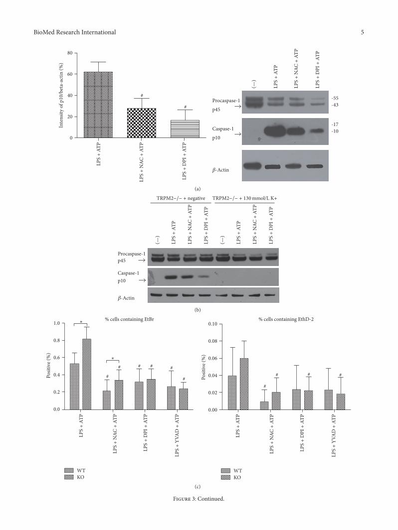
3.3. In TRPM2−/− Mice, the Activation of Caspase-1 andCaspase-1-Dependent Pyroptosis Was Inhibited by NAC Treat-ment or DPI Treatment. Both caspase-1 and caspase-1-dependent pyroptosis were more prominent in TRPM2−/−mice. Since ROS production and K+ efflux are commonsteps of NLR/caspase-1 complex activation, we hypothesizedthat TRPM2 negatively regulates LPS-ATP-induced caspase-1 and caspase-1-dependent pyroptosis through modulatingROS production and K+ efflux. So we treated BMDM ofTRPM2−/− mice with ROS inhibitor DPI or NAC (ROSscavenger) or DPI (NADPH oxidase inhibitor) before ATPstimulation and added 130mmol/L K+ into medium beforeLPS and ATP stimulation. As predicted, in TRPM2−/−mice,the activation of caspase-1 was reduced in LPS + NAC + ATPgroup and LPS + DPI + ATP group compared to LPS + ATPgroup (Figure 3(a)).
Previous study has found that high extracellular potas-sium concentration (130mM) can block the activation ofNALP3 inflammasome and inhibit the activation of caspase-1.In our study, in the condition of 130mmol/L extracellular K+,caspase-1 activation was disappeared, but procaspase-1 wasnot affected (Figure 3(b)).
The percentage of EtBr positive cells was significantlyreduced by DPI or NAC treatment but the percentage ofEthD-2 positive showed no significant difference (Figures3(c)-3(d)).

4 BioMed Research International
LPS
+ AT
P
LPS
+ YV
AD
+ A
TP
20
40
60
100
0
Posit
ive (
%)
80
DAPI + EtBrDAPI + EthD-2
LPS
+ AT
P
LPS
+ YV
AD
+ A
TP
WT
##
KO
∗
(a)
WT
KO
Negative TRITONLPS + ATP LPS + YVAD + ATP
(b)
WT
KO
Negative TRITONLPS + ATP LPS + YVAD + ATP
(c)
Figure 2: Caspase-1-dependent pyroptosis was increased in TRPM2−/− group compared to WT group. ∗ means that TRPM2−/− groupcompared to WT group; # means that LPS + YVAD + ATP compared to LPS + ATP in the same type mice. The percentage of EtBr positivecells was significantly increased in the TRPM2−/− group compared toWT group after LPS plus ATP treatment, but the percentage of EthD-2positive cells was similar between two groups.
BioMed Research International 5
#
#
20
40
60
0
LPS
+ AT
P
LPS
+ N
AC +
ATP
LPS
+ D
PI +
ATP
80In
tens
ity o
f p10
/bet
a-ac
tin (%
)
-55-43
-17-10
Procaspase-1
p45
Caspase-1
p10
LPS
+ AT
P
LPS
+ N
AC +
ATP
LPS
+ D
PI +
ATP
-Actin
(—)
(a)
Procaspase-1p45
Caspase-1p10
LPS
+ AT
P
LPS
+ N
AC +
ATP
LPS
+ D
PI +
ATP
(—)
LPS
+ AT
P
LPS
+ N
AC +
ATP
LPS
+ D
PI +
ATP
(—)
-Actin
−/− + negativeTRPM2 −/− + 130mmol/L K+TRPM2
(b)
∗
∗
LPS
+ AT
P
LPS
+ N
AC +
ATP
LPS
+ D
PI +
ATP
LPS
+ YV
AD
+ A
TP
Posit
ive (
%)
Posit
ive (
%)
WT
#
# # # #
#
KO
LPS
+ AT
P
LPS
+ N
AC +
ATP
LPS
+ D
PI +
ATP
LPS
+ YV
AD
+ A
TP
WTKO
#
# # #
% cells containing EthD-2% cells containing EtBr1.0
0.8
0.6
0.4
0.2
0.0
0.10
0.08
0.06
0.04
0.02
0.00
(c)
Figure 3: Continued.

6 BioMed Research International
WT
KO
TRITONLPS + ATP LPS + NAC + ATP LPS + DPI + ATP LPS + YVAD + ATP
WT
KO
TRITONLPS + ATP LPS + NAC + ATP LPS + DPI + ATP LPS + YVAD + ATPDAPI + EtBr
DAPI + EthD-2 (d)
Figure 3: In TRPM2−/− mice, the activation of caspase-1 and caspase-1-dependent pyroptosis was inhibited by NAC treatment or DPItreatment. # means that this group compared to LPS + ATP group in the same type mice. ∗ means that TRPM2−/− group compared toWT group. In TRPM2−/−mice, the activation of caspase-1 was reduced in LPS + NAC + ATP group and LPS + DPI + ATP group comparedto LPS + ATP group.
3.4. ASC Oligomerization Was Largely Increased in TRPM2−/−Group Compared to WT Group. ASC oligomerization isan objective index of pyroptosis. DSS can make chemicalcrosslinking between NLRP3 mediated ASC dimer, trimer,and oligomer that can be separated and by 5000×g low-speed centrifugation. We used western blotting to detect thedegree of ASC oligomerization. ASC oligomerization wassignificantly higher in TRPM2−/− group compared to WTgroup. And in both TRPM2−/− group and WT group, ASColigomerization was inhibited by NAC or DPI treatment(Figure 4).
4. Discussion
This study demonstrated that TRPM2 can negatively regu-late the LPS-ATP-induced caspase-1-dependent pyroptosis inbone marrow-derived macrophage and the possible mecha-nism might be related to the modulation of ROS production.
As we know, TRPM2 has a protective role in the regula-tion of inflammatory disorders/immune disease. Interleukin6 (IL-6), macrophage inflammatory protein 2 (MIP-2), andtumor necrosis factor (TNF) were increased in the lungsof TRPM2 KO mice, which suggested the protective roleof TRPM2 in lung inflammation [20]. In addition, TRPM2channels preserved mitochondrial biological function andfurther protected the hearts which suffered from ischemia-reperfusion injury [21]. However, the exact mechanism of
TRPM2 in immune regulation is still elusive. Our study pro-vides potential evidence that TRPM2may modulate caspase-1-dependent pyroptosis in inflammatory immune diseases.
To the best of our knowledge, pyroptosis is a double-edged sword in the progress of sepsis. On one hand, pyrop-tosis is advantageous for host defense against intracellularpathogens by eliminating replicative niche, releasing theintracellular pathogens into the extracellular space, whichis convenient for neighboring phagocytes to uptake and killthem as a result. On the other hand, pyroptosis is importantin the inflammatory process since it can make activatedmacrophages rapidly release large amounts of cytokines intothe extracellular space, and pyroptosis-induced depletionof immune cells is associated with cytopenia and immunesuppression [22–24]. Therefore, we speculate depletion ofimmune cells and inflammation promotion have the domi-nant role in LPS-ATP-induced pyroptosis of bone marrow-derived macrophage.
The question of how TRPM2 modulates LPS-ATP-induced pyroptosis of bone marrow-derived macrophagearises. By comparing the severity of mitochondrial dysfunc-tion between WT group and TRPM2 KO group, Miller et al.postulate the protection of TRPM2 against hearts ischemia-reperfusion injury is due to ameliorated mitochondrial dys-function and reduced ROS production [21]. However, ROScan be produced by several sources, including NADPH

BioMed Research International 7
Pellets
Lysates
WT KO
ASC oligomerASC trimer
ASC dimer
ASC monomer
ASC
150-100-
75-50-
37-
25-20-
LPS
+ AT
P
LPS
+ N
AC +
ATP
LPS
+ D
PI +
ATP
(—)
LPS
+ AT
P
LPS
+ N
AC +
ATP
LPS
+ D
PI +
ATP
(—)
-Actin
Figure 4: ASC oligomerization was largely increased in TRPM2−/− group compared to WT group. ASC oligomerization was significantlyhigher in TRPM2−/− group compared to WT group. And in both TRPM2−/− group and WT group, ASC oligomerization was inhibited byNAC or DPI treatment.
oxidase, mitochondria, and 5-lipoxygenase [25]. ROS isbelieved to be a common NLR/caspase-1 complex activator,and activated caspase-1 can mediate a large structure assem-bled byASC,which is termed as pyroptosis [26].The results inour study confirmed the involvement of ROS in the processof pyroptosis. Our result is similar with that from Di et al.who confirmed themechanism of TRPM2 in downregulatingROS production, which is that TRPM2 increases membranedepolarization and thereby inhibits electrogenic activity ofNADPH oxidase and blocks ROS production as a result [20].
It has been reported K+ efflux is necessary for caspase-1 activation induced by bacterial toxins and particulatematter [22]. So we treated LPS-ATP-stimulated BMDM ofTRPM2−/− with 130mmol/L K+ and found that there wasno caspase-1 activation and pyroptosis happened. Therefore,we confirmed that K+ efflux acts on the upstream of caspase-1and pyroptosis, and it is the requirement that TRPM2 regu-lates LPS-ATP-induced pyroptosis of bone marrow-derivedmacrophage.
5. Conclusions
Our study revealed that TRPM2 negatively regulates LPS-ATP-induced caspase-1-dependent pyroptosis of bonemarrow-derived macrophage by modulating ROS production.
Conflicts of Interest
The authors declare that there are no conflicts of interestregarding the publication of this article.
Authors’ Contributions
Haihong Wang and Xinyi Zhou contributed equally to thiswork.
Acknowledgments
The authors thank Professor Y. mori (Graduate School ofEngineering, Kyoto University, Kyoto, Japan) for providingTRPM2-KO mice. This work was supported by programsfrom National Natural Science Foundation of China (no.81401565 to Dr. Wang).
References
[1] D. Rittirsch, M. A. Flierl, and P. A. Ward, “Harmful molecularmechanisms in sepsis,” Nature Reviews Immunology, vol. 8, no.10, pp. 776–787, 2008.
[2] J. S. Boomer, K. To, K. C. Chang et al., “Immunosuppression inpatients who die of sepsis and multiple organ failure,” Journalof the American Medical Association, vol. 306, no. 23, pp. 2594–2605, 2011.
[3] C. Gabay, C. Lamacchia, and G. Palmer, “IL-1 pathways ininflammation and human diseases,” Nature Reviews Rheuma-tology, vol. 6, no. 4, pp. 232–241, 2010.
[4] T. Vanden Berghe, D. Demon, P. Bogaert et al., “Simultaneoustargeting of IL-1 and IL-18 is required for protection againstinflammatory and septic shock,” American Journal of Respira-tory andCritical CareMedicine, vol. 189, no. 3, pp. 282–291, 2014.
[5] W. Docke, F. Randow, U. Syrbe et al., “Monocyte deactivationin septic patients: restoration by IFN-𝛾 treatment,” NatureMedicine, vol. 3, no. 6, pp. 678–681, 1997.

8 BioMed Research International
[6] F. Venet, C.-S. Chung, G. Monneret et al., “Regulatory T cellpopulations in sepsis and trauma,” Journal of Leukocyte Biology,vol. 83, no. 3, pp. 523–535, 2008.
[7] M. J. Delano, P. O. Scumpia, J. S. Weinstein et al., “MyD88-dependent expansion of an immature GR-1 +CD11b+ popula-tion induces T cell suppression andTh2 polarization in sepsis,”The Journal of Experimental Medicine, vol. 204, no. 6, pp. 1463–1474, 2007.
[8] F. Pinheiro Da Silva and V. Nizet, “Cell death during sepsis:Integration of disintegration in the inflammatory response tooverwhelming infection,” Apoptosis, vol. 14, no. 4, pp. 509–521,2009.
[9] M. R. Pinsky, “Dysregulation of the immune response in severesepsis,” The American Journal of the Medical Sciences, vol. 328,pp. 220–229, 2004.
[10] B. A. Croker, J. A. O’Donnell, and M. Gerlic, “Pyroptotic deathstorms and cytopenia,” Current Opinion in Immunology, vol. 26,no. 1, pp. 128–137, 2014.
[11] Z.Hu, T.Murakami, K. Suzuki et al., “Antimicrobial cathelicidinpeptide LL-37 inhibits the LPS/ATP-induced pyroptosis ofmacrophages by dual mechanism,” PLoS ONE, vol. 9, no. 1,Article ID e85765, 2014.
[12] T. Fernandes-Alnemri, J. Wu, J.-W. Yu et al., “The pyropto-some: a supramolecular assembly of ASC dimers mediatinginflammatory cell death via caspase-1 activation,” Cell Death &Differentiation, vol. 14, no. 9, pp. 1590–1604, 2007.
[13] S. L. Fink and B. T. Cookson, “Caspase-1-dependent poreformation during pyroptosis leads to osmotic lysis of infectedhostmacrophages,”CellularMicrobiology, vol. 8, no. 11, pp. 1812–1825, 2006.
[14] V. Hornung, F. Bauernfeind, A. Halle et al., “Silica crystals andaluminum salts activate the NALP3 inflammasome throughphagosomal destabilization,” Nature Immunology, vol. 9, no. 8,pp. 847–856, 2008.
[15] V. Petrilli, S. Papin, C. Dostert, A. Mayor, F. Martinon, and J.Tschopp, “Activation of the NALP3 inflammasome is triggeredby low intracellular potassium concentration,” Cell Death &Differentiation, vol. 14, no. 9, pp. 1583–1589, 2007.
[16] A. Sumoza-Toledo and R. Penner, “TRPM2: a multifunctionalion channel for calcium signalling,” The Journal of Physiology,vol. 589, no. 7, pp. 1515–1525, 2011.
[17] A.-L. Perraud, A. Fleig, C. A. Dunn et al., “ADP-ribose gatingof the calcium-permeable LTRPC2 channel revealed by Nudixmotif homology,” Nature, vol. 411, no. 6837, pp. 595–599, 2001.
[18] J. Du, J. Xie, and L. Yue, “Modulation of TRPM2 by acidic pHand the underlying mechanisms for pH sensitivity,”The Journalof General Physiology, vol. 134, no. 6, pp. 471–488, 2009.
[19] Y.Hara,M.Wakamori,M. Ishii et al., “LTRPC2Ca2+-permeablechannel activated by changes in redox status confers susceptibil-ity to cell death,”Molecular Cell, vol. 9, no. 1, pp. 163–173, 2002.
[20] A. Di, X.-P. Gao, F. Qian et al., “The redox-sensitive cationchannel TRPM2 modulates phagocyte ROS production andinflammation,” Nature Immunology, vol. 13, pp. 29–34, 2011.
[21] B. A. Miller, N. E. Hoffman, S. Merali et al., “TRPM2 channelsprotect against cardiac ischemia-reperfusion injury: Role ofmitochondria,”The Journal of Biological Chemistry, vol. 289, no.11, pp. 7615–7629, 2014.
[22] T. Fernandes-Alnemri, J. Wu, J.-W. Yu et al., “The pyropto-some: a supramolecular assembly of ASC dimers mediatinginflammatory cell death via caspase-1 activation,” Cell Death &Differentiation, vol. 14, pp. 1590–1604, 2007.
[23] C. N. Casson and S. Shin, “Inflammasome-mediated cell deathin response to bacterial pathogens that access the host cellcytosol: Lessons from Legionella pneumophila,” Frontiers inCellular and InfectionMicrobiology, vol. 3, Article ID 00111, 2013.
[24] E. A. Miao, I. A. Leaf, P. M. Treuting et al., “Caspase-1-inducedpyroptosis is an innate immune effector mechanism againstintracellular bacteria,” Nature Immunology, vol. 11, no. 12, pp.1136–1142, 2010.
[25] E. Novo and M. Parola, “Redox mechanisms in hepatic chronicwound healing and fibrogenesis,” Fibrogenesis & Tissue Repair,vol. 1, no. 5, 2008.
[26] R. Munoz-Planillo, P. Kuffa, G. Martınez-Colon, B. L. Smith,T. M. Rajendiran, and G. Nunez, “K+ efflux is the commontrigger of NLRP3 inflammasome activation by bacterial toxinsand particulate matter,” Immunity, vol. 38, no. 6, pp. 1142–1153,2013.

Submit your manuscripts athttps://www.hindawi.com
Stem CellsInternational
Hindawi Publishing Corporationhttp://www.hindawi.com Volume 2014
Hindawi Publishing Corporationhttp://www.hindawi.com Volume 2014
MEDIATORSINFLAMMATION
of
Hindawi Publishing Corporationhttp://www.hindawi.com Volume 2014
Behavioural Neurology
EndocrinologyInternational Journal of
Hindawi Publishing Corporationhttp://www.hindawi.com Volume 2014
Hindawi Publishing Corporationhttp://www.hindawi.com Volume 2014
Disease Markers
Hindawi Publishing Corporationhttp://www.hindawi.com Volume 2014
BioMed Research International
OncologyJournal of
Hindawi Publishing Corporationhttp://www.hindawi.com Volume 2014
Hindawi Publishing Corporationhttp://www.hindawi.com Volume 2014
Oxidative Medicine and Cellular Longevity
Hindawi Publishing Corporationhttp://www.hindawi.com Volume 2014
PPAR Research
The Scientific World JournalHindawi Publishing Corporation http://www.hindawi.com Volume 2014
Immunology ResearchHindawi Publishing Corporationhttp://www.hindawi.com Volume 2014
Journal of
ObesityJournal of
Hindawi Publishing Corporationhttp://www.hindawi.com Volume 2014
Hindawi Publishing Corporationhttp://www.hindawi.com Volume 2014
Computational and Mathematical Methods in Medicine
OphthalmologyJournal of
Hindawi Publishing Corporationhttp://www.hindawi.com Volume 2014
Diabetes ResearchJournal of
Hindawi Publishing Corporationhttp://www.hindawi.com Volume 2014
Hindawi Publishing Corporationhttp://www.hindawi.com Volume 2014
Research and TreatmentAIDS
Hindawi Publishing Corporationhttp://www.hindawi.com Volume 2014
Gastroenterology Research and Practice
Hindawi Publishing Corporationhttp://www.hindawi.com Volume 2014
Parkinson’s Disease
Evidence-Based Complementary and Alternative Medicine
Volume 2014Hindawi Publishing Corporationhttp://www.hindawi.com