transformation-dependent a protein by - pnas.org · describedbylaskeyandmills(11). ... experiments...
TRANSCRIPT

Proc. Nati. Acad. Sci. USAVol. 75, No. 6, pp. 2767-2771, June 1978Cell Biology
Transformation-dependent secretion of a low molecular weightprotein by murine fibroblasts
(major excreted polypeptide/tumorigenicity/anchorage independence/radiolabeling/immunofluorescence)
MICHAEL M. GOTTESMANLaboratory of Molecular Biology, National Cancer Institute, National Institutes of Health, Bethesda, Maryland 20014
Communicated by Bert L. Vallee, February 21, 1978
ABSTRACT A protocol has been devised to radiolabelproteins secreted by murine fibroblasts in vitro. A radiolabeledpolypeptide of molecular weight 35,000 is released into mediumin relatively large amounts by transformed cells and in muchsmaller amounts by nontransformed fibroblasts. This majorexcreted polypeptide (MEP) is found in the medium of sponta-neously transformed mouse cells and in the medium of mousecells transformed by a DNA tumor virus, RNA tumor viruses,or methylcholanthrene. The appearance of MEP appears to bewell correlated with anchorage independence in these trans-formed cells.MEP can be localized within the cytoplasm of transformed
but not untransformed cells by indirect immunofluorescence.The presence of MEP within murine fibroblasts or in theirculture medium serves as a novel biochemical marker oftransformation. A biological role for this protein has not beenassigned.
Malignant cells in vivo interact with each other, with neigh-boring host cells, and with the host immune system and bloodsupply. Some of this interaction is at the level of the cell surface,but much of it is postulated to occur through the release ofspecific humoral factors by tumor cells. Thus, using conditionedmedium from transformed cells, investigators have found bi-ological activities such as plasminogen activator (1), plasmin-ogen-independent fibrinolysins (2), migration inhibitory factor(3), tumor angiogenesis factor (4), migration-stimulating ac-tivity (5), and multiplication-stimulating activity (6).
Studies of the proteins released by transformed cells havebeen hampered by the difficulty of obtaining large amountsof material free of contamination with serum proteins, cellproteins released by cell lysis, or proteins shed from the cellsurface during handling of the cells. Consequently, a pulse-chase protocol was devised in which proteins destined fortransport out of the cell could be radiolabeled and identified.By using this protocol, the major excreted polypeptides oftransformed and untransformed mouse fibroblasts have beenidentified. The secretion of one major excreted polypeptide(MEP) of molecular weight 35,000 is markedly increased in avariety of transformed mouse fibroblasts. The presence of thispolypeptide may serve as a biochemical marker of transfor-mation.
MATERIALS AND METHODSCell Lines. Cell lines were maintained in Dulbecco-Vogt
medium (National Institutes of Health Media Unit) supple-mented with 10% calf serum (Colorado Serum Company) onFalcon tissue culture dishes at 370 in a 5% CO2 atmosphere.Cells were trypsinized in 0.25% trypsin (Microbiological As-sociates) and were passaged using standard techniques. NIH
Swiss 3T3 mouse fibroblasts (NIH) and their transformed de-rivative, Kirsten NIH (KNIH), were from C. Scher (HarvardMedical School). Medium from the nonproducer line KNIHwas found to be free of viral particles and RNA-dependentDNA polymerase (reverse transcriptase) activity by E. Scolnick(National Cancer Institute). Abelson NIH and Harvey NIHwere from E. Scolnick (National Cancer Institute). BALB/c 3T3clone A31 and its derivatives Moloney BALB (Mol-BALB), SV-1(a BALB/c 3T3 fibroblast transformed by simian virus 40 andpassaged in vivo), methylcholanthrene-transformed BALB(Mc-BALB) and Swiss 3T3 were from the collection of I. Pastan(National Cancer Institute). Kirsten BALB (clone KA234) anda flat revertant of this strain, KA31-24 (7), were from C. Scher(Harvard Medical School). Primary mouse embryo fibroblastswere from B. Lovelace (National Cancer Institute). Spontaneoustransformants of NIH (SP-NIH) and BALB/c 3T3 lines wereobtained by cloning colonies that overgrew in the presence of10% serum.
Viral Antigens and Antisera. Viral antigens and antiserawere from J. Gruber, Viral Oncology Program, National CancerInstitute. Double immunodiffusion experiments (HylandOuchterlony plates, pattern D) were conducted between pu-rified MEP from KNIH cells and goat antisera prepared againstTween/ether-disrupted Kirsten murine sarcoma virus, Grossleukemia virus, Moloney leukemia virus, and Rauscher leuke-mia virus, and against purified Gross leukemia virus p30 andRauscher leukemia virus p30. Similar analysis was also madeusing rabbit antiserum against MEP versus purified Rauscherleukemia virus p30. The appearance of a precipitin band after72 hr of incubation at 40 was considered a positive response.Pulse-Chase Radiolabeling of Secreted Proteins. Freshly
trypsinized cells were grown in medium with 10% serum for48-72 hr until they reached a density of 1 X 10⁶ cells per 60-mmFalcon tissue culture petri dish. The medium was then removedand the cells were carefully washed three times with 3 ml ofmedium lacking amino acids and serum. They were then in-cubated for 15 min with 3 ml of medium lacking amino acidsand serum, but containing 10 ,uCi of mixed 14C-labeled aminoacids (New England Nuclear, NEC-445). This labeling mediumwas then discarded and the cells were washed two times with3 ml of medium containing amino acids but lacking serum. Thecells were then incubated for 3 hr in serum-free medium (achase time sufficient to allow for secretion of proteins synthe-sized during the pulse period as determined by following releaseof trichloroacetic acid-precipitable radioactive material intothe medium). The medium containing radiolabeled protein wascollected and centrifuged at 500 X g for 10 min to remove celldebris. Acid-precipitable radioactive material in the superna-
Abbreviations: MEP, major excreted polypeptide; NIH, 3T3 NIH Swissmouse fibroblasts; KNIH, Kirsten virus-transformed NIH; Mr, mo-lecular weight; LETS protein, large external transformation-sensitiveprotein; CSP, cell surface protein.
2767
The costs of publication of this article were defrayed in part by thepayment of page charges. This article must therefore be hereby marked"advertisement" in accordance with 18 U. S. C. §1734 solely to indicatethis fact.
Proc. Natl. Acad. Sci. USA 75 (1978)
tant was determined in a Beckman liquid scintillation counterusing Econofluor scintillant (New England Nuclear) aftercollection on glass fiber filters. Acid-precipitable radiolabeledmaterial in the cells was determined in the same manner afterlysis with 0.5 M NaOH.
Polyacrylamide Gel Electrophoresis and Fluorography.Samples were concentrated for polyacrylamide gel electro-phoresis by trichloroacetic acid precipitation. Two to fivemilliliters of medium was made 10% in trichloroacetic acid afteraddition of 20 ,ug of bovine serum albumin (Sigma) as carrier.After 30 min at room temperature, the samples were centri-fuged at 12,000 X g for 30 min and the supernatants were dis-carded. After careful drainage and removal of excess liquid,the pellets were briefly washed with 1 ml of diethyl ether toremove trichloroacetic acid and dried with a stream of air. Thepellets were redissolved in 30 Al of 0.15 M Tris-HCl, pH 6.8/10% (vol/vol) glycerol/1% sodium dodecyl sulfate/0.02 MEDTA/1% (vol/vol) 2-mercaptoethanol (sample buffer) andboiled for 2 min before application to the gels. A modificationof the Studier (8) technique of polyacrylamide gel electro-phoresis with a 5% stacking and 10% separating gel was used.The separating gel also contained 10% glycerol to enhance thesharpness and resolution of the protein bands (9). All electro-phoresis reagents were from Bio-Rad. All gels were calibratedwith the following molecular-weight (Mr) standards: filamin,Mr 250,000; phosphorylase a, Mr 95,000 (Sigma); bovine serumalbumin, Mr 68,000 (Sigma); pyruvate kinase, Mr 57,000(Sigma); rabbit skeletal muscle actin, Mr 43,000; tropomyosin,Mr 33,000; and hemoglobin, Mr 16,000 (Sigma). Filamin, actin,and tropomyosin were from P. Davies (National Cancer Insti-tute). Cell surface protein, Mr 220,000, was from K. Yamada(National Cancer Institute).
Fluorograms were prepared as described by Bonner andLaskey (10) and film images of the bands were quantitated asdescribed by Laskey and Mills (11). Densitometry scans weremade with a Joyce-Loebl densitometer and peak areas weredetermined using a Numonics electronic planimeter.
Preparation of Antibody to MEP. MEP was purified toapparent homogeneity from KNIH conditioned medium bySephadex and DEAE-cellulose chromatography. Antisera tothis purified antigen were prepared in two rabbits and gavesingle precipitin lines in double immunodiffusion experimentsagainst crude or purified MEP preparations. Antisera from bothrabbits specifically precipitated radiolabeled MEP from me-dium in which transformed cells had been grown. Details of theprotein purification, the amino acid analysis, and the prepa-ration and characterization of these antisera will be publishedelsewhere.
Indirect Immunofluorescence. Cells were grown for at least48 hr after trypsinization in 30-mm Falcon tissue culture petridishes, washed with Dulbecco's phosphate-buffered saline, fixedin 1% (wt/vol) formaldehyde in phosphate-buffered saline, andmade permeable with 80% (vol/vol) acetone before treatmentwith rabbit antiserum to MEP and rhodamine-labeled goatanti-rabbit gamma globulin (Cappel). Acetone was omitted inexperiments in which surface localization was examined.Rhodamine-labeled cells were observed and photographed withPolaroid type 107 film (60-sec exposure) on a Zeiss epifluo-rescence microscope.
RESULTSIdentification of major polypeptides secreted bytransformed and untransformed fibroblastsAnalysis of the proteins secreted by mouse fibroblasts duringthe 3 hr following a 15-min pulse of 14C-labeled amino acids
240.000-
95.000 --
e ,j tiF
" J.
68.000-
n 7.000 -
43.000_
MEP---33.000 -
-_ -.
._,_wzda._mo
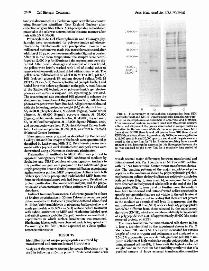
a b c dFIG. 1. Fluorography of radiolabeled polypeptides from NIH
(untransformed) and KNIH (transformed) cells. Samples were pre-pared for electrophoresis as described in Materials and Methods.After removal of medium, cells were lysed with 2% sodium dodecylsulfate, and aliquots of the lysates were boiled in sample buffer asdescribed in Materials and Methods. Secreted-proteins from NIH(lane a) and KNIH (lane b) and cell lysates from NIH (lane c) andKNIH (lane d) are shown. Approximately 6000 cpm were applied toa, 11,000 cpm to b, and 20,000 cpm to c and d, and the gels were ex-posed to the x-ray film for 72 hr. Proteins resulting from a smallamount of cell lysis can be detected in this fluorogranm because thegel was exposed to the x-ray film for a relatively long period oftime.
reveals several major differences between transformed anduntransformed cells. Fig. 1 compares an NIH Swiss 3T3 cell linewith its RNA tumor virus (Kirsten virus)-transformed deriva-tive. The banding patterns of the major radiolabeled poly-peptides in the medium as shown by polyacrylamide gel elec-trophoresis in sodium dodecyl sulfate are relatively simple forboth cell types (Fig. 1, lanes a and b), as compared to the pat-terns observed in the lysates of whole cells at the end of the 3-hrchase period (Fig. 1, lanes c and d). Furthermore, the mediumfrom both transformed and untransformed cells is enriched forspecific polypeptides that are not visible in the cell lysates atthe end of the chase period, indicating that these are not presentin the medium as a result of cell lysis. It is apparent that theuntransformed cell line (NIH) releases high Mr polypeptidessomewhat different from those released by the transformedderivative (KNIH), which also secretes a relatively large amountof a polypeptide with a Mr of approximately 35,000 (the majorexcreted protein, or MEP).The major bands from the untransformed cells shown in Fig.
1, lane a, are identified by the experiment shown in Fig. 2.Media from NIH and KNIH cells were incubated for variouslengths of time in trypsin and collagenase and analyzed on a7.5-15% exponential polyacrylamide gradient gel (12) to im-prove resolution of high molecular weight polypeptides. In theuntransformed cell line (Fig. 2, lanes a-d), the highest molecularweight band in the medium has a mobility similar to that of apurified sample of large external transformation-sensitive
2768 Cell Biology: Gottesman
Proc. Natl. Acad. Sci. USA 75 (1978) 2769
240,000- - ,m w _
95,000-
68,000 * n * v ft I
43,000- ti 0; ; A # 0
MEP- _A
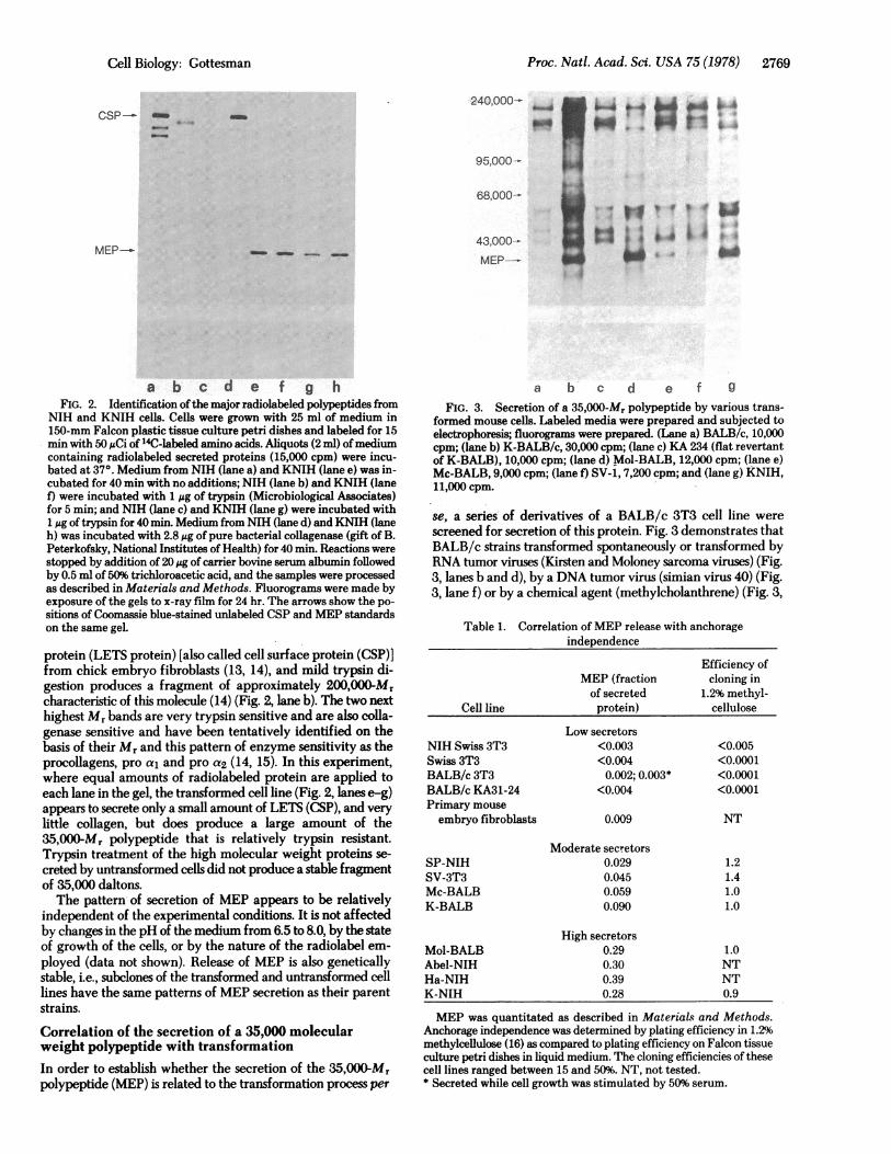
a b c d e f g hFIG. 2. Identification ofthe major radiolabeled polypeptides from
NIH and KNIH cells. Cells were grown with 25 ml of medium in150-mm Falcon plastic tissue culture petri dishes and labeled for 15min with 50 MCi of 14C-labeled amino acids. Aliquots (2 ml) ofmediumcontaining radiolabeled secreted proteins (15,000 cpm) were incu-bated at 37°. Medium from NIH (lane a) and KNIH (lane e) was in-cubated for 40 min with no additions; NIH (lane b) and KNIH (lanef) were incubated with 1 jtg of trypsin (Microbiological Associates)for 5 min; and NIH (lane c) and KNIH (lane g) were incubated with1 ,g of trypsin for 40 min. Medium from NIH (lane d) and KNIH (laneh) was incubated with 2.8 gg of pure bacterial collagenase (gift of B.Peterkofsky, National Institutes of Health) for 40 min. Reactions werestopped by addition of 20 1g of carrier bovine serum albumin followedby 0.5 ml of 50% trichloroacetic acid, and the samples were processedas described in Materials and Methods. Fluorograms were made byexposure of the gels to x-ray film for 24 hr. The arrows show the po-sitions of Coomassie blue-stained unlabeled CSP and MEP standardson the same gel.
protein (LETS protein) [also called cell surface protein (CSP)]from chick embryo fibroblasts (13, 14), and mild trypsin di-gestion produces a fragment of approximately 200,000Mrcharacteristic of this molecule (14) (Fig. 2, lane b). The two nexthighest Mr bands are very trypsin sensitive and are also colla-genase sensitive and have been tentatively identified on thebasis of their Mr and this pattern of enzyme sensitivity as theprocollagens, pro a, and pro a2 (14, 15). In this experiment,where equal amounts of radiolabeled protein are applied toeach lane in the gel, the transformed cell line (Fig. 2, lanes e-g)appears to secrete only a small amount of LETS (CSP), and verylittle collagen, but does produce a large amount of the35,OOOMr polypeptide that is relatively trypsin resistant.Trypsin treatment of the high molecular weight proteins se-
creted by untransformed cells did not produce a stable fragmentof 35,000 daltons.The pattern of secretion of MEP appears to be relatively
independent of the experimental conditions. It is not affectedby changes in the pH of the medium from 6.5 to 8.0, by the stateof growth of the cells, or by the nature of the radiolabel em-ployed (data not shown). Release of MEP is also geneticallystable, i.e., subclones of the transformed and untransformed celllines have the same patterns of MEP secretion as their parentstrains.
Correlation of the secretion of a 35,000 molecularweight polypeptide with transformationIn order to establish whether the secretion of the 35,000-Mrpolypeptide (MEP) is related to the transformation process per
a b c d e f g
FIG. 3. Secretion of a 35,000-Mr polypeptide by various trans-formed mouse cells. Labeled media were prepared and subjected toelectrophoresis; fluorograms were prepared. (Lane a) BALB/c, 10,000cpm; (lane b) K-BALB/c, 30,000 cpm; (lane c) KA 234 (flat revertantof K-BALB), 10,000 cpm; (lane d) Mol-BALB, 12,000 cpm; (lane e)Mc-BALB, 9,000 cpm; (lane f) SV-1, 7,200 cpm; and (lane g) KNIH,11,000 cpm.
se, a series of derivatives of a BALB/c 3T3 cell line werescreened for secretion of this protein. Fig. 3 demonstrates thatBALB/c strains transformed spontaneously or transformed byRNA tumor viruses (Kirsten and Moloney sarcoma viruses) (Fig.3, lanes b and d), by a DNA tumor virus (simian virus 40) (Fig.3, lane f) or by a chemical agent (methylcholanthrene) (Fig. 3,
Table 1. Correlation of MEP release with anchorageindependence
Efficiency ofMEP (fraction cloning inof secreted 1.2% methyl-
Cell line protein) cellulose
Low secretorsNIH Swiss 3T3 <0.003 <0.005Swiss 3T3 <0.004 <0.0001BALB/c 3T3 0.002; 0.003* <0.0001BALB/c KA31-24 <0.004 <0.0001Primary mouseembryo fibroblasts 0.009 NT
Moderate secretorsSP-NIH 0.029 1.2SV-3T3 0.045 1.4Mc-BALB 0.059 1.0K-BALB 0.090 1.0
High secretorsMol-BALB 0.29 1.0Abel-NIH 0.30 NTHa-NIH 0.39 NTK-NIH 0.28 0.9
MEP was quantitated as described in Materials and Methods.Anchorage independence was determined by plating efficiency in 1.2%methylcellulose (16) as compared to plating efficiency on Falcon tissueculture petri dishes in liquid medium. The cloning efficiencies of thesecell lines ranged between 15 and 50%. NT, not tested.* Secreted while cell growth was stimulated by 50% serum.
CSP- m
40_
MEP-_s
Cell Biology: Gottesman
_m _ _
Proc. Natl. Acad. Sci. USA 75 (1978)
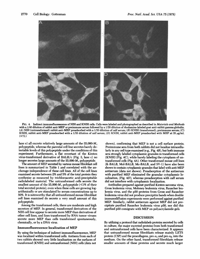
FIG. 4. Indirect immunofluorescence of NIH and KNIH cells. Cells were labeled and photographed as described in Materials and Methodswith a 1/40 dilution of rabbit anti-MEP or preimmune serum followed by a 1/20 dilution of rhodamine-labeled goat anti-rabbit gamma globulin.(A) NIH (untransformed) rabbit anti-MEP preadsorbed with a 1/20 dilution of calf serum; (B) KNIH (transformed), preimmune serum; (C)KNIH, rabbit anti-MEP preadsorbed with a 1/20 dilution of calf serum; (D) KNIH, rabbit anti-MEP preadsorbed with MEP at 25 sg/ml.(x72.)
lane e) all excrete relatively large amounts of the 35,OOO0Mrpolypeptide, whereas the parental cell line secretes barely de-tectable levels of this polypeptide under the conditions of thisexperiment. Furthermore, a flat revertant of the Kirstenvirus-transformed derivative of BALB/c (Fig. 3, lane c) nolonger secretes large amounts of the 35,000-Mr polypeptide.The amount of MEP secreted by various mouse fibroblast cell
lines is summarized in Table 1 and correlated with the an-chorage independence of these cell lines. All of the cell linesexamined secrete between 2% and 5% of the total protein theysynthesize as measured by trichloroacetic acid-precipitableradiolabeled material. The untransformed cells secrete thesmallest amount of the 35,000-Mr polypeptide (<1% of theirtotal secreted protein), even when these cells are growing log-arithmically or are stimulated by serum concentrations up to50%. It is noteworthy that all untransformed mouse fibroblastcell lines examined do secrete a very small amount of thispolypeptide.Among the transformed cells, there are moderate and high
secretors of MEP. In general, transformed derivatives of theNIH cell line appear to secrete more MEP than derivatives ofother cell lines, and lines transformed by RNA tumor virusessecrete more MEP than cells transformed spontaneously,chemically, or by a DNA virus.
Immunofluorescence localization of MEPBy using the technique of indirect immunofluorescence, MEPwas localized within transformed cells. Antisera from each oftwo rabbits showed very little localization on the surfaces oftransformed (KNIH) and untransformed (NIH) cells (data not
shown), confirming that MEP is not a cell surface protein.Preimmune sera from both rabbits did not localize intracellu-larly in any cell type examined (e.g., Fig. 4B), but both immunesera strongly labeled cytoplasmic granules in transformed cells(KNIH) (Fig. 4C), while barely labeling the cytoplasm of un-transformed cells (Fig. 4A). Other transformed mouse cell lines(K-BALB, Mol-BALB, Mc-BALB, and SV-1) have also beenshown to contain cytoplasmic granules that label with anti-MEPantiserum (data not shown). Preadsorption of the antiserumwith purified MEP eliminated the granular cytoplasmic lo-calization, (Fig. 4D), whereas preadsorption with calf serumdid not interfere with cytoplasmic localization.
Antibodies prepared against purified Kirsten sarcoma virus,Gross leukemia virus, Moloney leukemia virus, Rauscher leu-kemia virus, and the p30 proteins from Gross and Rauscherleukemia virus did not produce precipitin bands when doubleimmunodiffusion experiments were performed against purifiedMEP. Similarly, rabbit antiserum against MEP did not pre-cipitate purified Rauscher leukemia virus p30, nor did thispurified p30 comigrate with MEP on polyacrylamide gels.
DISCUSSIONBy utilizing a protocol that radiolabels proteins secreted by cellsin culture, the major excreted proteins from both transformedand untransformed cells have been characterized. It appearsthat untransformed mouse fibroblasts release mainly LETSprotein (CSP) and the procollagens, pro al and pro a2, into themedium. On the other hand, transformed fibroblasts releasesmaller amounts of these proteins and secrete much larger
2770 Cell Biology: Gottesman

Proc. Natl. Acad. Sci. USA 75 (1978) 2771
amounts of a 35,000Mr protein (MEP). On the basis of mass,MEP represents approximately 1% of the total protein syn-thesized by one transformed cell line (KNIH), and approxi-mately 30-40% of that cell's secreted protein. This is approxi-mately the same as the amount of LETS protein (CSP) or totalcollagen secreted by the untransformed parent cell line. It hasbeen previously observed that transformed mouse and chickcells synthesize decreased amounts of collagen (17, 18) and CSP(19). Adams et al. (20) concluded that the decrease in collagenand CSP biosynthesis in transformed chick cells was due toreduced levels of translatable mRNA for CSP and for collagen.The molecular basis for the increased MEP secretion seen aftertransformation has not yet been established.The secretion of MEP appears to be transformation specific.
This protein is released into the medium in large amounts (0.1to 1% of total protein synthesized) by all of the transformed cellsstudied regardless of the means of transformation, and in muchlower amounts (0.02% or less of total protein synthesis) by un-transformed cells. The growth stimulation associated withtransformation does not seem to be the mechanism responsiblefor increased release of MEP, because stimulation of the growthof cells obtained by plating at 20% of confluence or by growthin 50% serum results in only a slight increase in measurableMEP in the medium. Furthermore, complete inhibition of thegrowth of KNIH cells by sodium butyrate results in no decreasein the amount of MEP released by these cells (data not shown).It is noteworthy that primary mouse embryo fibroblasts, whichhave less density-dependent inhibition of growth than estab-lished 3T3 cell lines, make more MEP than the other non-transformed cells. Thus, in this case, MEP release correlates withdegree of growth control.MEP does not appear to be a product of an exogenous viral
genome, because cells transformed chemically or spontaneouslyor by a variety of unrelated tumor viruses secrete it. It is alsosecreted to a small extent by nontransformed cell lines. In ad-dition, it does not comigrate or crossreact with various murinesarcoma viral antigens, such as p30, and is secreted by non-virus-producing cell lines. These data do not rule out the pos-sibility that MEP is the product of a previously undescribedendogenous viral genome.
It is unclear whether the release of MEP is a primary or earlystage in the process of cell transformation, or whether it is asecondary effect. To date, it has not been possible to assign abiological role to MEP. Initial experiments indicate that thepurified protein does not have protease activity, nor does it havegrowth-promoting or migration-stimulating activity. Its ap-pearance in relatively large amounts suggests a stoichiometricor structural, rather than a catalytic, role for this protein.
Although the role of MEP in cell transformation is unclearat this time, the appearance of this protein in large amounts incell culture medium might prove to be a useful marker oftransformation. Because MEP release correlates with anchorage
independence for the cell lines studied, and because anchorageindependence has been well correlated for mouse fibroblastswith tumorigenicity in vivo (21), this easily detectable proteinmight prove to be a marker of tumorigenicity as well as oftransformation.
The author wishes to thank the members of the Biophysics ResearchLaboratory, Peter Bent Brigham Hospital, and the Laboratory ofMolecular Biology, National Cancer Institute, for many helpful dis-cussions. I am grateful to K. Yamada, F. Cabral, M. Sobel, I. Pastan,and E. Scolnick for reviewing this manuscript prior to publication; toE. Scolnick, C. Scher, I. Pastan, and B. Lovelace for cell lines; to M.Willingham for help with the immunofluorescence experiments; toR. Steinberg for photographic assistance; and to W. Levillain fortechnical assistance. I am especially indebted to Professor B. L. Valleeand the Monsanto Corporation for support during the early phases ofthis research.
1. Unkeless, J. C., Tobia, A., Ossowski, L., Quigley, J. P., Rifkin, D.B. & Reich, E. (1973) J. Exp. Med. 137,85-112.
2. Chen, L. B. & Buchanan, J. M. (1975) Proc. Natl. Acad. Sci. USA72, 1132-1136.
3. Hammond, M. E., Roblin, R. O., Dvorak, A. M., Selvaggio, S. S.,Black, P. H. & Dvorak, H. F. (1974) Science 185,955-957.
4. Klagsbrun, M., Knighton, D. & Folkman, J. (1976) Cancer Res.36, 110-113.
5. Burk, R. R. (1973) Proc. Natl. Acad. Sci. USA 70,369-372.6. Dulak, N. C. & Temin, H. M. (1973) J. Cell Physiol. 81, 153-
160.7. Greenberger, J. S. & Aaronson, S. A. (1974) Virology 57, 339-
346.8. Studier, W. (1973) J. Mol. Biol. 79, 237-248.9. Cabral, F. & Schatz, G. (1978) in Methods in Enzymology, eds.
Fleischer, S. & Packer, L. (Academic, New York), in press.10. Bonner, W. M. & Laskey, R. A. (1974) Eur. J. Biochem. 46,
83-88.11. Laskey, R. A. & Mills, A. D. (1975) Eur. J. Biochem. 56, 335-
341.12. Douglas, M. G. & Butow, R. A. (1976) Proc. Natl. Acad. Sci.
USA 73, 1083-1086.13. Hynes, R. 0. (1973) Proc. Natl. Acad. Sci. USA 70, 3170-
3174.14. Yamada, K. M. & Weston, J. A. (1974) Proc. Natl. Acad. Sci. USA
71,3492-3496.15. Burke, J. M., Balian, G., Ross, R. & Bornstein, P. (1977) Bio-
chemistry 16, 3243-3249.16. Stoker, M., O'Neill, C., Berryman, S. & Waxman, V. (1968) Int.
J. Cancer 3,683-693.17. Peterkofsky, B. (1972) Arch. Biochem. Biophys. 152, 318-328.18. Levinson, W., Bhatnagar, R. S. & Liu, T.-Z. (1975) J. Natl. Cancer
Inst. 55, 807-810.19. Olden, K. & Yamada, K. (1977) Cell 11, 957-969.20. Adams, S. L., Sobel, M. E., Howard, B. H., Olden, K., Yamada,
K. M., de Crombrugghe, B. & Pastan, I. (1977) Proc. Natl. Acad.Sci. USA 74,3399-3403.
21. Shin, S. I., Freedman, V. H., Risser, R. & Pollack, R. (1975) Proc.Natl. Acad. Sci. USA 72,4435-4439.
Cell Biology: Gottesman