transcriptome walking: a laboratory-oriented gui - biomed central
TRANSCRIPT

French BMC Research Notes 2012, 5:673http://www.biomedcentral.com/1756-0500/5/673
TECHNICAL NOTE Open Access
Transcriptome walking: a laboratory-orientedGUI-based approach to mRNA identification fromdeep-sequenced dataAndrew S French
Abstract
Background: Deep sequencing technology provides efficient and economical production of large numbers ofrandomly positioned, relatively short, estimates of base identities in DNA molecules. Application of this technologyto mRNA samples allows rapid examination of the molecular genetic environment in individual cells or tissues, thetranscriptome. However, assembly of such short sequences into complete mRNA creates a challenge that limits theusefulness of the technology, particularly when no, or limited, genomic data is available. Several approaches to thisproblem have been developed, but there is still no general method to rapidly obtain an mRNA sequence fromdeep sequence data when a specific molecule, or family of molecules, are of interest. A frequent requirement is toidentify specific mRNA molecules from tissues that are being investigated by methods such as electrophysiology,immunocytology and pharmacology. To be widely useful, any approach must be relatively simple to use in thelaboratory by operators without extensive statistical or bioinformatics knowledge, and with readily availablehardware.
Findings: An approach was developed that allows de novo assembly of individual mRNA sequences in two linkedstages: sequence discovery and sequence completion. Both stages rely on computer assisted, Graphical UserInterface (GUI)-guided, user interaction with the data, but proceed relatively efficiently once discovery is complete.The method grows a discovered sequence by repeated passes through the complete raw data in a series of steps,and is hence termed ‘transcriptome walking’. All of the operations required for transcriptome analysis are combinedin one program that presents a relatively simple user interface and runs on a standard desktop, or laptop computer,but takes advantage of multi-core processors, when available. Complete mRNA sequence identifications usuallyrequire less than 24 hours. This approach has already identified previously unknown mRNA sequences in twoanimal species that currently lack any significant genome or transcriptome data.
Conclusions: As deep sequencing data becomes more widely available, accessible methods for extracting usefulsequence information in the biological or medical laboratory will be of increasing importance. The approachdescribed here does not rely on detailed knowledge of bioinformatic algorithms, and allows users with basicknowledge of molecular biology and standard laboratory computing equipment, but limited software orbioinformatics experience, to extract complete gene sequences from deep-sequencing data.
Keywords: Deep-sequencing, Assembly, GUI, Transcriptome, Bioinformatics
Correspondence: [email protected] of Physiology and Biophysics, Dalhousie University, PO BOX15000, Halifax NS B3H 4R2, Canada
© 2012 French; licensee BioMed Central Ltd. This is an Open Access article distributed under the terms of the CreativeCommons Attribution License (http://creativecommons.org/licenses/by/2.0), which permits unrestricted use, distribution, andreproduction in any medium, provided the original work is properly cited.
French BMC Research Notes 2012, 5:673 Page 2 of 8http://www.biomedcentral.com/1756-0500/5/673
FindingsAvailability and requirementsProject homepage [1]: http://asf-pht.medicine.dal.ca/Downloads/Operating system: Windows (64-bit only).Programming language: Visual C++, Microsoft Vis-
ual Studio.Other requirement: 64-bit Intel processor (or equiva-
lent), 4 GB memory.License: Compiled installer package is freely available
and provided as Additional file 1, [2].Data: Two files containing data that can be used to
operate the program and perform the complete walkillustrated in the manuscript are available as Additionalfiles 2 and 3.
IntroductionTechnology for DNA sequencing is developing rap-idly, and sequencing of cDNA derived from cell ortissue RNA (RNA-Seq) allows relatively easy access tothe transcribed RNA, or transcriptome, of almost anytissue [3-6]. This opens many new opportunities forlaboratories that have traditionally relied on functionalor morphological techniques, to obtain mRNA datafor the tissues under investigation. This, in turn, canhelp to solve problems that require detailed molecularstructures of cellular proteins and their products.While acquisition of genomic and transcriptome data
becomes easier and more affordable, methods for pro-cessing the sequence data to obtain complete DNA orRNA sequences have not developed at the same pace[7,8]. At the time of writing, a typical sequencing run bythe Illumina process, for example, produces >108
sequences, or ‘reads’ of ~102 base length, randomly posi-tioned to the original molecules (‘reads’ are similar toshort sequences of cDNA produced by earlier sequen-cing technology, commonly called Expressed SequenceTags or ESTs). This amount of data presents significantanalysis and processing problems. When complete gen-omic data is available, it is possible to search for knownor putative sequences, but in the absence of such infor-mation, some form of de novo assembly is required.Complete de novo assembly of 108 reads by searchingfor overlaps between reads would require years by a sin-gle processor, leading to the development of alternativeapproaches. Curiously, many of these have relied on re-ducing the data to even shorter, fixed-length sequences,sometimes called k-mers, before constructing de Bruijngraphs [6,8,9]. However, while the field is progressing,the existence of many competing approaches, and thecomplexity of measuring effectiveness, indicates that denovo assembly is still immature, for both genomic andtranscriptomic data [6,7].
The present work grew out of a need in our own la-boratory for detailed molecular structures of several spe-cific proteins involved in sensory transduction and itsmodulation in a spider sensory organ [10]. While it wasrelatively easy to obtain the raw data of the transcrip-tome for the tissue, discovery and assembly of the spe-cific sequences of interest was not easily availablebecause of the absence of genomic information for this,or any other spider. The method that we developedattempts to deal with the large amount of data, whilesimultaneously allowing the operator to directly view asmuch information about the data as possible. In particu-lar, we attempted to make the large number of reads,and the extensive overlapping of the reads, into advan-tages, rather than disadvantages. The method is designedfor the task of identifying specific individual mRNAsequences, rather than a complete collection of tran-scribed genes. The presentation here used data fromIllumina RNA-Seq operations, but is sufficiently generalto be applied to other sequence data.The software described here provides a single program
(Additional file 1) for extracting complete mRNAsequences from commercial RNA-Seq data that can beused by operators with knowledge of basic molecularbiology but without detailed knowledge of bioinformat-ics. The program runs on a standard desktop or laptopcomputer and provides an easily understood graphicalmodel of the data being processed at each stage.
RNA-Seq dataTropical wandering spiders, Cupiennius salei were main-tained in a laboratory colony at room temperature (22 ±2°C) and a 13:11 h light:dark cycle. Eight legs from anadult male spider were autotomized following a protocolapproved by the Dalhousie University Committee on La-boratory Animals. Total RNA (81 μg) was extractedfrom the combined legs using a Qiagen RNeasy plusuniversal midi kit and following the manufacturer’sinstructions. Separation of mRNA, construction ofcDNA library and Illumina processing were performedby McGill University and Génome Québec InnovationCentre, Montréal, Québec. The cDNA fragments had anaverage length of 219 ± 50 bases. Illumina processinggave paired reads of 100 bases commencing from eitherend of each fragment. These will be referred to as pri-mary reads and their paired ends. The raw data con-sisted of 89,919,581 such pairs of 100 base reads withassociated quality values (phred values) in IlluminaCasava 1.8 structured files. The primary reads and pairedends were in two separate files. The raw data weregroomed to remove any sequence containing less than80 contiguous bases with Phred score > 19 (probabilityof error in base identification less than 1%), to yield filesof 60,110,040 and 70,141,080 reads of 80–100 bases.

French BMC Research Notes 2012, 5:673 Page 3 of 8http://www.biomedcentral.com/1756-0500/5/673
All operations on the data were performed within onecomputer program, written in Visual C++ using Micro-soft Visual Studio (Additional file 1). Data within theprogram was organized into software structures termedDataSets, with each DataSet containing one or more Se-quence software structures. Each Sequence structurecontained the sequence itself, matching Phred qualityvalues, and additional information defining its alignmentto a target sequence.Groomed data was held in standard disk files in
Casava 1.8 structured files. Visual C++ allows program-ming of multiple parallel software tasks, termed‘threads’. If multiple processors are available, threads canrun in parallel, increasing the data processing efficiency.Searching and walking operations were performed onboth data files simultaneously using a triple-threadorganization. Two identical threads performed the actualsearches on the primary reads and paired ends, transmit-ting their results to a supervisory thread that performedall executive functions and produced the user display.
Searching for specific sequencesTargets for searches were selected from fruit fly, Drosophilamelanogaster (http://www.ncbi.nlm.nih.gov, http://www.flybase.org) or brown dog tick, Rhipicephalus sanguineus,(Lees et al. 2010) sequences. Typically, 200 base segmentsfrom highly conserved regions were searched at low strin-gency (15–20 contiguous base matches). Searches were
Figure 1 Discovering putative matches to an initial target sequence (are shown as colored blocks, using the indicated color code. The target sesearch. Each matched read is shown as a single horizontal line, located horblocks at left show match orientation (normal - black, reverse complementhalf the 1,387 matching reads are shown. The reads are shown sorted intocriteria, including number of matched bases, maximum identical base matc
performed on the two files containing paired ends simul-taneously, using the triple-thread method.An example of a typical search for calcium-calmodulin
dependent protein kinase (CaM-kinase) is shown inFigure 1. The initial search was conducted against aDrosophila sequence [GenBank: BT050453.1]. This figurealso illustrates the graphical user interface that was usedfor all major operations on the data. Sequences were dis-played as color coded pixel blocks arranged in horizontallines, with the target sequence at the top of the display.All base positions were numbered relative to the 5’-end ofthe target sequence, commencing at the left side of thedisplay. The vertical order of sequences could be decidedby a range of sorting options. In this figure the readsequences were sorted by their matching position to thetarget. Note that the figure contains a mixture ofsequences from both of the Illumina paired end files, andthat both direct and reverse complement matches wereincluded, indicated by color-coded bars at left. However,each matched sequence now included a description of itsalignment to the target sequence that allowed the pro-gram to display the correct match alignment. This align-ment data stayed with the sequence as it progressedthrough further stages of processing, and was used bymany of the other possible operations that the programprovides (Table 1).Several alternate further steps were available at this
stage. The number of matching reads would usually besmall enough to allow a complete assembly by sequence
part of Drosophila CaM-kinase, [GenBank: BT050453.1]). All basesquence is above, with grey color indicating regions not used in theizontally by its match position to the target sequence. Indicator color- green) and pair end (primary - green, paired end - red). Less thanascending match position to the target, but sorting by several otherhes, etc. is possible.
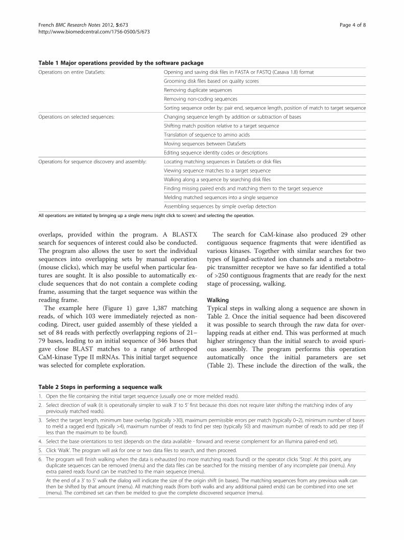
Table 1 Major operations provided by the software package
Operations on entire DataSets: Opening and saving disk files in FASTA or FASTQ (Casava 1.8) format
Grooming disk files based on quality scores
Removing duplicate sequences
Removing non-coding sequences
Sorting sequence order by: pair end, sequence length, position of match to target sequence
Operations on selected sequences: Changing sequence length by addition or subtraction of bases
Shifting match position relative to a target sequence
Translation of sequence to amino acids
Moving sequences between DataSets
Editing sequence identity codes or descriptions
Operations for sequence discovery and assembly: Locating matching sequences in DataSets or disk files
Viewing sequence matches to a target sequence
Walking along a sequence by searching disk files
Finding missing paired ends and matching them to the target sequence
Melding matched sequences into a single sequence
Assembling sequences by simple overlap detection
All operations are initiated by bringing up a single menu (right click to screen) and selecting the operation.
French BMC Research Notes 2012, 5:673 Page 4 of 8http://www.biomedcentral.com/1756-0500/5/673
overlaps, provided within the program. A BLASTXsearch for sequences of interest could also be conducted.The program also allows the user to sort the individualsequences into overlapping sets by manual operation(mouse clicks), which may be useful when particular fea-tures are sought. It is also possible to automatically ex-clude sequences that do not contain a complete codingframe, assuming that the target sequence was within thereading frame.The example here (Figure 1) gave 1,387 matching
reads, of which 103 were immediately rejected as non-coding. Direct, user guided assembly of these yielded aset of 84 reads with perfectly overlapping regions of 21–79 bases, leading to an initial sequence of 346 bases thatgave close BLAST matches to a range of arthropodCaM-kinase Type II mRNAs. This initial target sequencewas selected for complete exploration.
Table 2 Steps in performing a sequence walk
1. Open the file containing the initial target sequence (usually one or more
2. Select direction of walk (it is operationally simpler to walk 3’ to 5’ first becpreviously matched reads).
3. Select the target length, minimum base overlap (typically >30), maximumto meld a ragged end (typically >4), maximum number of reads to find pless than the maximum to be found).
4. Select the base orientations to test (depends on the data available - forw
5. Click ‘Walk’. The program will ask for one or two data files to search, and
6. The program will finish walking when the data is exhausted (no more maduplicate sequences can be removed (menu) and the data files can be seextra paired reads found can be matched to the main sequence (menu).
At the end of a 3’ to 5’ walk the dialog will indicate the size of the originthen be shifted by that amount (menu). All matching reads (from both w(menu). The combined set can then be melded to give the complete dis
The search for CaM-kinase also produced 29 othercontiguous sequence fragments that were identified asvarious kinases. Together with similar searches for twotypes of ligand-activated ion channels and a metabotro-pic transmitter receptor we have so far identified a totalof >250 contiguous fragments that are ready for the nextstage of processing, walking.
WalkingTypical steps in walking along a sequence are shown inTable 2. Once the initial sequence had been discoveredit was possible to search through the raw data for over-lapping reads at either end. This was performed at muchhigher stringency than the initial search to avoid spuri-ous assembly. The program performs this operationautomatically once the initial parameters are set(Table 2). These include the direction of the walk, the
melded reads).
ause this does not require later shifting the matching index of any
permissible errors per match (typically 0–2), minimum number of baseser step (typically 50) and maximum number of reads to add per step (if
ard and reverse complement for an Illumina paired-end set).
then proceed.
tching reads found) or the operator clicks ‘Stop’. At this point, anyarched for the missing member of any incomplete pair (menu). Any
shift (in bases). The matching sequences from any previous walk canalks and any additional paired ends) can be combined into one setcovered sequence (menu).
French BMC Research Notes 2012, 5:673 Page 5 of 8http://www.biomedcentral.com/1756-0500/5/673
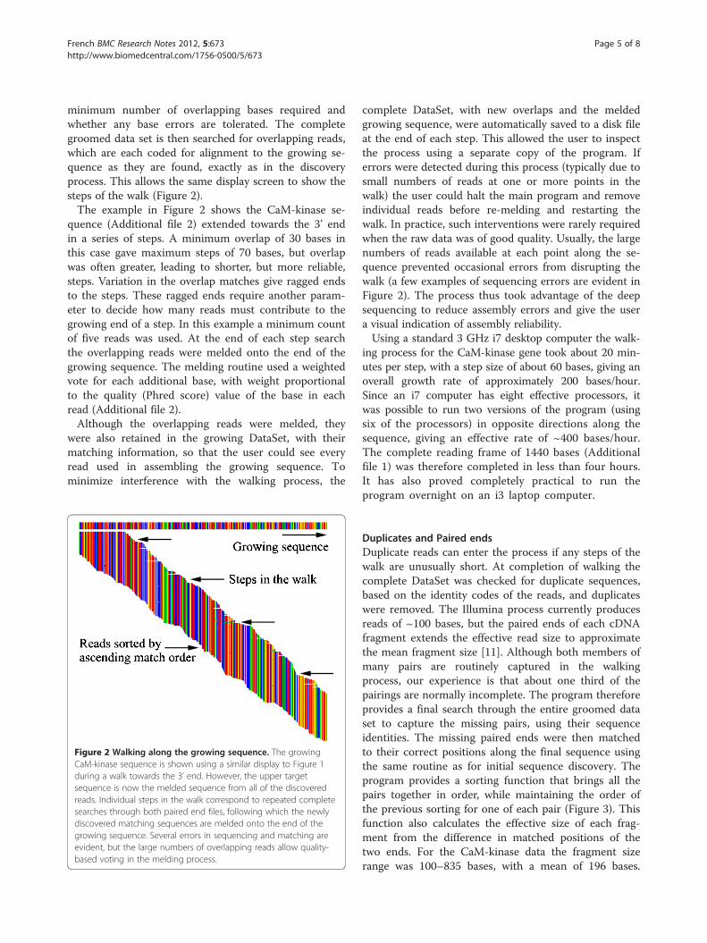
minimum number of overlapping bases required andwhether any base errors are tolerated. The completegroomed data set is then searched for overlapping reads,which are each coded for alignment to the growing se-quence as they are found, exactly as in the discoveryprocess. This allows the same display screen to show thesteps of the walk (Figure 2).The example in Figure 2 shows the CaM-kinase se-
quence (Additional file 2) extended towards the 3’ endin a series of steps. A minimum overlap of 30 bases inthis case gave maximum steps of 70 bases, but overlapwas often greater, leading to shorter, but more reliable,steps. Variation in the overlap matches give ragged endsto the steps. These ragged ends require another param-eter to decide how many reads must contribute to thegrowing end of a step. In this example a minimum countof five reads was used. At the end of each step searchthe overlapping reads were melded onto the end of thegrowing sequence. The melding routine used a weightedvote for each additional base, with weight proportionalto the quality (Phred score) value of the base in eachread (Additional file 2).Although the overlapping reads were melded, they
were also retained in the growing DataSet, with theirmatching information, so that the user could see everyread used in assembling the growing sequence. Tominimize interference with the walking process, the
Figure 2 Walking along the growing sequence. The growingCaM-kinase sequence is shown using a similar display to Figure 1during a walk towards the 3’ end. However, the upper targetsequence is now the melded sequence from all of the discoveredreads. Individual steps in the walk correspond to repeated completesearches through both paired end files, following which the newlydiscovered matching sequences are melded onto the end of thegrowing sequence. Several errors in sequencing and matching areevident, but the large numbers of overlapping reads allow quality-based voting in the melding process.
complete DataSet, with new overlaps and the meldedgrowing sequence, were automatically saved to a disk fileat the end of each step. This allowed the user to inspectthe process using a separate copy of the program. Iferrors were detected during this process (typically due tosmall numbers of reads at one or more points in thewalk) the user could halt the main program and removeindividual reads before re-melding and restarting thewalk. In practice, such interventions were rarely requiredwhen the raw data was of good quality. Usually, the largenumbers of reads available at each point along the se-quence prevented occasional errors from disrupting thewalk (a few examples of sequencing errors are evident inFigure 2). The process thus took advantage of the deepsequencing to reduce assembly errors and give the usera visual indication of assembly reliability.Using a standard 3 GHz i7 desktop computer the walk-
ing process for the CaM-kinase gene took about 20 min-utes per step, with a step size of about 60 bases, giving anoverall growth rate of approximately 200 bases/hour.Since an i7 computer has eight effective processors, itwas possible to run two versions of the program (usingsix of the processors) in opposite directions along thesequence, giving an effective rate of ~400 bases/hour.The complete reading frame of 1440 bases (Additionalfile 1) was therefore completed in less than four hours.It has also proved completely practical to run theprogram overnight on an i3 laptop computer.
Duplicates and Paired endsDuplicate reads can enter the process if any steps of thewalk are unusually short. At completion of walking thecomplete DataSet was checked for duplicate sequences,based on the identity codes of the reads, and duplicateswere removed. The Illumina process currently producesreads of ~100 bases, but the paired ends of each cDNAfragment extends the effective read size to approximatethe mean fragment size [11]. Although both members ofmany pairs are routinely captured in the walkingprocess, our experience is that about one third of thepairings are normally incomplete. The program thereforeprovides a final search through the entire groomed dataset to capture the missing pairs, using their sequenceidentities. The missing paired ends were then matchedto their correct positions along the final sequence usingthe same routine as for initial sequence discovery. Theprogram provides a sorting function that brings all thepairs together in order, while maintaining the order ofthe previous sorting for one of each pair (Figure 3). Thisfunction also calculates the effective size of each frag-ment from the difference in matched positions of thetwo ends. For the CaM-kinase data the fragment sizerange was 100–835 bases, with a mean of 196 bases.

Figure 3 Paired ends effectively increase read length. A similar CaM-kinase walk display to Figure 2, but now the reads have been sorted tobring paired ends together, while retaining the match position to the growing sequence for one of each pair. Each read is still positionedhorizontally according to its match position. The sorting algorithm also calculated the effective fragment length for each pair and gave estimatesof the mean and range of fragment lengths.
French BMC Research Notes 2012, 5:673 Page 6 of 8http://www.biomedcentral.com/1756-0500/5/673
These values can be compared with the initial bioanaly-zer estimate of 219 ± 50 for all fragmented cDNA.
Depth of coverage and mutationsAn optional rearrangement of the colored pixels in thedisplays produces a histogram of the number of readscontributing to each base in the final sequence (Figure 4).This function also provides a visual image of the numberof mutations or sequencing errors occurring at each pos-ition. For the CaM-kinase data the number of errors wassmall, but two alleles of the gene were immediately evi-dent with a mutation from A to G in 30/64 reads at lo-cation 912 of the reading frame (Figure 4), giving asilent mutation of CTA to CTG.
Figure 4 Histogram display of matched reads. The same CaM-kinase display converted into a histogram of base counts bycollapsing the display onto the X-axis (melded sequence). Thisalternative display gives a better visual indication of the number ofreads contributing to each base decision, and hence reliability. Inthis case, it also illustrates that two alleles are present in the reads.
Resulting amino-acid sequenceThe complete reading frame of the CaM-kinase data wastranslated by the program and entered into the protein-protein BLAST against the non-redundant protein data-base. The closest match (89% identical) was to calcium/calmodulin-dependent protein kinase II, isoform A, ofPeriplaneta americana, the American cockroach(Figure 5).
ConclusionsOur initial objective was to identify the amino acidsequences of a series of molecules whose existence inCupiennius salei was indicated by electrophysiologicaland immunocytochemical studies [2,12]. It is importantto note that these requirements were significantly differ-ent to those of researchers interested in comparativegenomics, or seeking quantitative data on gene transla-tion in different tissues or conditions. However, ourrequirements are common to many studies that seek tounderstand physiological processes, and the proteinsinvolved at each stage of those processes e.g. [3]. Follow-ing RNA separation from Cupennius salei, and Illuminasequencing, we found that very limited resources wereavailable for specific gene identification from the databecause of the lack of genomic data. We needed a rela-tively simple and straightforward method of findingmRNA sequences from the deep sequencing data thatcould be used by experimental laboratory memberswithout extensive bioinformatics training, and usingstandard laboratory desktop or laptop computers.The method that we have developed allows the user to
perform all the major steps in finding and completing a

Figure 5 Amino acid sequence comparison of the translated CaM-kinase reading frame by protein-protein BLAST against the non-redundant protein database. The closest match was to the calcium/calmodulin-dependent protein kinase II isoform A of the Americancockroach, Periplaneta americana. Other details are indicated in the figure.
French BMC Research Notes 2012, 5:673 Page 7 of 8http://www.biomedcentral.com/1756-0500/5/673
transcribed gene using a single program [2] on a labora-tory desktop computer. The only knowledge required isa basic understanding of mRNA transcription and trans-lation, combined with reasonable estimates of the num-ber of overlapping bases that should be expected fromreads that originate from the same fragmentary se-quence, or that come from genes with close homologyto similar genes in a related species that can provide atemplate.The single mutation in the CaM-kinase reading frame
(Figure 4) illustrates an important limitation of sequen-cing methods that produce short reads. While no othermutations were seen in this CaM-kinase reading frame,other mutations were present in the 3’ noncoding re-gion, and other Cupiennius genes that we have exploredhad multiple mutations within the reading frame. If suchmutations are further apart than the average cDNA frag-ment length it becomes impossible to decide whichmutations are associated with each other in the allelesthat produced the original data.As described above, the program provides a method of
finding an initial set of reads from the mRNA of interest.However, other approaches and information, such asgenomic data if available, could also be used for thisstep. The major development here is in the second stage,walking, where the numerous overlapping reads pro-vided by deep sequencing allow an easily comprehended,but highly reliable and efficient method of completingde novo synthesis of the complete sequence. In addition,
the graphical user display (Figures 1,2,3 and 4) providesa crucial resource by giving the user an immediate andaccessible understanding of the data being processedand its internal relationships. Using standard laboratorycomputers, a transcribed gene can usually be discoveredand sequenced reliably in a period of a few hours to oneday, which is a very acceptable time period for mostfunctional studies.
Additional files
Additional file 1: Sequence.exe Self-unzipping executable Windowsinstaller package of the program.
Additional file 2: Cupiennius_0162a.fastq Nucleotide sequence ofthe CaM-kinase reading frame.
Additional file 3: Cupiennius_0162b.fastq Set of nucleotidesequences that generated the reading frame. The two nucleotide filesare in FASTQ format with Casava 1.8 quality score coding. Sequences inthe Cupiennius_0162b.fastq file contain matching information to thereading frame. They can be viewed against the reading frame directly byopening them in the Sequence program.
Competing interestsThe author declares that he has no competing interests.
AcknowledgementsShannon Meisner and Audrey Li provided expert technical assistance in RNAseparation. Sequencing was performed by The McGill University andGénome Québec Innovation Centre. This work was supported by grantsfrom the Canadian Institutes for Health Research and the JuseliusFoundation of Finland.
French BMC Research Notes 2012, 5:673 Page 8 of 8http://www.biomedcentral.com/1756-0500/5/673
Received: 14 September 2012 Accepted: 22 November 2012Published: 5 December 2012
References1. Sequence.exe, a complete, self-unpacking Windows installer package is
available as Additional file 1, and the latest version of the package isavailable from the project homepage at: http://asf-pht.medicine.dal.ca/Downloads/Sequence.exe.
2. Torkkeli PH, Panek I, Meisner S: Ca2+/calmodulin-dependent protein kinaseII mediates the octopamine-induced increase in sensitivity in spider VS-3mechanosensory neurons. Eur J Neurosci 2011, 33:1186–1196.
3. Lees K, Woods DJ, Bowman AS: Transcriptome analysis of the synganglionfrom the brown dog tick. Rhipicephalus sanguineus. Insect Mol Biol 2010,19:273–282.
4. Trapnell C, Williams BA, Pertea G, Mortazavi A, Kwan G, van Baren MJ,Salzberg SL, Wold BJ, Pachter L: Transcript assembly and quantification byRNA-Seq reveals unannotated transcripts and isoform switching duringcell differentiation. Nat Biotechnol 2010, 28:511–515.
5. Wang Z, Gerstein M, Snyder M: RNA-Seq: a revolutionary tool fortranscriptomics. Nat Rev Genet 2009, 10:57–63.
6. Grabherr MG, Haas BJ, Yassour M, Levin JZ, Thompson DA, Amit I, AdiconisX, Fan L, Raychowdhury R, Zeng Q, Chen Z, Mauceli E, Hacohen N, Gnirke A,Rhind N, di Palma F, Birren BW, Nusbaum C, Lindblad-Toh K, Friedman N,Regev A: Full-length transcriptome assembly from RNA-seq data withouta reference genome. Nat Biotechnol 2011, 15:644–652.
7. Earl D, Bradnam K, St John J, Darling A, Lin D, Fass J, Yu HO, Buffalo V,Zerbino DR, Diekhans M, et al: Assemblathon 1: A competitive assessmentof de novo short read assembly methods. Genome Res 2011,21:2224–2241.
8. Zhao QY, Wang Y, Kong YM, Luo D, Li X, Hao P: Optimizing de novotranscriptome assembly from short-read RNA-Seq data: a comparativestudy. BMC Bioinformatics 2011, 12(Suppl 14):S2.
9. Zerbino DR, Birney E: Velvet: algorithms for de novo short read assemblyusing de Bruijn graphs. Genome Res 2008, 18:821–829.
10. French AS, Torkkeli PH, Seyfarth E-A: From stress and strain to spikes:mechanotransduction in spider slit sensilla. J Comp Physiol A 2002,188:739–752.
11. Chaisson MJ, Brinza D, Pevzner PA: De novo fragment assembly with shortmate-paired reads: does the read length matter? Genome Res 2009,19:336–346.
12. Pfeiffer K, Torkkeli PH, French AS: Activation of GABAA receptorsmodulates all stages of mechanoreception in spider mechanosensoryneurons. J Neurophysiol 2012, 107:196–204.
doi:10.1186/1756-0500-5-673Cite this article as: French: Transcriptome walking: a laboratory-orientedGUI-based approach to mRNA identification from deep-sequenced data.BMC Research Notes 2012 5:673.
Submit your next manuscript to BioMed Centraland take full advantage of:
• Convenient online submission
• Thorough peer review
• No space constraints or color figure charges
• Immediate publication on acceptance
• Inclusion in PubMed, CAS, Scopus and Google Scholar
• Research which is freely available for redistribution
Submit your manuscript at www.biomedcentral.com/submit